Scholarship@Western
Scholarship@Western
Electronic Thesis and Dissertation Repository
2-1-2016 12:00 AM
Development and Initial Validation of Novel Multi-Planar Neck
Development and Initial Validation of Novel Multi-Planar Neck
Strength Assessment and Neuromuscular Training Protocols
Strength Assessment and Neuromuscular Training Protocols
Theo H. Versteegh
The University of Western Ontario Supervisor
Dr Dave Walton
The University of Western Ontario
Graduate Program in Physical Therapy
A thesis submitted in partial fulfillment of the requirements for the degree in Doctor of Philosophy
© Theo H. Versteegh 2016
Follow this and additional works at: https://ir.lib.uwo.ca/etd Part of the Physical Therapy Commons
Recommended Citation Recommended Citation
Versteegh, Theo H., "Development and Initial Validation of Novel Multi-Planar Neck Strength Assessment and Neuromuscular Training Protocols" (2016). Electronic Thesis and Dissertation Repository. 3511. https://ir.lib.uwo.ca/etd/3511
This Dissertation/Thesis is brought to you for free and open access by Scholarship@Western. It has been accepted for inclusion in Electronic Thesis and Dissertation Repository by an authorized administrator of
Concussions are a serious health concern in today’s active society. There are many contributing factors to concussions but one that is starting to draw significant attention is the potential role the neck muscles play in mitigating concussive forces. There is evidence that stronger neck muscles may decrease an individual’s concussion risk. In order to fully define this role, an appropriate outcome measure for assessing neck strength is required. Once this is established, methods of training to improve neck strength can be evaluated for their effect on neck strength and subsequently effect on concussion risk. This thesis included three studies. Chapter 2 was a within session and between session test-retest agreement of a novel multi-planar neck-strength and upper kinetic chain assessment protocol using a hand-held
dynamometer in a healthy adult population. Chapter 3 examined this protocol to determine its preliminary validity. Due to the lack of an accepted ‘gold standard’ for neck strength
assessment, the validity was examined using three a priori hypotheses; face validity, known groups validity and convergent validity using EMG muscle activity. Chapter 4 is a pilot study investigating the effects of a training program using a novel neuromuscular neck-training device that has theoretical rationale on how to improve neck function to decrease concussion risk. This investigation demonstrated the device to be safe and potentially effective at
ii
Keywords
iii
Co-Authorship Statement
This thesis contains material from a published manuscript (Chapter 2) and two
manuscripts that will be prepared for submission (Chapters 3 and 4). Theo Versteegh was the primary author of all chapters contained in this thesis. All studies contained in this thesis were conceived, designed, analyzed, interpreted and written by the primary author with invaluable input and guidance from David Walton, an assistant Professor in the School of Physical Therapy, Faculty of Health Sciences, Western University. Joy MacDermid, a Professor in the School of Physical Therapy, Faculty of Health Sciences, Western University provided guidance on the design of the research program overall and Chapters 2-4; Jim Dickey, an Associate Professor in the School of Kinesiology, Faculty of Health Sciences, Western University provided guidance in EMG methodology, biomechanics, processing and analysis as well as overall methodological support for Chapters 2-4; Carolyn Emery,
Associate Dean (Research), Faculty of Kinesiology, University of Calgary provided input and guidance on proper methodology and analysis for Chapters 2-4; Lisa Fischer, Assistant Professor, Departments of Family Medicine and Faculty of Health Sciences, Western University provided clinical guidance and input for Chapters 2-4. Under the direct
iv
Acknowledgments
First and foremost, I would like to thank Dr. Dave Walton for his key role in support and guidance towards the completion of this thesis, without him this thesis would not be possible. I greatly appreciate your willingness to take me on as your first PhD student and allow me the freedom to study my personal choice of project independently of your own research interests. You were always there to provide direction and support while leaving the ultimate decisions up to me. For that I am truly grateful. Your wealth of knowledge and skill for research has continually impressed me throughout and has been invaluable in bringing this thesis through to fruition. I strongly look forward to collaborating with you in the future.
I would like to thank Dr. Joy MacDermid for co-supervising with Dr. Dave Walton, and trusting his judgment in taking me on as one of your PhD students. Thank you for your guidance and advice on ensuring Knowledge Translation as a key component of this research. I would also like to thank my Advisory Committee; Dr. Jim Dickey for his
immeasurable help in project synthesis, providing equipment and software, analyzing EMG, software programming and understanding biomechanical concepts. Thank you to Drs. Carolyn Emery and Lisa Fischer for their support and guidance in maintaining clinical application and expertise with concussion populations.
v
Table of Contents
Abstract ... i!
Co-Authorship Statement ... iii!
Acknowledgments ... iv!
Table of Contents ... v!
List of Tables ... viii!
List of Figures ... ix!
List of Appendices ... x!
List of Abbreviations ... xi!
1! Introduction ... 1!
! Neck strength ... 1!
1.1 ! Neck function ... 5!
1.2 ! Stiffness and anticipation ... 6!
1.3 ! Neck training ... 8!
1.4 ! Neuromuscular training ... 9!
1.5 ! Conclusion ... 11!
1.6 ! References ... 14!
1.7 2! Evaluating the Reliability of a Novel Neck-Strength Assessment Protocol for Healthy Adults Using Self-Generated Resistance with a Hand-Held Dynamometer ... 20!
! Introduction ... 20!
2.1 ! Methods ... 22!
2.2 2.2.1! Participants ... 22!
2.2.2! Testing protocol ... 23!
2.2.3! Data analysis ... 27!
! Results ... 28!
vi
2.4.1! Limitations ... 33!
! Conclusion ... 34!
2.5 ! Key Messages ... 35!
2.6 ! References ... 36!
2.7 3! Examining the validity of a novel neck strength assessment tool ... 38!
! Introduction ... 38!
3.1 3.1.1! Hypotheses ... 41!
! Methods ... 42!
3.2 3.2.1! Participants ... 42!
3.2.2! Testing protocol ... 44!
3.2.3! Data analysis ... 47!
3.2.4! Specific hypothesis testing ... 47!
! Results ... 48!
3.3 ! Discussion ... 54!
3.4 3.4.1! Limitations ... 55!
! Conclusion ... 56!
3.5 ! References ... 58!
3.6 4! Evaluating the effects of a novel neuromuscular neck training device on strength, performance and concussion risk: A pilot study ... 62!
! Introduction ... 62!
4.1 ! Methods ... 66!
4.2 4.2.1! Participants and Recruitment ... 66!
! Data Analysis ... 71!
4.3 ! Results ... 71!
4.4 ! Discussion ... 79!
vii
! Conclusion ... 82! 4.6
! References ... 83! 4.7
5! Discussion ... 86! ! Summary ... 86! 5.1
! Limitations ... 89! 5.2
! Future research ... 90! 5.3
! What this study adds ... 92! 5.4
! References ... 94! 5.5
viii List of Tables
Table 1.1: Selected isometric neck extension and flexion strength values of healthy male
subjects. ... 4!
Table 2.1: Participant characteristics ... 23!
Table 2.2: Intra-session Retest Reliability of Neck Strength Using a Handheld Dynamometer in a Healthy Population ... 29!
Table 2.3: Inter-session Retest Reliability of Neck Strength Using a Handheld Dynamometer in a Healthy Population ... 30!
Table 2.4: Mean strength values by sex of trial 1. ... 31!
Table 3.1: Anticipated pattern of EMG activity by direction ... 42!
Table 3.2: Demographic details. (SD) ... 50!
Table 3.3: Mean peak strength values in kgf. (95% CI) ... 51!
Table 3.4: Area under the curve of the Receiver Operating Characteristics (ROC) graph for each isometric test direction. ... 52!
Table 3.5: Percentage of peak muscle activity for sternocleidomastoid (SCM) and upper fibers of trapezius muscles in each test position (SD). ... 53!
Table 3.6: Tukey post hoc tests for multiple comparisons for anticipated muscle activity pattern by direction. ... 53!
Table 4.1: Subject demographics. (95% CI) ... 72!
Table 4.2: Pre and post training performance (n = 8). (95% CI) ... 74!
ix
List of Figures
Figure 2.1: MicroFET 2TM dynamometer ... 25!
Figure 2.2: Test positions: Calibration (A), forward flexion (B), extension (C), side-flexion (D), side-flexion with rotation (E), axial rotation (F). ... 26!
Figure 3.1: Test positions ... 46!
Figure 4.1: Neuromuscular training device ... 65!
Figure 4.2: Flow diagram of participants through stages of study. ... 68!
Figure 4.3: Mean peak velocities for each of the 14 training sessions with 95% confidence intervals. ... 76!
Figure 4.4: Pre and post isometric flexion strength values graph ... 77!
Figure 4.5: Pre and post isometric extension strength values graph ... 77!
Figure 4.6: Pre and post isometric side-flexion strength values graph (average between right and left sides) ... 78!
Figure 4.7: Pre and post isometric side-flexion/ rotation strength values graph (average between right and left sides) ... 78!
x
List of Appendices
APPENDIX A: Screening Protocol and Assessment Protocol ... 97!
APPENDIX B: Bland–Altman plots showing 95% levels of agreement for the various test positions from Chapter 2 ... 103!
APPENDIX C: Receiver Operating Characteristics Curves from Chapter 3 ... 107!
APPENDIX D: Copyright permission and reproduction of journal article ... 110!
ANOVA – Analysis of variance
AUC – Area under the curve
CC – Comparator cohort
CI – Confidence intervals
E:F – Neck extension strength to flexion strength ratio
EMG – Electromyography
FC – Football cohort
ICC – Intraclass correlation coefficient
kgf – Kilogram-force
MDC – Minimal detectable change
N*m – Newton metre
NFL – National Football League
NMT – Neuromuscular training
RMS – Root mean square
ROC – Receiver operating characteristics curve
xii RSF or LSF – Right or left side-flexion
RSF/ROT or LSF/ROT – Right or left side-flexion/rotation
SCM – Sternocleidomastoid muscle
SD – Standard deviation
SEM – Standard error or measurement
1
1 Introduction
The purpose of this thesis is two fold. The first is to present a new method of
assessing neck strength and examine its reliability and validity. The second is to examine the effects of a neuromuscular training device that is consistent with the current state of the literature on how to decrease the risk of concussion through training. This first chapter will provide the background and rationale for this thesis. An overview of neck strength assessment and neck function as it pertains to concussion risk is presented. Training principles to be incorporated into neck strengthening are also described. Lastly, a brief synopsis of thesis chapters 2-5 is provided.
A concussion is defined as “a complex pathophysiological process affecting the brain, induced by biomechanical forces.” 1(pg1) These biomechanical forces are multi-planar and most often consist of both linear and angular acceleration.2 Unfortunately, concussions are not an uncommon occurrence in the world of sports; an estimated 1.6 to 3.8 million sport and recreation-related concussions occur annually in the United States.3 The majority of preventive measures tend to focus on awareness, education, rule changes and enforcement, fair play, and improvements in equipment design.4-9 Strategies that an athlete or individual can initiate to minimize their own concussion risk are limited.
Neck strength 1.1
one-2
pound increase in neck strength, a student’s odds of concussion drop by 5% (OR = 0.95, 95% CI 0.92 to 0.98).10 The authors concluded that evaluating neck strength differences may be useful in developing a screening tool for determining an athlete’s concussion risk. Although these results are encouraging, a few caveats regarding the outcome measure used in this study need to be addressed.
The primary outcome measure in this study was neck strength assessed via a hand-held tension scale. This method was ‘validated’ by five athletic therapists, of varying levels of experience, by comparing the results from the device to the results gathered using a hand-held dynamometer, “currently the gold standard of measuring neck strength.” 10(pg317) Unfortunately, the description of the “gold standard” technique was vague and no reference was given to further describe the technique or support their “gold standard” claim. This is not only a weakness in this study but also a limitation in the current state of the literature. Out of four review papers which investigated various methods of examining neck strength, each investigation concluded that no gold standard is currently available, using hand-held dynamometry or otherwise.11-14
3
aforementioned review papers and others that separated young healthy male cohorts for appropriate comparison to this thesis’ population of interest.
Finally, the neck strength value used for the study by Collins and colleagues10 study was a composite score consisting of average flexion, extension, and right and left side-flexion results and did not include assessment of axial rotation strength. There is evidence to suggest that rotational acceleration forces in the transverse plane i.e. axial rotation, are some of the most damaging to the brain.15,16 Kleiven and colleagues15 used finite element modeling of equal magnitudes of rotational acceleration in each of the primary planes of motion to demonstrate that the most strain on the cerebral cortex are caused by axial rotation forces. This postulation is supported by Viano and colleagues,16 who reconstructed head impacts from National Football League (NFL) games using Hybrid III dummies and matched the head kinematics of known concussion impacts from game film. Using finite analyses, they calculated the head displacement, rotation and neck loads of each impact. From this analysis they concluded most NFL concussions occur from impact to the front of the helmet causing primarily axial rotation.
4
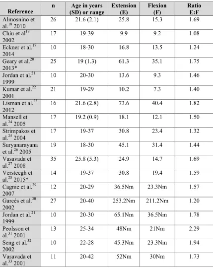
Table 1.1: Selected isometric neck extension and flexion strength values of healthy male subjects.
* Indicates hand-held dynamometry, all others fixed-frame (values in kgf or Nm where indicated). Ratio = extension strength/ flexion strength
Reference n (SD) or range Age in years Extension (E) Flexion (F) Ratio E:F Almosnino et
al.18 2010
26 21.6 (2.1) 25.8 15.3 1.69
Chiu et al19
2002 17 19-39 9.9 9.2 1.08
Eckner et al.17
2014 10 18-30 16.8 13.5 1.24
Geary et al.20
2013* 25 19 (1.3) 61.3 35.1 1.75
Jordan et al.21
1999 10 20-30 13.6 9.3 1.46
Kumar et al.22 2001
21 19-29 10.2 7.3 1.40
Lisman et al.23 2012
16 21.6 (2.8) 73.6 40.4 1.82
Mansell et al.24 2005
17 19.2 (0.9) 18.1 12.1 1.50
Strimpakos et al.25 2004
17 19-37 30.8 23.4 1.32
Suryanarayana
et al.26 2005 19 18-30 45.1 31.4 1.44
Vasavada et al.27 2008
35 25.8 (5.3) 24.9 14.7 1.69
Versteegh et al.28 2015*
14 19-37 30.8 19.4 1.59
Cagnie et al.29 2007
12 20-29 36.5Nm 23.3Nm 1.57
Garcés et al.30 2002
27 20-40 253.2Nm 211.2Nm 1.20
Jordan et al.21
1999 10 20-30 65.1Nm 36.5Nm 1.78
Peolsson et
al.31 2001 13 25-34 48Nm 21Nm 2.29
Seng et al.32
2002 10 22-28 45.3Nm 23.3Nm 1.94
Vasavada et
5
neck strength in the appropriate plane of motion was independently associated with decreased linear and angular head acceleration in that plane (r = 0.42 to r = 0.66). Of all strength values and planes tested, maximum isometric axial rotation strength showed the strongest association with decreased linear and angular head accelerations (r = 0.66, p < .01). These results, along with the conclusions from both Kleiven et al.15 and Viano et al.16 suggest the ability to measure axial rotation strength may help further define the role of neck strength in assessing concussion risk.
Neck function 1.2
The presence of neck pain is indicative of a dysfunction in the neck and a lack of optimum functional performance.34-36 The presence of headache, in some cases, may also be indicative of neck dysfunction.37,38 In a prospective cohort of over 3800 male hockey players aged 11-14, Schneider and colleagues39 showed that pre-season complaints of neck pain was the single highest risk factor for concussion (RR = 1.67, 95% CI 1.15 to 2.41), followed by complaints of headache (RR = 1.47, 95% CI 1.01 to 2.13).While not conclusively causal, this supports the importance of proper neck function in mitigating concussion risk.
Neck dysfunction may also be a source of confounding symptoms that are (mis) diagnosed as concussion. In a prospective cohort study of 15-35 year old male hockey players Hynes and Dickey40 determined that there is a strong association between
whiplash induced neck injuries and symptoms of concussion. Of 183 players, six received a whiplash injury while seven received a concussion injury. Irrespective of the
6
associated disorder symptoms (WAD classification system, 0 = no complaints to IV = most severe), with symptoms ranging from WAD I to III. More recently, Leddy and colleagues41 further confirmed this blend of symptomology between concussion and neck dysfunction. A convenience sample of 128 post-concussion disorder (PCD) patients (individuals who remained symptomatic for more than three weeks after sustaining a head injury) were classified as either cervicogenic/vestibular PCD (normal treadmill test, abnormal cervical/vestibular exam) or physiologic PCD (abnormal treadmill test, normal cervical/vestibular exam). The authors found no statistical method that could adequately distinguish the two groups from each other based on self-reported symptoms and thus concluded that symptoms after head injury do not discriminate between concussion and cervicogenic/vestibular injury.
Stiffness and anticipation 1.3
7
caused nearly a 20% decrease in the peak head angular velocity response to an impulse load in either direction of flexion or extension.
The previously discussed study by Eckner and colleagues17 also demonstrated the effect of anticipatory muscle contraction in mitigating peak head acceleration (both angular and linear) from an impulse load. They calculated a significant decrease in linear and angular acceleration of 12.3% and 9.7% respectively when the subjects anticipated the impulse load versus when the load was unanticipated. The authors concluded the ability to anticipate a hit coming and bracing the neck muscles as a means of lowering a player’s risk of concussion. This conclusion is synonymous with Mihalik and
colleagues44 who examined the relationship between collision type and anticipation level using video footage and instrumented helmets in 16 young hockey players. More
specifically, in medium-intensity head impacts (defined as 50th -75th percentile of Head Impact Telemetry severity profile (HITsp) – a similar metric as the HIC), players with good anticipation prior to collision had significantly less rotational acceleration (1215 rad/s2 95% CI 1112 to 1327 rad/s2) than players who had no anticipation prior to collision (1466 rad/s2 95% CI 1240 to 1731 rad/s2). Thus also suggesting that bracing for impact by contracting the neck muscles helps lower head acceleration in vivo.
Lastly, Schmidt and colleagues45 explored the effects of various muscle
8
stiffness and neuromuscular response are all potentially important protective mechanisms to study.
Neck training 1.4
To date, few studies have examined the effects of strength training on the head kinematic and muscular response to impulse loading. Using a pre-test and posttest
randomized control group design, Mansell and colleagues24 examined 36 collegiate level soccer players’ (17 men, 19 women) head kinematic (head acceleration or displacement), head/ neck stiffness and EMG response (peak activity, muscle activity area and onset latency for sternocleidomastoid (SCM) or upper fibers of trapezius (UFT)) to a weight drop impulse load applied to the head. The intervention group trained for eight weeks on an isotonic resistance-training machine. The training program consisted of three sets of 10 repetitions for each direction of flexion and extension with an intensity ranging from 55% to 70% of the individual’s 10-repetition maximum. Although this intensity is lower than what is suggested for maximizing strength development in trained athletes,46 the authors still showed modest improvements in flexion strength in the males and females (15%) and in female extension strength (22.5%). After completion of the training program head kinematic, head/ neck stiffness and neck EMG response to the impulse load was re-evaluated and compared to the matched control group. Despite the
improvements in neck strength, they found no effects of the training on head kinematic, head/neck stiffness or EMG activity.
9
strength training program on head kinematic (acceleration, displacement and time to peak acceleration) and absolute root mean square EMG (rmsEMG) response to a football dummy tackling drill. The eight-week neck strength-training program was characterized by two to three training sessions per week, in which each session consisted of three sets of 10 repetitions in the flexion, extension and right and left side-flexion directions. The exercises were performed on a 4-way neck machine, a similar apparatus to the one used by Mansell et al.24 but, unlike Mansell et al.,24 this training regimen produced more modest results after the eight weeks of training. The only statistically significant
improvements were found in extension and left side-flexion of 7% and 8% respectively. Lisman et al. also failed to show a significant effect of the training on either the head kinematic or EMG response to the dummy tackling drill. Both of these studies concluded that traditional resistance type training might not be appropriate for improving head kinematic and neuromuscular responses to sudden head accelerations. These authors,23,24 along with Schmidt et al.,45 proposed developing programs that incorporate enhancing neuromuscular control, dynamic stabilization and higher-speed or plyometric training i.e. neuromuscular training.
Neuromuscular training 1.5
10
program in preventing injuries, which includes but is not limited to compliance, duration, frequency and type of training.
Compliance is a significant determining factor for overall effectiveness. Hägglund and colleagues52 showed adolescent female soccer players who demonstrated high-compliance to a NMT program reduced their rate of anterior cruciate ligament injury by 88% when compared to controls (IRR = 0.12, 95% CI 0.01 to 0.85). This is in contrast to the low-compliance group who were not significantly different than their control
counterparts (IRR = 0.77, 95% CI 0.27 to 2.21). Similarly, Steffen and colleagues53 found that in a cohort of young female soccer players, individuals in the high-adherence group (IRR = 0.47, 95% CI 0.15 to 1.43) demonstrated a 72% decrease in the risk of injuries when compared to lower adherence groups (IRR = 1.90, 95% CI 0.88 to 4.09).
Longer duration and greater frequency of NMT is also associated with a lower risk of injury. A meta- and sub-group analysis by Sugimoto and colleagues54 showed that two or more NMT session per-week (OR = 0.35, 95% 0.23 to 0.53) tended to reduce injuries more than only one NMT session per-week (OR = 0.62, 95% CI 0.41 to 0.94). This review also showed that in female athletes who complete NMT sessions that are at least 20 minutes in length have a lower risk of ACL injury (Odds ratio (OR) = 0.35, 95% CI 0.23 to 0.53) when compared to athletes who complete sessions lasting less than 20 minutes (OR = 0.61, 95% CI 0.41 to 0.90).
11
in regards to injury prevention than programs that did not (RR = 0.45, 95% CI 0.35 to 0.57 versus RR = 0.74, 95% CI 0.61 to 0.90). It is also important to note that NMT is very different than passive or static stretching. Passive stretching is a technique that has not been demonstrated to prevent sport injuries.56 Furthermore, it has been demonstrated to decrease stiffness57,58 and has been shown to decrease the rate of force development in muscles.59 As research suggests greater neck stiffness and increasing the rate of force development of the neck muscles to be potentially mitigating factors of head
acceleration,16,42,60 passive stretching of the neck prior to sport participation should likely be avoided.
Although most studies on the effect of NMT on injury prevention only look at lower extremity injuries, there is some support for its use in the upper extremity as well.
Parkkari and colleagues61 demonstrated a decrease in the risk of upper extremity injury in NMT trained young male conscripts with moderate to high baseline fitness (n = 315) compared to the control cohort (n = 298) (adjusted hazards ratio 0.37, 95% CI 0.14 to 0.99).
Conclusion 1.6
12
This thesis proposes to develop an appropriate outcome measure for assessing neck strength in order to allow future research to more fully define the relationship between neck strength and concussion risk. This outcome measure must be safe to administer for both the assessor and the assessed. Second, it must be capable of measuring neck strength along all planes of motion, including axial rotation. Third it should be well described, easy to administer, portable, practical and not dependent on the skill or strength of the assessor. Ideally it should also not require any external equipment for stabilization. Finally, it should be reliable and demonstrate at least preliminary evidence of validity.
The second purpose is to present a method of neck training with a theoretical rationale that is consistent with the state of the literature on how to decrease an
individual’s concussion risk. This method needs to strengthen the neck muscles along all three planes of movement, specifically axial rotation. It should incorporate plyometric or ballistic type contractions. It should enhance dynamic stabilization and increase the rate of force development of the neck muscles. Most importantly, it should accomplish all of these criteria safely, without the risk of the training method causing a concussion.
13
References 1.7
1. McCrory P, Meeuwisse WH, Aubry M, et al. Consensus statement on concussion in sport: the 4th international conference on concussion in sport held in zurich, november 2012. British Journal of Sports Medicine.
2013;47(5):250-258. doi:10.1136/bjsports-2013-092313.
2. Meaney DF, Smith DH. Biomechanics of concussion. Clinics in Sports Medicine. 2011;30(1):19–31–vii. doi:10.1016/j.csm.2010.08.009.
3. Langlois JA, Rutland-Brown W, Wald MM. The epidemiology and impact of traumatic brain injury. Journal of Head Trauma Rehabilitation.
2006;21(5):375-378. doi:10.1097/00001199-200609000-00001.
4. Tator C. Sport Concussion Education and Prevention. Journal of Clinical Sport Psycholology. 2012;6:293-301.
5. Benson BW, McIntosh AS, Maddocks D, Herring SA, Raftery M, Dvorak J. What are the most effective risk-reduction strategies in sport concussion? British Journal of Sports Medicine. 2013;47(5):321-326.
doi:10.1136/bjsports-2013-092216.
6. Benson BW, Hamilton GM, Meeuwisse WH, McCrory P, Dvorak J. Is protective equipment useful in preventing concussion? A systematic review of the literature. British Journal of Sports Medicine. 2009;43(Suppl_1):i56-i67. doi:10.1136/bjsm.2009.058271.
7. Daneshvar DH, Baugh CM, Nowinski CJ, McKee AC, Stern RA, Cantu RC. Helmets and mouth guards: the role of personal equipment in preventing sport-related concussions. Clinics in Sports Medicine. 2011;30(1):145-163. doi:10.1016/j.csm.2010.09.006.
8. Smith AM, Stuart MJ, Greenwald RM, et al. Proceedings from the ice hockey summit on concussion: a call to action. PM&R. 2011;3(7):605-612. doi:10.1016/j.pmrj.2011.05.013.
9. Smith AM, Jorgenson M, Sorenson MC. Hockey Education Program (HEP): a statewide measure of fair play, skill development, and coaching excellence. Journal of ASTM International. 2009;6(4):1-14.
10. Collins CL, Fletcher EN, Fields SK, et al. Neck strength: a protective factor reducing risk for concussion in high school sports. Journal of Primary Prevention. 2014;35(5):309-319. doi:10.1007/s10935-014-0355-2. 11. Strimpakos N, Oldham JA. Objective measurements of neck function. a
2001;6(1):39-51. doi:10.1179/108331901786161573.
12. Strimpakos N. The assessment of the cervical spine. Part 2: Strength and endurance/fatigue. Journal of Bodywork & Movement Therapies.
2011;15(4):417-430. doi:10.1016/j.jbmt.2010.10.001.
13. de Koning CH, Heuvel S, Staal JB, Smits-Engelsman BC, Hendriks EJ. Clinimetric evaluation of methods to measure muscle functioning in patients with non-specific neck pain: a systematic review. BMC Musculoskeletetal Disorders. 2008;9(1):142. doi:10.1186/1471-2474-9-142.
14. Dvir Z, Prushansky T. Cervical muscles strength testing: methods and clinical implications. Journal of Manipulative and Physiological Therapeutics. 2008;31(7):518-524. doi:10.1016/j.jmpt.2008.08.008. 15. Kleiven S. Evaluation of head injury criteria using a finite element model
validated against experiments on localized brain motion, intracerebral acceleration, and intracranial pressure. International Journal of Crashworthiness. 2006;11(1):65-79. doi:10.1533/ijcr.2005.0384. 16. Viano DC, Casson IR, Pellman EJ. Concussion in professional football.
Neurosurgery. 2007;61(2):313-328.
doi:10.1227/01.NEU.0000279969.02685.D0.
17. Eckner JT, Oh YK, Joshi MS, Richardson JK, Ashton-Miller JA. Effect of neck muscle strength and anticipatory cervical muscle activation on the kinematic response of the head to impulsive loads. The American Journal of Sports Medicine. 2014;42(3):566-576. doi:10.1177/0363546513517869. 18. Almosnino S, Pelland L, Stevenson JM. Retest reliability of force-time
variables of neck muscles under isometric conditions. Journal of Athletic Training. 2010;45(5):453-458. doi:10.4085/1062-6050-45.5.453.
19. Chiu TTW, Lo SK. Evaluation of cervical range of motion and isometric neck muscle strength: reliability and validity. Clinical Rehabilitation. 2002;16(8):851-858.
20. Geary K, Green B, Delahunt E. Intrarater reliability of neck strength measurement of rugby union players using a handheld dynamometer. Journal of Manipulative and Physiological Therapeutics. 2013;36(7):444-449. doi:10.1016/j.jmpt.2013.05.026.
21. Jordan A, Mehlsen J, Bülow PM, Ostergaard K, Danneskiold-Samsøe B. Maximal isometric strength of the cervical musculature in 100 healthy volunteers. Spine. 1999;24(13):1343-1348.
23. Lisman P, Signorile JF, Del Rossi G, Asfour S. Investigation of the effects of cervical strength training on neck strength, emg, and head kinematics during a football tackle. International Journal of Sports Science and Engineering. 2012;06(03):131-140.
24. Mansell J, Tierney RT, Sitler MR, Swanik KA. Resistance training and head-neck segment dynamic stabilization in male and female collegiate soccer players. Journal of Athletic Training. 2005;40(4):310-319.
25. Strimpakos N, Sakellari V, Gioftsos G, Oldham J. Intratester and intertester reliability of neck isometric dynamometry. Archives of Physical Medicine and Rehabilitation. 2004;85(8):1309-1316. doi:10.1016/j.apmr.2003.08.104. 26. Suryanarayana L, Kumar S. Quantification of isometric cervical strength at
different ranges of flexion and extension. Clinical Biomechanics. 2005;20(2):138-144. doi:10.1016/j.clinbiomech.2004.10.003.
27. Vasavada AN, Danaraj J, Siegmund GP. Head and neck anthropometry, vertebral geometry and neck strength in height-matched men and women. Journal of Biomechanics. 2008;41(1):114-121.
doi:10.1016/j.jbiomech.2007.07.007.
28. Versteegh T, Beaudet D, Greenbaum M, Hellyer L, Tritton A, Walton D. Evaluating the reliability of a novel neck-strength assessment protocol for healthy adults using self-generated resistance with a hand-held
dynamometer. Physiotherapy Canada. 2015;67(1):58-64. doi:10.3138/ptc.2013-66.
29. Cagnie B, Cools A, De Loose V, Cambier D, Danneels L. Differences in isometric neck muscle strength between healthy controls and women with chronic neck pain: the use of a reliable measurement. Archives of Physical Medicine and Rehabilitation. 2007;88(11):1441-1445.
doi:10.1016/j.apmr.2007.06.776.
30. Garcés GL, Medina D, Milutinovic L, Garavote P, Guerado E. Normative database of isometric cervical strength in a healthy population. Medicine & Science in Sports & Exercise. 2002;34(3):464-470.
31. Peolsson A, Öberg B, Hedlund R. Intra- and inter-tester reliability and reference values for isometric neck strength. Physiotherapy Research International. 2001;6(1):15-26.
32. Seng KY, Peter V, Lam PM. Neck muscle strength across the sagittal and coronal planes: an isometric study. Clinical Biomechanics. 2002;17(7);545-547.
34. Kristjansson E, Treleaven J. Sensorimotor function and dizziness in neck pain: implications for assessment and management. Journal of Orthopaedic & Sports Physical Therapy. 2009;39(5):364-377.
doi:10.2519/jospt.2009.2834.
35. Treleaven J, Jull G, Atkinson L. Cervical musculoskeletal dysfunction in post-concussional headache. Cephalalgia. 1994;14(4):273–9–discussion257. 36. Treleaven J, Jull G, Sterling M. Dizziness and unsteadiness following
whiplash injury: characteristic features and relationship with cervical joint position error. Journal of Rehabilitation Medicine. 2003;35(1):36-43. 37. Bogduk N, Govind J. Cervicogenic headache: an assessment of the evidence
on clinical diagnosis, invasive tests, and treatment. The Lancet Neurology. 2009;8(10):959-968.
38. Jull G, Barrett C, Magee R, Ho P. Further clinical clarification of the muscle dysfunction in cervical headache. Cephalalgia. 1999;19(3):179-185.
doi:10.1046/j.1468-2982.1999.1903179.x/abstract.
39. Schneider KJ, Meeuwisse WH, Kang J, Schneider GM, Emery CA. Preseason reports of neck pain, dizziness, and headache as risk factors for concussion in male youth ice hockey players. Clinical Journal of Sport Medicine. 2013;23(4):267-272. doi:10.1097/JSM.0b013e318281f09f. 40. Hynes LM, Dickey JP. Is there a relationship between whiplash-associated
disorders and concussion in hockey? A preliminary study. Brain Injury. 2006;20(2):179-188. doi:10.1080/02699050500443707.
41. Leddy JJ, Baker JG, Merchant A, Picano J. Brain or strain? Symptoms alone do not distinguish physiologic concussion from cervical/vestibular injury. Clinical Journal of Sport Medicine 2015;25(3): 237-242.
42. Dirisala V, Karami G, Ziejewski M. Effects of neck damping properties on brain response underimpact loading. International Journal for Numerical Methods in Biomedical Engineering. 2011;28(4):472-494.
doi:10.1002/cnm.1480.
43. Simoneau M, Denninger M, Hain TC. Role of loading on head stability and effective neck stiffness and viscosity. Journal of Biomechanics.
2008;41(10):2097-2103. doi:10.1016/j.jbiomech.2008.05.002. 44. Mihalik JP, Blackburn JT, Greenwald RM, Cantu RC, Marshall SW,
Guskiewicz KM. Collision type and player anticipation affect head impact severity among youth ice hockey players. Pediatrics. 2010;125(6):e1394-e1401. doi:10.1542/peds.2009-2849.
Marshall SW. The influence of cervical muscle characteristics on head impact biomechanics in football. The American Journal of Sports Medicine. 2014;42(9):2056-2066. doi:10.1177/0363546514536685.
46. Peterson MD, Rhea MR, Alvar BA. Maximizing strength development in athletes: A meta-analysis to determine the dose-response relationship. Journal of Strength and Conditioning Research. 2004;18(2):377-382. doi:10.1519/R-12842.1.
47. Emery CA, Roy T-O, Whittaker JL, Nettel-Aguirre A, van Mechelen W. Neuromuscular training injury prevention strategies in youth sport: a systematic review and meta-analysis. British Journal of Sports Medicine. 2015;49(13):865-870. doi:10.1136/bjsports-2015-094639.
48. Hübscher M, Zech A, Pfeifer K, Hänsel F, Vogt L, Banzer W.
Neuromuscular training for sports injury prevention: a systematic review. Medicine & Science in Sports & Exercise. 2010;42(3):413-421.
doi:10.1249/MSS.0b013e3181b88d37.
49. Herman K, Barton C, Malliaras P, Morrissey D. The effectiveness of
neuromuscular warm-up strategies, that require no additional equipment, for preventing lower limb injuries during sports participation: a systematic review. BMC Medicine. 2012;10(1):75. doi:10.1186/1741-7015-10-75. 50. Leppänen M, Aaltonen S, Parkkari J, Heinonen A, Kujala UM. Interventions
to prevent sports related injuries: a systematic review and meta-analysis of randomised controlled trials. Sports Medicine. 2014;44(4):473-486. doi:10.1007/s40279-013-0136-8.
51. O'Driscoll J, Delahunt E. Neuromuscular training to enhance sensorimotor and functional deficits in subjects with chronic ankle instability: A
systematic review and best evidence synthesis. Sports Medicine, Arthroscopy, Rehabilitation, Therapy & Technology. 2011;3(1):19. doi:10.1186/1758-2555-3-19.
52. Hägglund M, Atroshi I, Wagner P, Waldén M. Superior compliance with a neuromuscular training programme is associated with fewer ACL injuries and fewer acute knee injuries in female adolescent football players: secondary analysis of an RCT. British Journal of Sports Medicine. 2013;47(15):974-979. doi:10.1136/bjsports-2013-092644.
53. Steffen K, Emery CA, Romiti M, et al. High adherence to a neuromuscular injury prevention programme (FIFA 11+) improves functional balance and reduces injury risk in Canadian youth female football players: a cluster randomised trial. British Journal of Sports Medicine. 2013;47(12):794-802. doi:10.1136/bjsports-2012-091886.
neuromuscular training intervention to reduce anterior cruciate ligament injuries in female athletes: meta- and sub-group analyses. Sports Medicine. 2014;44(4):551-562. doi:10.1007/s40279-013-0135-9.
55. Rössler R, Donath L, Verhagen E, Junge A, Schweizer T, Faude O. Exercise-based injury prevention in child and adolescent sport: a systematic review and meta-analysis. Sports Medicine. 2014;44(12):1733-1748.
doi:10.1007/s40279-014-0234-2.
56. Thacker SB, Gilchrist J, Stroup DF, Kimsey CD JR. The impact of stretching on sports injury risk: a systematic review of the literature. Medicine &
Science in Sports & Exercise. 2004;36(3):371-378. doi:10.1249/01.MSS.0000117134.83018.F7.
57. Morse CI, Degens H, Seynnes OR, Maganaris CN, Jones DA. The acute effect of stretching on the passive stiffness of the human gastrocnemius muscle tendon unit. The Journal of Physiology. 2008;586(1):97-106. doi:10.1113/jphysiol.2007.140434.
58. Ryan ED, Herda TJ, Costa PB, et al. Determining the minimum number of passive stretches necessary to alter musculotendinous stiffness. Journal of Sports Sciences. 2009;27(9):957-961. doi:10.1080/02640410902998254. 59. Young W, Elliott S. Acute effects of static stretching, proprioceptive
neuromuscular facilitation stretching, and maximum voluntary contractions on explosive force production and jumping performance. Research quarterly for exercise and sport. 2001;72(3):273-279.
doi:10.1080/02701367.2001.10608960.
60. Gilchrist I, Storr M, Chapman E, Pelland L. Neck muscle strength training in the risk management of concussion in contact sports: critical appraisal of application to practice. Journal of Athletic Enhancement. 2015;4(2). doi:10.4172/2324-9080.1000195.
2 Evaluating the Reliability of a Novel Neck-Strength Assessment Protocol for Healthy Adults Using Self-Generated Resistance with a
Hand-Held Dynamometer
Introduction 2.1
Assessing muscle strength is a fundamental part of patient care for physiotherapists. The value of a reliable tool to assess muscle strength has been emphasized, both to determine functional impairment and to develop appropriate therapeutic interventions. A review of the literature has shown a lack of neck-strength assessment protocols that evaluate side-flexion and rotation along with flexion and extension and that are both portable and reliable.1 Currently, fixed-frame dynamometry is the most widely recognized method of reliably assessing isometric neck strength. This method uses a large wall or frame-mounted machine with a fixed base, which are expensive and
generally impractical for most clinical settings.2 In contrast, hand-held dynamometers are portable, relatively inexpensive, and easy to use. Hand-held dynamometry has been shown to be an objective and reliable measure of strength for several different
movements of the extremities in healthy adults.3-6 Normative reference values have also been determined for these various movements. Although previous research has used hand-held dynamometry to assess neck strength, a review article1 noted a lack of consistency in the methodology and description of the testing procedure and a lack of normative values. The number of articles reporting comprehensive strength
measurements in all planes of the neck is also limited. Of particular note is the difficulty in clinical assessment of neck rotation strength,7 which has traditionally been limited to clinically inaccessible lab-based measurement equipment.
One of the challenges of using hand-held dynamometry to assess muscle strength is that results are influenced by the strength of the tester, which may compromise
reliability.8 If the tester is significantly weaker than the person being tested, the results will only be as high as the force the tester is capable of generating; even if the tester is able to generate sufficient resistance, the stronger the person being tested, the more difficult it becomes for the tester to generate this resistance along the proper vector in a consistent and safe manner, which further decreases the reliability of the results. A person may also be apprehensive about providing full resistance against someone pushing on the side of his or her head.
Our study therefore provides a standardized and functional isometric strength-testing protocol that allows assessment of strength in all planes of the neck, including rotation, using self-generated resistance and a hand-held dynamometer. Given that the resistance is self-generated through the upper kinetic chain (including the shoulder, elbow, wrist, and hand), the test inherently assesses the neck up to the strength limit of the upper kinetic chain. We believe that simultaneous functional assessment of strength about the neck and upper kinetic chain could function as a useful clinical evaluation for people with neck pain and may have potential as a prognostic tool after neck injuries.
Methods 2.2
2.2.1 Participants
Participants were recruited for this study from the Health and Rehabilitation Sciences programme and the Master of Physical Therapy programme at Western University, as well as from the university community through word of mouth and electronic recruitment (letter of information posting on a Facebook class page; class group email). Volunteers were eligible for inclusion if they were healthy adults aged between 18 and 60 years; able to speak and understand English at a conversational level; free of neck, shoulder, elbow, and wrist pain (self-reported); and able to pass the cervical screening protocol (see Appendix A) with no positive results.
Table 2.1: Participant characteristics
Sex$ n$ Mean$(SD)$age,$y$ Age$Range$y$
Men$ 14! 25.29!(5.41)! 19*37!
Women$ 16! 23.94!(1.29)! 23*28!
2.2.2 Testing protocol
After providing written informed consent, potentially eligible participants were screened by a group of four physiotherapy student examiners (in their 2nd year of the MPT programme), who used a screening protocol to identify any gross cervical
consists of a plastic unit housing a force gauge and a soft, cushioned pad that is applied to the long bone of the joint to be tested, as shown in Figure 2.1.
To measure neck strength, participants were seated comfortably on a stool with their feet flat on the floor. They sat with no back or arm rests to prevent bracing the trunk against a chair. One of the four physiotherapy students then guided each participant through the testing procedure. For calibration purposes, the isometric peak force
Figure 2.1: MicroFET 2TM dynamometer
All test positions were performed with the neck in neutral; proper positioning was augmented by the use of a mirror. In each ‘‘make’’ test position, the participants were instructed to build up to their maximum cervical muscle force over three seconds,
maintaining the static neck position (a ‘‘make’’ test is an isometric strength test in which the tester matches the maximum resistance produced by the testee, maintaining the length of the muscle, and a ‘‘break’’ test is an eccentric test in which the tester exceeds the maximum resistance produced by the testee and causes lengthening of the muscle). The peak force produced in Trial 1 for each test position was recorded. Participants could stop the test at any point during the assessment and were instructed to stop should any pain or dizziness arise. On completing the protocol, participants rested comfortably in a
Figure 2.2: Test positions: Calibration (A), forward flexion (B), extension (C), side-flexion (D), side-side-flexion with rotation (E), axial rotation (F).
strength-testing protocol. Finally, participants returned to the lab after 6–8 days for a second visit to determine inter-session reliability. This second visit was no longer than 10 minutes and consisted of a single trial using the same data-collection process as in the first testing session (Trial 3).
2.2.3 Data analysis
The statistic of interest was the intra-class correlation coefficient type 2,1 (ICC [2,1], absolute). We chose this statistic because it assumes the same group of raters
(participants themselves) randomly sampled from the population of possible raters (random effects) and allows for generalizability beyond this study for other participants using themselves as raters. For clinical and research purposes,10 we expected an ICC (2,1), absolute, of at least 0.8, with 95% confidence that the true value is greater than 0.4. Using these values and a formula presented by Walter and colleagues,11 we calculated that a sample size of 27 would provide 80% power for detecting a true difference between 0.8 and 0.4 where one exists. Therefore, we set a target sample size of 30 to ensure sufficient power for our study. To determine the level of reliability, we adapted the scheme previously reported by Meyers and Blesh,12 who defined the degrees of reliability based on ICC (2,1), absolute, values as follows: 0.90–0.99, high reliability; 0.80–0.89, good reliability; 0.70–0.79, fair reliability; and < 0.69, poor reliability.
confident that a true change has occurred. Bland–Altman plots with 95% limits of agreement were produced for the various test positions across trials (Appendix B).
Results 2.3
All participants completed the full test procedure; none reported experiencing any discomfort during or after testing.
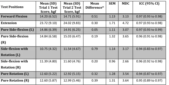
Table 2.2: Intra-session Retest Reliability of Neck Strength Using a Handheld Dynamometer in a Healthy Population
Test%Positions% Trial%1%Test%Mean%(SD)% Score,%kgf%
Mean%(SD)% Trial%2%Test%
Score,%kgf%
Mean%
Difference*% SEM% MDC% ICC%(95%%CI)%
Forward%Flexion% 14.20&(6.52)& 14.71&(5.91)& 0.51& 1.13& 3.13& 0.97&(0.93&to&0.98)& Extension% 23.72&(9.10)& 24.02&(9.83)& 0.30& 1.71& 4.72& 0.97&(0.93&to&0.98)& Pure%SideEflexion%(L)% 14.86&(6.39)& 14.91&(6.25)& 0.05& 1.11& 3.07& 0.97&(0.93&to&0.99)& Pure%SideEflexion%
(R)%
14.84&(6.58)& 15.03&(6.47)& 0.19& 1.32& 3.65& 0.96&(0.91&to&0.98)&
SideEflexion%with% Rotation%(L)%
10.75&(4.32)& 11.54&(4.67)& 0.79& 1.14& 3.17& 0.94&(0.83&to&0.97)&
SideEflexion%with% Rotation%(R)%
11.39&(4.80)& 11.60&(4.76)& 0.20& 0.96& 2.66& 0.96&(0.92&to&0.98)&
Pure%Rotation%(L)% 12.60&(5.22)& 12.92&(5.15)& 0.32& 1.28& 3.54& 0.94&(0.87&to&0.97)& Pure%Rotation%(R)% 12.60&(5.87)& 12.99&(5.46)& 0.39& 1.31& 3.64& 0.95&(0.89&to&0.97)&
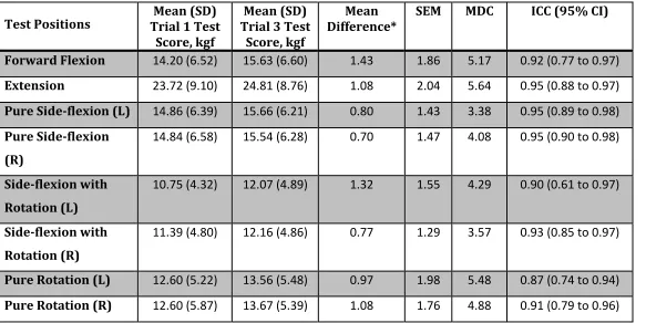
Table 2.3: Inter-session Retest Reliability of Neck Strength Using a Handheld Dynamometer in a Healthy Population
Test%Positions%%% Trial%1%Test%Mean%(SD)% Score,%kgf%
Mean%(SD)% Trial%3%Test%
Score,%kgf%
Mean%
Difference*% SEM% MDC% ICC%(95%%CI)%
Forward%Flexion% 14.20&(6.52)& 15.63&(6.60)& 1.43& 1.86& 5.17& 0.92&(0.77&to&0.97)& Extension% 23.72&(9.10)& 24.81&(8.76)& 1.08& 2.04& 5.64& 0.95&(0.88&to&0.97)& Pure%SideEflexion%(L)% 14.86&(6.39)& 15.66&(6.21)& 0.80& 1.43& 3.38& 0.95&(0.89&to&0.98)& Pure%SideEflexion%
(R)%
14.84&(6.58)& 15.54&(6.28)& 0.70& 1.47& 4.08& 0.95&(0.90&to&0.98)&
SideEflexion%with% Rotation%(L)%
10.75&(4.32)& 12.07&(4.89)& 1.32& 1.55& 4.29& 0.90&(0.61&to&0.97)&
SideEflexion%with% Rotation%(R)%
11.39&(4.80)& 12.16&(4.86)& 0.77& 1.29& 3.57& 0.93&(0.85&to&0.97)&
Pure%Rotation%(L)% 12.60&(5.22)& 13.56&(5.48)& 0.97& 1.98& 5.48& 0.87&(0.74&to&0.94)& Pure%Rotation%(R)% 12.60&(5.87)& 13.67&(5.39)& 1.08& 1.76& 4.88& 0.91&(0.79&to&0.96)&
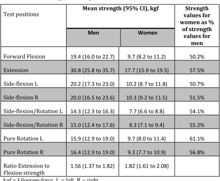
Table 2.4: Mean strength values by sex of trial 1.
Test%positions! Mean!strength!(95%!CI),!kgf%! values!for!Strength! women!as!%!
of!strength! values!for!
men!
Men$ Women!
Forward%Flexion% 19.4!(16.0!to!22.7)! 9.7!(8.2!to!11.2)! 50.2%! Extension% 30.8!(25.8!to!35.7)! 17.7!(15.9!to!19.5)! 57.5%! Side3flexion%L% 20.2!(17.3!to!23.0)! 10.2!(8.7!to!11.8)! 50.7%! Side3flexion%R% 20.0!(16.5!to!23.6)! 10.3!(9.2!to!11.5)! 51.5%! Side3flexion/Rotation%L% 14.3!(12.3!to!16.3)! 7.7!(6.6!to!8.8)! 54.1%! Side3flexion/Rotation%R% 15.0!(12.4!to!17.6)! 8.3!(7.1!to!9.4)! 55.2%! Pure%Rotation%L% 15.9!(12.9!to!19.0)! 9.7!(8.0!to!11.4)! 61.1%! Pure%Rotation%R% 16.4!(12.9!to!19.0)! 9.3!(7.7!to!10.9)! 56.8%! Ratio%Extension%to%
Flexion%strength% 1.56!(1.37!to!1.82)! 1.82!(1.61!to!2.08)! ! kgf = kilogram-force, L = left, R = right
Average neck strength in Trial 1 ranged from 14.3 to 30.8 kgf for men and from 7.7 to 17.7 kgf for women; women’s mean strength ranged from 50.2% to 61.1% of men’s. The mean extension-to-flexion ratio in Trial 1 was 1.56 for men and 1.82 for women (see Table 2.4). Although Bland–Altman plots are best used to compare different
Discussion 2.4
Our results are consistent with reliability findings from studies using large
fixed-frame dynamometers to assess isometric neck strength.13-16
For instance, Peolsson and
Öberg13
examined the intra- and inter-tester reliability of isometric neck strength in 30
healthy participants using a David Back Clinic (DBC 140), a large fixed-frame
dynamometer, and found high ICCs (ranging from 0.85 to 0.97) for the tested movements
of flexion, extension, and lateral flexion. Chiu and Lo14
also studied the reliability of
isometric neck strength using another large fixed-frame dynamometer, the Multi Cervical
Rehabilitation Unit. Their results demonstrated that intra-session test–retest reliability
was high for all tested positions of neck flexion (ICC = 0.98), neck extension (ICC =
0.98), left side-flexion (ICC = 0.97), and right side-flexion (ICC = 0.95), all values very
similar to those found in our study (flexion = 0.97, extension = 0.97, left side-flexion =
0.97, right side-flexion = 0.96). Comparing our findings with those of Peolsson and
Öberg13
and Chiu and Lo14
illustrates that the same level of reliability achieved with
large, expensive fixed-frame dynamometry can be achieved using the protocol presented
here and the more cost-effective MicroFET 2TM
.
Although evaluating validity was not a goal of our study, it is notable that we found
ratios of extension to flexion strength and comparative strength of men and women that
are in line with those found in studies using fixed-frame dynamometry. Using various
fixed-frame dynamometry systems, prior studies have found women to be 40%–70% 7,13,15-17 as strong as men; our study found a range of 50.2%–57.5% for the same movements. In
those fixed-frame dynamometry studies that reported the extension-to-flexion ratio,
men.7,13,15-17
Although the values in our study are consistent with those of fixed-frame
dynamometry, further studies are needed to formally test the validity of the protocol
presented here.
Our protocol avoids a known shortfall of using hand-held dynamometry—the
influence of the tester’s strength on the reliability of the test8—by having the person being tested provide the resistance. It has also been suggested that measuring neck
strength using a break test in people with neck pain is difficult because participants fear
evoking pain during the assessment.1
Our study suggests that assessing neck strength
using a closed-kinetic-chain make test is likely to reduce participants’ fears during
maximal strength testing because the participant’s own hand is providing the resistance to
neck movement. This consideration will be especially important when assessing
individuals with neck dysfunction. Our protocol allows participants to stop quickly at any
time if they experience pain or discomfort without first informing the therapist, which
makes this test inherently safer and easier to administer.
2.4.1 Limitations
Our study has several limitations. First, our convenience sample of 30 participants
had a very narrow age range (19–37 years for men, and only 23–28 years for women);
future studies should include a sample with a larger age range. Second, the study assessed
a healthy cohort of participants, which limits its applicability to a population with
pathology. We intend to continue collecting normative values for comparison purposes in
future clinical studies. Furthermore, future directions will investigate this protocol as a
neck injuries.
The proposed assessment protocol also has some limitations. To perform the test, the
participant must have sufficient range of motion of the shoulder, elbow, and wrist; the
protocol cannot be used effectively if any of these are lacking. The participant must also
be able to generate sufficient force to overcome the tested neck movement. This
limitation is addressed by having participants perform a calibration test consisting of
compressing the dynamometer between their two hands, without interlocking their
fingers, in front of their head. For example, if the participant is able to generate 50 kgf for
the calibration test and only 18 kgf as a maximum for the side-flexion and rotation
components, then it is arguably safe to say that the strength of the side-flexion or rotation
movement is the value found with that test. If, however, the calibration value is 18 kgf
and the side-flexion and or rotation test also measures approximately 18 kgf, then it is
possible that the maximum force of those movements was not determined because the
participant may not have been able to generate enough force to overcome his or her own
neck strength.
Conclusion 2.5
Our study provides a standardized protocol for assessing neck strength in all planes
using a MicroFET 2TM
. The results suggest that all five test positions of the neck and
upper-quadrant strength assessment procedure can be performed using hand-held
dynamometry with good to high reliability. Moreover, self-generated resistance using a
MicroFET 2TM to measure neck strength could be a reliable evaluation procedure for a
Key Messages 2.6
What is already known on this topic 2.6.1.1
Reliable methods of assessing neck strength currently exist, but these methods have several limitations. Many of them require large, expensive fixed-frame dynamometry systems that are not practical for use in most clinics. Protocols that use portable hand-held dynamometry lack standardization and depend on the therapist’s being stronger than the patient. They also commonly rely onbreak tests that can cause apprehension, pain, and safety concerns for the participant or patient.
What this study adds 2.6.1.2
References 2.7
1. de Koning CH, Heuvel S, Staal JB, Smits-Engelsman BC, Hendriks EJ.
Clinimetric evaluation of methods to measure muscle functioning in patients with non-specific neck pain: a systematic review. BMC Musculoskeletetal Disorders. 2008;9(1):142. doi:10.1186/1471-2474-9-142.
2. Dvir Z, Prushansky T. Cervical muscles strength testing: methods and clinical implications. Journal of Manipulative and Physiological Therapeutics.
2008;31(7):518-524. doi:10.1016/j.jmpt.2008.08.008.
3. Kelln BM, McKeon PO, Gontkof LM, Hertel J. Hand-held dynamometry: reliability of lower extremity muscle testing in healthy, physically active,young adults. Journal of Sport Rehabilitation. 2008;17(2):160-170.
4. Kolber MJ, Beekhuizen K, Cheng M-SS, Fiebert IM. The reliability of hand-held dynamometry in measuring isometric strength of the shoulder internal and external rotator musculature using a stabilization device. Physiotherapy Theory and
Practice. 2007;23(2):119-124. doi:10.1080/09593980701213032.
5. Van Meeteren J, Mens J, Stam HJ. Reliability of strength measurement of the hip with a hand-held dynamometer in healthy women. European journal of physical medicine & rehabilitation. 1997;7(1):17-20.
6. Schaubert KL, Bohannon RW. Reliability and validity of three strength measures obtained from community-dwelling elderly persons. Journal of Strength and Conditioning Research. 2005;19(3):717-720. doi:10.1519/R-15954.1. 7. Strimpakos N, Sakellari V, Gioftsos G, Oldham J. Intratester and intertester
reliability of neck isometric dynamometry. Archives of Physical Medicine and Rehabilitation. 2004;85(8):1309-1316. doi:10.1016/j.apmr.2003.08.104. 8. Wikholm JB, Bohannon RW. Hand-held dynamometer measurements: tester
strength makes a difference. Journal of Orthopaedic & Sports Physical Therapy. 1991;13(4):191-198. doi:10.2519/jospt.1991.13.4.191.
9. Tong HC, Haig AJ, Yamakawa K. The Spurling test and cervical radiculopathy. Spine. 2002;27(2):156-159.
10. Weir JP. Quantifying test-retest reliability using the intraclass correlation coefficient and the SEM. Journal of Strength and Conditioning Research. 2005;19(1):231-240. doi:10.1519/15184.1.
12. Meyers CR, Blesh TE. Measurement in Physical Education. Ronald Press Company; 1974.
13. Peolsson A, Öberg B, Hedlund R. Intra- and inter-tester reliability and reference values for isometric neck strength. Physiotherapy Research International. 2001;6(1):15-26.
14. Chiu TTW, Lo SK. Evaluation of cervical range of motion and isometric neck muscle strength: reliability and validity. Clinical Rehabilitation. 2002;16(8):851-858.
15. Garcés GL, Medina D, Milutinovic L, Garavote P, Guerado E. Normative database
of isometric cervical strength in a healthy population. Medicine & Science in Sports & Exercise. 2002;34(3):464-470.
16. Vasavada AN, Li S, Delp SL. Three-dimensional isometric strength of neck muscles in humans. Spine. 2001;26(17):1904-1909.
17. Cagnie B, Cools A, De Loose V, Cambier D, Danneels L. Differences in isometric neck muscle strength between healthy controls and women with chronic neck pain: the use of a reliable measurement. Archives of Physical Medicine and
3 Examining the validity of a novel neck strength assessment tool
Introduction 3.1
It is estimated that there are up to 3.8 million sports and recreation-related
concussions each year in the United States.1 Given this high incidence, healthcare
workers are looking for simple and valid methods of assessment and screening that may
help establish individuals’ concussion risk.2 A pilot study assessing anthropometric
measurements of over 6,600 high school athletes suggests that neck flexion, extension
and lateral flexion strength may be a protective factor in reducing concussion risk.3
Specifically, for every one-pound increase in neck strength, odds of concussion decreased
by 5%. Since axial rotation strength was not measured, it is not known if it is also
associated with concussion risk. Given that concussions are caused by multi-planar linear
and rotation forces,436 it may be of benefit to measure neck strength in all primary planes
of motion (flexion/extension, lateral flexion and axial rotation).7,8 A systematic review by
Dvir and Prushansky9 found only 6 of 16 methods of assessing neck strength assessed
axial rotation strength. Strimpakos10 has suggested axial rotation strength is not
frequently included because of the practical difficulty in assessing this movement. The
methods that do exist are neither portable nor practical. An accurate and reliable means of
assessing neck strength that includes all three primary planes of movement may help
further define the role of the neck muscles in concussion risk, and provide additional
There have been a number of studies that have examined isometric neck strength
that have led to four review papers evaluating these approaches.9312 Each of these four reviews concluded that there is currently no gold standard for neck strength assessment.
Most studies used a form of fixed frame dynamometry to assess neck strength. These
devices are large and may be cost-prohibitive for most smaller or non-specialized clinics.
Other approaches used custom-built machines that are not widely available.
Problematically, the use of different measurement apparatuses has led to vastly different
normative strength values for samples from similar populations, in some cases differing
by 10-fold between studies.13,14 Even the ratios of extension strength to flexion strength (E:F) within these different studies range from values indicating extension is 10% to over
100% greater than flexion.13,15 Inconsistent methods and results make comparisons between studies and defining translatable normative strength values difficult. However,
Strimpakos10 points out that neck extensors can produce higher forces than flexion or lateral flexion muscles and that this trend can be used as an indicator of valid results. It is
also expected that strength values from the right and left side should be symmetrical (i.e.
side-flexion, rotation).9
Other studies have used operator-applied hand-held dynamometry and portable
strain gauges as a method of assessing neck strength.16319 However, these approaches also
have limitations. For example, Wikholm and Bohannon20 found that inter-rater reliability was influenced by the strength difference between the examiner and the subject; weaker
examiners demonstrated less consistency in scores. This becomes particularly challenging
Since these reviews, a method of assessing neck strength using a hand-held dynamometer has been presented that addresses these shortcomings. Versteegh and colleagues21 proposed a method of evaluating neck strength using a hand-held dynamometer and self-generated resistance by the subject. By having the subjects generate their own resistance, it can be argued that there is an element of added safety insofar as resistance applied to the neck can be rapidly modulated. This method also eliminates the need for external stabilization as the subjects’ use their own hand and arm or arms to generate the resistance, which should naturally engage the torso for stability. As a result, this test is probably best conceptualized as an evaluation of overall kinetic chain activity influenced most strongly by neck strength. Notably, this method also provides an easy means of assessing neck rotation strength with a hand-held device, which to our knowledge has not been previously examined.
3.1.1 Hypotheses
1. Face validity: The E:F strength ratio obtained from this new testing method should be greater than 1 and within the range of ratios obtained from other tools reported in the literature.10 In accordance with published literature, extension strength should also be significantly stronger than each of the unilateral strength tests. Strength values for flexion, side-flexion/rotation and axial rotation should not be significantly different between the right and left sides in healthy subjects.9
2. Known Groups validity: A sample of male football players who train with a neck strengthening machine as part of their standard training protocol will show significantly higher peak isometric neck strength on the new protocol than will a group of age- and sex-matched non-football players who do not routinely train neck strength. When ability to discriminate between the two groups (sensitivity vs. 1-specificity) is plotted using a Receiver Operating Characteristic (ROC) curve, the area under the curve should be statistically greater than parity (0.5) for all directions tested. 3. Convergent Validity: the peak EMG activity of the upper fibers of
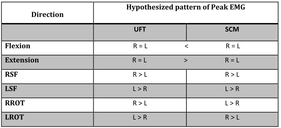
Table 3.1: Anticipated pattern of EMG activity by direction
Direction! Hypothesized!pattern!of!Peak!EMG!
! UFT$ SCM$
Flexion! !R!=!L!!!!!!!!!!!!!!!!!!!!!!<!!!!!!!!!!!!!!!!!!!!!!R!=!L!
Extension! R!=!L!!!!!!!!!!!!!!!!!!!!!!>!!!!!!!!!!!!!!!!!!!!!!R!=!L!
RSF! R!>!L! R!>!L!
LSF! L!>!R! L!>!R!!
RROT! R!>!L! L!>!R!
LROT! L!>!R! R!>!L!
UFT = upper fibers of trapezius, SCM = sternocleidomastoid, R = right side muscle, L = left side muscle, RSF=right side-flexion, LSF = left side-flexion, RROT = right rotation, LROT = left rotation.
Methods 3.2
This was a cross-sectional observational study of two known groups.
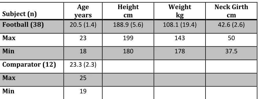
3.2.1 Participants
Participants were recruited for the football cohort (FC) from the spring camp
roster of the Western University Varsity Football Team. The age and sex-matched
comparator cohort (CC) were drawn from the Health and Rehabilitation Sciences
program and the university community at Western University, London, Ontario, Canada.