2 8 0 6 0 9 8 0 3 4
ACTIVATION OF NEUTROPHILS, PLATELETS AND
THE COAG ULATION SYSTEM IN INTERM ITTENT
CLAUDICATION
PAUL S. G. EVERS
Rose Cottage Shop Lane East Lavant
Chichester West Sussex
P 0180B A
Local Supervisor: Professor C N McCollum
University Department of Surgery Withington Hospital
Manchester
University of London Supervisor:
'ACCESSION iVUMBER
Dr A Goodall
Department of Chemical Pathology Royal Free Hospital
London Currently:
Division of Chemical Pathology University of Leicester
The Glenfield Hospital Trust Leicester
ProQuest Number: U642168
All rights reserved
INFORMATION TO ALL USERS
The quality of this reproduction is dependent upon the quality of the copy submitted.
In the unlikely event that the author did not send a complete manuscript and there are missing pages, these will be noted. Also, if material had to be removed,
a note will indicate the deletion.
uest.
ProQuest U642168
Published by ProQuest LLC(2015). Copyright of the Dissertation is held by the Author.
All rights reserved.
This work is protected against unauthorized copying under Title 17, United States Code. Microform Edition © ProQuest LLC.
ProQuest LLC
789 East Eisenhower Parkway P.O. Box 1346
ABSTRACT
Intermittent claudication is a common condition, affecting 2-5% o f the population. The prognosis for the affected limb is relatively benign, but morbidity and mortality from associated cardiovascular events is high, with up to 50% of patients suffering a myocardial infarction or stroke in the five years following diagnosis. Multivariate analysis has suggested that claudication itself may be a risk factor for these events.
The pathophysiologies of claudication and cardiovascular events are reviewed, with particular emphasis on the role o f neutrophils, platelets and the coagulation system. From this we developed a hypothesis that claudication may activate, or increase the potential to be activated of, neutrophils, platelets and the coagulation system; and that this may contribute to the increased cardiovascular morbidity and mortality seen in claudicants. The aim of this work was to investigate activation, and, in particular, the activation potential o f these elements in claudicants, before and after exercise.
CO NTENTS
CHAPTER ONE: INTRODUCTION
SECTION ONE: INTERMITTENT CLAUDICATION
1.1.1 Intermittent Claudication
a) Definition
b) Historical Perspectives c) Aetiology
d) Epidemiology
1.1.2 Natural History
a) Local disease b) Systemic disease
c) Possible mechanisms for increased morbidity and mortality d) Intermittent claudication as an independent risk factor
1.1.3 Pathophysiology O f Excess Cardiovascular Morbidity And Mortality In Intermittent Claudication
a) Atherosclerosis
b) Mechanisms leading to cardiovascular events c) Pathology of cardiovascular events
1.1.4 Pathophysiology O f Intermittent Claudication
a) Physiology of clinical symptoms b) Ischaemia- reperfusion
SECTION TWO: NEUTROPHILS, PLATELETS AND THE COAGULATION SYSTEM IN ATHEROSCLEROSIS AND INTERMITTENT CLAUDICATION
1.2.1 Neutrophils In Atherosclerosis And Intermittent Claudication
1.2.1.1 Role o f white blood cells in atherosclerostic disease a) Atherogenesis
b) Progression and complications o f atherosclerosis
1.2.1.2 Neutrophils in intermittent claudication a) Population/ Static studies
b) Dynamic studies
1.2.2 Platelets In Atherosclerosis And Intermittent Claudication 1.2.2.1 Role o f platelets in atherosclerostic disease
b) Progression and complications of atherosclerosis c) Platelet activation in cardiovascular disease d) Problems of cause and effect
e) Evidence for causality
12.2.2 Platelets in intermittent claudication a) Population/ Static studies
b) Dynamic studies
1.2.3 Coagulation System In Atherosclerosis And Interm ittent Claudication
a) Haemostatic system b) Coagulation system
1.2.3.1 Role of the coagulation system in atherosclerostic disease a) Atherogenesis
b) Progression and complications o f atherosclerosis 1.2.3.2 The coagulation system in intermittent claudication
a) Population/ Static studies b) Dynamic studies
SECTION THREE: SUMMARY, HYPOTHESIS AND AIMS
1.3.1 Summary
1.3.2 Hypothesis
1.3.3 Aims
CHAPTER
TWO:
NEUTROPHIL
ACTIVATION
IN
INTERMITTENT CLAUDICATION
n. 1 Research Question: Does claudication activate or prime neutrophils?
n.2 Method
a) Patients and controls
b) Study design - Exercise protocol
c) Laboratory methods - White cell counts and differentials - Plasma Elastase
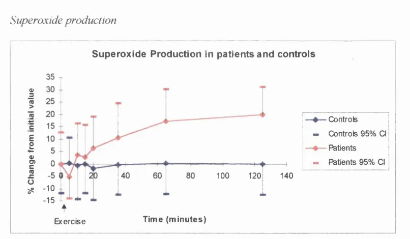
- Superoxide release
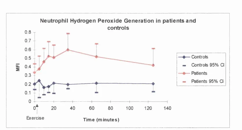
- Hydrogen peroxide generation - Thromboxane B]
n.3
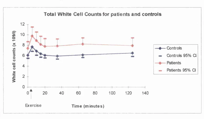
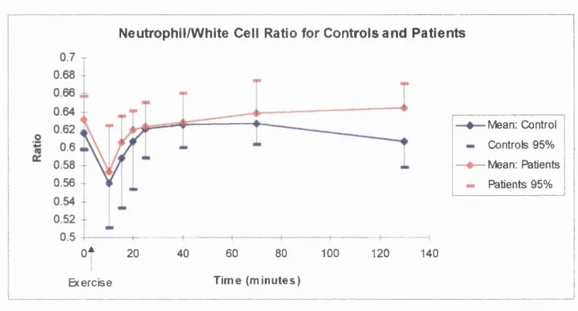
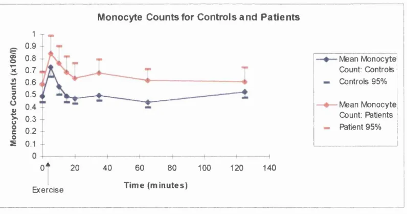
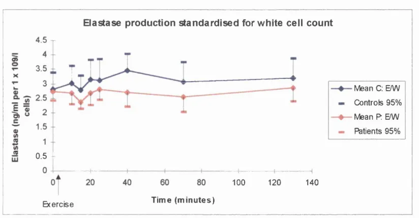
Resultsa) White cell counts and differentials b) Plasma Elastase
c) Superoxide release
d) Hydrogen peroxide generation e) Thromboxane 8 %
n.4
Discussiona) Implications o f white cell counts and differential results b) Implications of elastase results
c) Neutrophil activation results
d) Mechanism of neutrophils activation e) Implication of thromboxane results
f) Implications o f increased neutrophil activation after claudication
CHAPTER THREE:
PLATELET
AND
COAGULATION
SYSTEM ACTIVATION IN INTERMITTENT CLAUDICATION
III, 1 Research Question; Does claudication activate or prime platelets and/or the coagulation system?
in.2 Methods
in.2.1 Flow Cytom etry
a) Validity o f Flow cytometry in assessing platelet activation b) Principles of the technique
c) Whole blood assay
d) Standardisation of method
in.2.2 G eneral a) Subjects b) Study design
c) Measurements and laboratory techniques i. Physical measurements
ii. Microalbuminuria iii. Platelet size iv. Platelet markers
- Surface glycoproteins - Plasma markers V. Platelet aggregation
vi. Markers of coagualtion activation d) Statistical analysis
III. 3 Results
c)Platelet size d)Platelet markers
Surface glycoproteins Plasma markers e)Platelet aggregation
f)Markers o f coagulation activation ni.4 Discussion
a) Study design
b) Physical measurements c) Microalbuminuria d) Platelet count and size
e) Markers o f platelet activation f) Coagulation Markers
CHAPTER FOUR:
DISCUSSION,
CONCLUSION
AND
FUTURE WORK
IV. 1 Discussion IV. 2 Conclusion
IV. 3 Future Work
REFERENCES
APPENDIX ONE:
DataLIST OF ABREVEATIONS
5-HT 5-Hydroxy-tryptamine
ABPI Ankle Brachial Pressure Index ADP Adenosine Di-Phosphate ANOVA Analysis O f Variance
APTT Activated Partial Thromboplastin Time ATP Adenosine Tri-Phosphate
BSA Bovine Serum Albumin Cl Confidence Intervals PTG Beta-Thromboglobulin DCFH-DA Dichlorofluoroscin Diacetate DNA Deoxyribonucleic acid ECG Electrocardiogram
EDTA Ethylene Diamine Tetra Acetic Acid ELISA Enzyme-linked Immunosorbent assay EDGF Endothelial cell Derived Growth Factor FDPs Fibrin Degradation Products
FpA Fibrinopeptide A
FITC Fluorescein Isothiocyanate Fb Fibrinogen
GPRP Glycyl-propyl-L-arginyl-L-proline H2O2 Hydrogen Peroxide
IHD Ischaemic Heart Disease IGF-1 Insulin-like Growth Factor-1 IgG Immunoglobulin G
LDL Low Density Lipoprotein Lp(a) Lipoprotein (a)
LTB4 Leucotriene B4
MI Myocardial Infarction
MDGF Macrophage Derived Growth Factor MFI Maximum Fluorescence Intensity MoAb Monoclonal Antibody
OPD o - Phenylenediamine PAF Platelet Activation Factor PDGF Platelet Derived Growth Factor PAI-1 Plasminogen Activation Inhibitor-1 PF4 Platelet Factor 4
PGI2 Prostacyclin
PRT Pressure Recovery Time PRP Platelet Rich Plasma PPP Platelet Poor Plasma PBS Phosphate Buffered Saline PMA Phorbol Myristate Acetate PVD Peripheral Vascular Disease TAT Thrombin-Anti-Thrombin TIA Transient Ischaemic Attack SO Superoxide
t-PA Tissue Plasminogen Activator TxÀ2 Thromboxane A2
TxBz Thromboxane B2
u-PA Urinary Plasminogen Activator vWF von Willebrand Factor
C H A PT E R ONE:
IN TRO DUC TIO N
SECTION ONE: INTERMITTENT CLAUDICATION
1 .1.1 INTERM ITTENT CLAUDICATION
a) Definition Intermittent claudication describes a clinical syndrome of cramp-like muscular pain, usually affecting the lower limbs, induced by exercise. This increases steadily causing a limp (Latin, claudicare = to limp) and eventually the patient is forced to stop. The pain is relieved by a few minutes rest, but if exercise is resumed the pain recurs after a similar distance. Commonly the pain occurs in the calf muscles but is also described in the thigh and buttocks. The essential features of claudication are: the pain is always experienced in the functional muscles group; is reproducibly precipitated by a consistent amount of exercise and is relieved by rest\
c) Aetiology Claudication develops whenever blood flow to the exercising muscle mass is unable to meet the increased metabolic demands. It is caused by a fixed arterial occlusion, or significant stenosis, proximal to the affected muscle bed. At rest the blood supply is adequate to meet the metabolic demands of the tissue. However, upon exercise this demand rises markedly, the normal exercise-induced increase in muscle blood flow is impaired and cannot meet the increased need. As a result the muscle becomes hypoxic, the interstitial pH falls and waste products build up resulting in pain. The most common cause of this impaired blood flow is atherosclerosis, which will be discussed later. Less common causes include thromboangiitis obliterans, or Buerger’s disease, vasculitis, fibrosis (retroperitoneal or due to radiation), cystic advential disease, popliteal entrapment syndrome or late effects of arterial trauma^.
proven lower extremity arterial occlusion denied claudication despite extensive questioning/ Hence, the symptom of claudication is relative: the patient must have a significant arterial stenosis or occlusion and exercise sufficiently to induce muscle ischaemia/ The most commonly used questionnaire to for intermittent claudication was devised in 1962 by Professor G Rose at the London School o f Hygiene and Tropical Medicine^^. Despite only moderate sensitivity it remains the most valid way of making international comparisons of the prevalence o f intermittent claudication'^’^^’^'^. The reported prevalence of claudication varies widely. A review of international population studies revealed a range from 0.9% in N America to 6.9% in Moscow in men aged 40 - 60 years. This may represent some geographical difference, but problems with translation, age stratification and health worker effects make comparisons difficult"^. Reported prevalences of 0.9% in sedentary office workers compared with 2.1% in agricultural workers^ suggests an occupational component.
Prevalence also varies with age^’^^’^^. McDaniel and Cronenwett found claudication occurred in about 1.8% o f people under 60 years, in 3.7% of those between 60 and 70 years, and in 5.2% of those over 70 years^^. These figures have been confirmed in a further international r e v i e w ^ T h e prevalence of claudication is more common in men than in women^’^’^^’^^’^^ and this difference becomes more marked with increasing disease severity^^.
older group (>65 years). However, only a third o f those with detectable disease reported symptoms of claudication.
1.1.2 NATURAL HISTORY O F INTERM ITTENT CLAUDICATION
Interest in peripheral arterial disease and its natural history occurred wdth the development of vascular surgery. Consequently there is very little data on the natural history o f patients with intermittent claudication without the influences of surgery\
a) Local disease Morbidity associated with the local effects o f peripheral vascular disease is usually determined by assessing any deterioration of symptoms and by measuring the amputation rate. Early studies tend to suggest progression is unusual. Boyd prospectively followed up 1440 patients with intermittent claudication and reported an amputation rate of 12.2% after 10 years^"^. Further studies, although smaller and with shorter follow-up times, reported lower amputation rates around 7%, with approximately 2 0% o f patients suffering a deterioration o f their
influence of surgical management, Dormandy et al reviewed ten studies, which were not primarily surgical. They concluded in approximately 75% of patients the disease will stabilise shortly after presentation and 25% will deteriorate^^. However, stable symptoms do not necessarily indicate stable disease. The underlying atheromatous process almost certainly progresses, although the patient adapts psychologically and physiologically, and may develop further collateral circulation"^’^^. The incidence of amputation depends on the population studied. In hospital based studies patients tend to have more severe disease, their symptoms having been sufficient to warrant referral. This bias is important because numerous studies have shown that the severity of arterial disease, assessed objectively at presentation, is the most important factor predicting future outcome^^’^^’^^’^^. Thus, hospital based work records higher amputation rates, around 5-7%, compared to population studies, approximately 1- 2%^'^^. Furthermore, outcome is influenced by risk factor management and surgical intervention. Both of which are more likely to occur in hospital based work.
Unfortunately, despite a relatively optimistic outlook for the local disease, most claudicants will die prematurely from events secondary to generalised atherosclerosis.
b) Systemic disease Atherosclerosis is a generalised disease, a fact which is emphasised by the incidence and severity of associated coronary and cerebral vascular disease found in claudicants.
i) Concurrent disease Early studies looking at the incidence o f concurrent coronary disease have been limited by a lack of suitable screening tools. The
(ECG) ranges from 19%^^ to 47%^^. Use of more sophisticated tests such as Treadmill stress testing^^ and Dipyridamole-stress thallium imaging^^ gave levels of 62% and 63% respectively. Only one study has used the ‘gold standard’ o f coronary angiography and reported some degree of coronary artery atherosclerosis in 90% of patients undergoing surgery for claudication^^. Importantly, 14% of patients with no history or ECG findings of coronary artery disease had severe surgically correctable coronary lesions. However, the patients in this study were all being assessed pre- operatively and hence represent a group with more severe peripheral vascular disease. A more realistic figure suggests approximately 50% o f patients presenting with claudication have coronary artery disease sufficient to be detected by simple clinical techniques'*. The incidence o f concurrent cerbrovascular disease also varies with the screening technique used. Duplex ultrasound, used by many as the only pre-operative assessment of carotid disease prior to carotid endarterectomy, detected significant cerebrovascular disease in 52% o f claudicants pre-operatively. Interestingly, 25% of these had >60% stenosis"'^.
ii) Non-fatal coronary and cerebrovascular events In view of the above it is not suprising that claudicants have a high incidence o f cardiovascular events.
placebo for at least 1 year. They recorded 36 (1.8%) non-fatal MI, 12 (0.6%) fatal MI, 27 (1.4%) non-fatal major strokes and 8 (0.4%) fatal strokes"^^.
iii) Fatal coronary and cerebrovascular events Follow-up studies of claudicants report a loss of 10 years in life expectancy'^'^’'^^’'^^. In male claudicants followed-up in population studies the five year cumulative mortality rates ranged from 4.8%^^^ to 17%"^*. Higher rates still are seen in hospital based studies. In a review by Dormandy et al overall mortality at 5, 10 and 15 years is given as approximately 30%, 50% and 70% respectively^^. In studies where patient mortality was compared to a parallel, age and sex matched control population, the overall risk of dying within 5 years was 2 - 3 times greater in claudicants; the difference being greater in surgical series^’ Mortality rates tend to be higher in men than women^’^^’^°, but the relative risk of death was similar^^. Mortality also increases with age, although the relative risk fell with age^’^^’^^’^°. Similar findings have been noted in subjects followed up for 10 years^^’^^’^^’^^’"^^ and 21 years^\ Ischaemic heart disease is the commonest cause o f mortality, accounting for between 35 and 60% of deaths. Cerebrovascular disease causes between 7 and 17%, and other vascular events (such as ruptured aneurysms and visceral infarction) around 8% o f deaths
i) Associated vascular disease Intermittent claudication is a manifestation of a generalised condition, and hence, it is not suprising that claudicants have an increased mortality from Ml and stroke; claudication acts as a marker o f coronary and cerebrovascular disease. However, the answer may not be as straightforward as this. Reports of ischaemic heart disease (IHD) show that mortality rates are three times higher in-patients with coronary heart disease and claudication when compared to patients with coronary heart disease alone^^. Although this suggests an independent effect on mortality due to claudication, associated risk factors must be taken into account.
ii) Associated risk factors The development of peripheral vascular disease is associated with certain risk factors, in particular smoking, hypertension and diabetes"^’^’^’^’^^’^^’^^’^^^"^. Although there is some discrepancy between the risk factors linked with IHD, cerebrovascular disease and claudication, the above three are strongly associated with each group. Any group of claudicants is therefore likely to have increased levels of risk factors associated with cardiovascular death. Thus, claudication maybe considered as a marker for risk factors for raised cardiovascular mortality rather than a causal agent.
both these studies were uncontrolled and hospital based. The close association of claudication and other presentations o f atherosclerosis make it very difficult, if not impossible, to answer this question. Interestingly, Brevetti et al found the more severe the peripheral vascular disease, as measured by Ankle Brachial Pressure Index (ABPI), the higher the cardiovascular morbidity^^.
d) Interm ittent claudication as an independent risk factor An alternative possibility is that claudication may act, at least in part, as an independent risk factor for cardiovascular events or death. Although an earlier study^ found no independent role for claudication, subsequent work has identified claudication as an independent risk factor for death^®’^^’^^’^ . The Whitehall Study, which followed 18 388 subjects for 17 years, showed increased cardiovascular mortality in claudicants after adjusting for cardiac ischaemia, blood pressure, serum cholesterol, smoking and glucose tolerance'^^. Furthermore, the Frammingham Study found claudication imposed a penalty after MI in men, and was a significant predictor o f survival following first MI in women^^. These findings were confirmed by the SPRINT Study Group which followed 3 695 patients after their first MI. After controlling for risk factors and cardiac status, intermittent claudication was independently linked to re-infarction^^. The Lausanne Stroke Registry reported that claudication predicts stroke following MI^^. Similarly, studies of patients with Transient Ischaemic Attacks or non-disabling stroke have found (TIAs) claudication to independently predict further stroke, MI and vascular death^°.
of this increased mortality relates to associated disease and risk factors, there is evidence for intermittent claudication acting as independent risk factor for mortality. Although it would be logical to assume a similar relationship for non-fatal cardiovascular events such data is not available. Intermittent claudication is a common condition and a better understanding of the mechanism of any independent effect may help target future therapeutic strategies. Investigation o f the mechanism of such an independent effect is likely to be difficult; the effect may well be small and the effect o f confounding variables, such as risk factors and concurrent disease, comparatively large.
1.1.3 PATHOPHYSIOLOGY OF EXCESS CARDIOVASCULAR M ORBIDITY AND M ORTALITY IN CLAUDICANTS
To consider possible ways in which claudication may act independently to increase mortality and morbidity, the pathophysiology of atherosclerosis, cardiovascular events and claudication itself will be discussed.
a) Atherosclerosis “Atherosclerosis is a degenerative disease, characterized by the accumulation of cells, matrix fibres, lipids, and tissue debris in the intima, which may result in the narrowing of the lumen and obstruction of blood flow or ulceration, embolisation, and thrombosis''^^
hypercholesterolaemia and smoking. Lesser factors including diabetes, obesity, hypertriglyceridaemia, sedentary lifestyle, stress and family history have a more variable correlation with atherosclerotic disease. Although commonly found in patients with atherosclerosis they cannot be separated as independent predictors of disease^ \ Interestingly, one study has found fibrinogen to be a better predictor for the development of ischaemic heart disease than cholesterol^^.
i) Aetiology The pathogenesis of the atherosclerotic plaque remains obscure. Whilst many theories abound, each presented as discrete entities, more
Endothelial dysfunction Endothelial dysfunction can also arise due to exogenous factors, including toxins, chemical irritation (e.g., factors from cigarette smoke), immunological trauma, viruses and hypertension. Such dysfunction may then manifest itself as increased passage of lipid into the subintimal space and expression o f adhesion proteins for monocytes on the endothelium^"^. Thus, by one or more o f the above mechanisms there is an ingress of lipid, particularly LDL, and monocytes into the sub-endothelial space.
Smooth Muscle Cell Proliferation Further maturation involves proliferation of smooth muscle cells and the increased production <of collagen and proteoglycans to form the ‘fibrous’ part of the mature fibro-fatty atherosclerotic lesion. Smooth muscle cells migrate into the sub-endothelial space from the media, in response to chemotactic agents^^’^^, including Platelet Derived Growth Factor (PDGF) and Insulin-like Growth Factor-1 (IGF-l)^"^. Here, they undergo proliferation and probably transformation from contractile to secretory phenotype^"^. Smooth muscle proliferation is induced by a variety o f proposed mitogens^"^, o f which PDGF maybe the most important^^. This cationic protein, found in the a-granules o f platelets, was isolated from experiments studying platelet deposition on areas of denuded endothelium. It was found to be mitogenic and chemotactic for smooth muscle cells^^. Subsequent work has shown PDGF, or PDGF-like molecules, can be produced by macrophages, endothelial cells and smooth muscle cells**. Interestingly, hypercholesterolaemic serum may trigger production of PDGF by normal endothelium, possibly explaining the smooth muscle cell proliferation which maybe found at sites of fatty streak formation^^.
Into the above synopsis of the two main atherogenesis theories has to be integrated the work suggesting a role for platelets and the coagulation system, which are discussed in more detail in Section Two. Whilst, it is possible to conceive links between most of the current views on atherogenesis, the fine details and exact mechanisms are far from clear.
progression is characterised by foam cell necrosis, f ormation o f an extracellular lipid core, smooth muscle cell proliferation and the deposition of connective tissue. Further progression occurs with plaque Assuring and thrombosis which can either further narrow or completely occluded the lumen. Despite what appears to be an inexorable sequence of events in pathological terms, the natural history o f the disease can follow a different course. It has been shown that the arterial lumen expands in response to narrowing by atherosclerotic plaques^^’^^. This is a natural adaptive response of the artery to reduced blood flow; mediated through reduced shear stress^ When the plaque occupies 40% of the total artery lumen a critical point is reached, the mechanism fails and narrowing begins^"^. Furthermore, despite appearances at operation or post-mortem, intra-luminal pressure often results in a smooth luminal contour^^. Hence, quite marked disease in pathological terms may produce very little in the way of clinical effects. However, some cases show both clinical and pathological deterioration, even with modification o f associated risk factors. Whilst some o f this maybe due to gradual disease progression, much of the clinical events relating to atherosclerosis are thought to arise from ‘lesion complications’, namely thrombosis; with sudden narrowing or occlusion of vessels or formation o f emboli. Why some cases of atherosclerosis lead to clinical disease and others do not is not clear.
progression, ii) acute thrombosis, or iii) thromboembolism. The majority of work in this area has been done looking at the coronary circulation, however, excepting that thromboembolism maybe more significant in stroke, the principles are probably similar in all thrombotic disease states.
i) Disease progression Arterial occlusion may arise through continued disease progression either by continued action of the initial insult, mural thrombosis
on small plaque fissures or thrombosis in the absence of plaque disruption. It may be that all three of these processes act simultaneously, but there is good evidence that mural thrombosis at the site of plaque disruption is important in the progression of atherosclerosis. Autopsy studies of the coronary arteries in patients with IHD, who died from their disease or unrelated causes, reveal fresh mural thrombi, layered thrombi over plaque fissures as well as previously healed fissures with various stages of thrombosis and thrombus organisation^^’^^. Use of antibodies has demonstrated products related to platelets and fibrin in advanced and fibrous plaques^^’^^, and experimental work has shown the incorporation of mural thrombi into atherosclerotic p l a q u e s T h r o m b o s i s may also occur in the absence of plaque disruption. Evidence for this comes from the finding that severely stenotic plaques tend to be very fibrotic and s t a b l e I t has been proposed that haemodynamic factors, such as shear stress may lead to platelet aggregation and result in thrombotic occlusion o f such lesions^^^'
105
critical and/or occlusive, the majority of blood ilow is through collaterals and occlusion has little clinical effect^^^'^®^. In conditiions o f critical stenoses in both normal and collateral circulation occlusion may produce infarction, but the majority o f events are thought to arise due to the formation o f an occluding thrombus on a ruptured atherosclerotic plaque.
ii) Occluding thrombosis on ruptured atherosclerotic plaques Whilst occlusive thrombosis due to plaque disruption is probably the more severe end of the spectrum, which includes disease progression through mural thrombosis, there are certain interesting differences. It is well established that thrombosis secondary to plaque rupture plays a major part in myocardial infarction^^’^®*'^^^. A prospective angiographic study has shown that severe stenoses are three times more likely to occlude, but that this rarely causes infarction. In contrast, 85% o f infarct-related lesions were considered not haemodynamically significant^ Thus, it appears disruption of smaller plaques is more important in the pathogenesis o f These smaller plaques tend to be soft with a high concentration of cholesterol at the base; thinning o f the fibrous cap overlying the lipid core precedes rupture^^^'^^^. The pathophysiology o f plaque rupture is unclear. Studies by Davies et al showed fissures frequently occur at the junction o f the fibrous cap and the normal vessel wall. It is postulated that a lack of underlying collagen support makes the cap more prone to rupture^ In addition, stress from disordered blood flow and sudden changes in intraluminal pressure or tone may contribute to rupture^*^.
formed, and probably results in mural thrombus or very transient occlusive thrombus. Conversely, deep rupture exposes collagen and tissue factor, and produces a larger, relatively persistent occlusive thrombus’ Studies have shown increased platelet deposition, from shear-induced aggregation, with increased stenosis. This suggests that acute platelet response to plaque disruption depends in part on the degree of stenosis following disruption’^^. Another local factor influencing thrombosis is the presence of residual, partly lysed, thrombus. This not only narrows the lumen, increasing shear rates, but is strongly thrombogenic, causing increased platelet deposition and activation of the coagulation s y s t e m I n addition, to the physical effect o f arterial occlusion factors released due to plaque disruption, platelet aggregation and thrombosis’^ can produce arterial vasoconstriction thus further impairing myocardial oxygen supply and demand.
collaterals. At a histological level, the dead parenchymal cells swell and then autolyse; any diapedesed red cells haemolysing. There is then a brisk inflammatory response with infiltration of neutrophils and macrophages, in response to chemotactic agents released from the dying cells. Subsequently, dead cells are removed by phagocytes and replaced with granulation tissue and eventually mature scar tissue^
The clinical effects of infarction obviously depend on site and size. The site of an infarct is determined by the anatomy of the vasculature and the site of the occluding lesion. The size of an infarct is also determined by these factors, but in addition the general level of perfusion, the presence of collateral vessels and tissue pressure play a part. Furthermore, the size of an infarct can evolve. The pathophysiology behind this is thought to relate to the presence of a peri-infarct area, or penumbra. Cells in this area may show morphological and/or functional abnormalities. However, they differ from cells central to the infarct area in that they are salvageable; the damage being reversible. Such peri- infarct tissue is vulnerable to further insult, as the oxygen supply is just enough to maintain viability. Any further impairment in blood supply or oxygen delivery can lead to increased cell death and an enlargement of the infarct size. Interestingly, the presence of neutrophils within and around the infarction can have a detrimental effect on the survival of the peri-infarct tissue, and this is discussed further in Section Two.
1.1.4 PATHOPHYSIOLOGY O F INTERM ITTENT CLAUDICATION
b) Ischaemia-reperfusion Ischaemia is a common clinical event with potentially serious consequences. Interruption of the blood supply to a tissue deprives the cells of oxygen, the fuel essential for the generation of the high-energy phosphate bonds required for normal cell function. Lack of oxygen leads to anaerobic metabolism with a build-up of lactic acid and a reduction in the generation o f Adenosine Tri-phosphate (ATP) molecules. A depletion of cellular energy results in a failure of homeostatic mechanisms and eventual cell death. Re-establishing blood supply, which restores the energy supply and removes toxic metabolites, is necessary for recovery from ischaemic injury. However, there is good evidence showing that reperfusion of ischaemic tissue causes more tissue damage than ischaemia alone. Parkes and Granger demonstrated that the histological changes o f injury were worse after 3 hours ischaemia and 1 hour reperfusion than after 4 hours ischaemia^ This phenomenon is called the ischaemia-reperfusion injury.
i) Pathophysiology Ischaemia-reperfusion injury is thought to be initiated by the generation o f oxygen free radicals. Ischaemia leads to an accumulation of
and migration is unclear. Oxygen derived free radicals cause lipid peroxidation and damage cell membranes. This may induce release of arachidonic acid metabolites^^^'^^^ and cytokines^^^, activation of complement^^®, and altered expression cellular adhesion molecules^^\ Many of these inflammatory mediators are chemotactic for, and cause activation of, neutrophils^^^’^^^. These activated neutrophils may adhere and act locally causing damage by the release of free radicals, peroxidase and proteolytic enzymes^^^’^^^ including elastase, collagenase and cathepsin G. Local injury is characterised by oedema, increased microvascular permeability and reduced, or occluded, micro-circulation. Alternatively, neutrophils may circulate, impact in distant microcirculations and cause remote tissue i n j u r y O n e manifestation of this injury is non-cardiogenic pulmonary oedema, due to abnormal pulmonary capillary permeability to p r o t e i n ^ E x p e r i m e n t a l evidence suggests this is due to the sequestration of activated neutrophils with the pulmonary vasculature. Although the precise mechanisms behind this pulmonary sequestration are unclear, it does demonstrate that ischaemia reperfiision generates circulating activated neutrophils.
compared to controls^^^’^"^®. Neutrophil activation hais been suggested by the reduced filterabilty of neutrophils and the raised levels of semim lysozyme^'^®’^'^^ released upon neutrophil degranulation, fbllovWng exercise in claudicants. Experimental work has shown increased neutrophil adhesion to endothelial cells, increased microvascular permeability and endothelial cell swelling in model of claudication^'^^’^'^^. Interestingly, there was evidence of increased permeabilty and endothelial cell swelling in systemic sites. Clinical studies of claudicants have shown increased urinary excretion of albumin compared to c o n t r o l s ^ Th i s is in keeping with the idea of increased microvascular permeability, possibly mediated by oxygen derived free radicals which are known to increase vascular permeability^^®’^^^. Thus, there is evidence to suggest not only a local ischaemia-reperfusion injury but also a remote effect. Whilst the size o f the response is small compared to cases of prolonged limb ischaemia, the process is repetitive occurring many times each day.
SECTION TWO: NEUTROPHILS, PLATELETS AND THE
COAGULATION
SYSTEM IN ATHEROSCLEROSIS
AND
INTERMITTENT CLAUDICATION.
L2.1 NEUTROPHILS AND INTERMITTENT CLAUDICATION
White blood cells act, with the complement and immunoglobulin systems, to protect the body against infection. Broadly speaking they can be divided into two groups: phagocytes and immunocytes. Neutrophils make up the majority of the phagocyte group. A large circulating cell, neutrophils have a characteristic dense lobulated nucleus, with pale cytoplasm containing many granules. Their function is to migrate to areas of infection or inflammation, and through a process of phagocytosis and digestion destroy bacteria and cellular debris. In atherosclerotic events neutrophils appear to be producing an inflammatory response to ischaemia and infarction, and “inadvertently” aggravating the situation.
L2.1.1 Role O f Neutrophils In Atherosclerotic Disease
role of the T-lymphocytes is unclear. In the absence of evidence for a specific antigen-led immune response, they are thought to act as part of the inflammatory process. Animal models of atherogenesis have shown neutrophil adhesion and migration in response to endothelial injury^"^, and neutrophil activation has been linked to increased vascular permeabihty, thought to be an early event in atherosclerosis^"'^. However, there is little evidence for a role for neutrophils in the pathogenesis of atherosclerosis in humans.
b. Progression and Complications of Atherosclerosis
There is increasing evidence that white cells, in particular neutrophils, play an important role in vascular disease and are integral to the mechanism of tissue injury in ischaemia and infarction'"'^’
i) Epidemiological observations The Caerphilly study, which looked at prognostic factors in nearly 800 men, found a positive correlation between white blood
or died within 1 year^^^. In these studies it could be argued that the raised WBC count reflects the size of the first infarct; the bigger the infarct, the bigger the inflammatoiy response and hence WBC count. In turn, the larger the infarct the greater the morbidity and mortality, thus linking the WBC count and clinical picture^ However, results from the incident studies above suggest this cannot be the entire stoiy. There is a strong correlation between smoking and WBC count^^^, but Friedman et al^^^ concluded that only two-thirds of the predictive value of WBC count could be explained by the effect of smoking. Further studies controlled for associated risk factors, including smoking, have confirmed WBC count as a strong independent risk factor for coronaiy heart disease^^, myocardial infarction^reinfarction^^^’^^^, sudden cardiac death^"^^'^"^^’^^^’^^ and all-cause mortality^^^.
Interestingly, a decline in WBC count has been reported to reduce the risk of coronaiy heart disease^^. This protective nature of reduced white cell counts is supported by epidemiological studies of the Yemenite Jews. This group commonly has benign constitutional neutropenia and a low level of vascular events^^\
ii) Clinical studies Epidemiological studies have been supported by evidence fi-om hospital reports. Cole et al reported patients with a WBC count of greater than 15 x
Furthermore, WBC count is also reported to correlate with the risk of re-thrombosis following peripheral vascular bypass grafting'^^ and the presence of peripheral vascular disease
Thus, the correlation between WBC count and vascular disease is likely to be meaningful, an elevation of WBC count being associated with an increased risk of vascular disease and thrombotic events.
iii) Pathophysiology o f white blood cell action The mechanism behind this increased risk unclear. One possibility is that the WBC count is a marker for one of
the disease processes (recognised or unrecognised) which cause vascular injury. However, there is increasing evidence that WBC, especially neutrophils, play a pathogenic role in the process of ischaemia and infarction.
a) Ischaemia and Infarction Interruption of the blood supply to a tissue leads to cellular dysfunction, oedema and eventual cellular death, with loss of membrane integrity
or without interventional reperfiision. The majority of this evidence comes from animal models of myocardial ischaemia and reperfiision, which have shown animals made neutropenic developed smaller infarcts^^^ and fewer arrythmias^^^. Depletion of circulating neutrophils using antiserum^^^ or white cell filters^^"^’^^^, blockade of neutrophil adhesion using antibodies to CD llb^^*, CD 18^^^ or ICAM-1^^, and inhibition of activating and chemotactic factors such as leucotriene B4 (LTB^)^^^ and
thromboxane A j (TxA2)^^\ all reduce neutrophil accumulation and subsequent infarct
size. A monoclonal antibody to CD 18 has also been shown to reduce the cerebral infarct size in an animal stroke model^*^.
b) Mechanisms o f white cell action Plugging of microvessels by leucocytes contributes to the no-reflow phenomenon, seen following ischaemia and attempted or
biological reperfiision^*^’^^, which contributes to the progression of tissue damage during myocardial infarction^^^’^^, and stroke and cerebral ischaemia^^^’^^^. Neutrophils are strongly linked to vascular disease because of their size, dramatic response to activation and prevalence; they are the most numerous leucocyte population^*^. They may interfere with cell survival in the peri-infarct zone in a number of ways, including physical obstruction, release o f chemical agents and interaction with other blood components. It seems likely that these mechanisms probably act in concert.
Physical aspects With a diameter of around 8pm, neutrophils have to
deform considerably to pass through the average capillary with a diameter of about 3 -
6pm. They are considerably stiffer than red cells due to their actin cytoskeleton and high
disproportionate to their number’ Studies of capilllary transit by white cells shows slowing and transient stopping of flow, even under normal conditions’^ ’^’, and this doesn’t appear to have any adverse effects. In low p)erfusion states, such as the peri- infarct region, leucocytes become trapped and cause prolonged disturbance of microvascular flow’"’^’’^^. Although, even a relatively sttiff 'neutrophil will eventually pass through the capillary because it’s cytoplasm is viiscoelastic. Neutrophil activation increases these rheological effects’ further impaiiring microvascular perfusion’^^. Furthermore, activated neutrophils can adhere to thie endothelium’^^’’^- resulting • in capillary plugging’"’^ ’^^'’^^ as the low perfusion pressurre fails to overcome the combined effects of adhesion and neutrophil stiffness’"’^. They cam also produce narrowing o f pre- capillary arterioles or post-capillary venules’^^’^’’^ amd formation of aggregates^’” which can impact in slightly larger vessels.
Biochemical aspects Neutrophils are attracted to the site of infarction, and thence activated, by the products of cellular death. Further neutrophil activation occurs in the peri-infarct tissue in response to free radical release, generated by ischaemia reperfusion. In turn, these activated neutrophils release enzymes and free radicals directly onto the endothelial cell, resulting in membrane and cellular dysfunction, and even death^'^’^^^’^^’^^^. Lysozymal enzymes, like elastase, degrade matrix molecules and, along with factors such as LTB4, produce increased endothelial permeability^^*’^^\ This results
,in, endothelial cell swelling, and tissue and interstitial oedema, which can further impair microcirculation^"^^. The products of neutrophil activation can disrupt the microcirculation still further. Leucotriene B4 initiates aggregation, the release of free
radicals and lysosomal enzymes^^\ The action of TXA2 is usual offset by prostacyclin
(PGI2) produced by endothelial cells. However, PGI2 generation is reduced by lipid
peroxides^tilting the balance towards vasoconstriction and platelet aggregation. This maybe further exacerbated by the 5-lipoxygenase metabolites of arachidonic acid, leucotriene D4, C4 and E4^^^. Factors including LTB4, TxA.2, complement and platelet
activating factor are strongly chemotactic for neutrophils and induce further accumulation and adhesion^^*’^^^. Experimental work, using an animal infarction model, has shown that inhibition of activating and chemotactic factors such as LTB4^^^ and TxA2^^\ reduces
neutrophil accumulation and infarct size. Post-infarction studies in patients have reported increased elastase^®^ and LTB4 levels^’
work showing that neutrophil depletion protects ischaemic myocardium against platelet deposition^^^. Leucocytes also interact with erythrocytes and components of the coagulation-fibrinolysis system to promote intravascular thrombosis^^^. In turn, platelets involved in the initial thrombotic event can activate neutrophils via release o f 5- Hydroxytryptophan (5HT), adrenaline and Platelet Activation Factor (FAF)^^*.
Hence, although neutrophil accumulation at the site of an infarct is part of the natural inflammatory/healing mechanism, their presence can be highly detrimental. Impairment of the microcirculation, especially in the vulnerable peri-infarct area, can exacerbated hypoxia, lead to cell death and increase the infarct size. This in turn may lead to increased morbidity and mortality.
L2.1.2 Neutrophils In Intermittent Claudication
Studies of the role of neutrophils in claudication can be divided into two types. Those considering measurements in patients at rest, i.e., static or population studies, and those measuring factors before and after an episode of claudication, i.e., dynamic studies.
a) Population/ Static Studies
The Edinburgh Artery Study reported increased plasma leucocyte elastase levels in claudicants compared to controls, and a positive correlation with the severity of peripheral arterial disease, but made no mention o f WBC counts^^\ This has been suggested to represent a chronic, low level white cell, in particular neutrophil, activation^^^. Studies of smaller numbers have reported reduced filterability or deformability^^^'^^^ in claudicants endorsing the idea of chronic activation. Indirect evidence for this activation, via release of free radicals, has come from reports of increased lipid peroxide levels^^^’^^^ and reduced antioxidant capacity in claudicants^^^.
b) Dynamic Studies
Evidence of increased neutrophil activation following claudication has come from numerous studies^^"^. Ciuffetti et al showed reduced white cell filterability in claudicants after exercise, which was negatively correlated to the degree of ischaemia measured using trancutaneous oximetry^^^. Neumann et al found increases in neutrophil count, proportion activated and neutrophil rigidity in the femoral vein of the claudicating leg. These changes were subsequently seen in systemic blood samples^^^. Other reports of decreased filterability^^^’^'^^’^^^’^^^, plus studies showing increased elastase^^^ and serum lysozyme activity^"^^ support the idea that exercise activates neutrophils in claudicants. Experimental work, using an animal model of claudication, has shown increased neutrophil-endothelial cell adhesion and endothelial cell swelling, both locally and
systemically^'^^’^'^^.
Activation is thought to occur due to free radicals generated in response to the ischaemia and subsequent reperfusion^^^ of the skeletal muscle of the legs during claudication^"^. Evidence to support the generation of free radicals during claudication has come from reports of increased lipid peroxides^malondialdehyde^"^ and plasma oxidant activity^^^ following claudication. Ciuffetti et al noted malondialdehyde levels, a marker of free radical production, correlated with decreases in leucocyte filterability, a marker of white cell activation^"^. In addition, Capecchi et al showed improved filterability of whole blood following exercise in claudicants treated with a xanthine oxidase inhibitor^"^*; reducing free radical generation from ischaemia-reperfusion^^^.
myocardial infarction or stroke. A limiting factor in many of the studies is the short-term nature of the data, i.e., assessment of activation is performed within 0-5 minutes post exercise. There is little data on the time course of the activation, which has important implication for a proposed risk of increased mortality and morbidity. In addition, studies measuring released factors may simply be measuring activation within the limb, and as such have no implication on systemic effects.
L2.2 PLATELETS IN ATHEROSCLEROSIS AND INTERMITTENT CLAUDICATION
components, plasma proteins such as fibrinogen and von Willebrand factor and the activation or expression of certain platelet membrane glycoproteins. Shape change is associated with elongation and the formation of projections; this initially reversible change helps cover the damaged area. Upon degranulation this change becomes permanent. Secretion or degranulation occurs in response to numerous stimuli including collagen, thrombin, adenosine diphosphate (ADP), adrenaline and TxA2. Factors released
include platelet factor 4 (PF4), beta-thromboglobulin (pTG) and PDGF from alpha granules, and various adenine nucleotides from the dense granules. These factors and others promote aggregation and coagulation^^^. An integral part of adhesion and aggregation is the expression of various glycoproteins on the surface membrane.
L2.2.1 Role Of Platelets In Atherosclerosis
a) Atherogenesis
PDGF, which was shown to stimulate smooth muscle cell proliferation^^*. Further evidence to support a role for platelets in atherogenesis came from findings that platelet consumption at injury sites correlated with plaque formation^^^ and that inhibition of platelet fimction^^^’^^, or thrombocytopaenia^^"^’^^^, resulted in smaller plaques. In addition, pigs with von Willebrand’s disease (a condition of impaired platelet adhesion) showed less atherosclerosis^^\
However, subsequent work has suggested endothelial denudation is unlikely to occur in human atherogenesis, and that the ‘'injury” is probably more fimctional, such as increased permeability, than structural**. The role of platelets in such non-denuding injury is less certain. Platelet adhesion to apparently normal endothelium has been reported^^^, but most studies have been unable to demonstrate this^°^. Furthermore, PDGF is produced by several other cells including monocytes and smooth muscle cells, and these sources are thought to be more important in the mitogenic effect on smooth muscle cells**. Faggiotto et al have reported platelets in the fatty streaks generated in hypercholesterolaemic primates^^l However, the general lack of evidence for a role for platelets in non-denuding injury, particularly in hyperlipidaemic models, has meant platelets have fallen out of favour in modem theories of atherogenesis.
b) Progression and Complications of Atherosclerosis
i) Platelets and Disease Progression Jcprgensen et al have shown experimental mural thrombi become organised into lesions ranging from those rich in smooth muscle cells, to advanced lipid containing plaques^^^ Post-mortem studies in humans have also found evidence that atherosclerotic lesions can grow through mural thrombi^’^^^’^^’^^^, and histological studies of atherosclerotic vessels has shown large amounts of fibrin and fibrin degradation products^^. Angiographic^^^*^^^, angioscopic^^^ and pathological^’^®^’^^^’^^^ studies have all linked plaque rupture with mural thrombosis. The precise mechanism of rupture remains unclear although lipid content^^’^^^ and position^ macrophage action^^’’^^^ and haemodynamics^^^’^^’ have all been postulated as causative agents. Upon plaque rupture platelets adhere to the exposed connective tissue and are integral in precipitating thrombosis. In cases of non-occlusive thrombosis the thrombus becomes incorporated and undergoes fibrotic organisation, thus contributing to the plaque^'^^'^^^'^ i5^64^?4 pi^telets have also been reported to aggregate in response to high shear rates^^^, such as those found across pre-existing stenoses or those newly formed by mural thrombosis. This mechanism may further contribute to mural thrombosis and plaque progression.
ii) Platelets and Complications o f Atherosclerosis The clinical complications of atherosclerosis usually arise due to thrombotic occlusion of a vessel, but also occur in response to spasm or thrombo-embolism^^. Occlusive thrombi are nearly always found in arteries supplying infarcted tissue, and the majority arise on fissured plaques^'^^'^ 15.276-279
minimal efifect^^. It is unclear exactly what determines whether fissuring of a plaque leads to complete occlusion or to partial occlusion with plaque progression. However, the depth of f i s s u r i n g ^ t h e area of disruption, blood flow^^^ and platelet activity^^ may have an effect. Thrombo-embolism may arise from a shallow fissure in conditions of high flow, where a newly formed mural thrombus breaks off and embolises distally producing acute ischaemia^. Vasoconstriction is also thought to play an important role, particularly in acute coronaiy syndromes^^’^^^, and has been shown to contribute to coronary arterial occlusion post-thrombolysis^^^. Platelets activated by plaque rupture release vasoactive amines, which cause arterial spasm^^*^^^^^, particularly in the presence of an existing stenosis^^^. This not only reduces the luminal diameter, but also may contribute to platelet aggregation by generating high shear rates^^^'^^^'^^. Haemodynamic, scintigraphic and artériographie studies in patients with angina at rest or acute myocardial infarction have shown that some episodes of ischaemia or infarction are caused by primary reduction in coronary flow due to either increased arterial tone^^*’^^^’^^ or to phasic platelet aggregation at the site of stenosis^^'^^^. Inhibition of platelet release agents TxA] and serotonin has been shown to reduce vasospasm and platelet aggregation in a model of coronary thrombosis^^'^^^. In clinical studies, patients with unstable angina demonstrated increased transcardiac levels of TxA.2^^^ suggesting a vasospastic component to the
condition^^. Furthermore, Maseri et al used electrocardiographic, haemodynamic and angiographic monitoring to suggest the importance of vasospasm in ischaemic heart
disease^*^.
catecholamine activity^^^’^*^’^^^, hypercholesterolaemîa^'^’^^^ and inherited coronary artery disease^^^, all of which are linked to cardiovascular thrombotic events. Hyper-reactive or stimulated platelets may result in an enhanced response to plaque rupture. In turn, this may cause a more aggressive thrombotic response and increase the likelihood of complete vessel occlusion^ The importance of platelets is further supported by the beneficial effects of aspirin in acute, and chronic, coronary syndromes^®^, and by the on going research into blocking alternative routes of platelet activation^
c) Platelet Activation In Cardiovascular Disease
The majority of work investigating the role of platelets in cardiovascular disease has been in coronary artery disease. Studies of platelet adhesiveness, circulating platelet aggregate ratios and platelet survival time in coronary heart disease have yielded conflicting results, partly due to methodology and population differences. Furthermore, marked inter- laboratoiy variation and poor reducibility of results has meant their use in assessing platelet function has largely been abandoned
myocardial infarction compared to controls. They also found increased responses in patients with no infarction but a history of vascular disease^^"^.
Markers of platelet release, pTG and PF4, have also been studied in ischaemic heart disease. Although the majority of work has shown increased pTG or PF4 levels following myocardial infarction^^^'^^^, patient values show considerable overlap with controls^^^'^^^. Patients with coronary artery disease, but no recent symptoms, tend to have normal levels of although there are some reports of elevated levels^^\ Angina is associated with an increase in pTG and/or PF4 initially, but a rapid return to
There is also some trend towards higher levels in more severe disease. Mehta and Mehta found higher increases in patients with coronary heart disease following a positive stress test compared to those with a negative test^^\ However, Nichols et al found no correlation of pTG and PF4 levels with the angiographic degree of coronary artery disease^^^.
Thromboxane A2 is a potent vasoconstrictor and platelet proaggregant. Elevated levels of
its metabolite have been reported in patients with unstable angina^^^’^'^'^'^^, variant angina^^^’^^^ and recent myocardial infarction^"*^. Conversely, Sobel et al found no increase in Thromboxane B2 (TXB2) during coronary spasm^^^ and Chierchia et al
reported failure of a TxA2 blocker to relieve coronary spasm^^^. Hirsch et al found
elevated levels of TxB2 in patients following recent angina^^^, and others have reported
with atrial pacing, but basal levels were significantly higher in patients with coronary arteiy disease compared to controls. They also reported increased levels of TxB2 in
coronary sinus blood, but no increase in PTG or PF4, suggesting generation of thromboxane locally but no a-granule release^^"^.
Data on platelet fimction in cerebrovascular disease is limited compared to that on coronary artery disease. Adhesion^^^'^^^, aggregation^^^’^^^'^^^, and platelet aggregate ratio^^’^^'^^^ studies have produced conflicting results. Elevated levels of pTG have been found following stroke or returning towards normal levels several months after the event^^^’^^"^’^^^. Platelet survival time tends to be reduced following acute stroke or TIA, returning to normal during recovery. All these result may represent the effect of a thrombotic event, and, as yet, there is no data suggesting hyperresponsive platelets as a predictor for cerebral thrombotic events.
In summary, there is good pathological, experimental and clinical evidence to support a role for platelets in the pathophysiology of acute thrombotic events. However, their role in stable, chronic disease, symptomatic or not, is less clear. Differences in techniques and methodology, patient investigation and study populations make comparison of studies difficult. There appears to be reasonable evidence to support platelet activation in or after symptomatic disease, but platelet behaviour in resting subjects remains unclear.
d) Problems Of Cause And Effect
Hyper-responsive platelets as a causal agent in cardiovascular disease are biologically feasible^^^. However, any differences in platelet function in the resting disease state are likely to be very small. Traditional methods of assessing platelet responsiveness may be too insensitive, or prone to preparation errors, to detect such a difference. Newer, more sensitive techniques, requiring minimal preparation may allow more detailed assessment of platelet function in cardiovascular disease. Furthermore, prospective studies of subjects “pre-disease” or with risk conditions for cardiovascular disease are required to support a causal role.
e) Evidence For Causality
activation correlated with the risk of acute ischaemic events following coronary angioplasty^^^.
Leaving aside the question of platelet activation and the cause of cardiovascular ischaemia and thrombosis, there is good evidence to suggest that coronary ischaemia, i.e., angina, is associated with platelet activation itself. We reasoned skeletal muscle ischaemia might also stimulate platelets, perhaps priming them to be hyper-responsive to subsequent stimuli, such as coronary artery plaque rupture.
L2.2.2 Platelet Activation In Intermittent Claudication
Awareness of the role of platelets in the development of atherosclerosis and its thrombotic complications has lead to numerous studies looking at platelet activation in ischaemic heart, cerebrovascular and peripheral vascular disease. Such studies can be divided into:
a) Population Studies
Enhanced platelet activation and reactivity in patients with peripheral vascular disease (PVD) compared to controls has been demonstrated using measures of TxB2, PTG, PF4,
chronic disease^^^. Ambrus et found increased circulating aggregates but no difference in aggregation studies with a range of agonists. They postulated that the sensitised or activated platelets tended to form aggregates, which were then rapidly removed from the circulation. The remaining platelets then tended to have reactivity similar to normal individuals. This possibility exists in all ex-vivo studies comparing platelet activation in patient and controls. Increased platelet aggregation with ADP, in patients with peripheral vascular disease, has been reported by Zahavi et al^^^ and o t h e r s ^ b u t Celia et al^^^ and others^^^’^^^ found conflicting results. While increased heparin induced platelet aggregation^^^ and aggregation using a whole blood technique'^^ has been noted in patients with severe peripheral vascular disease. Elevated levels of thromboxane have also been noted in peripheral vascular disease^^^’'^^\
b) Dynamic Studies
i) Normals Platelet count increases in exercise due to release of stored platelets in the spleen, lung and bone. Studies on the effects of exercise on platelet aggregation and markers of platelet activation have produced conflicting results. Differences in methodology, exercise parameters, both duration and intensity, and subject fitness make comparisons between studies difficult"^^^. Of interest is the finding that platelet activation may relate to anaerobic metabolism during exercise, and that activation is higher above the anaerobic threshold'^'^’'^^^. Furthermore, adrenaline released in response to exercise and pain has been shown to sensitise platelets to activation by physiological agonists"^^^. Claudicants function under anaerobic conditions every time they develop pain on exercise and this may explain any finding of activation in patients. However, the majority of studies of platelet function in normal subjects use strenuous, exhaustive or extended exercise making direct comparison with patient studies very difficult.
and Di Perri'^^^ also noted an increase in TxB2. Importantly, these changes were less
pronounced, at the same distance, after a 3 week training programme"^^^. However, in a similar study Minar et af^^ found no increase in pTG or PF4 on claudication. Edwards et al reported increased levels of TxB2 between 15 and 30 minutes post-exercise, compared
to controls^**.
L2.3. THE COAGULATION SYSTEM IN ATHEROSCLEROSIS AND INTERMITTENT CLAUDICATION
a) Haemostatic System
The normal haemostatic response is a protective mechanism, essential to life, which occurs upon vascular injury. It involves a complex, closely linked interaction between blood vessel wall, circulating platelets and blood coagulation factors.
Briefly, the injured vessel, and the surrounding vessels, vasoconstrict slowing blood flow to the area and reducing blood loss. This is a reflex action, enhanced by vasoactive amines, TxÀ2 released from platelets and possibly by products o f fibrin
Extension of the process is limited by normal endothelium, probably through the action o f prostacyclin.
This rapid response produces a temporary control o f bleeding or coverage o f the injury site. The unstable platelet plug is now re-inforced by cross-linked fibrin strands formed by the activation of the coagulation system.
b) Coagulation System
The function o f the coagulation system is to form a cross-linked fibrin polymer mesh, which stabilises the platelet plug, the system is comprised of two parts: a coagulation system to form fibrin, and a control system to limit the extent of its formation.
i) Formation o f Fibrin Blood coagulation involves a biological amplification system in which a few initiator substances sequentially activate a
aggregation and the binding of thrombin to platelets, act to maximise coagulation at the site o f injury. The amplification system is so powerful that one mol of activated factor VII can generate up to 2 x 10^ mol of fibrin. If uncontrolled the process could produce complete vessel occlusion and life-threatening thrombosis.
ii) Control o f the coagulation system This is achieved by a number of mechanisms:
Blood Flow Flowing blood removes any circulating factors, thus maintaining low concentrations of factors outside of the thrombus.
Inhibitors Activated factors, in particular thrombin, are inactivated by circulating inhibitors. The most powerful of these is anti-thrombin HI, which binds thrombin to form an inactive stable complex. Other inhibitors include, a
i-macroglobulins, a2-antiplasmin and ai-antitrypsin. Thrombin itself inhibits its own
generation by binding to thrombomodulin on endothelial cells and activating protein C. This acts with protein S to destroy co-factors V and Vm. In addition, products of fibrinolysis (see following) acts as competitive inhibitors o f thrombin and fibrin polymerisation.
and VIII, limiting thrombus extension by destruction of fibrin and by blocking its formation. In turn, t-PA is inactivated by Plasminogen Activator Inhibitor-1 (PAI-1), whilst plasmin is inactivated by a2-macroglobulins and a2-antiplasmin, thus limiting
widespread destruction of fibrinogen and other coagulation factors.
1.2.3.1 The Role O f The Coagulation System In Atherosclerosis
Thrombosis, and activation of the coagulation system, plays an integral part in atherosclerosis and it’s complications. However, although invoked in the aetiology and pathogenesis o f atherosclerosis this role remains controversial'^^'^’'^^^.
a) Atherogenesis
Von Rokitansky, in 1852, first suggested a role for the coagulation system in atherosclerosis when he postulated that atheroma was caused by mural deposits of fibrin"^^^. His theory was taken up and modified by Duguid"^^^, and others"^^^"^^^ who suggested atheroma was the results of microthrombosis on vessel walls. In the 1950’s Astrup'^^^ and Copley"^^"^ proposed impaired fibrinolysis may result in persistent fibrin deposits at the site o f injury which promote atherosclerosis, which is in keeping with the modem “intimai injury hypothesis” of Ross^^^’^^^.