Stomatal development in Arabidopsis and grasses:
differences and commonalities
LAURA SERNA*
Facultad de Ciencias del Medio Ambiente, Universidad de Castilla-La Mancha, Toledo, Spain
ABSTRACT Stomata, found on the epidermis of all terrestrial plants, consist of two specialized cells called guard cells, which surround a tiny pore. Major advances have been made in our understanding of the genetic control of stomatal development in Arabidopsis and grasses. In Arabidopsis, three basic-helix-loop-helix (bHLH) genes control the successive steps that lead to stomatal formation. SPEECHLESS (SPCH) drives the cell division that initiates the stomatal cell lineage, MUTE induces the formation of the immediate stomatal precursor cell, and FAMA causes the stomatal precursor cell to divide into the two guard cells. Recent results demonstrate that these genes share functions with their grass homologs, and that MUTE is expressed later in development than its grass counterparts. Other differences in stomatal development between these two plant groups are exemplified by the PANGLOSS1 (PAN1) gene of maize. PAN1, which encodes a leucine-rich repeat receptor-like kinase with an inactive kinase domain, promotes polarization of the subsidiary mother cell and orients its cell division plane. Because such events do not exist in Arabidopsis, it is likely that the PAN1-like genes of Arabidopsis and PAN1 are paralogs. Together, these results indicate that distinctions in the regulation of gene expression and protein function are both responsible for the divergence of stomatal development between Arabidopsis and grasses.
KEY WORDS:
Arabidopsis, bHLH, grasses, PAN1, stomata
Introduction
The evolution of a hydrophobic cuticle that covers all above-ground plant surfaces and that is interrupted by stomatal pores allowed plants to survive on land (Edwards et al., 1998). Stomatal pores, which seem to have arisen once during evolution, are present in the sporophyte generation of all land plants except for the liverworts (Raven, 2002). They are surrounded by two guard cells that swell following the influx of water from adjacent epider-mal cells (Taiz and Zeiger, 2006). When the guard cells swell, the pore opens. Guard cells also contract upon water loss, which induces stomatal closure. The main function of stomata in vascu-lar plants is to facilitate the capture of atmospheric CO2 for photosynthesis, while keeping water loss to a minimum (Taiz and Zeiger, 2006).
It has been proposed that the stomata of vascular plants have never been replaced by other structures because they have been essential for adaptation to the terrestrial environment throughout land plant evolution. However, both the structure and the
distribu-BIOLOGY
www.intjdevbiol.com*Address correspondence to: Laura Serna. Facultad de Ciencias del Medio Ambiente, Universidad de Castilla-La Mancha. Real Fábrica de Armas, Avda. Carlos III, s/n, 45071 Toledo, Spain. Fax: +34-925-268840. e-mail: [email protected]
Accepted: 21 May 2010. Final author corrected PDF published online: 21 January 2011
ISSN: Online 1696-3547, Print 0214-6282
© 2011 UBC Press Printed in Spain
Abbreviations used in this paper: BASL, BREAKING OF ASYMMETRY IN THE STOMATAL LINEAGE; bHLH, basic helix-loop-helix; EPF, EPIDERMAL PATTERNING FACTOR; PAN1, PANGLOSS1; SCREAM, SCRM; SPCH, SPEECHLESS, TMM, TOO MANY MOUTHS.
tion of guard cells vary among plant groups. Kidney-shaped guard cells are characteristic of most dicots and non-graminaceous monocots, and dumbbell-shaped guard cells are typical in grasses and most other monocots (Weyers and Meidner, 1990). In grasses, leaves exhibit parallel venation, and stomata are arranged in linear files that make contact with non-stomatal-forming files. In contrast, dicots have reticulate venation, and stomata are scat-tered across the leaf surface (Hickey, 1979; Judd et al.,1999; Fig. 1). These differences in stomatal patterns between grasses and dicots are a consequence of the developmental processes that give rise to stomata.
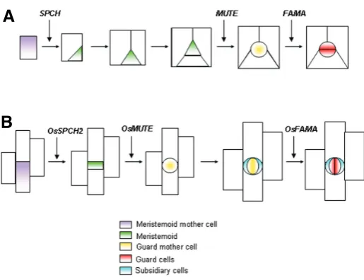
plant starts with the division of an epidermal cell termed the meristemoid mother cell (Bergmann and Sack, 2007; Serna, 2007; Fig. 2A). This asymmetric cell division produces a small, triangular meristemoid cell and a larger cell. Meristemoids can divide asymmetrically in an inward spiral up to three times, always producing a larger cell and a smaller meristemoid that maintains its stem cell character. After these asymmetric cell divisions, the meristemoid loses its stem cell activity and acquiresa subangular orrounded shape, whichgives rise to the guard mother cell. The guard mother cell divides symmetrically to produce a pair of guard cells, which do not divide further. Although stomatal development occurs in a basipetal manner, with stomata maturing towards the leaf apex, new meristemoids arise among mature stomata, allow-ing intercalary growth.
In grasses, stomatal development starts with an asymmetric division, which yields the guard mother cell (Stebbins and Shah, 1960; Fig. 2B). Before it produces the paired guard cells, the lateral neighbors of the guard mother cell, termed subsidiary mother cells, divide asymmetrically to produce the two subsidiary cells. The guard mother cell then undergoes a symmetric cell division to generate paired guard cells, which are flanked by the subsidiary cells. In contrast to what is observed in dicots, stomatal development in grasses initiates at the leaf base, while cells expand and mature only near the apex.
During the last few years, an increasing number of genes regulating Arabidopsis stomatal development have been discov-ered (Peterson et al., 2010). Many of them function in a signalling cascade, which is initiated at the cell surface through the activa-tion of the membrane receptors TOO MANY MOUTHS (TMM) and/or ERECTA family members by members of the EPIDERMAL PATTERNING FACTOR family (EPF1, EPF2, STOMAGEN/ EPFL9, CHALLAH/EPFL6), and/or a substrate processed pro-teolytically by the subtilase STOMATAL DENSITY AND DISTRI-BUTION1 and transduced through cytoplasmic MAP kinases (YODA, MKK4/ MKK5, and MPK3/MPK6) towards the nucleus. These components negatively regulate the development of
su-pernumerary stomata and enhance spacing among stomata. In the nucleus, these MAP kinases regulate the activity of the basic helix-loop-helix (bHLH) proteins SPEECHLESS (SPCH), MUTE, and FAMA, which act in concert with the bHLH-Leu zipper proteins SCREAM1 and SCREAM2 promoting stomatal develop-ment. The recent availability of genome sequences from grasses like rice (Oryza sativa) and maize (Zea mays) has permitted the analysis of genes that are homologs of Arabidopsis genes impli-cated in stomatal development (Liu et al., 2009), as well as the analysis of genes that most probably function exclusively in grasses (Cartwright et al., 2009). This review mainly focuses on the functions of SPCH, MUTE, FAMA and their homologs in grasses, highlighting the differences and commonalities of sto-matal development in these two plant groups. The role of PANGLOSS1 (PAN1) gene of maize, whichencodes a leucine-rich repeat receptor-like kinase with an inactive kinase domain (Cartwright et al., 2009), is also highlighted.
Orthologs and paralogs of
SPCH-
like genes
In Arabidopsis, stomatal development starts with an asymmet-ric cell division. Mutations in the Arabidopsis gene SPCH that abolish its expression prevent the initiation of stomatal develop-ment and give rise to an epidermis consisting of only jigsaw puzzle-shaped pavement cells (MacAlister et al., 2007; Pillitteri et al., 2007). Thus, SPCH, which encodes a bHLH protein, is thought to drive the first asymmetric cell division that initiates stomatal development (MacAlister et al., 2007; Pillitteri et al., 2007). In support of this role, the overexpression of SPCH results in numerous extra cell divisions (MacAlister et al., 2007; Pillitteri et al., 2007). In addition to regulating the first asymmetric cell division that accounts for stomatal development, SPCH appar-ently maintains the stem cell activity of meristemoids (MacAlister et al., 2007): the missense mutation spch-2, which affects the carboxy terminus of the protein, reduces the number of cells entering the stomatal pathway and appears to trigger premature stomatal formation in the pedicel epidermis. SPCH promoter activity is observed in undifferentiated cells and persists in sto-matal lineage cells, including guard cells (MacAlister et al., 2007; Pillitteri et al., 2007). However, the encoded protein is restricted to the early stages of the stomatal lineage, suggesting that the SPCH protein might be post-transcriptionally downregulated (MacAlister et al., 2007). These expression patterns are consis-tent with a role for SPCH in determining the entrance into the stomatal lineage and perhaps a later role in maintaining the stem cell activity of meristemoids.
In contrast to the single SPCH gene of Arabidopsis, poplar and Ricinus, Liu et al. found two SPCH-like genes in both O. sativa (OsSPCH1 and OsSPCH2) and Z. mays (ZmSPCH1 and ZmSPCH2). Like SPCH, OsSPCH2, ZmSPCH1 and ZmSPCH2 contain an N-terminal acidic region and, also like SPCH, the four grass homologs have a conserved mitogen-activated protein kinase (MAPK) target domain (Liu et al., 2009). The MAPK target domain of SPCH is phosphorylated by MAPKs (Lampard et al., 2008). Interestingly, the SPCH phosphorylation sites are con-served in the grass homologs (Lampard et al., 2008; Liu et al., 2009), suggesting that the SPCH function is also conserved in grasses. Certainly, in the case of OsSPCH2, this is supported by the observation that overexpression of SPCH under the control of Fig. 1. The epidermis of Arabidopsis and rice leaves.(A) Arabidopsis.
Cells are randomly arranged, with stomata (paired guard cells) scattered across the surface. (B)Rice. Cells are arranged in linear files, with some files bearing stomata flanked by two subsidiary cells.
the Cauliflower Mosaic Virus 35S (35S) promoter leads to a phenotype identical to that induced by OsSPCH2 overexpres-sion, which consists of ectopic divisions in pavement cells (MacAlister et al., 2007; Liu et al., 2009). Furthermore, rice plants homozygous for the osspch2-1 insertion, like Arabidopsis plants harboring the weak spch-2 allele, have a reduced number of stomata (MacAlister et al., 2007; Liu et al., 2009). However, in contrast to the spch-2 mutant, osspch2-1 plants occasionally also develop stomatal patterning defects, and expression of OsSPCH2 under the SPCH promoter fails to rescue the Arabidopsisspch phenotype (Liu et al., 2009). Therefore, although OsSPCH2 and SPCH appear to share some functions, their regulatory roles are not identical (Liu et al., 2009). OsSPCH2 controls patterning and it presumably has no role in the maintenance of meristemoid stem fate, given that rice meristemoids develop into guard mother cells without undergoing repeated rounds of asymmetric cell division. Remarkably, although OsSPCH2seems to control stomatal de-velopment, its expression has not been detected in leaves (Liu et al., 2009). Overexpression of OsSPCH1 by the 35S promoter does not confer an apparent phenotype (Liu et al., 2009), suggest-ing that the OsSPCH1 function might have diverged from that of both OsSPCH2 and SPCH. In support of this view, OsSPCH1 lacks the N-terminal acidic region that characterizes SPCH pro-teins (Liu et al., 2009). In addition, the OsSPCH1 transcript was not detected in leaves (Liu et al., 2009). Mutation analysis of the OsSPCH1 gene and cross-species tests will confirm (or annul) this apparent divergence.
In summary, these findings strongly suggest that Arabidopsis SPCH and rice OsSPCH2 share some functions. Interestingly, OsSPCH1, which most probably originated as an OsSPCH2 gene duplication, seems to have diverged from OsSPCH2 and lost its function, and perhaps it acquired a new one. Certainly, when a gene duplicates, one copy generally retains the ancestral func-tion, whereas the other copy is free to accumulate mutations (Ohno, 1970). Data on the role of SPCH-like genes in maize will reveal valuable information to extend the discussion on the molecular evolution of stomatal development to grasses.
MUTE
-like genes may act earlier in grasses than in
Arabidopsis
After a few cell divisions, meristemoids lose their stem cell identity and become guard mother cells, which then produce paired guard cells. MUTE plays an essential role in this transition by repressing the stem cell activity of meristemoids; the loss-of-function mute-1 mutant does not develop stomata but forms meristemoids that abort after excessive asymmetric cell divisions (MacAlister et al., 2007; Pillitteri et al., 2007). The MUTE promoter is active in a subset of meristemoids, with residual activity in guard mother cells and immature stomata, whereas MUTE protein is restricted solely to a subset of meristemoids (MacAlister et al., 2007; Pillitteri et al., 2007). It is therefore probable that, in this subset of meristemoids, MUTE represses stem cell activity and induces guard mother cell formation. The fact that MUTE overex-pression converts all epidermal cells into stomata is consistent with the function of this gene (MacAlister et al., 2007; Pillitteri et al., 2007).
Liu et al. found that, as in Arabidopsis, MUTE-like genes are present in grasses as single-copy genes. In addition, OsMUTE cDNA driven by the MUTE promoter partially rescues the mute-1 phenotype by inducing the development of guard cells at the edges of leaves, while meristemoids remain arrested in the central regions of leaves (Liu et al., 2009). Like OsMUTE, ZmMUTE expressed under the control of the MUTE promoter also comple-ments the Arabidopsismute-1 mutant (Liu et al., 2009). These lines of evidence suggest that the function of MUTE-like genes is conserved in grasses and Arabidopsis. Interestingly, the stomatal phenotype of Arabidopsis overexpressing either ZmMUTE or OsMUTE is similar to that caused by MUTE overexpression (at least when expression is at very high levels), in that all of the epidermal cells in cotyledons and leaves are guard cells (MacAlister et al., 2007; Pillitteri et al., 2007; Liu et al., 2009). This finding also supports the designation of these genes as orthologs.
However, it remains undetermined whether MUTE-like genes abolish the stem cell character of meristemoids in grasses, and if so, how do they do this? Interestingly, grass meristemoids de-velop directly into guard mother cells without undergoing re-peated rounds of asymmetric cell division. Grass MUTE-like genes may be expressed earlier in development than their Arabidopsis ortholog, that is, before the stem cell character of the meristemoids is manifested. Certainly, Liu et al. demonstrated thatboth OsMUTE and ZmMUTE are expressed when cell files are forming. Also consistent with this suggestion, OsMUTE::GUS expression in Arabidopsis is not restricted to a subset of meristemoids, but instead resembles the broad early expression pattern of the SPCH::GUS construct (MacAlister et al., 2007; Fig. 2. Stomatal development and bHLH genes. (A) Arabidopsis.
SPEECHLESS(SPCH) initiates stomatal development by inducing the first asymmetric division, which gives rise to the first meristemoid. Two or three divisions after SPCH, MUTE represses the stem cell character of meristemoids and induces guard mother cell formation. FAMA then drives the symmetric division that gives rise to the two guard cells. (B) Rice. OsSPCH2 regulates the first asymmetric cell division in the sto-matal lineage, which produces a meristemoid. Immediately afterwards, OsMUTE represses the stem cell character of the meristemoid and triggers guard mother cell differentiation. Consequently, meristemoids do not manifest stem cell properties. The lateral neighbors of the guard mother cell then divide asymmetrically to form stomatal subsidiary cells. Finally, OsFAMA causes the guard mother cell to divide, producing two guard cells. Adapted from (Serna, 2007; Liu et al., 2009).
A
Pillitteri et al., 2007; Liu et al., 2009). This result suggests that premature expression of OsMUTE (and ZmMUTE) may force guard mother cell formation after the first asymmetric cell division, preventing the stem cell activity of rice (and maize) meristemoids from persisting. If this is true, loss-of-function mutants of OsMUTE or ZmMUTE might be expected to have a phenotype similar to that of the loss-of-function mute-1 mutant. Also, an obvious and testable corollary is that MUTE expressed under the control of the OsMUTE (or ZmMUTE) promoter in Arabidopsis should force meristemoids to develop directly into guard mother cells without undergoing repeated rounds of asymmetric cell division.
In contrast to both SPCH and FAMA, MUTE lacks both an N-terminal extension and a MAPK target domain. However, maize and rice MUTE orthologs exhibit multiple potential MAPK phos-phorylation sites (Liu et al., 2009). It will be interesting to establish if there is any relationship between the presence of these MAPK phosphorylation sites in both OsMUTE and ZmMUTE, with re-spect to differences between the MUTE and OsMUTE (or ZmMUTE) expression patterns. Anyway, modifications in tran-scriptional regulation comprise an important component for the evolution of organismal phenotypes (Wray et al., 2003; Wray, 2003).
The role of FAMA-like genesis conserved in rice
In all plant species examined, the guard mother cell undergoes a symmetrical cell division to produce the two guard cells that form the stoma. In Arabidopsis, this transition from the guard mother cell to two fully differentiated guard cells is controlled by the FAMA
gene, which encodes a member of the bHLH family of transcrip-tion factors (Ohashi-Ito and Bergmann, 2006). Plants homozy-gous for an insertion mutation in FAMA (fama-1), which lack the FAMA transcript, exhibit clusters of guard mother cells or imma-ture guard cells instead of fully differentiated stomata (Ohashi-Ito and Bergmann, 2006). These clusters of small and narrow epider-mal cells are referred to as tumors (Ohashi-Ito and Bergmann, 2006). Thus, FAMA stops the proliferation of guard mother cells, which prevents tumor formation, and triggers the formation of paired guard cells. The expression of FAMA is restricted to guard mother cells and differentiating guard cells, and the FAMA protein localizes to the nuclei of these cells (Ohashi-Ito and Bergmann, 2006).
Both maize and rice encode a single FAMA homolog, which are very similar (more than 90%) to the Arabidopsis FAMA sequence (Liu et al., 2009). Like FAMA, these protein homologs contain an N-terminal acidic domain (Liu et al., 2009) that is required to activate transcription (Ohashi-Ito and Bergmann, 2006). Liu et al. also showed that plants homozygous for an insertion mutation in the OsFAMA gene (osfama-1), which also lack OsFAMA tran-scripts, develop two guard mother cells or immature guard cells instead of fully differentiated stomata. In contrast to the fama-1 mutation, the osfama-1 allele does not cause numerous tumor-like cells to form in stomatal cell rows (Liu et al., 2009). Thus, OsFAMA controls stomatal differentiation but does not appear to prevent proliferation. In support of this interpretation, the heter-ologous expression of OsFAMA in Arabidopsis under the control of the FAMA promoter partially rescues the fama-1 phenotype, producing stomata that mature but that are arranged in tumors.
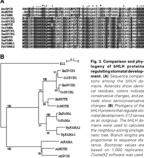
Fig. 3. Comparison and phy-logeny of bHLH proteins regulating stomatal develop-ment. (A) Sequence compari-sons among the bHLH do-mains. Asterisks show identi-cal residues, colons indicate conservative changes, and pe-riods show semiconservative changes. (B) Phylogeny of the bHLH proteins that regulate sto-matal development. E12 serves as an outgroup. The bHLH do-mains were used to calculate the neighbour-joining phyloge-netic tree. Branch lengths are proportional to sequence dis-tance. Bootstrap values are based on 1,000 replicates. ClustalX2 software was used.
Two explanations may account for these observations: 1) OsFAMA may have lost (or FAMA may have gained) part of its function in restricting cell division, and 2) the FAMA and OsFAMA functions are fully conserved, and rice guard mother cells, but not Arabidopsis guard mother cells, have cell cycle control features that pre-vent cell divisions upstream of OsFAMA.
Overexpression of OsFAMA under the control of the 35S promoter, like that of FAMA, induces the produc-tion of unpaired guard cells (Liu et al., 2009). Guard mother cells may develop into guard cells without undergoing a symmetrical division (Ohashi-Ito and Bergmann, 2006). Thus, the levels of both FAMA and OsFAMA may be essential for regulating cell division, with high levels repressing cell division and forcing guard mother cells to directly differentiate into guard cells (Ohashi-Ito and Bergmann, 2006).
Characterization of the FAMA-like gene function in rice suggests that the ability of FAMA-like genes to drive the transition from guard mother cells to fully differentiated guard cells is conserved, at least in Arabidopsis and rice. More data on the role of FAMA -like genes in maize and in other plant species are required to further understand the function of FAMA -like genes in grasses.
PAN1
and subsidiary cell formation in maize
In grasses, before guard mother cells produce paired guard cells, subsidiary mother cells polarize towards
A
the guard mother cell (Cho and Wick, 1990; Gallagher and Smith, 2000; Panteris et al., 2006). This polarization is marked by the migration of the nucleus towards the guard mother cell and the formation of dense patches of cortical F-actin at the guard mother cell contact site. Mutations in two maize genes, PAN1 and PAN2, cause defects in these two processes (Cartwright et al., 2009). They also cause aberrant subsidiary mother cell divisions (Cartwright et al., 2009). Thus, both PAN1 and PAN2 promote the polarization of the asymmetric subsidiary mother cell division and orient its cell division plane (Cartwright et al., 2009). PAN1, which encodes a member of the leucine-rich repeat receptor-like kinase family, localizes within subsidiary mother cells to sites where these cells contact guard mother cells (Cartwright et al., 2009). Analysis of PAN1-dependent protein phosphorylation revealed that PAN1 has an inactive kinase domain (Cartwright et al., 2009). Consistent with these data, PAN1 lacks several amino acid residues that are critical for kinase activity (Hanks et al., 1995; Cartwright et al., 2009).
In Arabidopsis, the TMM gene also directs asymmetric cell division in stomatal development (Nadeau and Sack, 2002). TMM contains leucine-rich repeat and transmembrane domains, but it does not have a kinase domain (Nadeau and Sack, 2002). However, in contrast to PAN1, TMM does not promote the polarization of these asymmetric cell divisions, nor is it asym-metrically localized (Nadeau and Sack, 2002). Like PAN1 (and PAN2), the ArabidopsisBREAKING OF ASYMMETRY IN THE STOMATAL LINEAGE (BASL) gene controls both the polariza-tion of stomatal lineage cells and orients their asymmetric cell divisions (Dong et al., 2009). Mutations in BASL disrupt the physical asymmetry of stomatal linage division and cause the production of more stomata (Dong et al., 2009). BASL, whichis expressed in stomatal lineage cells, encodes a novel protein for which homologs are found only in plants (Dong et al., 2009). Also like PAN1, BASL exhibits a polar localization at the cell periphery in asymmetrically dividing cells (Cartwright et al., 2009; Dong et al., 2009). But unlike PAN1, BASL forms a crescent distal to the nucleus and it also accumulates in the nucleus (Cartwright et al., 2009; Dong et al., 2009).
TMM functions in a well-characterized signal transduction pathway (Bergmann and Sack, 2007). However, the up- and downstream components of the pathways in which PAN1 and BASL operate are incomplete. The absence of PAN1 patches and the reduction of PAN1 levels in the pan2 mutant suggest that PAN1 functions downstream of PAN2, whose molecular nature remains unknown (Cartwright et al., 2009). Future goals include unravelling the components of the pathways in which PAN1 and BASL function, and identifying the PAN2 gene product. Studies on the role of PAN1-like genes in Arabidopsis and in other grasses, and of TMM- and BASL-like genes in grasses, may reveal valuable information on the roles of these genes in the evolutionary context. Given that the asymmetric cell divisions that produce the subsidiary cells in grasses do not exist in Arabidopsis, it is likely that the PAN1 and PAN1-like genes of Arabidopsis are paralogs.
Concluding remarks
Classification of plant bHLH proteins has allowed inferring that members of the same subfamily usually play redundant roles
(Pires and Dolan, 2010). Strikingly, Pires and Dolan (2010) found that members of subfamily Ia proteins, which include SPCH, MUTE, FAMA, OsMUTE, OsFAMA and OsSPCH2, play non-overlapping roles. An attractive hypothesis considers that the bHLH genes encoding these proteins might have derived from a single bHLH gene in the ancestor of land plants, which would trigger the first asymmetric cell division and immediately guard cells formation. Interestingly, the moss Physcomitrella patens does not have SPCH- or MUTE-like genes, but has two FAMA-like genes (Peterson et al., 2010). Consistently, stomata in moss develop through a single asymmetric cell division to produce the guard mother cell, which then divides to produce the paired guard cells (Payne, 1979). Then, it is likely that MUTE- and SPCH-like genes arose from duplications and divergences of FAMA-like genes, which allowed an increase in the complexity of stomatal development. In agreement with this view, the bHLH proteins of Arabidopsis and grasses fall into different evolutionary lineages (Fig. 3). Interestingly, FAMA is less similar to ZmFAMA and OsFAMA than is PpFAMA1 and PpFAMA2 (Fig. 3), which sup-ports that stomatal development of Physcomitrella patens is more similar to that of grasses than that of Arabidopsis.
Knowledge of the functions of SPCH, MUTE and FAMA in Arabidopsis has facilitated understanding the roles of their orthologs in grasses (Table 1). Differences in the expression of MUTE-like genes appear to have contributed to the divergent patterns of stomatal development between Arabidopsis and grasses. The presence of homologs of the Arabidopsis signalling genes (for example, YODA and TMM) in grasses (Peterson et al., 2010) suggests that a similar signalling cascade guides stomatal development in both groups. Similarly, the presence in rice of homologs of the Arabidopsis bHLHs ICE1/SCREAM (SCRM) and SCRM2, which seem to control stomatal development through their physical interaction with SPCH, MUTE, and FAMA (Kanaoka et al., 2008; Liu et al., 2009), suggests that similar multimeric complexes trigger stomatal development in the two plant groups. In contrast, the development of trichomes in Arabidopsis and grasses most likely depends on different transcriptional
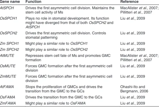
regula-Gene name Function Reference
AtSPCH Drives the first asymmetric cell division. Maintains the stem cell activity of Ms
MacAlister et al., 2007; Pillitteri et al., 2007
OsSPCH1 Plays no role in stomatal development. Its function might have diverged from that of both OsSPCH2 and
AtSPCH
Liu et al., 2009
OsSPCH2 Drives the first asymmetric cell division. Controls stomatal patterning
Liu et al., 2009
Zm SPCH1 Might play a similar role to OsSPCH1 Liu et al., 2009
Zm SPCH2 Might play a similar role to OsSPCH2 Liu et al., 2009
AtMUTE Represses stem cell fate of Ms and promotes GMC formation
MacAlister et al., 2007; Pillitteri et al., 2007
OsMUTE Forces GMC formation after the first asymmetric cell division
Liu et al., 2009
ZmMUTE Forces GMC formation after the first asymmetric cell division
Liu et al., 2009
AtFAMA Stops the proliferation of GMCs and drives the transition from the GMC to the GCs
Ohashi-Ito and Bergmann, 2006
OsFAMA Drives the transition from the GMC to the GCs Liu et al., 2009
ZmFAMA Might play a similar role to OsFAMA Liu et al., 2009 TABLE 1
bHLH GENES REGULATING STOMATAL DEVELOPMENT
tory networks (Serna and Martin, 2006).
The acquisition of the PAN1 function in grasses (or its hypo-thetical loss in Arabidopsis) may also have contributed to diver-gences in stomatal development between Arabidopsis and grasses. Determining whether thecommonalities and differences relative to stomatal development extend beyond these plant groups is essential for an in-depth understanding of the molecular evolution of stomatal development. Some interesting models of the roles that these genes may have in plants other than Arabidopsis and grasses have been proposed (Peterson et al., 2010). Future work will determine if these models reflect reality.
Acknowledgements
This work is supported by a Grant (BIO2008-02149) from the Spanish Ministry of Science and Innovation and partially by a Grant (PCI08-0041-1136) from the Autonomic Government of Castilla-La Mancha. I thank D. C. Bergmann for generously providing the grasses bHLH sequences.
References
BERGMANN, D.C. and SACK, F.D. (2007). Stomatal development. Annu Rev Plant Biol 58: 163-181.
CARTWRIGHT, H.N., HUMPHRIES, J.A. and SMITH, L.G. (2009). PAN1: A receptor-like protein that promotes polarization of an asymmetric division in maize. Science 323: 649-651.
CHO, S.O. and WICK, S.M. (1990). Distribution and function of actin in the developing stomatal complex of winter rye (Secale cereale cv. Puma).
Protoplasma 157: 154-164.
DONG, J., MACALISTER, C.A. and BERGMANN, D.C. (2009).nBASL controls asymmetric cell division in Arabidopsis. Cell 137: 1320-1330.
EDWARDS, D., KERP, H. and HASS, H. (1998). Stomata in early land plants: an anatomical and ecophysiological approach. J Exp Bot 49: 255-278. GALLAGHER, K. and SMITH, L.G. (2000). Roles for polarity and nuclear
determi-nants in specifying daughter cell fates after an asymmetric cell division in the maize leaf. Curr Biol 10: 1229-1232.
HANKS, S.K. and HUNTER, T. (1995). Protein kinases 6. The eukaryotic protein kinase superfamily: kinase (catalytic) domain structure and classification.
FASEB J 9: 576-596.
HICKEY, L. (1979). A revised classification of the architecture of dicotyledonous leaves. In Anatomy of the dicotyledons (Eds. C.R. Metcalfe and L. Chalk). Second Edition, Vol. 1. Clarendon Press, Oxford, pp. 25-39.
JUDD, W.W., CAMPELL, C.S., KELLOGG, E.A. and STEVENS, P.F. (Ed.) (1999).
Plant systematics: A phylogenetic approach. Sinauer Associates, Sunderland, MA. KANAOKA, M.M., PILLITTERI, L.J., FUJII, H., YOSHIDA, Y., BOGENSCHUTZ, N.L., TAKABAYASHI, J., ZHU, J.-K. and TORII, K.U. (2008). SCREAM/ICE1
and SCREAM2 specify three cell-state transitional steps leading to Arabidopsis
stomatal differentiation. Plant Cell 20: 1775-1785.
LAMPARD, G.R., MACALISTER, C.A. and BERGMANN, D.C. (2008). Arabidopsis
stomatal initiation is controlled by MAPK-mediated regulation of the bHLH SPEECHLESS. Science 322: 1113-1116.
LIU, T., OHASHI-ITO, K. and BERGMANN, D.C. (2009). Orthologues of Arabidopsis Thaliana stomatal bHLH genes and regulation of stomatal development in grasses. Development 136: 2265-2276.
MACALISTER, C.A. et al. (2007). Transcription factor control of asymmetric cell divisions that establish the stomatal lineage. Nature 445: 537-540.
NADEAU, J.A. and SACK, F.D. (2002). Control of stomatal distribution on the
Arabidopsis leaf surface. Science 296: 1697-1700.
OHASHI-ITO, K. and BERGMANN, D.C. (2006). Arabidopsis FAMA controls the final proliferation/differentiation switch during stomatal development. Plant Cell
18: 2493-2505.
OHNO, S. (Ed.) (1970). Evolution by gene duplication. Springer-Verlag. PANTERIS, E., APOSTOLAKOS, P. and GALATIS, B. (2006). Cytoskeletal
asym-metry in Zea mays subsidiary cell mother cells: a monopolar prophase micro-tubule half-spindle anchors the nucleus to its polar position. Cell Motil Cytosk-eleton 63: 696-709.
PAYNE, W.W. (1979). Stomatal patterns in embryophytes - Their evolution, ontogeny and interpretation. Taxon 28: 117-132.
PETERSON, K.M., RYCHEL, A.L. and TORII, K.U. (2010). Out of the mouths of plants: the molecular basis of the evolution and diversity of stomatal develop-ment. Plant Cell 22: 296-306.
PILLITTERI, L.J., SLOAN, D.B., BOGENSCHUTZ, N.L. and TORII, K.U. (2007). Termination of asymmetric cell division and differentiation of stomata. Nature
445: 501-505.
PIRES, N. and DOLAN, L. (2010). Origin and diversification of basic-helix-loop-helix proteins in plants. Mol Biol Evol 27: 862-874.
RAVEN, J.A. (2002). Selection pressures on stomatal evolution. New Phytol 153: 371-386.
SERNA, L. and MARTIN, C. (2006). Trichomes: different regulatory networks lead to convergent structures. Trends Plant Sci 11: 276-280.
SERNA, L. (2007). bHLH proteins know when to make a stoma. Trends Plant Sci
12: 483-485.
STEBBINS, G.L. and SHAH, S.S. (1960). Developmental studies of cell differentia-tion in the epidermis of monocotyledons. I. Cytological features of stomatal development in the Gramineae. Dev Biol 2: 177-200.
TAIZ, L. and ZEIGER, E. (Ed.) (2006). Plant Physiology. Fourth Edition. Sinauer Associates, Inc.
WEYERS, J.D.B. and MEIDNER, H. (Ed.) (1990). Methods in stomatal research. Longman Scientific and Technical, Harlow.
WRAY, G.A., HAHN, M.W., ABOUHEIF, E., BALHOFF, J.P., PIZER, M., ROCKMAN, M.V. and ROMANO, L.A. (2003). The evolution of transcriptional regulation in eukaryotes. Mol Biol Evol 20: 1377-1419.
Further Related Reading, published previously in the
Int. J. Dev. Biol.
See our recent Special Issue Placenta edited by Joan S. Hunt and Kent L. Thornburg at: http://www.ijdb.ehu.es/web/contents.php?vol=54&issue=2-3
Chromatin remodeling in plant development
José A. Jarillo, Manuel Piñeiro, Pilar Cubas and José M. Martínez-Zapater Int. J. Dev. Biol. (2009) 53: 1581-1596
Evolution and pleiotropy of TRITHORAX function in Arabidopsis
Zoya Avramova
Int. J. Dev. Biol. (2009) 53: 371-381
Arabidopsis monomeric G-proteins, markers of early and late events in cell differentiation
Mariette Bedhomme, Chantal Mathieu, Amada Pulido, Yves Henry and Catherine Bergounioux
Int. J. Dev. Biol. (2009) 53: 177-185
Epidermal differentiation: trichomes in Arabidopsis as a model system
Swen Schellmann and Martin Hülskamp Int. J. Dev. Biol. (2005) 49: 579-584
Clonal analysis by transposon excision of stomatal clusters in Arabidopsis
L Serna and C Fenoll
Int. J. Dev. Biol. (1996) 40: S131-S132