POST MORTEM MICROSTRUCTURAL CHANGE TO THE SKELETON
Lynne Sevon Bell
A Thesis Submitted f or the Degree of Doctor of Philosophy
Faculty of Science
Department of Anatomy and Developmental Biology University College London
University of London
October 1995
ABSTRACT
The microstructural impact of diagenetic or post mortem alteration has been assessed in predominately human skeletal tissues. The method of assessment selected was microscopical analysis, mainly using backscattered electron imaging in a scanning electron microscope and, to a lesser extent, confocal reflection microscopy. The microstructural morphologies of post mortem alteration were investigated in archaeological material, both normal and pathological, from terrestrial and marine contexts. Further studies were undertaken on a case-by-case basis on skeletal material which offered some unique pathology, environmental context, spatial relationship, time
variable, or mortuary practice. Additionally, the effect of diagenetic change on mitochondrial DNA (mtDNA) recovery and the potential location of DNA within the skeletal tissues were Investigated. Two quantitative
studies were undertaken to validate and measure the observed mineral density changes.
The investigations showed that post mortem alteration or diagenetic change to skeletal material can be extensive, and can occur shortly after death. Diagenesis did not represent a post burial phenomenon as the term
diagenesis suggests, but was found to have begun above ground in a range of exposural contexts. The implication of gut bacteria in the promotion of early bacterially-related microstructural change was strong, and it is proposed that body status at the point of, or soon after, death Is
information within a stratigraphic matrix. Characterizing the post mortem microstructural and density changes to bone has helped to elucidate the preservational status of mtDNA in terms of its relative retrieval in
archaeological specimens, and the potential location of mtDNA in bone. It is proposed that the shift in mineral density that was found in
ACKNOWLEDGEMENTS
This thesis could not have been completed without the unstinting support of my doctoral supervisor Sheila J. Jones. Throughout the long duration of this study she has always been ready to offer encouragement, tactful
criticism, endless patience, gramrnatic enlightenment and kindness. For all of this I am most grateful.
I also gratefully acknowledge the help and guidance Alan Boyde has given throughout this study. Certainly many aspects of this work could not have been undertaken without him. In addition, I would like to offer warmest thanks to Mo Arora and Roy Radcliffe f or their expert technical assistance and friendship.
Many other people have generously provided their time, interest and skeletal material. Of these people I particularly wish to thank Ann
Stirland who not only introduced me to the Mary Rose material, but who has remained the best of colleagues and the best of friends. Peter Andrews and Theya Molleson have always given freely of their ideas and material, and provided me with many inspirational moments. Mark Skinner provided
essential forensic material at a critical time. Andy Elkerton has provided valuable stratigraphic information on the Mary Rose wreck. Erika
Brothwell, Rosalie David, Chris Dean, Paul Dieppe, Helen Donohue, Ebba During, Roger Flinn, Mark Garrison, Robert Hedges, Marianne Hester, Mike Mc Carthy, Douglas Owsley, John Paddock, Juliet Rogers, Margaret Rule, Alan Saville, Derek Stirland and Noreen Tuross.
To my friends who have collectively endured this project I am particularly grateful. I owe special thanks to Jane Gill for her expert help with the Figures and to Lynx Training for allowing me multifarious access to their resources. I gratefully acknowledge the financial assistance of the Medical Research Council.
TABLE OF CONTENTS
Top page 1
Abet ract 2
Acknowledgements 4
List of Figures 9
List of Tables 14
CHAPTER 1: MICROSTRUCTURAL CHANGES TO SKELETAL TISSUES AND RELATED
ISSUES 15
1. 1 Defining definitions 15
1.2 The micromorphology of diagenesis in the skeletal tissues 20
1. 3 The micromorphology of diagenesis in teeth 20
1.4 The micromorphology of diagenesis in bone 26
1.5 Terrestrial and marine decomposition 40
1.6 Ante mortem microbial changes to the skeleton 44
1.7 Aims of this study 46
CHAPTER 2: THE MORPHOLOGY OF DIAGENETIC CHANGE IN HUMAN BONE AND TEETH 48
2. 1 PART I: Soil-buried contexts 48
2.2 Introduction 48
2.3 Diagenetic morphology in archaeological normal and
pathological human bone 48
2. 4 MaterIals and methodology 49
2.5 Results and discussion 51
2. 6 Conclusion 75
2.7 PART II: Terrestrial versus marine 76
2.8 Introduction 76
2.9 Materials and methodology 76
2. 10 Results and discussion 77
2.11 Conclusion 82
CHAPTER 3: PAGET' S DISEASE: POST MORTEM ALTERATION AND BONE PATHOLOGY 99
3.1 Introduction 99
3.2 Aims of this study 100
3.3 MaterIals and methodology 100
3.4 Results 101
3.5 Discussion 102
3.6 Conclusion 109
CHAPTER 4: THE MARY ROSE WRECK: A UNIQUE SEA BURIAL 110
4.1 Introduction 110
4.2 Historical background 110
4.3 Deposltional history 112
4. 4 Microstructural changes to marine substrates 113
4.5 The aims of this study 114
4.6 Materials and methodology 114
4.7 Results 115
4.8 Discussion 131
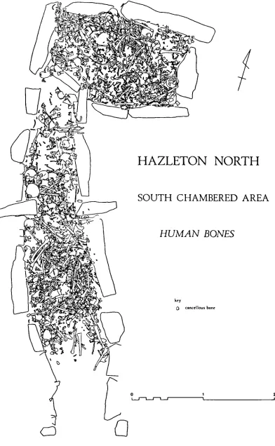
CHAPTER 5: h NEOLITHIC LONG CAIRN: LONG TERM SKELETAL EXPOSURE 135
5. 1 Introduction 135
5.2 Historical background 135
5.3 Microstructural evidence of exposure 137
5.4 Aims of this study 139
5.5 Materials and methodology 139
5.6 Results 142
5.7 Discussion 143
5.8 Conclusion 160
CHAPTER 6: THE SPEED OF POST W)RTEM CHANGE TO THE HUMAN SKELETON AND ITS
TAPHONO4IIC SIGNIFICANCE 161
6. 1 Introduction 161
6.2 Aims of this study 163
6.3 Materials and methodology 163
6.4 Results 165
6.5 Discussion 167
6.6 ConclusIon 180
CHAPTER 7: BUTCHERY AND DELIBERATE MUMMIFICATION 181
7. 1 Introduction 181
7.2 Butchery in an archaeological context 181
7.3 Mummification in an archaeological context 182
7.4 Aim of this study 184
7.5 Materials and methodology 185
7.6 Results 187
7.7 Discussion 188
7.8 Conclusion 193
CHAPTER 8: SKELETAL DNA 194
8. 1 PART I: Microstructural preservation and DNA recovery 194
8.2 Introduction 194
8.3 Aim of this study 196
8.4 Materials and methodology 196
8,5 Results 197
8.6 Brief summary of DNA results 199
8.7 DiscussIon and conclusion 206
8.8 PART II: Mineralised osteocytes: a possible location of
bone DNA 207
8.9 Introduction 207
8. 10 Materials and methodology 213
8. 11 Results 213
8. 12 Discussion and conclusion 221
CHAPTER 9: QUANTITATION 223
9. 1 Introduction 223
9. 2 Bone 223
9.3 Bone mineral density 224
9.4 SEM/BSE imaging and bone density 225
9.5 AIms of this study 228
9.6 Materials and methodology 228
9. 7 Results 231
9.8 Discussion 237
246 265 266 267 268 270
CHAPTER 10: SUW'IARY AND CONCLUSIONS 240
REFERENCES APPENDICES
Appendix 4. 1 Appendix 4. 2 Appendix 7. 1 Appendix 10
Data set of tubule diameters Data set of maximum ingress
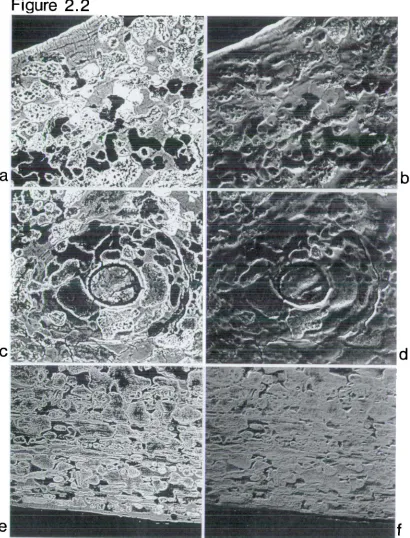
Figure 2. 1 2. la 2. lb 2. ic 2. id 2. le 2. if Figure 2.2 2. 2a 2. 2b 2. 2c 2. 2d 2. 2e 2. 2f Figure 2. 3
2. 3a 2. 3b 2. 3c 2. 3d 2. 3e 2. 3f
Figure 2. 4 2. 4a 2. 4b 2. 4c 2. 4d 2. 4e 2. 4f Figure 2.5 2. 5a 2. 5b 2. 5c 2. 5d 2. 5e 2. 5f
Figure 2. 6 2. 6a 2. 6b
LIST OF FIGURES
62 Image of modern cranial bone
Topographical image of above
Image of modern bone periosteal aspect Topographical image of above
Image of modern mid-cortical bone Topographical image of above
Image of archaeological bone which has been diagenetically altered
Topographical image of above
Single osteon diagenetically altered Topographical image of above
Bone at periosteal aspect affected by diageenesis Topographical image of above
Diagenetically altered trabeculum
Cortical field of bone altered by diagenesis Extensive diagenetic remodelling of cortex
Extensive diagenetic remodelling of single osteon Circumferential lamellae intact above field of diagenetically altered bone
Primary osteonal bone affected by diagenesis, located on periosteal aspect
High power view of altered primary osteonal bone Topographical image of above
De and remineralisation centred within single osteon
Diagenetic foci centred on osteocyte lacunae Non-survival of circumferential lamellae Distribution of diagenetic foci around single ost eon
High power view of 2. 4f
Single osteon which has increased in overall density due to diagenesis
Diagenetic foci orientated around vascular spaces in pathological specimen
High power view of above
Normal alveolar crest, dentine and cementum Diagenetic alteration to cementum and alveolar bone
Montage of soil-buried tooth Montage of marine-buried tooth
65
6B
71
74
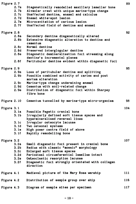
Figure 2.7 2. 7a 2. lb 2. 7c 2. 7d 2. 7e 2. 7f Figure 2.8 2. 8a 2. 8b 2. 8c 2. 8d 2. 8e 2. 8f Figure 2.9 2. 9a 2. 9b 2. 9c 2. 9d 2. 9e
Figure 3. 1 3. la 3. lb 3. ic 3. id 3. le 3. if Figure 3. 2
3. 2a 3. 2b 3. 2c 3. 2d 3. 2e 3. 2f 89 Diagenetically remodelled maxillary lamellar bone
Alveolar crest with unique marine-type change Unaffected dentine, enamel and calculus
Enamel white-spot lesion
Microcavitation of carious lesion Unaffected field of dentine and enamel
Secondary dentine diagenetically altered
Extensive diegenetic alteration to dentine and cement urn
Normal dent me
Preserved intergiogular dentine
Diagenetic demineralisation foci streaming along dentine's incremental planes
Peritubular dentine evident within diagenetic foci
Loss of peritubular deritine and splitting Possible combined activity of caries and post mortem alteration
Marine-type change undermining enamel Cementum with soil-related change
Distribution of diagenetic foci within Sharpey fibre bone
92
95
Figure 2. 10 Cementum tunnelled by marine-type micro-organism 98 104 Possible Pagetic cranial bone
Irregularly defined soft tissue spaces and hypermineralised reversal lines
Irregular osteocyte lacunae Two osteonal systems
High power centre field of above Rapidly remodelling bone
Small diagenetic foci present in cranial bone Radius with classic NmosaicN morphology
Enlarged soft tissue spaces
Periosteal circumferential lamellae intact Osteoclastic resorption lacunae
Diagenetic foci strongly orientated with collagen direct ion
107
Figure 4. 4 122 4. 4a Enamel undermined by post mortem tunnelling
4. 4b Dentine totally remodelled
4. 4c Internal reflection artefact of tunnel direction
Figure 4. 5 125
4. 5a Alveolar bone invaded by tunnelling 4. 5b Maximum ingress measurement
4.5c Maximum post mortem tubule diameter
Figure 4. 6 Distribution of tubule diameters 126
Figure 4.7 Cumulative silt phase schematic of ship 129 Figure 4.8 Graphical representation of maximum ingress 130
Figure 5. 1 Contour map of long cairn 136
Figure 5. 2 Spatial representation of total bone scatter 138 Figure 5. 3 Spatial representation of specimen location 14.0
Figure 5. 4 146
5.4a Dentine invaded by bacterial-type attack 5. 4b Cementum unaffected
5. 4c Post mortem change as reflective globular features
Figure 5. 5 149
5. 5a Post mortem enlargement of alveolar bone
5. 5b Dentine tunnelled to enamel dentine junction (EDJ) 5. 5c Dentine extensively remodelled post mortem
Figure 5. 6 152
5. 6a Enlargement of osteocytic spaces in alveolar bone 5. 6b Centre field of above
5. 6c Apical cementum altered post mortem
Figure 5. 7 155
5. 7a Tooth extensively remodelled post mortem
5. 7b Dentine extensively remodelled to the EDJ post mortem
5. 7c Radicular dentine extensively altered post mortem 5. 7d Peritubular dentine preserved within a post mortem
deniineralisat ion focus
Figure 5. 8 157
5.8a Post mortem bacterial foci changing direction at the CDJ 5.8b Unaffected tooth
5.8c Region of intergiobular dentine 5.8d Unaffected cementocytes
Figure 6. 1 170
Figure 7. 2 7. 2a 7. 2b 7. 2c 7. 2d 7.2e Figure 8. 1
8. la 8. lb 8. ic 8. id 8. le 8. if 8. ig 8. ih 8. ii
Figure 8. 2 8. 2a 8. 2b 8. 2c 8. 2d
6. lb 3 ii post mortem. Intracorticel demineralisation 6. Ic 15 in post mortem. Bacterial-type remodelling 6. id 15 in post mortem. Bacterial-type foci located on
osteocyte lacunae Figure 6.2
6.2a 15 in post mortem. Bacterial-type change in two ost eons
6.2b Small diagenetic focus
6.2c 2 years (yrs) post mortem. Marine-type tunnelling to tooth
6. 2d 2 yrs post mortem. Tunnels penetrating dentine Figure 6. 3
6. 3a 7 yrs post mortem. Radicular cementum with two localised post mortem foci
6. 3b 70 yrs post mortem. Diffuse peripheral band of demirieralisation to periosteal aspect of a rib 6. 3c High power view of above
Figure 7. 1 Schematic of modern carcass division in Britain
173
173
182 191 Extensive post mortem alteration to bone
Small localised post mortem voids
Extensive diagenetic change to trabecular bone Peripheral demineralisation at periosteal aspect Small intracortical diffuse demineralisation focus
Diagenetic change to bone at periosteal aspect Mid-cortical osteonal systems exhibiting localised diagenetic foci
Single trabeculum with containing small diagenetic foci
Only the circumferential lamellae are unaffected Mid-cortical bone with moderate diagenetic change Cortical bone close to medullary aspect
diagenetically altered
Bone below periosteal aspect extensively diagenetically remodelled
Mid-cortical region altered post mortem Medullary aspect bone extensively altered post mortem
Extensive diagenetic remodelling next to periosteal aspect
Small area of mid-cortical bone extensively altered post mortem
Medullary aspect and adjoining trabeculae altered post mortem
Mid-cortical region of intact bone bordering a diagenetically altered region of bone
202
8. 2e Specimen with normal micromorphiogy
8. 2f Small focus of demineralisation at periosteal aspect Figure 8. 3
8. 3a 8. 3b 8. 3c 8. 3d 8. 3e Figure 8. 4
8. 4a 8. 4b 8. 4c 8. 4d 8. 4e 8. 4f Figure 9. 1
Figure 9. 2
Figure 9. 3 Figure 9. 4 Figure 9. 5
217 Two 'mineral infilled osteocyte lacuriae' (MIOL)
Field of MIOL
Layed and partially infilled MIOL
A single MIOL abutting a diagenetic focus Higher power view of MIOL from above
220 Field of MIOL close to band of peripheral demineralisation
MIOL with internal structure similar to cell nucleus As above
Field of MIOL As above
MIOL in bone close to calcified cartilage
Diagram of specimen set-up and radial motion for 229 ml crot omography
Tomogram of Barton femur with high density 233 diagenet Ic component
Tomogram of Barton femur recalibrated 234
Diagenetic shift in sample group 235
LIST OF TABLES
Table 1. 1 Stages of Decomposition for Land and Water 41
Table 4. 1 Translated Data Grades 128
Table 6. 1 Forensic Sample Group 164
CHAPTER 1: MICROSTRUCTURAL CHANGES TO SKELETAL TISSUES ANI) RELATED ISSUES
1. 1 Defining definitions
The aim of this dissertation is to assess the microstructural impact of post mortem change to primarily human bone and teeth. The study is therefore a taphonoinic one, of a phenonmerion called diagenesis which can occur in forensic, archaeological and fossil skeletal material. The terms or nomenclature used in this study originate historically from overlapping disciplines and as such vary in their use and comprehension. A short discussion of this nomenclature follows by way of clarification.
Taphonomy means literally 'the laws of burial'. Arguably, the German geologist Weigelt, whose classic treatise published in 1927 described how organisms die, decay and become encased in sediment, was the first to outline taphonomic study, calling it "biostratinomy". However,
historically, the term "taphonomy" was first coined by the Russian
vertebrate palaeontologist Efremov (1940) who outlined a more systematic approach to the study of' the transitionary phases between death, entombment and final lithification. Efremov wanted to establish predictive laws in order to understand just why and how fossil groupings display bias in terms of' species representation. He suggested that if one could account for the factors which control successful fossilisation, then the resultant bias could be understood and corrected for, allowing for the theoretical reconstruction of fossil communities (Efremov, 1940).
on uniformitarianism. Uniformitarianism, outlined by L.yell in "Principles of Geology" in 1830, stated that a) processes are uniform throughout time and b) natural laws are constant in space and time. (This theory opposed the Catastrophist ruling paradigm which asserted that geological strata were created out of environmental catastrophies, the most recent being the Noachian Flood, which all occurred after Creation 4004 BC (Daniel, 1975). ) Taphonomy Is theoretically based upon the latter aspect of
uniformitarlanism and has been called methodological uniformitarianism (Gould, 1965). It has also been referred to as actualistic and
neotaphonomic, which essentially means that methodologi.cal inferences can be made about past events by direct analogy to the present, since the processes which governed events in the past will proceed identically to those in the present (Lyman, 1994). Hence, 'actualistic' experiments which model or observe a taphonomic event In either a cross-sectional or
longitudinal time-frame will have legitimate relevance to interpreting comparable historical skeletal assemblages.
Diagenesis Is a geological term and refers to the physical, physicochemical and biochemical events which can occur during the formation of a
sedimentary deposit. The events referred to are here understood to mean:-• . compaction, desslcation, deformation, corrosion, bleaching, oxidation reduction, crystal inversion, recrystallization, intercrystalline bonding, cementation, decementation, mineral growth and mineral replacement, particle accretion, flocculation, sediment mixing, boring, decomposition and synthesis of organic compounds. . . incorporation of blota. . . and excludes the effects of high temperature and pressure." (Lapedes, 1978: 153)
common use of the term diagenesis within archaeological literature and refers to any microstructural post mortem alteration to skeletal material. Unfortunately, this has introduced an implicit and potentially erroneous assumption that post mortem alteration begins after burial and Is
controlled by soil cofactors stretched Into geological time (Plepenbrink & Schutowskl, 1987). Indeed, part of this problem hangs upon the question of when exactly diagenesis begins. Recently, workers have taken to stating that diagenesis begins at the point of burial (Lyman, 1994; Retallack, 1990) and that any changes which occur prior to that are 'blostratinomic'. Essentially this is the correct view and considered earlier by Lawrence
(1971) as a necessary distinction. However, the theoretical practicality of such an approach is questionable. Purdy (1968) readily admitted in his study of carbonate diagenesis that diagenesis Is a "vague" concept which is difficult to define, particularly at the beginning and end of its effects on sediments: "the impossibility of clearly distinguishing between
diagenesis and metamorphism because diagenesis is, In fact, the beginning of metamorphism.... (and) the distinction between depositional and post-depositional processes is equally arbitrary" (Purdy, 1968: 184). Rolfe and Brett (1969) attempted to solve this problem by subdividing diagenesis into three stages, where the earliest stage is referred to as "syndiagenesis", which Involves only the activity of bacteria In shallow sediments
metabolizing organic matter in skeletal tissue. This subdivision, although refining terminology, goes no further towards solving the problem other than stating that diagenetic processes begin postburial. Indeed, the entomological and microbial aspects of decay, whether above or below ground, may be considered Identical processes and therefore descriptively
This problem is further added to by the prefixture of "forensic", "archaeological" and UfossliN to date skeletal material. A forensic specimen can generally be defined as an individual who has been dead, buried or unburied, for up to 50-100 years and is also of no interest to the County Coroner (Burial Act 1657, section 25). In other parts of the world this generality may vary usually for legal reasons, but also for political reasons, as in the reburial issues surrounding the Israeli, Amerindian, Aboriginal and Moon skeletal collections (Watzman, 1995; tJbelaker & Grant, 1989).
An archaeological specimen is more difficult to define due to the historical origins of archaeology. A discussion of the origins of
archaeology would be lengthy and inappropriate here, but generally, modern archaeology is concerned with all aspects of human social organisat ion where there is evidence of people and their material culture (Hodder, 1991; Daniel, 1975). It no longer necessarily involves the study of human
evolution as it did during the 1800's, nor is it treasure hunting as it was at the turn of the century. Instead, it has become a global discipline which examines evidence of social orgariisation dating from Mesolithic hunter gatherer societies onwards. In Britain this can be clearly defined as beginning 10,000 years BP (at the end of the last glaciation), but other parts of the world may pre or post date this.
A fossil can mean all things to all people. The nineteenth century biblical scholars believed that Creation began 4004 years BC, and consequently found fossils to be the irksome protagonists of new
Jehovah's Witnesses will argue that fossils have been placed in geological deposits, by God, in order to test our faith - an argument difficult to dispute! Indeed, one notable and extant professor has called enamel a "living fossil", it being effectively dead and almost totally mineralized within the living mouth (Boyde, pers. comm.). However, from a geological standpoint a fossil constitutes any evidence of past life which is, or was, situated in a geological deposit (Lapedes, 1978). Fossils fall within four main
groups:-1. Unaltered remains such as frozen woolly maminoths or animals and insects preserved in oil seeps and oil shales.
2. Petrified fossils produced by perimineralization, where mineral is added to natural microscopic voids within specimens; by replacement of minerals by substitution at a molecular level; and by distillation where organic tissues are carbonised via the liberation of carbon dioxide and water, leaving only a carbon copy.
3. Molds, casts and imprints of animals etc are created when a creature is surrounded by a deposit and latterly lost leaving only an impression of Itself.
4. Tracks, trails and burrows are the final subgroup where life has passed over or through a deposit and subsequently that feature is preserved as an interface or morphological discontinuity within the deposit.
(After Lapedes, 1978: 247-8) Hence, fossils can be thought of as specimens of some great antiquity which have been preserved as distinct features or inclusions within geological deposits and relatively predate the beginning of the Mesolithic period. However, two major inconsistancies remain: one is the problem of when
secondly, archaeological specimens are usually inhumations from cemetery sites, and as such, are intrusively buried deep within a sediment, and therefore considered to be both archaeological and fossil.
This thesis will use all the terms referred to in this section. It will as a result try to unravel some of the processes and contradictions manifest in an eclectic area of study.
1.2 The microrphology of diagenesis
In the previous century and well into this one, the internal
micromorphology of diagenesis, or post mortem alteration to bone and teeth, was observed only as a curiosity. Latterly, diagenesis has come to
represent an irksome source of contamination for various archaeological and forensic techniques which rely heavily on the anorganic integrity of
skeletal tissues. As a source of information in itself, diagenesis has had few admirers, and much of the microscopic work has concentrated on the taphonomic relevance of surface changes to the periosteal circumferential lamellae of bone (Andrews, 1990; Teaford, 1988).
1.3 The micro.orphology of diagenesis in teeth
results revealed that the micro-organism, considered to be a fungus, was capable of penetrating the dentine via the neck and peripheral aspect of the root but had no effect on enamel. Penetration of the dentine occurred within 13-17 days of immersion, resulted in canals with transverse
diameters of 7 microns and invaded dentine without any regard to its structure. Wedl repeated his experiment with a section of horse rib and found the same invasive tunnelling by the micro-organism. He then examined several fossil species of teeth in order to determine the antiquity of this alteration, and also to evaluate the relationship between tooth morphology and distribution of the micro-organisms' attack. He found that some of the fossil specimens exhibited the previously observed post mortem alteration and that the enamel remained unaffected by such changes no matter what the structural arrangement of the dental tissues. He concluded that the change itself must have occurred after death since the morphology of the attack was different from that of any known caries; that it was caused by a fungus; that the fungus fed on the collagenous fraction of the skeletal tissues and therefore could not compromise enamel, it being almost entirely mineral; and that this particular post mortem change was itself of great antiquity.
teeth of over 90 million years and found similar microscopic tunnelling. Later Duckworth (1901) passed brief comment on post mortem microscopic tunnelling in a human tooth fragment, and in 1903 Hopewell Smith re-examined Tomes' cited specimen and, unsurprisingly, observed exactly the same changes to microstructure as those reported by Tomes.
saprophytic agent f or the branching type of tunnelling and extensively dismissed the notion of in vivo caries as a possible cause.
Syssoeva (1958) undertook the first systematic study to ascertain the speed with which post mortem alteration to teeth could occur. He examined a total of 196 teeth from soil-buried individuals who had been dead from between 6 months to 70 years and buried for 10 years or more. He observed macroscopic changes to teeth, although no obvious change to microstructure were evident in these teeth, or in any others of the sampled group.
However, an x-ray study did show a slight decrease in density in specimens which had been in the ground f or 65-70 years. Hence, Syssoeva's study appeared to show that post mortem alteration, if it were to occur at all, would begin no earlier than 70 years after burial and possibly some
considerable time thereafter. It is curious however, that the unspecified macroscopic changes to teeth observed, were not detectable microscopically.
Work by Clement (1958, 1963) and Falin (1961) addressed post mortem
alteration to teeth only as a subjunct to assessing evolutionary aspects of tooth development from the early anthropoids through to the Bronze Age period. Both workers found evidence of Sognnaes' commonest type of
tunnelling (some of which were infilled with mineral presumed to originate from the soil), and agreed that mechanisms governing the presence and
severity of such changes could not be time-dependent. As a result of these findings Falin suggested that the work of Syssoeva (1958) needed
Werelds examined 300 human teeth excavated from a sandy soil (1961), 72 human teeth from a clay soil (1962) and a set of human teeth deliberately exposed to soil end marine burial (1967). The age of his specimens spanned approximately 1000 years. The sandy specimens yielded the same tunnelling as described by previous workers (a type of mycetes was suggested as the causative agent), but Werelds also felt he had identified a new type of post mortem change, described as an apparent "disaggregation of the dentine" (1961: 60) and later described it as a "multitude of galleries"
(1962: 589). In his examination of the clay based specimens he found the same array of post mortem change to dentine and cementum - buried for approximately 300 years - and concluded that time was not a pertinent
factor in this process of change. Werelds then buried and immersed freshly extracted teeth in an attempt, similar to Wedl's (1864) work, to induce post mortem alteration experimentally. The soil buried specimens exhibited post mortem alteration in the third year of burial and the water immersed specimens suffered tunnelling within a few months (Werelds, 1967). Hence, Werelds demonstrated, as did Wedl, that post mortem alteration could occur soon after death and as a corollary that the presence and severity of such changes were unlikely to have a linear relationship with time.
Electron microscopic work undertaken to investigate the survival of fossil dentine collagen (Isaacs & Little, 1963; Doberenz, 1967; Doberenz &
contaminant: the cultures proved negative. Cold incineration with ionized oxygen, which would have converted any carbon to carbon dioxide, also proved negative. The authors felt it unlikely that the bacteria were carious in origin since no regional breakdown in the structure of the surrounding dentine, indicative of present day disease, was apparent. It was felt that the most likely period for the introduction of the bacteria was soil related, either during decomposition or at a much later stage.
A new post mortem alteration to enamel was detected and described as
"pseudocaries" by Poole and Tratman (1978). Twelve Mesolithic human teeth excavated in a limestone cave were examined using ordinary light and
polarized microscopy. Macroscopically, teeth appeared free of any carious lesions; however, on microscopic inspection, zones of demineralisation
(morphologically characteristic of caries) affected the surface area of whole crowns, and extended pulpally approximately one third to one half of the total enamel thickness. This alteration was considered to have been caused by slow demineralisation and created internal lesions that were identical to those created by oral micro-organisms, but the distribution of this change was totally unlike that of dental caries. The dentine situated beneath such lesions showed no disaggregation or reparative response. In addition, post mortem tunnelling extended both peripulpally and
perpendicular to dentine tubule direction, with tunnel diameters of 2-5 microns, and was only detected in specimens free of enamel pseudocaries. The authors considered post mortem alteration to the affected dental
the observed acidification of the enamel. The authors mention, almost as an aside, that they buried a set of human and dog teeth in a Bristol cemetery f or 6 months, but that on reinspection no post mortem alteration was evident.
At this point work which considered the internal micromorphology of post mortem alteration to dental tissues either stopped or went unreported. Instead, work shifted toward ascertaining the elemental and organic
composition of dental tissues. This change of emphasis in archaeologically related research was largely technique driven and itself originated from the emerging science-based 'New Archaeology'. However, the shift was largely a taphonomic and geochemical one, and as such, post mortem
alteration to skeletal tissues became known as diagenetic change, with all the incumbant problems associated with that term.
1.4 The icro..orphology of diagenesis in bone
Historically, the first histological observation of post mortem change within bone, as in teeth, was made by Wedi (1864). In addition to his experimental work on teeth with a fungal type isolate, he innoculated a section of horse rib and observed that similar tunnelling occurred within four days. The tunnels had the same morphology as those previously
observed in dentine, but their distribution was not detailed by Wedl. He also observed extensive tunnelling in the fossil rib bone of a warbler
deeper than 250 microns and appeared unaffected by the collagenous arrangement of the bone. Other fossil material studied by Roux (1887), Schaffer (1895, 1889, 1890) and Thomasett (1931) extended and confirmed Wedl's initial observations that post mortem tunnelling, alongside other post mortem changes, could be found in a range of species, including dinosaurs, mammals and fish.
Graf (1949) undertook a histological study to assess the preservation of soft and bony tissue in human Egyptian mummy and Swedish skeletalized soil-buried material and made some interesting observations. He found a range of preservation states: all bone belonging to the mummy material was intact; whereas, the skeletonized material from different soil contexts showed a "derangement of the Haversian systems" (ibid: 245). This
alteration was different in morphology from that described by Wedl, and was figured by Graf as a picture of enlarged osteocyte lacunae, with
order to study early post mortem changes, Graf attempted a small "flower pot" experiment where a cadaver rib was acquired, allowed to sit unfixed for a fortnight, and then potted on f or a year. When examined no
discernible change to bone was evident, although "the endoplasma of the osteocytes and their nuclei were still to be seen.... but necrotic in appearance.... erythrocytes in blood vessels had fused to formo.'amorphous mass....white corpuscles were clearly outlined but shrunken....remnants of bone marrow were observed and contained primitive blood cells" (ibid: 246-7). Gref concludes that post mortem alteration to bone is not a time dependent phenomenon and that the cellular components of bone can potentially survive partially intact for some centuries.
Other histological observations by Morganthaler and Baud (1956), Enlow and Brown (1956), Cook et al. (1962), Little et al. (1962), Berg (1963) and Sergi et al. (1972) extended and confirmed Gref's work. They all found evidence f or a range of post mortem alteration dating from the Triassic and Eocene through to approximately 1800 AD. Morgarithaler and Baud observed minute canaliculae which disrupted osteon lamellar structure, some of which were filled with whet appeared to be brownish septate filaments. They were unable to cultivate any of these filaments. Cooke et al. (1962) commented that under polarized light specimens which were unaffected by tunnelling
(Ic had good morphology) exhibited "the pattern of concentric striations imposed on the living osteon by the fibrillar organisation of
protein will still have occurred. Similar collagen degradation was
observed in human bones dating approximately to 1800 AD by both Little et al. (1962) and Berg (1963). Berg (1963) asserted that such changes to bone microstructure occur gradually over 1000-2000 years post mortem. Sergi et al. (1972) made ultrathin sections of bone and found that unaltered bone had very thin needle-like crystals (width 8.5 nm) typically aligned with the major axis of the lamellae, but in some areas the crystals changed direction and formed obtuse angles with previous and following groups, giving an overall impression of collapsed tissue. In areas of bone which had undergone considerable and diffuse or "mottled" post mortem alteration, large irregular and interconnecting voids opened up with bordering walls which were very thin (only a few crystals thick) and "labyrinth-like" in structure (crystallite size and orientation is not reported). Again there Is general agreement amongst the authors that the post mortem process of
change Is not a time related one.
Marchiafava et al. (1974) undertook a morphological Investigation into the "osteoclastic activity" of a Mucor fungus on buried bone. Initially
fragments of human vertebrae, dissected from cadavers, were buried in flower pots with garden earth. The earth was kept wet at 20C. After a few days a white mould masked the surface of the bone fragments. Three fungal isolates were obtained from this mould and subsequently reseeded individually onto autoclaved bone. Only Mucor grew under these controlled conditions and bone changes were observed within 15-20 days. The
tubules within bone which showed no evidence of decalcification resorption and was smooth up to the free edge. In the second pattern of liwasion, bone matrix was undergoing fungal decalcification: closely packed collagen fibrils with clearly recognisable 64 nm periodicity were alongside small aggregates of crystals. The affected tissue appeared as thin strips
surrounding the tubules created by the fungal hyphae which showed evidence of aging, containing intrahyphal lipid droplets. Exactly how Mucor managed to tunnel into bone without using decalcifying agents remained a puzzle. However, the authors postulate that aging hyphae may have utilised a
possible combination of acid, pectinase and cellulase, as is the case with pathogenic fungi.
In 1978 two papers by Stout (1978) and Stout and Teitelbaum (1978) advanced an argument for standard microscopic screening of bone as an error-check for other investigative techniques involving archaeological bone. After examining both cadaver and archaeological human bone using basic staining technology, polarizing light (Stout & Teltelbaum, 1978) and
microradiography (Stout, 1978), the authors realised that post mortem alteration to bone could be not only extensive intracortically but also cause a general reduction in overall mineral density. Stout casts doubt over the efficacy of various histological aging techniques which rely on osteon integrity and also on photon-absorptiometry due to marked changes to mineral density.
sufficient for histological analysis, and he conceived a four-tiered classification of post mortem change. Hackett believed that the micro-organisms responsible originated from the soil, initially scavenging on the decomposing soft tissues of the body, and later progressing to bone
intracortically via the endosteal and periosteal aspects. Invasion of osteonal systems themselves was via Haversian canals, but Hackett saw no evidence that the "destructive foci" were influenced by osteocyte lacunae and associated cenaliculae. Hackett considered that the micro-organisms, once within bone, fed on collagen via a process of systematic
demineralisation. Mineral redeposition was noted as " cuffing" around smaller foci, the composition of which is considered to be reconstituted bone mineral. This process of demineralisation and remineralisatlon was considered by Hackett to be a seasonally episodic event, which was aided by the corresponding rise and fall of moisture levels within soil: "burials 100 years old might be expected to have experienced many major annual tides" (Hackett, 1981: 248). Hackett ruled out secondary feeding by one set of micro-organisms on another and proposed that foci, demineralised and remineralised by the same micro-organisms, enlarge in area with a
consequent overall decrease in mineral density. Hence, the seasonal creep of water allows for various changes in mineral density as a driving co-factor to episodic attacks by the same micro-organisms. Once a bone is exhumed and allowed to dry out, the bone's micromorphology is considered by Hackett to be "fixed".
Hackett's classificatory system is considered in detail
morphological type. Although Wedi's specimen was water-immersed and similar to tunnelling described by Marchiafava et al. (1974), the
environmental history of Hackett's specimens was not well documented. The tunnelling had a uniform diameter of 5-10 microns and exhibited no
remineralisation at the free edge. The distribution of the tunnelling was peripheral, spreading inwards from the periosteal and endosteal aspects of the cortices, and was unaffected by the cellular and collagenous network of bone. A fungus was considered responsible.
2. Linear longitudinal: this type of tunnelling had similar dimensions to Wedi ie. 5-10 microns, but occasionally formed "cuffed" rims. Hackett stated that microradiography showed the tunnels to be dark, and that these tunnels "stream" together in many longitudinal arrays, being limited only by cement lines. Occasionally, tunnels passed transversely across
lamellae. In addition, when tunnels left the mid-zone of the cortex and approached the outer region, the morphology of post mortem change altered to large round dark foci, 30-50 microns in diameter, which were fibrillar with refractile rims. These foci were often concentrated within a single osteon. Hackett considered that bacteria were responsible.
3. Budded: "frond-like" tunnels followed osteonal canals and often filled them and were approximately 30 mIcrons in diameter. Budded side shoots formed at irregular intervals and measured 80-90 microns across. This type of change was considered to be produced by episodic demineralisation and remineralisation events, which led to further episodes of demineralisation. Consequently, large amounts of mineral were removed, resulting in the
formation of voids or empty tunnels. Again, this morphology was considered by Hackett to result from bacterial activity.
foci were usually rounded in shape and curved in transverse section, following the curvature of osteonal systems. They were often found near the surfaces of the cortices, localising around osteonal systems,
occasionally crossing cement lines, and ranged in size from small foci of 10-20 microns and larger ones ranging between 60-250 microns. Within a single focus, lamellation was visible, suggesting that collagen may have survived in some form. Bony bars, usually of 3-5 microns, surrounded the circumference of each lamellate focus. This lamellate pattern was
considered by Hackett to have the potential to survive for centuries, assuming that the focal destruction began soon after burial. Again, bacteria were considered the causal agent.
Hackett also undertook a small experimental investigation. He did obtain changes to bone within a year, but the experiment itself was uncontrolled and so was uninformative.
Sea water burial effects have been investigated by Arnaud et al. (1978) and by Ascenzi and Silverstrini (1984) and their studies are considered
together here for simplicity. Both confirmed that human bone, both archaeological and recent, deposited in sea water may exhibit tunnelling features. Arnaud et al. examined archaeological material (900 AD) and found many peripherally invasive tunnels with diameters similar to Marchiafava et al. (1974). Ascenzi and Sllverstrini undertook an
diameters of 6-10 microns, and again similar to those induced by
Marchiafava et al. (1974), although bacteria and algae were not considered directly involved. Instead, protozoans of the amoebic type were found to contain aggregations of bone crystals intracellularly when observed by transmission electron microscopy. The authors suggested that cells may initially cause solubilization of the hydroxyapatite, but could not explain the resultant take-up of hydroxyapatite crystals.
Work by Piepenbrink (1986) assessed post mortem change in archaeological human bone from two distinct environmental contexts. The 1986 study encompassed microbiology, x-ray diffraction and histology. In brief, Piepenbrink isolated four fungi from freshly excavated archaeological bone and subsequently re-innoculated them onto samples of histologically normal bone from the same specimens. The results were inconclusive as none of the innoculants produced any tunnelling, although Stachybotrys cylindrospora did produce a combination of non-identified antibiotics which fluoresced yellow green concentrically around Haversian canals. Histologically, Piepenbrink found varying degrees of Hackett's Nlamellateu form of post mortem change and identified these areas as apatitic. Piepenbrink
gravel contexts). He also discovered a similar distribution of staining in bone innoculated with a fungus, Cryosporium.
In 1987 Piepenbrink and Schutkowski assessed the microscopical integrity of archaeological human bone from a dry desert context. The results from microradiography illustrated the many "micro-fissures" present as cracks which partially bisected osteonal systems and tracked along cement lines. Large crystals of calcite were identified within these cracks, alongside a significant decrease in phosphorus relative to calcium. The authors
commented that light microscopy proved ineffective for histological analysis, microradiography being a more powerful method for highlighting structural changes and relative mineralisation states. A curious
alteration in mineralisation was observed within lamellar groups where one lamella may be hyper or hypomineralised relative to another. The authors were unsure as to whether this change was an in vivo pathological condition or a post mortem alteration.
authors point out that if this were a product of diagenesis, then the distribution of the fluorescence would only occur at sites of natural porosity le Haversian canals and osteocyte lacunae, and this was not the case. Similar results were obtained by Cook et al. (1989) where the same distribution of yellow green fluoresence in Egyptian material (400-500 AD) was attributed to the ingestion of grain contaminated by a Streptomyces. Perhaps most controversial, and entertaining, was an argument which took place in Nature, over the apparent identification of syphilis in a
Pleistocene bear using a fluorescing polyclonal antibody method (Rothschild & Bruce, 1987). Clearly incensed, Neiburger (1988) attacked the results stating that polyclonals are notoriously non-specific and the observed fluorescence was more likely to be some other pathological condition, or, nothing more than a post mortem artefact. Rothschild's (1988) retort
accused Neiburger of having a poor understanding of pathology, little faith in the power of modern scientific techniques and less than no understanding of the durability of antigens over millions of years.
employed standard stain technology in light microscopy to assess its usefulness in determining the internal microstructure of human bone. In terms of diagenetic change many of his observations confirm the work of previous authors such as Hackett (1981), and have added further information on the presence of iron compounds and fungal inclusions within the vascular and cellular spaces of bone.
An important study was undertaken in Japan by Yoshino et al. (1991) which investigated the speed of diagenetic change in human bone microstructure in three different environmental contexts. Fifty-one human bone samples of known date, ranging from 0-15 years post mortem, were used. Of this group, 33 were exposed, 14 buried in soil (depth not given) and 4 immersed in sea water (depth not given). Information on the preparation of bone samples is not provided. On retrieval, the samples were either embedded without
fixation or dehydration for microradiography and SEM or fixed, decalcif led and embedded for TEM and UV fluorescence microscopy. Of the three
environmental contexts, air, soil and marine, the bone from the air context was least affected by post mortem alteration to microstructure. Only one specimen, exposed f or 15 years, exhibited a post mortem change which extended inwards from the subperiosteal aspect as a discrete band of
demineralisation to an approximate depth of 200 microns. Inside this band was located a mass of micro-organisms. Of the soil-buried material, the first changes to microstructure were noted in one sample at 2.5 years, although changes generally began 5 years post mortem. This change
0.5-1 micron tubules the walls contained a combination of degenerative collagen fibrils and bacterial waste. Haversian canals were also observed to be packed with bacteria, although no ].ytic erosion was noted. In the marine specimens post mortem tubules were detected in samples left for 4-5 years. The distribution of this change was peripheral extending
approximately 100 microns inwards from the subperiosteal aspect, penetrated by tubules ranging in diameter from 5-10 microns. These tubules were
separated by thin walls of collagen fibrils, but contained none of the inner labyrinth-like smaller tubules of the soil specimens. Also, it was generally noted for all 3 environments that UV-fluorescence decreased linearly over the 15 year period. The authors conclude that soil arid marine post mortem changes differ in type and morphology; and bacterial changes to bone cannot occur until the body has reached the point of
skeletonization - considered here as 5 years - whereupon soil bacteria can gain access.
the authors equated with degradation of the protein/mineral bond. However, no tunnelling was observed in any of the experimental group.
Maat (1993) reported the extraordinary survival of fossilized red blood cells, sickle cells, fungal spores and bacterial cocci In human skeletal material (2200 BP) from Kuwait. The author ascribes the unusual survival of the blood cells to an extremely high evaporation rate of groundwater, which caused a substantial and constant ascending flow of groundwater in the desert conditions (the opposite of temperate and tropical conditions); and also to anoxic burial conditions. The latter reason is puzzling, since one would assume that ascending water from a freshwater lens would serve to oxygenate rather than deoxygenate the burial environment. However, the survival of blood cells alongside microbial spores, when all other tissues have been decomposed, serves to illustrate the complex nature of
preservation and decomposition.
Hence, the findings of workers studying post mortem alteration to bone microstructure concur with those of the dental studies previously outlined. Post mortem alteration to bone is considered only to occur after
skeletonization, when either aerobic, soil, lacustrine or marine micro-organisms can gain access. Given the variation in skeletonization rates
(Galloway et al., 1989), time is subsequently thought to be the least
important co-factor involved in post mortem change. Post mortem alteration to bone has also been shown to be extensive and exhibit the same
1.5 Terrestrial and marine decoLposition
This
section is not intended to be an exhastive appraisal of the literatureconcerning the decomposition process, but is instead a concise synthesis of work relevant to bacterial degradation. To this end the differences
between terrestrial and marine decomposition are generalised, with particular regard to putrefaction and blood lividity.
After death the decomposition of a body will progress through several
stages prior to skeletonization. These stages are represented in Table 1. 1 f or terrestrial and water contexts (Simpson & Knight, 1985). Putrefaction has been defined as
the:-"...gradual dissolution of the tissues Into gases, liquids
and salts. This transformation Is caused mainly by proteolytic and other enzymes produced by bacteria." (Gordon et al., 1988: 43). Putrefaction is Intially mediated by the body's own indigenous microflora and this can be seen to manifest itself as marbling" of the skin within 2-3 days after death. This marbling appears as a green discolouration of the flanks and abdomen and progresses to the superficial branching veins of the chest, neck and groin appearing red In colour (Simpson & Knight, 1985).
This
effect is caused by the translocation of anaerobic gas-formingbacteria from the small Intestine into the entire vascular system via the portal vein, where the blood is Initially haemolysed (causing red
Water
1 5C (average) per hour 0 75C (average) per lroiir At 5 6 hours, body lee/c cold At8 10 hours body us cold
Cutrs anserina and whitening of skin
No lividity until at rest
Slight retardation on y
Onset delayed by cod and often lasting longer
Rigor may still be present Skin markedly wrinkled in hands and feet sodden
Rigor passing off 2 4 days
Discoloration at root of neck
Neck and face discoloured and swollen.
Body floats 5-8 days (period halved in hot weather)
Table 1. 1
STAGES OF DECOMPOSITION FOR LAND AND WATER
Par od Land
Hours Coo/wig
0 12 1C (approx ) per hour
12 24 0 5C (approx) per hour 10 12 t3ndylee/scold 20 74 Bodyiscold
Hours Lividity
3 5 Poctmoriemlivdrtydeveloping
flours Chemical ci aug05
12 72 Vitreous humour Kr ses steadily Hours Rigor mortis
5 7 Rigor appearing in face, jaw and a,eck muscles
7 9 Spread to arms and trunk, and reaching legs
1 2 1 8 Rigor fully eslabf shed 14 36 Iligor passing away in same order
t.iays Putrefaction
2 Green staining in flanks
2 3 Green and purple staining over abdomen and some distension. 3 4 Marbling of veins Further spread of
stains into neck and limbs i 6 Gaseous swelling and distuption
internally Skin b ebs
Weeks
Abdomen ii stended to light Decomposition well established n tension Swelling ol body marked, trunk but little distension and blabbing w th purple transurdate Culis peeling and hair loosening widespread All organs disrupted by easily pulled out
gas Nails pulled Out with difficulty
3 Vesictes bursting and tissues Face bloated Eyes and tongue sofening and d Sr Ipling protruding
Fyes bulging
O qans and cavities bursting Distig rratuois to eatreme
4 General slimy lug iefaction and Body greatly swollen with gases disruption ol al soft tissues and organs creprrarrt
Hair easily wiped away Naifs (fingers easily toes lass easrly) pulled away
Casts 01 hands and feat separating
Months Au./uporero ( f c d lions cuilurt,le) I S Adipocere of facx head and
breasts
Ad flocere of arms lags and internally
Slightly slower adipocere development nn proporl on to I owei temperature
exogenous bacteria in internal body decomposition (Gordon et al, 1988; Simpson & Knight, 1985).
It is a common misconception that blood immediately clots after death. The reverse Is true and In most cases It remains permanently Incoagulable
(Gordon et al., 1988). This phenomenon is known as post mortem lividity and causes blood to leak from capillaries into the body creating bluish areas, which appear similar to bruising. The blood remains liquid due to the release of fibrinolysin (Inhibits the clotting action of fibrin) from small calibre vessels within 30-60 minutes after death (Gordon et al., 1988), and this must facilitate the transmlgration of motile bacteria
throughout the vasculature. Indeed, bacteria can also produce fibrinolysin as a kinase, known as plasmin, and this additionally will contribute to post mortem blood liquidity. During the 1930's a Russian worker called Yudin (1936) made use of the post mortem liquidity of blood and transfused cadaver blood to living patients. He reported that he lost only 7 patients out of a total 1000 through bacterially contaminated blood! This practice continued for 30 years and was only picked up latterly by American workers in the 1960's when a report from Kevorkian and Byisma (1961) described undertaking 4 successful transfusions of cadaver blood. This practice ceased on the creation of the live donor scheme for blood collection.
The given order of decomposition of the Internal organs Including the circulatory system varies according to author. Simpson and Knight (1985) stated that the Intestines, stomach, liver blood, heart blood and
muscles, uterus and prostate. However, Gordon et al. (1988) reported that the eyeballs liquify first, that the heart muscle is not immediately
decomposed and that the capsules of the liver, spleen and kidneys resist putrefaction the longest.
The speed at which the decay process proceeds largely depends upon the temperature of the body after death. In a temperate climate, a fully
clothed body will cool to match the external environment within 18-24 hours and will match the process of decomposition out-lined in Table 1. 1 (Simpson & Knight, 1985). For an unclothed body, or a body which is buried, or a body which has been submersed in cold water, cooling is much faster so that bacterial growth is slowed. If a body is stored below 4°C, bacterial
growth is fully inhibited. Dehydration prior to death can also inhibit decomposition, since water is an important prerequisite for bacterial growth. Conversely, extreme humidity will cause rapid decomposition, whilst extreme aridity will cause rapid drying and hence mummification
(Gordon et al., 1988; Micozzi, 1991). If the body has suffered traumatic damage to vasculature and/or the soft tissues, either ante or post mortem, then the progress of decomposition may be faster, due to predation by external microfauna and inegafauna (Lyman, 1994; Gordon et al., 1988; Smith,
1986; Simpson & Knight, 1985).
head and neck and by 10 days the skin will have become sodden and begun to peel away. After 4 weeks have elapsed the body starts to disarticulate
(Simpson & Knight, 1985). Gordon et al. (1988) state that stagnant water, or water with slow movement, will increase the speed of decomposition.
Clearly, the rate of decomposition is highly variable depending on body status and the depositing environment, and as a consequence,
skeletonization is enhanced or delayed accordingly (Galloway et al., 1989). Previous work has consistently implicated bacteria and fungi as causal agents of microstructural change to the skeletal tissues. The micro-organisms responsible have been assumed to be exogenous in origin, gaining access at a point near or after skeletonization. However, this section has served to illustrate that indigenous bacteria are known to migrate to all parts of the body, including the skeleton (Haines & Scott, 1940; Ingram, 1952), very early on in the decay process, and as a result, need to be taken into account.
1.6 Ante rtem microbial changes to the skeleton.
e.g. Helicobacter pylon on the gastric mucosa. Only bacterial activity in the mouth, which localises on teeth and surrounding soft tissues, can cause microstructural change to the skeleton, in the form of tooth caries. In all other pathological situations involving a bacterial insult to the bony skeletal tissues, the inflammatory response will always involve either bone formation and/or bone resorption.
Tooth caries is a pathology caused by bacteria which can effect the
niicrostructure of enamel, dentine and cementum. The mechanism of change to microstructure is caused by the combined actions of bacterially produced organic acids, proteolytic enzymes and chelative agents. The attack Is thought to be mediated by a consortia of bacteria which attach themselves to an organic film covering the enamel called the acquired pellicle (Newman & Poole, 1974). CoccAl bacterial forms appear to colonize the tooth
surface first, followed by filamentous forms. This build-up of bacteria and other interbactenial matrix Is called plaque and begins to mineralise to become calculus. Phasic layering has been identified in calculus indicating time-related incremental growth (Jones, 1987). The internal microinorphology of calculus replicates bacterial forms and has been
identified in archaeological material (Dobney & Brothwell, 1988). Canious microstructural changes to enamel are caused by demineralisation of mineral
dentine and comprises a combination of demineralisation and collagenous degradation. The microstructural appearance is one of localised diffuse demineralisation of the cementum and with similar changes to dentine.
Jones & Boyde (1987) reported observing a striking reduction in density of dentine, due to carious insult, which tracked at right angles to dentine tubule direction i.e. following and emphasizing the incremental plane and dentine tubule branching. Once this type of root damage has occurred, no natural restoration of the dentine is possible, although secondary dentine may form on the pulpal aspect. Hence, any carious damage caused to enamel, or dentine or cementum will remain permanently and will therefore be the only ante mortem site of bacterially driven microstructural degradation of the skeletal tissues which can be observed post mortem.
1.7 Aims of this study
The central aim of this dissertation is to assess the microstructural impact of diagenetic or post mortem change to predominately human skeletal tissues. The method of assessment will be microscopical, utilizing
scanning electron microscopy in backscattered electron mode and, to a lesser extent, conf.ocal reflection microscopy. The intention is to
CHAPTER 2: THE MORPHOLOGY OF DIAGENETIC CHANGE IN HUMAN BONE AND TEETH
2.1 PART L Soil—buried contexts 2.2 Introduction
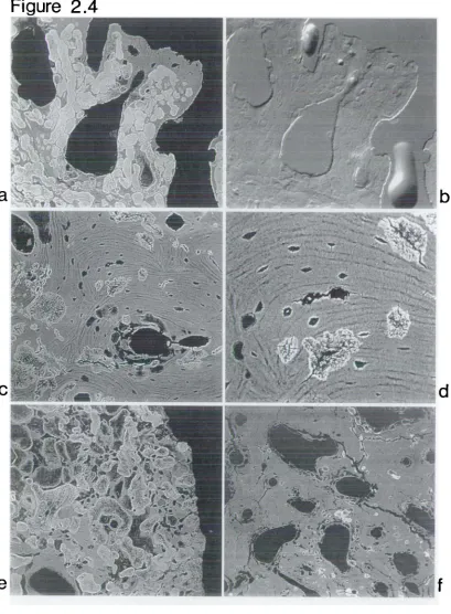
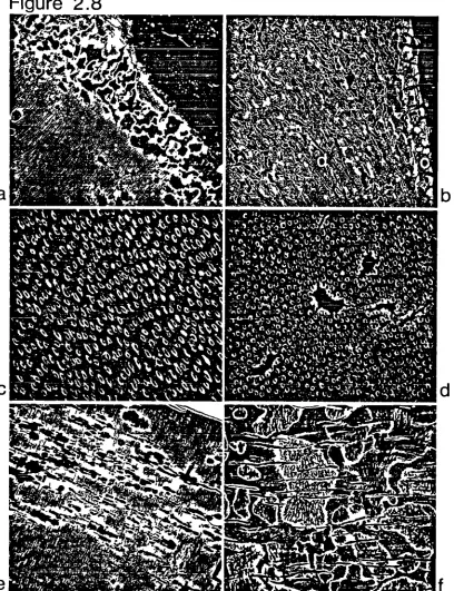
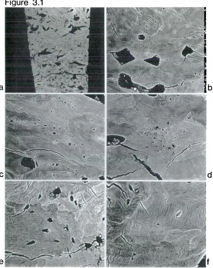
This chapter is divided into two parts end sets out to establish the usefulness of backscettered electron (BSE) imaging in a scanning electron microscope (SEM) for the characterisation of diagenetic change to archaeological human bone and teeth from terrestrial and marine contexts.
2.3 Diagenetic morphology in archaeological normal and pathological human bone This study was undertaken to consider the morphological effects of diagenesis on normal and pathological archaeological bone with particular regard to any
relative density variations found intracortically. SEM/BSE imaging has been little applied to bones and teeth
in
the study of archaeological materials. To date most workin
the archaeological field has been concerned with the atomic number '2) composition of metals, ceramics and glassware (Meeks, 1988), even though scanning electron microscopy has been utilised extensively to study surfaces on many different types of archaeological material (see Olsen, 1988). Dobney & Brothwell (1988) applied the same technique to the structure of dental calculus in archaeological samples. Other workers have employed the technique in wider studies and,in
particular, to tooth enamel structure during primatedisease, fluorosis, osteomalacia and osteogenesis imperfecta cBoyde et al., 1986). BSE imaging was considered to be an entirely appropriate method to adopt for this archaeological bone study.
2.4 Mater1l and methodology
The skeletal material used in this study was adult human femora and tibiae taken from archaeological and modern sources. The archaeological material was taken from soil-buried medieval contexts and was adult but of unknown age and sex. One sample consisted of presumed normal bone in that it had no obvious gross pathology and was taken from a macroscopically well preserved skeleton. The remaining archaeological bone examined was considered pathological, it being affected by the non-specific condition of 'periostitis', and was drawn from a miscellaneous group of bone. Two specimens were examined from this latter group: the first (specimen 1), an adult femur, macroscopically and on X-ray presented with slight striations of new bone distributed anteriorly and
Mid-shaft transverse and longitudinal sections, 10mm 2 , were taken from the
femora and similarly from the mid-shaft medial and lateral aspects of the tibiae. The sections were cut using a wet diamond-edged circular saw and then allowed to air-dry. They were then placed Into ref lux columns containing 50:50
chloroform and methanol to remove any residual water present through natural humidity within the bone. Ordinarily, reflux is used to remove any fatty
material and water present in fresh specimens (see Boyde et al, 1986). Whilst this procedure is not necessarily required for deproteinized or anorganic
specimens - vacuum embedding being a perfectly adequate alternative - it was done so that the results could, if necessary, be compared with fresh material. Reflux continued for two weeks. The sections were then removed and put through three changes of liquid polymethylmethacrylate (PMMA) every 24 hours and
subsequently placed
in
an oven set at 32°C until the PMMA solidified. The methylmethacrylate had been prepared by the 'flash distil' method outlined by Boyde et al. (1986) which prevents bubblingin
the methacrylate. Latterlyspecimens were embedded
in
methacrylate which had been distilled after Howell & Boyde (1994). The embedded specimens were polished using graded abrasives and finished with a water-dispersed 1 micron diamond abrasive on a rotary lap. Each block was mounted individually on an aluminium stub and the block face received a sputter coating of silver or carbon to render it electrically conductive and therefore suitable for SEM study in the backscattered electron imaging mode.four-segmented ring configuration and the topographical images were obtained by subtracting the east and south quadrants simultaneously from the north and west. Topographical images were necessary for the identification of bubbles, holes, edges or scratches which may have contributed to the overall signal artefactually by altering the escape volume and hence trajectories of backscattered electrons
(Howell & Boyde, 1994). The images of flat sections gave qualitative relative assessments of density changes within the specimen alongside any morphological changes. Such changes in density appear as a density map composed of dark areas which are less dense and light areas which are relatively more dense. This density dependence is the result of backscattered high energy electrons, which have an approximate escape depth of 1 micron, being released from the specimen so that backscattering increases steeply with increasing Z values. Hence, any local variations
in
Z composition within the specimen will give variations in intensity and so image, due to modulations in the BSE signal (Boyde et al., 1983, 1986; Watt, 1985).2.5 Results and discussion
All the prepared archaeological specimens, both normal and pathological, showed dramatic diagenetic change. In all specimens, the extensive changes had removed, changed or obscurred the characteristic morphology and density associated with adult human bone. Such changes therefore placed limitations on the