CHARACTERIZATION OF BIOSURFACTANTS PRODUCED BY
BACILLUS SUBTILIS USING FRESH AND WASTE COOKING OIL
ENRICHED MEDIUM
Wan Nur Afini Binti Wan Adlin1
Vivien Jong Yi Mian2+
Ang Chung Huap3
Wen-Chien Lee4
1,2,3Faculty of Applied Sciences, Universiti Teknologi MARA Sarawak, Kota Samarahan, Sarawak, Malaysia.
4Department of Chemical Engineering, National Chung Cheng University, Min-Hsiung Township, Taiwan.
(+ Corresponding author)
ABSTRACT
Article History
Received: 10 March 2020 Revised: 13 April 2020 Accepted: 18 May 2020 Published: 15 June 2020
Keyword
s
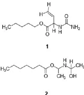
Biosurfactant Bacillus subtilis Lipopeptide Waste cooking oil Fresh cooking oil pentyl 2-ureidobut-3-enoate (1-hydroxyethyl)amino)methyl heptanoate.This study aims to compare and analyze the chemical structures of bacterial biosurfactants produced by an indigenous Bacillus subtillis strain cultured in minimal salt medium (MSM) enriched with fresh cooking oil and used cooking oils that are cheaper and easy to obtained in huge quantities. Optimization of biosurfactants was successfully conducted in two MSMs enriched with 2% (v/v) fresh cooking oil and 2% (v/v) waste cooking oil respectively. The initial pH of growth medium was fixed at pH 7.64 and the whole fermentation process was maintained in a constant temperature of 30 °C. Chemical structures analysis of biosurfactants produced at 1L scale in bioreactor were conducted using spectroscopic methods such as Nuclear Magnetic Resonance (NMR) and Fourier-Transform Infrared spectroscopy (FTIR). The chemical compound isolated from the biosurfactant produced from the waste cooking oil was pentyl 2-ureidobut-3-enoate (1) while (1-hydroxyethyl)amino)methyl heptanoate (2) was isolated from the biosurfactant produced from fresh cooking oil. This study demonstrated the ability of Bacillus subtilis to produce a low-cost biosurfactant characterized as lipopeptide.
Contribution/ Originality:
This study used cheaper yet less toxic cooking oils as substrate for biosurfactant production. Furthermore, we also successfully identified the impact of used and fresh cooking oils on the rate of bacterial growth, rate biosurfactant productions and chemical structures of biosurfactants produced using FTIR and NMR method.1. INTRODUCTION
Biosurfactants are common surface-active secondary metabolites of microorganisms, with amphipathic properties due to the presence of both hydrophilic head and lipophilic hydrocarbon tail [1].Commonly used as emulsifiers and additives, biosurfactants such as lectin have been found to improve the taste and flavor of foods without compromising consumer health [2].
Journal of Asian Scientific Research
ISSN(e):2223-1331
ISSN(p):
2226-5724
DOI: 10.18488/journal.2.2020.103.156.164 Vol. 10, No. 3, 156-164.
In recent years, some microbial biosurfactants were also successfully being developed into bioactive compounds possessing either antibacterial or antifungal properties.
Both carbon and nitrogen sources used for microbial growth can caused variation within biosurfactants molecular structures produced by the same bacterial strains [3]. To date, majorities of microbial biosurfactants were produced from growth medium that utilizing agro-industrial wastes, including bran, straw of wheat and rice, hull of soy, corn and sugarcane molasses as main carbon source [4-6].
Unlike synthetic surfactants derived from petroleum, microbial biosurfactants are more environmentally friendly due to their low toxicity and biodegradable nature [4, 7].
The main objective of this study is to characterize the molecular structures of biosurfactants produced by Bacillus subtillis growing in two different types of cooking oils: fresh cooking oil and waste cooking oil using NMR and FTIR analysis. These data are essential to optimize the subsequent pilot study for larger scale biosurfactant production.
2. MATERIALS AND METHODS
2.1. Bacterial genomic DNA Extraction
For DNA isolation, bacterial cells were obtained from one-day old culture growing in LB broth. Cell pellets were then collected through high speed centrifugation before suspended in 20 mM Tris-HCL, 2mM EDT A, 1% Triton X-00, 20 mg/mL Lysozyme or 0.2 mg/ml lysotaphin, pH 8.0 (10^9 cells add 100 μl). The mixture was then incubated at 37°C for 60 mins. Bacterial genomic DNA was extracted using Genomic DNA Isolation Kit (Protech Technology, Taiwan).
2.2. 16S rDNA Sequencing
PCR amplification for bacterial 16S rDNA was carried out using universal PCR primers, 8F (5’-AGA GTT TGA TCC TGG CTC AG-3’) and 1492R (5’-TAC GGT TAC CTT GTT ACG ACT-3’) in a reaction mixture that contains 3.0μL of 10X PCR buffer, 0.3μL of dNTP mix, 1.0μL each of PCR primers, 0.3μL of Taq DNA polymerase, and 0.3μL of bacterial genomic DNA. Sterile ultrapure water was added to the reaction mixture to achieve a final mixture volume of 30μL. PCR was then conducted in Takara PCR Thermal Cycler with an initial denaturation of 94°C for 5 mins, followed by 40 repeating cycles of denaturation at 94°C for 30 sec, primers annealing at 55°C for 30 sec, and DNA extension at 72 °C for 2.3 mins. The PCR was completed with a final extension of PCR products at 72 °C for 5 mins. PCR product obtained in this study was sent to Mission Biotech for sequencing and DNA sequences were then BLAST with data deposited in NCBI GenBank.
2.3. Initial Biosurfactant Production
Initially, bacterial isolates were cultured in a 100 mL minimal salt medium (MSM) containing (w/v) 0.2 g/L NaCl, 2.0 g/L KH2PO4, 0.2 g/L MgSO4, 0.1 g/L CaCl2.2H2O, 2.5g/L NaNO3, 3.0 g/L yeast extract and 10.0 g/L
CMC. Following that, another six replicates of these 100 mL MSM were prepared for biosurfactant production. For the first three MSM, 1%(v/v) fresh cooking palm oil was added as main carbon source while the remaining three MSM were added with 1%(v/v) waste cooking oil, also serving as main carbon source for the bacteria isolate.
Subsequently, 10 mL of bacterial culture from previous MSM were added into each of these six MSM before incubated at 30°C for 4 hours. Bacterial growths in all the MSM were monitored at every 1-hour interval using spectrophotometer.
2.4. Upscale Production of Biosurfactants
culture mixtures were then transferred into two separate 5 L bioreactors with constant temperature set at 30°C, 200 rpm and pH of 7.64. Bacterial growth was maintained and observed based on OD readings of spectrophotometry. Fermentation was stopped once the OD value has stop rising.
2.5. Oil Spreading Assay
Oil spreading assay was performed to detect the presence of crude biosurfactants during the upscaling production. This assay was carried out by adding one drop of the crude biosurfactant onto the surface of distilled water covered with 20 μL cooking oil in a Petri dish. The biosurfactant was detected by observing the presence of clear zones produce on the cooking oil.
2.6. Extraction of Crude Biosurfactants
Following the upscale fermentation, culture medium within the 5L bioreactor were then collected. At first, growth mediums were centrifuged at 12000 rpm at 25 °C for 10 mins to remove unwanted bacterial cells. The biosurfactants were then precipitated by acidification process, until the pH of mixture reached 2.0. Then, the precipitates were further collected through centrifugation. Eventually, these 200 mL of the cell-free broth from both bioreactors were collected and extracted twice with an equal volume of ethyl acetate. The solvent was further removed by reduced-pressure distillation and the dried product was washed with distilled water.
2.7. Structural Characterization of Biosurfactants
The infra-red light was used for the irradiation of molecules that gives the characteristic frequencies of every molecule for the identification of chemical compound. Infra-red spectra give information about functional groups of a molecule [8]. Perkin Elmer FTIR spectroscopy analysis was performed to evaluate if the biosurfactant induces modifications on cell surface functional groups [9].
Structural elucidation was performed using FT-NMR Bruker 400 MHz Nuclear Magnetic Resonance (NMR) spectroscopic analysis. It was based on transitions in atoms and chemical shifts in their frequency of absorption. It will allow more accurate structure and purity analysis than IR spectroscopy. Each sample of biosurfactant was dissolved in deuterated solvents. The supernatant was inserted into NMR tube after centrifugation 400 μL for the analysis [10].
3. RESULT AND DISCUSSION
3.1. Identification of Bacterial Strain
Figure-S1. PCR amplicons amplified with 8F and 1492R primers.
3.2. Bacterial Growth Rate Calculation
Prior to fermentation in bioreactors, OD readings was collected to monitor the bacterial growth in initial MSM culture. This essential stage will provide crucial information for optimizing the fermentation duration for maximizing biosurfactant production in later stages. Growth data Supplementary Table S1 indicated an exponential growth of bacteria only after 5 hours of initial fermentation. The total amount of biomass (in gram) produced during this stage is approximately 0.4440 g.
Meanwhile, for bacterial growth in the bioreactors, enriched with either fresh cooking oil or waste cooking oil, it was observed that there was continuous bacterial growth even after four hours of incubation period Supplementary Table S2. The amount of biomass obtained from the fermentation in the bioreactors were approximately 0.2456g. Growth data suggested at least four hours of incubation period are required for Bacillus subtilis used in this study to produce biosurfactant.
Besides the continuous OD readings, pH of growth medium in the bioreactors were also being continuously monitored Supplementary Table S3. An initial 3-day (Day-1 to Day-3) pH monitoring indicated a stable decrement of pH, suggesting the presence of fatty acid and biosurfactants within fermentation medium.
Table-S1. OD Value of bacterial growth taken per hour.
Time (hours) 1 2 3 4 5
OD Value 0.397 0.384 0.836 3.22 9.760
Table-S2. OD Value of biosurfactants taken per hours during four hour scaling up process.
Time (hours) 1 2 3 4
OD Value 0.108 0.141 0.237 0.418
Table-S3. pH readings recorded during biosurfactant production (day-basis).
Time (days) 1 2 3
pH Value 7.64 6.87 6.86
3.3. Effect of Different Carbon Sources for Biosurfactant Production
3.4. Effect of Additional Nitrogen Sources in Biosurfactant Production
Nitrogen sources is also another important supplement for bacterial growth and enhancing the production rate of biosurfactants. In fermentative processes, the C:N ratio will affect the build-up of metabolites [11]. High C:N ratio (low nitrogen levels) will limit the bacterial growth, leading to production of secondary metabolites. In contrast, excessive nitrogen leads to the synthesis of cellular material used in biosurfactant production[12]. In this study, NaNO3 was used and proven to be an effective limiting agent for enhancing the cellular metabolism for
biosurfactant production.
3.5. Oil Spreading Assay
Oil spreading assay is important for detecting the presence of biosurfactants. The oil displacement area is directly proportional to the surface-active compound in the solution [13]. The assay conducted in this study indicated a positive result for biosurfactant presence in culture medium using both fresh and waste cooking oils. Presence of clear zones in the oil droplets spread on the water surface, together with emulsification effects Supplementary Figure S2 suggested that the biosurfactants produced by Bacillus subtilis used in this study were highly effective in dispersing hydrocarbon-based contaminants.
Figure-S2. Positive results of oil spreading assay showing clear zone formation and emulsifying
effects on the oil droplets spread on distilled water in Petri dishes.
3.6. Physical Properties of Biosurfactants
The biosurfactants Figure 1 obtained from waste cooking oil has slightly darker brown colour compared to that from fresh cooking oil but both have strong smell and sticky in texture. The biosurfactants are very polar and fully dissolved in water but partially dissolved in acetone-methanol.
3.7. Structural Characterization of Suggested Biosurfactant Obtained from the Waste Cooking Oil
The FTIR absorption spectra Supplementary Figure S3, the strong absorption bands in the region 3291.38 cm -1 was due to the stretching vibrations of –OH group. The absorption peaks at 2921.43 cm-1 and 2852.25 cm-1
indicated the presence of methylene groups confirming the presence of aliphatic chains. The absorption peaks 1744.51 cm-1 confirmed the presence of carbonyl (-C=O) group. The peak at 1093.38 cm-1 revealed the presence of
amine (C-NH2) group and the absorption peak at 1160.49 cm-1 was assigned to the stretching vibrations due to C-O
in ester linkage. The bands at 1464.61 cm-1 and 1415.70 cm-1 are due to aliphatic chains.
The 1H NMR spectrum Supplementary Figure S4 revealed obtained from the 1 Figure 1 from waste cooking
oil indicated that the purified surfactant was a possible lipopeptide due to the presence of a long aliphatic chain (CH2
at 1.43-0.75 ppm). The chemical shift at 5.19 ppm was consistent with a proton attached to the C-3 of the hydroxyl fatty acid (3-HFA) residue and indicated that this carbon was attached to amino acid residue by an ester bond Supplementary Table S4. The intense singlet at 3.85 ppm, is similar to 1H NMR spectrum of lipopeptide
monoesters reported by other researchers, which suggests the existence of a methoxy group on the Glu or Asp amino residues [14]. The 1H NMR analysis detected an ester carbonyl group at 5.19 ppm, indicating the presence
of a lactone ring in the structure of the 1 Figure 1 [15].
Table-S4. The 1H NMR signals and the functional groups of the biosurfactant component (WCO).
δ ,ppm Functional groups
5.19 Representing ester carbonyl group 3.85 Chemical shifts of methoxy group
3.57 R-C=O-O-C-H
3.48 R-N-H
1.77 R-C=C-C-H
1.43 CH2 long aliphatic chain
1.16 The proton signals in the region belong to saturated R-CH2-R chains
1.03 The proton signals of the methyl groups, R-CH3
0.93 Terminal branching in the fatty acyl chain [-(CH3)2-CH-].
0.75 The proton signals of the methyl groups, R-CH3
Figure-S4. The 1H NMR signals of biosurfactant from waste cooking oil.
3.8. Structural Characterization of Suggested Biosurfactant Obtained from the Fresh Cooking Oil
In the FTIR spectrum Supplementary Figure S5, the strong absorption bands in the region 3368.82 cm-was
due to the stretching vibrations of –OH and –NH groups. The absorption peaks 1587.23 cm-1 confirmed the
presence of carbonyl (-C=O) group in 2 Figure 1. The absorption peaks at 1351.70 cm-1 was appeared due to the
presence of alkyl (-CH3 and –CH2) groups. The sharp peak at 1050.94 cm-1 revealed the presence of amine (C-NH2)
group.
The result obtained with 1H NMR Supplementary Figure S6 further suggested the lipopeptide nature of 2
Figure 1. The alpha-hydrogen (Hαs) of the amino acids showed resonance from 4.7-3.8 ppm. A δ = 1.20 ppm was
observed, which indicated a terminal branching in the fatty acyl chain [-(CH3)2-CH-] Supplementary Table S5.
Other multiplets in the upfield region arise as a result of the side chain protons of the amino acids, and remaining spectra confirmed the presence of β-hydroxy fatty acid [16].
In this study, potential biosurfactant bacterial strain Bacillus subtillis was identified. The biosurfactant production were optimized using waste and fresh cooking oil. The study aimed at comparing the effect of various carbon and nitrogen sources to the biosurfactant production. The optimization of biosurfactant production was done through upscaling of Bacillus subtillis in bioreactor at specific parameter to produce large scale of biosurfactant. The oil spreading assay was done to test the presence of the biosurfactant. The result showed a positive result for both biosurfactant produced from waste and fresh cooking oil. The pH had shown to give effect to the biosurfactant production which the pH between 5.5 and pH 6.0 gave the best result for the biosurfactant production. Besides, the use of vegetable oils added with molasses gave the highest yield of biosurfactant. On the other hand, sodium nitrate proved to be more effective as nitrogen sources to biosurfactant.
Table-S5. The 1HNMR signals and the functional groups of the biosurfactantcomponent (FCO).
δ ,ppm Functional groups
4.15 (CH2)2CH(OH)
4.01 R-C=O-O-C-H
3.88 R-N-H
3.10 HO-C-H
1.20 The proton signals in the region belong to saturated R-CH2-R chains
Figure-S5. FTIR analysis ofbiosurfactant produced from the fresh cooking oil.
Figure-S6. The 1H NMR signals of biosurfactant from fresh cooking oil.
Funding: This study received no specific financial support.
Competing Interests: The authors declare that they have no competing interests.
Acknowledgement: The authors highly acknowledges to several individuals and organizations
especially Dr Vivien Jong Yi Mian, Dr. Ang Chung Huap and Universiti Teknologi MARA Sarawak for guidance and relentless support in improving my research. They are highly thankful to National Chung Cheng University especially Professor Wen-Chien Lee and their students for their assistance and technical support in my research.
REFERENCES
[1] J. C. Mata-Sandoval, J. Karns, and A. Torrents, "High-performance liquid chromatography method for the characterization of rhamnolipid mixtures produced by Pseudomonas aeruginosa UG2 on corn oil," Journal of Chromatography A, vol. 864, pp. 211-220, 1999. Available at: https://doi.org/10.1016/s0021-9673(99)00979-6.
[3] A. Fiechter, "Biosurfactants: moving towards industrial application," Trends in Biotechnology, vol. 10, pp. 208-217, 1992. Available at: https://doi.org/10.1016/0167-7799(92)90215-h.
[4] M. Benincasa, "Rhamnolipid produced from agroindustrial wastes enhances hydrocarbon biodegradation in contaminated soil," Current Microbiology, vol. 54, pp. 445-449, 2007. Available at: https://doi.org/10.1007/s00284-006-0610-8.
[5] M. Nitschke and G. M. Pastore, "Biosurfactant production by Bacillus subtilis using cassave-processing effluent,"
Applied Biochemistry and Biotechnology, vol. 112, pp. 163-172, 2004. Available at: https://doi.org/10.1385/abab:112:3:163.
[6] H. Rashedi, M. M. Assadi, B. Bonakdarpour, and E. Jamshidi, "Environmental importance of rhamnolipid production from molasses as a carbon source," International Journal of Environmental Science & Technology, vol. 2, pp. 59-62, 2005. Available at: https://doi.org/10.1007/bf03325858.
[7] I. M. Banat, R. S. Makkar, and S. S. Cameotra, "Potential commercial applications of microbial surfactants," Applied Microbiology and Biotechnology, vol. 53, pp. 495-508, 2000. Available at: https://doi.org/10.1007/s002530051648. [8] A. Peele, T. C. Vekateswarulu, J. Tammineedi, L. Kanumuri, R. B. Kumar, D. V. Ramu, and P. K. Vidya, "Role of
biosurfactants in bioremediation of oil pollution-a review," Petroleum, vol. 4, pp. 241-249, 2018. Available at: https://doi.org/10.1016/j.petlm.2018.03.007.
[9] F. J. De Freitas, E. A. a. Vieira, and M. Nitschke, "The antibacterial activity of rhamnolipid biosurfactant is pH dependent," Food Research International, vol. 116, pp. 737-744, 2018.
[10] R. Consonni, D. Polla, and L. Cagliani, "Organic and conventional coffee differentiation by NMR spectroscopy," Food Control, vol. 94, pp. 284-288, 2018. Available at: https://doi.org/10.1016/j.foodcont.2018.07.013.
[11] M. Robert, M. Mercade, M. Bosch, J. Parra, M. Espuny, M. Manresa, and J. Guinea, "Effect of the carbon source on biosurfactant production by Pseudomonas aeruginosa 44T1," Biotechnology Letters, vol. 11, pp. 871-874, 1989. Available at: https://doi.org/10.1007/bf01026843.
[12] L. Santa Anna, G. Sebastian, N. Pereira, T. Alves, E. Menezes, and D. Freire, "Production of biosurfactant from a new and promising strain of Pseudomonas aeruginosa PA1," Applied Biochemistry and Biotechnology, vol. 91, pp. 459-467, 2001. Available at: https://doi.org/10.1385/abab:91-93:1-9:459.
[13] M. Morikawa, Y. Hirata, and T. Imanaka, "A study on the structure–function relationship of lipopeptide biosurfactants," Biochimica et Biophysica Acta (BBA)-Molecular and Cell Biology of Lipids, vol. 1488, pp. 211-218, 2000. Available at: https://doi.org/10.1016/s1388-1981(00)00124-4.
[14] J.-S. Tang, H. Gao, K. Hong, Y. Yu, M.-M. Jiang, H.-P. Lin, W.-C. Ye, and X.-S. Yao, "Complete assignments of 1H and 13C NMR spectral data of nine surfactin isomers," Magnetic Resonance in Chemistry, vol. 45, pp. 792-796, 2007. Available at: https://doi.org/10.1002/mrc.2048.
[15] X.-Y. Liu, S.-Z. Yang, and B.-Z. Mu, "Production and characterization of a C15-surfactin-O-methyl ester by a lipopeptide producing strain Bacillus subtilis HSO121," Process Biochemistry, vol. 44, pp. 1144-1151, 2009. Available at: https://doi.org/10.1016/j.procbio.2009.06.014.
[16] S. Joshi, Y. Al-Wahaibi, S. Al-Bahry, A. Elshafie, A. Al-Bemani, A. Al-Bahri, and M. Al-Mandhari, "Production, characterization, and application of Bacillus licheniformis W16 Biosurfactant in enhancing oil recovery," Frontiers in Microbiology, vol. 7, pp. 1853-1853, 2016. Available at: https://doi.org/10.3389/fmicb.2016.01853.