Current Perspective of Beta Thalassemia and Its
Treatment Strategies
Shaukat Ali1,*, Shumaila Mumtaz1, Hafiz Abdullah Shakir2, Hafiz Muhammad Tahir1 and Tafail Akbar Mughal1
1Department of Zoology, Government College University, Lahore, 54000-Lahore, Pakistan
2Department of Zoology, University of the Punjab, Lahore, Pakistan
*Corresponding author: Department of Zoology, Government College University, Lahore, 54000-Lahore, Pakistan. Ph: +923054190596. Email: [email protected].
Abstract: Thalassemia is genetic blood disease cause by absence or decrease of one or more of the globin chain synthesis. Beta thalassemia is characterized by one or more mutations in beta globin gene. Absence or reduced amount the of beta globin chains cause ineffective erythropoiesis which leads to anemia. Beta thalassemia has been further divided into three main forms: Thalassemia minor/silent carrier, major and intermedia. More severe form is thalassemia major in which patients depend upon blood transfusion for survival and high level of iron occur as a consequence of consistent blood transfusion. Over loaded iron invokes the synthesis of reactive oxygen species that are toxic in redundancy and triggering the impairment to vascular, endocrine and hepatic system. Thalassemia can be diagnosed and detected through various laboratory tests such as blood smear, prenatal testing (genetic testing of amniotic fluid), DNA analysis (genetic testing) and complete blood count. Treatment of thalassemia intermedia is symptomatic but it can also be managed by splenectomy and folic supplementation. While thalassemia major can be treated by transplantation of bone marrow, regular transfusion of blood and iron chelation treatment, stimulation of fetal hemoglobin production, hematopoietic stem cell transplantation and gene therapy.
Key words: Thalassemia; iron overload; chelation therapy; gene therapy; blood transfusion 1. Introduction
Thalassemia is categorized by abnormal production or reduction in rate of formation of normal alpha (α) globin or beta (β) globin subunits of hemoglobin (Hb) A. Hemoglobin A comprises of
normal adult such as HbA, HbA2 and HbF that consist of α2; β2, α2; δ2 and α2; γ2 respectively as shown in Figure 1 [2].
Thalassemia is categorized as β, α, δ γ, δβ as well as γδβ, depending upon which globin chain is affected. While α and β thalassemia are two major categories of thalassemia and occurrence depends upon two genes for β globin and four genes for alpha thalassemia [3, 4]. It is produced by more than numerous hundred modifications in the consistent DNA segment. The unpaired globin chains are not stable.They precipitous in cells which lead to immature destruction of precursors of RBCs and shortening of life-span of mature RBCs in blood. Hemoglobin breaks down in iron and heme that catalyze chemical reactions and produce the reactive oxygen species (ROS), which cause the impairment of hepatocytes as well as functions of islets of Langerhans [1].
Beta-thalassemia is described by absent or reduction in rate of synthesis of βglobin chain [5]. It was first time defined by Cooley and Lee in 1925 [6]. The β thalassemia is a consequence of substitutions of bases on introns, exons as well as on the promoter regions of β globin genes while αthalassemia is a consequence of deletions that remove α gene [7]. It is further categorized according to decreased (β+) or absent (β0) globin chain production which might lead to
microcytic and hypochromic anemia as well as wide range of syndromic forms [8]. 3. Types of Beta thalassemia
Beta-thalassemia major
Beta-thalassemia intermedia
Thalassemia intermedia is a hetrogenous genetic mutation in which individual have little bit ability for production of β chain of hemoglobin (B+/B+, B+/B0). In some situations both α and β mutations co-occur [5]. It occurs between 2 and 6 years, have milder anemia and level of
hemoglobin varies between 7 and 9-10 g/dL and transfusion of blood is not needed. The sufferer can survive without or only occasionally require blood transfusion as represented in Table I [11].
When bone marrow expand with increasing age, patients may develop many complications such as growth retardation, bone abnormalities infertility and high concentration of iron in several soft tissue owing to elevated the iron immersion in gastro intestinal tract as a result of hemolysis [12].
Beta thalassemia minor
It is also termed as thalassemia carrier/trait that occurs when one copy of β globin gene is
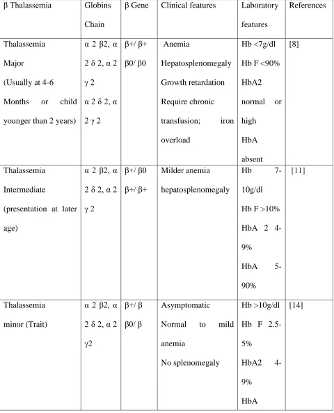
Table I. Beta thalassemia genotypes clinical features/laboratory features.
β Thalassemia Globins
Chain
β Gene Clinical features Laboratory
features
References
Thalassemia Major
(Usually at 4-6 Months or child younger than 2 years)
α 2 β2, α 2 δ 2, α 2 γ 2 α 2 δ 2, α 2 γ 2
β+/ β+ β0/ β0 Anemia Hepatosplenomegaly Growth retardation Require chronic transfusion; iron overload
Hb <7g/dl Hb F <90% HbA2 normal or high HbA absent [8] Thalassemia Intermediate
(presentation at later age)
α 2 β2, α 2 δ 2, α 2 γ 2
β+/ β0 β+/ β+
Milder anemia hepatosplenomegaly
Hb
7-10g/dl Hb F >10% HbA 2 4-9%
HbA
5-90%
[11]
Thalassemia minor (Trait)
α 2 β2, α 2 δ 2, α 2 γ2
β+/ β β0/ β
Asymptomatic
Normal to mild anemia
No splenomegaly
Hb >10g/dl Hb F 2.5-5%
HbA2 4-9%
HbA >90%
4. Hereditary transmission
Beta thalassemia is a congenital autosomal recessive condition. Children are obligate heterozygotes when parents are affected and bring mutation in a single copy of β globin chromosome. At beginning, every offspring of heterozygous parentages has 25% chance of being unaffected and not carrier, 25% possibility of being affected and 50% possibility of being an asymptomatic carrier [5, 15].
5. Pathophysiology
6. Medical Indices/Complications Iron overload
Hepatitis
Patients of thalassemia have a high threat of virus-related contagion. For example, pathological hepatitis due to prolong receiving blood and blood transfusion products. Incidence of hepatitis B for thalassemic patients and donors can greatly reduce due to availability of vaccines but hepatitis C is a greatly difficult among these patients due to lack of a vaccine [31,32].
Osteoporosis
7.Diagnosis
Thalassemia can be diagnose and detect through various laboratory tests such as blood smear, prenatal testing (genetic testing of amniotic fluid), DNA analysis (genetic testing) complete blood count (CBC), iron studies and hemoglobinopathy [35]. DNA analysis test is used to help detect mutations in genes of βand αglobin-chain. It can also help to determine carrier status of thalassemia but it not done routinely. Some mutations shows no symptoms while some decline the formation of βglobin chain in αthalassemia as well as some completely prevent the making of beta globin [36].
Hematologic diagnosis
Thalassemia intermedia is categorized by MCV between 50 and 80 fL, Hb level from 7 to 10 g/dl as well as MCH from 16 and 24 pg. Thalassemia carrier/silent is categorized by increased Hb A2 level with decline MCH and MCV. Thalassemia major is described by decline MCV > 50 < 70 fl, MCH > 12< 20 pg. and (>10 g/dl) Hb level [5, 15].
8. Prevention
alteration in the population of homozygote. Some republics such as Turkey, Lebanon, Iran, Canada, Egypt, Malaysia, Pakistan and China also have Thalassemia Control Programs [40]. 9. Treatment
No treatment is needed to the people who have thalassemia traits. They might be deliberate genetic counseling because mutant gene can pass to their offspring (Mustafa et al, 2016). Individuals with βthalassemia intermedia will experience a mild anemia in their life. They may be sentient like normal people however consistent monitoring will require and blood transfusion can be occasionally needed. Iron supplementation is not given, while folic acid supplementation is often recommended [41]. Individuals with thalassemia intermedia will also experience a hypersplenism which may cause growth retardation and worsening anemia and various other mechanical disturbances due to splenomegaly. Treatment of thalassemia intermedia is symptomatic but it can also be managed by splenectomy [42].
Splenectomy
Iron chelation therapy
Transplantation of bone marrow
Relocation of bone marrow is still remains the main conclusive treatment, reachable for patients of thalassemia [52]. Main efficacious transplantation of bone marrow was completed in 1980s. The results in young patients are 3 % mortality rate and 87% thalassemia free survival. But BMT has few disadvantages such as human leukocyte Antigen matched compatible donor is required for this remedial process [53]. The best results with very young individuals are: rejection rate is 23 %, mortality rate is 7% and thalassemia frees survival rate is 70 %. Treatment for thalassemia through bone marrow transplantation is still not available for all Indian patients [54].
In low socio-economic countries, the current management available for majority of β-thalassemia major patient is effective chelating therapy and management of complications of overloaded iron as well as consistent transfusion of packed red cells [55].
Blood transfusion
and activity of life [57] Frequency of blood transfusion depends on numerous causes such as level of hematocrit and Hb as well as weight of the patient [47]. Blood transfusion therapy should not transfer RBCs more than 15 to 20 ml/kg daily to evade a profligate rise in volume of blood. Efficiency of transfusion therapy should be examined via pre and post transfusion level of hemoglobin as well as hematocrit because these quantities can enable to monitor iron intake and requirement of RBCs [5].
Gene therapy
but HSPCs derived from iPSC is not yet feasible for HSCT because these cells have limitation for multiply in bone marrow.
Transplantation of hematopoietic stem Cell
bone marrow of healthy persons are sequestered and transmitted to patients of thalassemia. Nearly 80 percent of transplant recipients were successful by this treatment [61]. Graft vs host disease (GVHD) is the most significant and hazardous problem in transplantation of bone marrow which might be leads to death of transfer recipients [62].
Induction of fetal hemoglobin production
Individuals with long term manifestation of thalassemia, induction fetal hemoglobin enhance the lifetime of RBCs. There are various drugs which are used for stimulation of production of fetal hemoglobin such as hydroxyl urea. Hydroxyurea might be used for cure of both thalassemia and sickle cell disease. Hydroxyurea enhances the production of gamma-globin and also improves hematological and quantifiable signs in patients of thalassemia intermedia [9, 63]. Hydroxyurea acts as a cytotoxic compound for synthesis phase of the cell cycle and also a ribonucleotide reductase inhibitor [64]. It regulates and increase expressions of fetal hemoglobin gene 2 related to apoptosis and cell cycle as well as suppress the countenance of GATA-1 gene. It can also induce proliferation of progenitor cells and enhance the amount of erythropoietin [65].
10. Conclusions
Author contributions
SA, SM, HAS, HMT and TAM reviewed the literature and wrote the manuscript.
Disclosure
References
1. Adly, A. A. M.; Sherif, N. H.; Ismail, E. A. R.; Zaher, Y. A.; Farouk, A.;
El-Refaey, A. M.; Wahba, M. S. Vascular dysfunction in patients with young β-thalassemia:
relation to cardiovascular complications and subclinical atherosclerosis. Clin. Appl.
Throm/Hemost. 2015, 21, 733-744.
2. Thein, S. L. Molecular basis of β thalassemia and potential therapeutic targets. Blood
Cells Mol. Dis. 2018, 70, 54-65.
3. Sirachainan, N.; Chuansumrit, A.; Kadegasem, P.; Sasanakul, W.;
Wongwerawattanakoon, P.; Mahaklan, L. Normal hemostatic parameters in children and
young adults with α-thalassemia diseases. Throm. Res. 2016, 146, 35-42.
4. Gibbs, G.; Burdick, C.O. Separating thalassemia trait and iron deficiency by even simpler
inspection. Am. J. Clin. Patholol. 2009, 132, 643-644.
5. Galanello, R.; Origa, R. Beta-thalassemia. Orphanet J. Rare Dis. 2010, 5(1), 11.
6. Franco, S. S.; De Falco, L.; Ghaffari, S.; Brugnara, C.; Sinclair, D. A.; Iolascon, A.;
Rimmelé, P. Resveratrol accelerates erythroid maturation by activation of FoxO3 and
ameliorates anemia in beta-thalassemic mice. Haematologica 2014, 99, 267-275.
7. Stauder, R.; Valent, P.; Theurl, I. Anemia at older age: etiologies, clinical implications,
and management. Blood 2018, 5, 505-514.
8. Lei, M.; Sun, L.; Luo, X. S.; Yang, X.; Yu, F.; Chen, X.; Wang, Z. (Distinguishing iron
deficiency anemia from thalassemia by the red blood cell lifespan with a simple CO
9. Tari, K.; Ardalan, P.; Abbaszadehdibavar, M.; Atashi, A.; Jalili, A.; Gheidishahran, M.
Thalassemia an update: molecular basis, clinical features and treatment. Int. J. Biomed.
Public Health 2018, 1, 48-58.
10. Borgna-Pignatti, C. A. T. E. R. I. N. A.; Rugolotto, S. I. M. O. N. E.; De Stefano, P.;
Zhao, H. U. A. Q. I. N. G.; Cappellini, M. D.; Del Vecchio, G. C.; Piga, A. Survival and
complications in patients with thalassemia major treated with transfusion and
deferoxamine. Haematologica 2004, 89, 1187-1193.
11. Birgens, H.; Ljung, R. The thalassaemia syndromes. Scand. J. Clin. Lab. Invest. 2007,
67, 11-26.
12. Taher, A. T.; Musallam, K. M.; Cappellini, M. D.; Weatherall, D. J. Optimal
management of β thalassaemia intermedia. Brit. J. Haematol. 2011, 152, 512-523.
13. Urbinati, F.; Madigan, C.; Malik, P. Pathophysiology and therapy for
haemoglobinopathies; part II: Thalassaemias. Exp. Rev. Mol. Med. 2006, 8, 1-26.
14. Moi, P.; Faà, V.; Marini, M. G.; Asunis, I.; Ibba, G.; Cao, A.; Rosatelli, M. C. A novel
silent β‐thalassemia mutation in the distal CACCC box affects the binding and
responsiveness to EKLF. Brit. J. Haematol. 2004, 126, 881-884.
15. Canatan, D.; Koç, N. The effect of transfusion on pulmonary function tests in patients
with thalassemia. Turk J Haematol. 2004, 3, 137-139.
16.Shariati, L.; Modaress, M.; Khanahmad, H.; Hejazi, Z.; Tabatabaiefar, M. A.; Salehi, M.; Modarressi, M. H. Comparison of different methods for erythroid differentiation in the
K562 cell line. Biotechnol. Lett. 2016, 38, 1243-1250.
17. Sangkhae, V.; Nemeth, E. Regulation of the iron homeostatic hormone hepcidin. Adv.
18.Hoban, M.D.; Cost, G.J.; Mendel, M.C.; Romero, Z., Kaufman, M.L.; Joglekar, A.V.; Ho, M.; Lumaquin, D.; Gray, D.; Lill, G.R.; Cooper, A.R. Correction of the sickle cell
disease mutation in human hematopoietic stem/progenitor cells. Blood 2015, 125,
2597-2604.
19.Chakrabarti, A.; Bhattacharya, D.; Deb, S.; & Chakraborty, M. Differential thermal stability and oxidative vulnerability of the hemoglobin variants, HbA2 and HbE. PloS
one, 2013, 8, 818-820.
20.Ganz, T.; Nemeth, E. Hepcidin and disorders of iron metabolism. Ann. Rev. Med. 2011, 62, 347-360.
21.Camaschella, C.; Nai, A. Ineffective erythropoiesis and regulation of iron status in iron loading anaemias. Brit. J. Haematol. 2016, 172, 512-523.
22. Kim, A.; Nemeth, E. New insights into iron regulation and erythropoiesis. Curr. Opin.
Hematol. 2015, 3, 199.
23.Finianos, A.; Matar, C.; Taher, A. Hepatocellular carcinoma in β-thalassemia patients:
Review of the literature with molecular insight into liver carcinogenesis. Int. J. Mol. Sci.
2018, 19, 4070.
24. Preza, G. C.; Ruchala, P.; Pinon, R.; Ramos, E.; Qiao, B.; Peralta, M. A.; Nemeth, E.
Minihepcidins are rationally designed small peptides that mimic hepcidin activity in mice
and may be useful for the treatment of iron overload. J. Clin. Invest. 2011, 121,
4880-4888
25.Evans, R. W.; Rafique, R.; Zarea, A.; Rapisarda, C.; Cammack, R.; Evans, P. J.; Hider, R. C. Nature of non-transferrin-bound iron: studies on iron citrate complexes and
26.Hershko, C. Pathogenesis and management of iron toxicity in thalassemia. Ann. N. Y, Acad. Sci. 2010, 1202, 1-9.
27.Wang, C. Y.; Jenkitkasemwong, S.; Duarte, S.; Sparkman, B. K.; Shawki, A.; Mackenzie, B.; Knutson, M. D. ZIP8 is an iron and zinc transporter whose cell-surface expression is
up-regulated by cellular iron loading. J. Biol. Chem. 2012, 287, 34032-34043.
28.Fernandes, J. L.; Loggetto, S. R.; Veríssimo, M. P.; Fertrin, K. Y.; Baldanzi, G. R.; Fioravante, L. A.; Coelho, O. R. A randomized trial of amlodipine in addition to standard
chelation therapy in patients with thalassemia major. Blood 2016, 128, 1555-1561.
29.Glickstein, H.; El, R. B.; Link, G.; Breuer, W.; Konijn, A. M.; Hershko, C.; Cabantchik, Z. I. Action of chelators in iron-loaded cardiac cells: accessibility to intracellular labile
iron and functional consequences. Blood 2006, 108, 3195-3203.
30.Brissot, P.; Ropert, M.; Le Lan, C.; Loréal, O. Non-transferrin bound iron: a key role in iron overload and iron toxicity. Biochim. Biophys. Acta. (BBA)-Gen. Subj. 2012, 1820,
403-410.
31. Soliman, A.; Yassin, M.; Al Yafei, F.; Al-Naimi, L.; Almarri, N.; Sabt, A.; De Sanctis,
V. Longitudinal study on liver functions in patients with thalassemia major before and
after deferasirox (DFX) therapy. Mediterr. J. Hematol. Infect. Dis. 2014, 6, 322-330
32. Mousa, S.M.O.; Afifi, M.F.; Saedii, A.A.; El-Setohy, A.A. Ischemia modified albumin in
children with transfusion-dependent β-thalassemia: a new marker for an old
problem. Egypt. Haematol. 2016, (2), 45.
33. Terpos, E.; Voskaridou, E. Treatment options for thalassemia patients with
34.Mancuso, A. Hepatocellular carcinoma in thalassemia: A critical review. World J.
Hepatol. 2010, 2, 171.
35.Alqahtani, R. S.; Bedaiwi, A. A.; Alburkani, A. M.; AlFahed, M. M.; Alhoraibi, R. A.; Tarawah, A. M. Knowledge and response of the community to premarital screening
program (Sickle Cell Anemia\Thalassemia). AlMadinah Saudi Arabia J. Appl. Hematol.
2018, 9, 59.
36.Sagar, C. S.; Kumar, R.; Sharma, D. C.; Kishor, P. Alpha hemoglobin stabilizing protein: Its causal relationship with the severity of beta thalassemia. Blood Cells Mol. Dis. 2015,
55, 104-107.
37.Origa, R.; Comitini, F. Pregnancy in thalassemia. Mediterr. J. Hematol. Infect. Dis. 2019, 11, 120.
38.Mavrou, A.; Kouvidi, E.; Antsaklis, A.; Souka, A.; Kitsiou Tzeli, S.; Kolialexi, A. Identification of nucleated red blood cells in maternal circulation: a second step in
screening for fetal aneuploidies and pregnancy complications. Prenatal Diagnosis:
Published in Affiliation With the International Society for Prenatal Diagnosis 2007, 27,
150-153.
39.Colah, R.; Gorakshakar, A.; Nadkarni, A. Global burden, distribution and prevention of β-thalassemias and hemoglobin E disorders. Exp. Rev. Hematol. 2010, 3(1), 103-117.
40.Madan, N.; Sharma, S.; Sood, S. K.; Colah, R.; Bhatia, H. M. Frequency of β-thalassemia trait and other hemoglobinopathies in northern and western India. Ind. J. Hum. Genet.
41. Kumar, R.; Sharma, D. C.; Kishor, P. (Hb E/β-Thalassemia: The Second Most Common
Cause of Transfusion-Dependent Thalassemia in the Gwalior-Chambal Region of Central
India. Hemoglobin 2012, 36, 485-490
42.Origa, R. Combination therapies in iron chelation. Thalassemia Reports, 2014, 4(3). 43.Pecorari, L.; Savelli, A.; Guna, C. D.; Fracchia, S.; Borgna-Pignatti, C. The role of
splenectomy in thalassemia major. An update. Acta Pediatrica Mediterr. 2008, 24, 57-60.
44.Ikeda, M.; Sekimoto, M.; Takiguchi, S.; Kubota, M.; Ikenaga, M.; Yamamoto, H.; Tatsuta, M. High incidence of thrombosis of the portal venous system after laparoscopic
splenectomy: a prospective study with contrast-enhanced CT scan. Ann. Surg. 2005, 241,
208.
45.Sarker, N. R.; Ghosh, A. K.; Saha, S. K.; Shahriar, A. Recent advances in the management of Thalassaemia: A Review Update. J. Shaheed Suhrawardy Med. Coll.
2014, 6, 31-37.
46.Adewoyin, A.S; Oyewale, O.A. Complications of allogeneic blood transfusion: Current approach to diagnosis and management. Int. Blood Res. Rev. 2015, 3, 135-151.
47.Taher, A.T.; Weatherall, D.J.; Cappellini, M.D. Thalassaemia. The Lancet, 2018, 391, 155-167.
48.Crisponi, G.; Nurchi, V. M.; Lachowicz, J. I. Iron Chelation for Iron Overload in Thalassemia. Essential Metals in Medicine: Therapeutic Use and Toxicity of Metal Ions
in the Clinic 2019, 19, 49.
49.Piga, A.; Gaglioti, C.; Fogliacco, E.; Tricta, F. Comparative effects of deferiprone and deferoxamine on survival and cardiac disease in patients with thalassemia major: a
50.Cappellini, M. D.; Porter, J.; El-Beshlawy, A.; Li, C. K.; Seymour, J. F.; Elalfy, M.; Lin, K. H. Tailoring iron chelation by iron intake and serum ferritin: the prospective EPIC
study of deferasirox in 1744 patients with transfusion-dependent
anemias. Haematologica 2010, 95, 557-566.
51.Cappellini, M. D.; Cohen, A.; Piga, A.; Bejaoui, M.; Perrotta, S.; Agaoglu, L.; Capra, M. A phase 3 study of deferasirox (ICL670), a once-daily oral iron chelator, in patients with
β-thalassemia. Blood 2006, 107, 3455-3462.
52.Rivière, I.; Dunbar, C. E.; Sadelain, M. Hematopoietic stem cell engineering at a crossroads. Blood 2012; 119, 1107-1116.
53.Sabloff, M.; Chandy, M.; Wang, Z.; Logan, B. R.; Ghavamzadeh, A.; Li, C. K.; Hale, G. A. HLA-matched sibling bone marrow transplantation for β-thalassemia major. Blood
2011, 117, 1745-1750.
54.Sharma, D.C.; Singhal, S.; Woike, P.; Tomar, A.S.; Rawat, N.; Arya, A.; Gaur, R. Red Blood Cells Alloimmunization and Transfusion Strategy in Transfusion Dependent B-Thalassemia Patients. IOSR-JDMS, 2016, 15, 10-14.
55.Ganguly, S.; Bradley, J. P.; Patel, J. S.; Tilzer, L. Role of transfusion in stem cell transplantation: a freedom-from-transfusion (FFT), cost and survival analysis. J. Med.
Econo. 2010, 13, 55-62.
56.Cihan, M.K.; Belen, B.; Bolat, F., Bülbül, Ö.G.; Korgalı, E.Ü.; Koçak, Ü.. The Impact of Transfusion and Chelation on Oxidative Stress in Immigrant Syrian Children with
β-Thalassemia. Ind. J. Hematol. Blood Transfus. 2017,4, 552-558.
57.Kadhim, K.A.; Baldawi, K.H.; Lami, F.H. Prevalence, incidence, trend, and
58. Breda, L.; Kleinert, D. A.; Casu, C.; Casula, L.; Cartegni, L.; Fibach, E.; Rivella, S. A
preclinical approach for gene therapy of β‐thalassemia. Ann. N. Y. Acad. Sci. 2010, 1202,
134-140.
59. Takahashi, K.; Yamanaka, S. Induction of pluripotent stem cells from mouse embryonic
and adult fibroblast cultures by defined factors. Cell 2006, 126, 663–676.
60. Angelucci, E.; Matthes-Martin, S.; Baronciani, D.; Bernaudin, F.; Bonanomi, S.;
Cappellini, M.D. Hematopoietic stem cell transplantation in thalassemia major and sickle cell disease: indications and management recommendations from an international expert panel. Haematologica 2014, 99, 811-820.
61.Bernardo, M.E.; Piras, E.; Vacca, A.; Giorgiani, G.; Zecca, M.; Bertaina, A. Allogeneic hematopoietic stem cell transplantation in thalassemia major: results of a reduced-toxicity conditioning regimen based on the use of treosulfan. Blood 2012, 120, 473-476.
62.King, A.; Shenoy, SEvidence-based focused review of the status of hematopoietic stem cell transplantation as treatment of sickle cell disease and thalassemia. Blood 2014, 123 3089-3094.
63.Wilber, A.; Hargrove, P.W.; Kim, Y.STherapeutic levels of fetal hemoglobin in erythroid progeny of β-thalassemic CD34+ cells after lentiviral vector-mediated gene transfer.
Blood 2011, 117, 2817–26.
64.Fard, A.D.; Hosseini, S.A.; Shahjahani, M.; Salari, F.; Jaseb, K. Evaluation of novel fetal hemoglobin inducer drugs in treatment of β-hemoglobinopathy disorders. Int. J. Hematol
65. Tang, D.C.; Zhu, J.; Liu, W.; Chin, K.; Sun, J.; Chen, L. The hydroxyurea-induced small