Dissertation zur Erlangung des Doktorgrades
der Fakultät für Chemie und Pharmazie
der Ludwig
‐
Maximilians
‐
Universität München
"Acid-labile surface modification of
four-arm oligoaminoamide pDNA polyplexes
balances shielding and gene transfer activity
in vitro
and
in vivo
"
Linda Beckert
aus
Dresden, Deutschland
3 Erklärung
Diese Dissertation wurde im Sinne von § 7 der Promotionsordnung vom 28. November 2011 von Herrn Prof. Dr. Ernst Wagner betreut.
Eidesstattliche Versicherung
Diese Dissertation wurde eigenständig und ohne unerlaubte Hilfe erarbeitet.
München, 05.07.2016
...
Linda Beckert
Dissertation eingereicht am: 05.07.2016
1. Gutachter: Prof. Dr. Ernst Wagner
2. Gutachter: Prof. Dr. Olivia Merkel
5
7
Table of content
1. Introduction ... 11
1.1. Gene therapy ... 11
1.2. Challenges in DNA delivery via non-viral carriers ... 12
1.3. Design of bioresponsive non-viral carriers for gene delivery ... 15
1.3.1. Bioresponsive stability of non-viral carriers ... 15
1.3.2. Bioresponsive shielding of the surface of non-viral carriers ... 19
1.3.3. Targeting of non-viral carriers ... 22
1.4. Aims of the thesis ... 24
2. Materials and Methods ... 26
2.1. Materials ... 26
2.1.1. Reagents and Solvents ... 26
2.1.2. Equipment for solid-phase assisted peptide synthesis (SPPS) ... 30
2.1.3. Cell culture ... 30
2.1.4. Nucleic acids ... 31
2.2. Methods ... 31
2.2.1. Synthesis of 3-(azidomethyl)-4-methyl-2,5-furandione (AzMMMan) .... 31
2.2.2. Synthesis of Fmoc/Boc-protected succinyl-pentaethylene hexamine (Fmoc/Boc-Sph) ... 32
2.2.3. Solid-phase assisted peptide synthesis ... 32
2.2.3.1. Loading of resins ... 33
2.2.3.2. General procedure of SPPS ... 34
2.2.3.3. Cleavage of oligomers and targeting ligand ... 35
2.2.3.4. Kaiser Test ... 36
2.2.4. Synthesis of shielding polymers ... 36
8 2.2.4.2. Synthesis of bifunctional FolA-PEG(5000)-AzMMMan and
bifunctional FolA-PEG(5000)-NHS via FolA-PEG6-cysteamine ... 36
2.2.4.3. Synthesis of bifunctional FolA-PEG(5000)-AzMMMan and bifunctional FolA-PEG(5000)-NHS via NHS-FolA ... 37
2.2.4.4. Synthesis of monovalent pHPMA-AzMMMan and monovalent pHPMA-TT ... 38
2.2.4.5. Synthesis of multivalent pHPMA-AzMMMan and multivalent pHPMA-TT ... 38
2.2.4.6. Polyplex formation ... 38
2.2.4.7. Measurement of particle size and zeta potential ... 39
2.2.4.8. Agarose gel shift assay ... 39
2.2.4.9. Ethidium bromide exclusion assay (EtBr assay) ... 39
2.2.4.10. Oligomer buffering capacity ... 39
2.2.4.11. Cell culture ... 40
2.2.4.12. Luciferase gene expression ... 40
2.2.4.13. Cell viability assay (MTT) ... 41
2.2.4.14. Cellular uptake: flow cytometry studies ... 41
2.2.4.15. Intracellular localization: laser scanning microscopy (LSM) ... 42
2.2.4.16. In vivo delivery and expression ... 42
2.2.4.17. Statistical analysis... 42
3. Results ... 43
3.1. Designing new sequence-defined oligomers: evaluation of stabilizing effects of lysine residues on DNA polyplexes ... 43
3.1.1. Biophysical characterization of oligomers concerning DNA binding ability, particle size, zeta potential and buffering capacity ... 47
3.1.2. In vitro characterization of DNA polyplexes ... 52
3.2. Design and biophysical characterization of shielded polyplexes ... 54
3.2.1. Biophysical characterization of shielded polyplexes ... 55
9
3.2.3. In vivo evaluation of shielded polyplexes ... 66
3.3. FolA receptor-directed pCMVL/784 polyplexes for gene transfer in vitro ... 68
3.3.1. Synthesis of the post-PEGylation reagents FolA-PEG(5000)-AzMMMan and FolA-PEG(5000)-NHS ... 69
3.3.2. Biophysical characterization of FolA-targeted, stably or pH-sensitively shielded pCMVL/784 polyplexes ... 74
3.3.3. In vitro characterization of FolA-targeted, stably or pH-sensitively shielded pCMVL/784 polyplexes ... 77
4. Discussion ... 80
4.1. Evaluation of new sequence-defined oligomers concerning stabilizing effects of lysine residues on DNA polyplexes ... 80
4.2. Evaluation of pH-sensitively or stably modified PEG or pHPMA-shielded DNA polyplexes ... 83
4.3. Evaluation of FolA-targeted pH-sensitively or stably PEGylated DNA polyplexes ... 86
5. Summary ... 90
6. References ... 92
7. Appendix ... 105
7.1. Abbreviations ... 105
7.2. Analytical data ... 107
7.3. Summarized spectral data ... 108
7.4. Publication ... 110
7.5. Poster presentation ... 110
11
1. Introduction
1.1. Gene therapy
Gene therapy is a fast-growing field of medical research aiming at the introduction of therapeutic genes in the human body to repair malfunctions. In this concept, a disease-causing gene is silenced or restored making gene therapy an interesting alternative for the treatment of gene-associated defects like cancer or inflammatory diseases [1-3]. Genes are carriers of the genetic information, which provide the biological code that determines the specific functions of a cell. The origins of gene therapy can be traced back to the science of genetics. Researchers like Francis Crick established this branch of biology upon the realization that: "Almost all aspects of life are engineered at the molecular level, and without understanding molecules we can only have a very sketchy understanding of life itself." Consequently, major milestones in understanding the basis of life were the elucidation of the mechanisms of genetic transmission [4], the identification of the genetic material as DNA [5] and the clarification of the DNA structure [6]. Those findings enabled the first concept of gene therapy proposed by Friedman and Roblin in 1972 [7]. It was preceded by the development of new sequencing technologies and the deciphering of the human genome [8-10] that enabled the identification of different gene-associated diseases [11-13]. However, it took more than 16 years and several drawbacks for the approval of the first gene therapy trial in 1990 by the US Food and Drugs Administration (FDA) [14, 15]. This trial focused on the treatment of the severe combined immune deficiency (SCID), a monogenetic disorder characterized by adenosine deaminase deficiency [16]. Four years later cancer gene therapy was introduced into the clinical research. The first FDA-approved cancer trial was performed with a carrier expressing antisense IGF-I RNA aiming at the treatment of glioblastoma multiform. This strategy was efficient due to a strong immune and apoptotic reaction as a result of the anti-tumor mechanism of IGF-I antisense [17]. However, gene therapy research was halted in the USA and EU owing to an excessive immune reaction in one patient causing his death in 1999 [15] and the development of leukemia-like illness of two other patients in 2002 [18].
12 Irrespectively of the great potential, the main challenge for the implementation of gene therapeutics from bench to bedside is the achievement of highly efficient, safe and target-specific gene delivery carriers [20]. Regarding efficiency and specificity, nature offers a good example, i.e. viruses. Currently viral carriers are the most investigated gene delivery systems, resulting in more than 1800 clinical trials worldwide within 20 years since the first approved trial in 1990 [21-24]. As an alternative to virus-based carriers, non-viral gene carriers have been developed [25]. Non-viral approaches include physical methods such as microinjection [26], electroporation [27, 28], gene gun [29] or magnetofection [30] as well as chemical methods enabling "lipoplex" or "polyplex" formation [31]. In case of chemical methods, complex formation is facilitated by electrostatic interaction between the negatively charged nucleic acid and positively charged lipids or polymers. When designing non-viral gene carriers a high structure flexibility is ensured beyond the limitation of natural occurring building blocks offering the opportunity of tailor-made properties. Moreover, those carriers can be produced in large scale at low cost. Together with the lack of immunogenicity and the lower toxicity compared to the viral counterparts, non-viral vectors represent an attractive alternative for gene delivery. Nevertheless, non-viral carriers are generally considered as less efficient in gene transfer with gene expression levels beyond clinical relevance. In order to refine non-viral carriers significant efforts should be made in the clarification of the delivery pathway as well as in the elucidation of individual obstacles faced by the carrier.
1.2. Challenges in DNA delivery via non-viral carriers
13
Figure 1.1. Illustration of major barriers faced by non-viral DNA-based gene delivery carriers. A) DNA is condensed by a polycation, to form stable polyplexes. Additionally, the surface of the polyplexes is modified by a targeting-shielding moiety. B) After i.v. administration the polyplex faces various extracellular barriers such as unspecific cell-uptake, aggregation by blood components, degradation, self-aggregation and complement activation. C) After cellular uptake via receptor-mediated endocytosis, polyplexes have to overcome various intracellular barriers such as degradation, endosomal entrapment, DNA entrapment within the polyplex, nuclear trafficking and nuclear entry to accumulate in the nucleus.
14 developed in order to protect the nucleic acid in the extracellular environment and facilitate the transport within the intracellular space to its target. Figure 1.1.A illustrates the formation of polyplexes comprising of a positively charged polymer, also referred as polycation, a negatively charged DNA and a shielding-targeting moiety attached to the surface of the formed polyplexes.
Up to date, nearly two thirds of gene therapy clinical trials worldwide have addressed several types of cancer inter alia gliomas [34], liver [35] and pancreatic [36] cancer. The success in cancer gene therapy strongly depends on polyplex properties such as size and charge, which have a significant influence on the pharmacokinetics, biodistribution and intratumoral penetration of polyplexes after intravenous (i.v.) administration. For instance polyplexes with a mean size of 10-100nm are of particular interest for tumor specific targeting. The small size enables particle passage through capillary gaps within tumor vasculatures. As a consequence of the fast tumor cell growth, the newly formed tumor cells differ in their anatomical and pathophysiological characteristics from normal tissue. Thus, vascular permeability is enhanced facilitating an increased transport rate and accumulation of polyplexes into tumor tissue. This phenomenon is referred to as "enhanced permeability and retention (EPR) effect" of polyplexes in solid tumors [37].
The positive surface charge of the polyplexes facilitates cellular uptake but also mediates self-aggregation of the polyplexes, unspecific cell-uptake and binding to extracellular components such as the complement system [38-40]. As a part of the immune system the complement system enhances the clearance of pathogens from the organism. As a consequence of the activation of the complement system by polyplexes, the opsonization of latter by the C3 complement promotes the clearance by the reticuloendothelial system (RES) [38]. Additionally, the positive surface charge can lead to aggregation of polyplexes with blood components as well as degradation by enzymes (Fig. 1.1.B).
15 charged plasma membrane. On the other hand, cell entry can be enabled by specific interactions between ligands and receptors. In particular, receptor-mediated endocytosis is the major cell entry pathway [41, 42]. Thus, after the polyplex was taken up via endocytosis, it then has to circumvent endosomal entrapment before it is degraded within the endolysosomal vesicles. This degradation is caused by the progressively acidification of polyplexes during the endocytic pathway [43-45]. Therefore, carriers have been designed with a strong buffering capacity within the pH range from 5.0 to 7.4 enabling endosomal release also referred to as "proton sponge" effect [46]. Here, the protonation of the basic polymer leads to an influx of chloride counter-ions followed by water, which triggers the swelling of the endosome and causes the rupture of the endosomal membrane.
After the polyplex has been successfully released into the cytoplasm, nuclear trafficking presents another barrier to successful gene delivery. On the one hand, nucleases present in the cytoplasm are a threat to the integrity of polyplexes. On the other hand, the decreased mobility of macromolecules and the arbitrary release from the endosome within the cytoplasm hinders polyplex diffusion toward the nucleus [47]. In addition, the nucleic acid has yet to be released from the non-viral carrier. In most cases it remains unclear whether this is achieved just after endosomal release or just before the nucleic acid enters the nucleus [48].
Eventually, the nuclear envelope is the last barrier that has to be overcome by the DNA. The nuclear envelope consists of two chemically distinct lipid bilayers, the inner and outer nuclear membrane, separated by the perinuclear space. Nuclear pore complexes stretch the nuclear envelope and enable active transport of macromolecules from the cytoplasm to the nucleus. However, the upper size limit of nuclear pores (around 25nm) makes it unlikely that standard carriers such as polyethylenimine (PEI), which forms polyplexes in a size range between 50-100nm, are able to cross the nuclear envelope. Consequently, only 1-10% of DNA is found within the nucleus depending on applied DNA dose, cell type or detection method. It is assumed that passive nuclear uptake in proliferating cells is the main administration route of DNA into the nucleus enabled by the breakdown of the nuclear envelope [47].
16 delivery efficiency. Ideally, the carrier is suitable for transporting different types of nucleic acids and able to self-assemble with the nucleic acid. Additionally, it exhibits sufficient polyplex stability and solubility. Moreover, the carrier should have few toxic properties and induces no immune response. Therefore, non-viral carriers capable of changing their properties e.g. charge and conformation, upon environmental changes in a dynamic bioresponsive behavior have been designed. Additionally, shielding agents are incorporated within the polyplex surface to reduce unspecific interactions and nucleic acid degradation during blood circulation. Furthermore, targeting ligands are attached to polyplexes mediating efficient cell targeting and intracellular uptake. Moreover, linker molecules sensitive to changes of environmental parameters such as pH [49, 50], enzymatic activity [51, 52] or redox potential [53] are introduced between the shielding moiety and the surface of the polyplex to enable membrane fusion and endosomal release, respectively. Those strategies have shown reasonable success in preventing particle aggregation, lowering carrier toxicity, increasing polyplex circulation time and improving systemic targeted gene transfer. However, future developments should focus on further optimization of non-viral gene delivery carriers to achieve gene expression levels comparable of those obtained by the use of their viral counterparts. The following sections present an overview of different strategies for the design of functional polymers for polyplex formation with a focus on three key functions: polyplex stability, polyplex surface shielding and targeting ligands.
1.3. Design of bioresponsive non-viral carriers for gene delivery
1.3.1.Bioresponsive stability of non-viral carriers
Successful gene delivery requires stable binding of nucleic acid molecules to non-viral carriers to avoid degradation and premature release within the extracellular environment. Moreover, cellular uptake is only possible if the polyplex remains stable and intact during the passage through the extracellular space. However, as the polyplex reaches its site of action stability should be weakened to an extent that nucleic acid is released. In literature this conflicting effect of polyplex stability is referred to as “package and release dilemma” [54]. Thus, it still remains an enormous
17 and molecular weight has shown to increase polyplex stability, it did not solve the "release dilemma". Therefore, the controlled dissociation of polyplexes triggered by changes in the microenvironmental parameters such as pH, redox potential, light or temperature have shown to be promising approaches. Various barriers in the polyplex delivery pathway have been elucidated enabling structure-activity relationship studies. This in turn provides valuable information for the design of non-viral carriers that achieve an optimal balance between nucleic acid protection and release.
18 evaluated. Consequently, carriers with a strong buffering capacity within the pH range from 5.0 to 7.4 have been shown to overcome the endosomal entrapment by the "proton sponge" effect. Oligomers containing building blocks of even-numbered protonable secondary amines like Gtt and Sph mediated significantly higher endosomal buffering compared to the odd-numbered building block Stp [56].
In addition, polyplex stability has shown to increase after a hydrophobic dioleic motif, cysteine residue(s) and/or an oligotyrosine motif were incorporated within the sequence of the carrier. For instance, the introduction of fatty acids in a t-shaped oligomer exhibit higher polyplexes stability due to hydrophobic interactions [57, 58]. Another study showed, that the incorporation of a oligotyrosine motif and the combination with another stabilizing component increased DNA polyplex stability and transfection efficiency by 100-fold [59]. Moreover, the introduction of cysteines within the structure of a sequence-defined oligomer led to the formation of cross-linkages. As a consequence, the rather small size of the synthesized oligomer (Mw≤ 3100Da)
is compensated and premature nucleic acid release into serum is avoided.
However, at its target site, the polyplex has to disassemble to release nucleic acid. This can be triggered by the degradation of the oligomer [60], competitive displacement of nucleic acids by other polyanions such as intracellular RNA [61] or reduction of cleavable bonds between the nucleic acid and the oligomer [62]. Consequently, non-viral carriers have been developed containing disulfide bonds to enable a redox-responsive controlled release of nucleic acid. Those carriers are capable of distinguishing between extra- and intracellular cytosolic locations based on differences in the respective redox potentials. Hence, the increased polyplex stability provided by cross-links due to disulfide formation is beneficial during extracellular delivery phase and intracellular uptake. After entering the cytosolic environment disulfide bonds are reduced facilitating efficient cargo release [63, 64]. Additionally, the oligocationic carrier is disassembled into smaller, usually non-toxic fragments.
19
1.3.2.Bioresponsive shielding of the surface of non-viral carriers
The formation of highly positively charged polyplexes prevents on one the hand particle aggregation, but on the other it causes non-specific interactions with negatively charged groups of plasma proteins, vessel endothelium and blood cells after intravenous administration. Adsorption of blood components to the polyplex surface leads to the formation of large aggregates which primarily accumulate in the lung [65] due to the physical retention of large aggregates in narrow pulmonary capillaries. Additionally, those complexes are recognized by the immune system and are rapidly eliminated through RES [66, 67]. These facts including significant toxicity limit the use and efficiency of simple polycationic polyplexes, and herein steric stabilization and protection of the complexes is mandatory for systemic application. In this regard, a technical breakthrough was the modification of the particle surface with hydrophilic polymers. Those polymers function as ‘shielding agents' to mask surface charge and provide steric protection of the delivery system resulting in a prolonged blood circulation [68]. Moreover, unspecific cell-uptake is reduced and polyplex accumulation within solid tumors is increased due to the EPR effect [69]. Additionally, the surface modification of polyplexes by shielding agents significantly decreases particle-particle interaction and promotes complex stability [70].
The most prominent example of a shielding polymer is polyethylene glycol (PEG) which has been attached to several FDA-approved pharmaceuticals to improve the delivery profile [71]. The broad scope of application is based on its low toxicity as well as its non-immunogenicity. PEG is a highly hydrophilic, uncharged, widely soluble polymer and has been used in different structures (branched, star, comb, linear) and sizes. Regarding non-viral gene delivery, PEG has been successfully attached to multiple carriers demonstrating enhanced gene delivery efficiency in vitro and in vivo
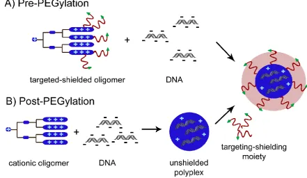
20 A common concept for the attachment of the shielding polymer to the polyplex surface is the "pre-PEGylation" concept (Fig. 1.2.A) [77]. It relies on the modification of a cationic carrier with the shielding polymer before nucleic acid complex formation. "Post-PEGylation" is another concept applied for the formation of shielded polyplexes. Thereby, DNA polyplexes are formed prior to the addition of the shielding polymer (Fig. 1.2.B) [78].
Figure 1.2. Illustration of A) pre-PEGylation and B) post-PEGylation approach applied for polyplex surface shielding. In the "pre-PEGylation" concept the shielding polymer is attached to the polycation prior to nucleic acid complexation, whereas in the "post-PEGylation" approach polyplexes are formed prior to the attachment of the shielding polymer.
21 occurs in presence of the stimuli. However in absence, the PEG shield remains intact in order to prolong blood circulation and avoid particle-particle aggregation or unspecific cell-uptake. Exploiting pH changes within the tumor tissue or endosomes acid-labile linkers, such as acetals [82-84], dialkylmaleic acids [50] or pyridylhydrazones [78, 85], have been introduced between the shielding moiety and the surface of the polyplex. These surface-modified delivery systems are intended to remain shielded during circulation, i.e. at physiological pH. However, the pH-labile bond is cleaved after the polyplex has entered a slightly acidic environment such as in tumor tissue or in an intracellular endosomal vesicle. Thus, the delivery system with its original endosomolytic capability is restored enabling membrane fusion or disruption to accomplish endosomal release. Previous studies have shown an up to 100-fold enhanced gene transfer by introducing a pH-sensitive hydrazone linker into PEG/PEI/DNA polyplexes in vitro and in vivo [78, 85].
In addition, enzymatic cleavable linkers attached between the polyplex surface and the shielding polymer presents an attractive tool to mediate location-specific cleavage of the PEG shield. In this respect, attention has recently turned to matrix metalloproteinases (MMPs) due to their over-expression within tumor tissues facilitating tumor site-specific cleavage of the PEG shield [86]. In healthy tissues the level of MMPs is rather low and therefore the PEG shield remains attached to the surface of the polyplex. To demonstrate the feasibility of this strategy PEG-cleavable polyplex micelles based on MMP-cleavable peptide-linked block copolymers have been synthesized [87]. Here, PAsp(DET) (poly(aspartamide)) with a flanking N-(2-aminoethyl)-2-aminoethyl group is bound via a MMPs cleavable peptide linker (GPLGVRG) to PEG. In the presence of matrix metalloproteinase-2 the PEG layer is efficiently cleaved and the initial positive surface charge of the carrier re-exposed. Consequently, polyplexes show a higher cellular uptake, an improved endosomal escape and a high-efficiency in gene transfection. The original purpose of PEG, to prolong blood circulation, still remains.
22 unspecific cell interactions at pH 7.4. However, after polyplexes have entered the extracellular tumor environment or endosomes, the pH changes to approximately pH 6.6 resulting in sulfonamide protonation and release from the nanoparticles. Therefore, the initial positive surface charge of PEI is restored, which enables sufficient interactions with cell membranes and cellular uptake [49].
Together, these studies indicate that polyplexes designed to response to different location-specific changes such as disulfide reducing potential, pH or enzyme concentrations represents a feasible strategy to overcome the PEG dilemma.
1.3.3.Targeting of non-viral carriers
Specific in vivo targeting to malignant cells by therapeutic genes presents an attractive concept to accomplish delivery tasks. In most cases, cell-specific targeting is enabled by specific interactions between a ligand, attached to the polyplex surface, and a receptor, exposed on the cell surface. However, cell-specific targeting is a key property that promotes nucleic acid transfer from the site of administration to the surface of the target cells and promotes cell-internalization. Additionally, side effects arising from toxicities toward mammalian cells are shown to be decreased by targeting malignant cells specifically. Furthermore, the receptor-ligand interaction stimulates the receptor-mediated endocytosis of target-specific polyplexes, which is known to be more efficient compared to adsorptive endocytosis [41, 42]. Various ligands are suitable for targeted gene delivery. Those can be categorized into several classes e.g. small molecules like folic acid (FolA), proteins such as transferrin (Tf) or epidermal growth factor (EGF) or asialoglycoproteins as well as peptides like CMBP1 or sugar derivatives such as N-acetylgalactosamine. When designing a target-specific carrier several aspects must be taken into account including the level of receptor expression, tissue specificity of ligands, ligand internalization, binding affinity, surface charge or degree of interaction with e.g. proteins. Thus, it is essential to select an appropriate ligand for successful application of a targeted non-viral carrier. Almost 30 years ago Wu et al. pioneered liver-cell specific targeting by an asialoglycoprotein targeted non-viral gene delivery system in vitro and in vivo [88, 89]. Meanwhile, various ligands have been incorporated within non-viral gene delivery systems and many studies have demonstrated successful cell-type specific
23 A commonly used ligand for targeted gene delivery to a wide variety of cell types is the iron-transporting serum glycoprotein transferrin (Tf). The interaction of Tf with the transferrin receptor (TfR) triggers the cellular iron uptake. TfR as a biological target is of special interest since it is over-expressed on the surface of many human cancers. Therefore, Tf has been conjugated to various non-viral carriers such as PEI [77, 96], polypropylenimine (PPI) dendrimers [97], polyamidoamine (PAMAM) dendrimers [98] or poly-L-lysine (PLL) [99, 100] in order to achieve TfR-targeted gene delivery in cancer cells in vitro and in vivo.
The epidermal growth factor (EGF) is another promising ligand for tumor cell-specific targeting because it binds with high affinity and specificity to the epidermal growth factor receptor (EGFR). In several tumors including glioblastoma, epithelial tumors and lung cancers as well as hepatocellular carcinoma EGFR is upregulated or overactivated due to mutations resulting in uncontrolled proliferation. Thus, various EGFR-targeting molecules including recombinant EGF proteins, EGFR-binding peptides and antibodies are conjugated to gene delivery systems, to analyze the targeting efficiency and subsequently the gene expression levels. In this regard, using EGF-targeted PEI-based polyplexes, high-level transgene expression was found in vitro [42, 101]. For example, after systemic administration of EGF-targeted polyplexes a 50-fold increased transgene expression in hepatocellular carcinomas compared to normal liver tissue was reported [102].
Recently, a c-Met binding ligand cMBP2 was pioneered in terms of non-viral gene delivery. The over-expressed receptor tyrosine kinase HGFR/c-Met found in epithelial derived tumors serves as binding site for the natural ligand hepatocyte growth factor (HGF) as well as the c-Met binding peptide cMBP2, activating different cell-signaling pathways including those involved in tumor progression. In this regard, the activation of c-Met signaling presents an interesting target for cancer treatment. This has been successfully demonstrated by Kos et al. showing an enhanced selective gene transfer to hepatocarcinoma cells by targeting the c-Met receptor by cMBP2 in vitro
24
1.4. Aims of the thesis
The successful implementation of nucleic acids as medical agents depends on the delivery efficiency of the non-viral carrier. While polymer-based gene carriers are promising due to the high-level of flexibility in structural design, overall efficiency is still several magnitudes lower in comparison to viral gene carriers. Whereas viruses have developed distinct mechanisms to transport DNA successfully into mammalian cells, most non-viral carriers are still unable to deliver DNA over extra- and intracellular barriers. To compensate for this disadvantage many research groups focus on the identification of structure-function relationships of DNA delivery carriers. We have generated a library of well-defined oligomer-based carriers with different architectures and functionalities, to identify individual structural requirements to the carrier. Based on this, this thesis focused on the optimization of polyplex stability and biological properties of a four-arm structured benchmark oligomer (compound ID:
606) to improve DNA delivery.
First, a small library of eight oligomers was to be synthesized via the solid-phase approach. In the sequence of these oligomers lysine residues had to be introduced at different sites to assess the influence of the incorporated lysine residues on polyplex stability in comparison to the benchmark oligomer 606. Additionally, the biophysical and biological properties of these stabilized particles had to be evaluated to identify the best oligomer.
25 the lysine residues within the oligomeric structure serve as attachment points for the shielding material. Two of the most commonly used shielding polymers, monovalent PEG (5kDa) or monovalent and multivalent pHPMA (10kDa, 20kDa, 30kDa), should be studied in parallel. The effect of shielding polymer chain length and architecture on polyplex shielding was to be evaluated. Furthermore, the shielding ability of pHPMA and PEG ought to be compared. To test the utility of such acid-labile carriers, the delivery efficiency was to be compared to that of acid-stably shielded polyplexes containing N-hydroxysuccinimide (NHS) groups in case of PEG and carbonylthiazolidine-2-thione (TT) groups in case of pHPMA. A detailed physicochemical, in vitro and in vivo analysis was to be performed to evaluate the effect of the pH-sensitive shielding compared to the analogous stable shielding on transfection efficiency.
26
2. Materials and Methods
2.1. Materials
2.1.1.Reagents and Solvents
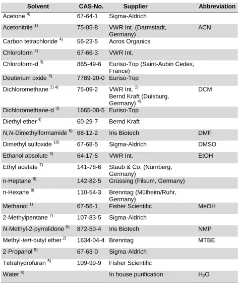
All reagents and solvents used for experiments described in this thesis are summarized in Table 2.1. and Table 2.2.
Table 2.1. Reagents with CAS numbers, sources of supply and abbreviation (abbr.) used for experimental procedures.
Reagents CAS-No. Supplier Abbr.
Resins Fmoc-L-Lys(Boc)-Wang resin Sigma-Aldrich (München, Germany) 1-amino-ethane-2- thiol(cysteamine)-2-chlorotrityl resin Iris Biotech (Marktredewitz, Germany) Fmoc-Ala-Wang Sigma-Aldrich
(München, Germany)
Amino acids
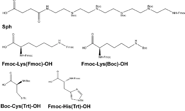
Boc-L-Cys(Trt)-OH 21947-98-8
Iris Biotech Fmoc-L-Glu-OtBu 84793-07-7
Fmoc-L-His(Trt)-OH 109425-51-6 Fmoc-L-Lys(Boc)-OH 71989-26-9 Fmoc-L-Lys(Fmoc)-OH 78081-87-5
SPPS
Acetic anhydride 108-24-7 Sigma-Aldrich
Benzotriazol-1-yl-oxytripyrrolidinophospho
nium-hexafluorophosphate
128625-52-5 Multisyntech GmbH (Witten, Germany)
PyBOP
1,8-diazabicyclo[5.4.0] undec-7-en
6674-22-2 Sigma-Aldrich
Diisopropylethylamin 7087-68-5 Iris Biotech DIPEA Di-tert-butyldicarbonate 24424-99-5 Sigma-Aldrich
Ethyl trifluoroacetate 383-63-1 Sigma-Aldrich Fmoc-OSu 82911-69-1 Iris Biotech
2-(1H-benzotriazol-1-yl)-
1,1,3,3- tetramethyluronium-hexafluorophosphate
94790-37-1 Multisyntech GmbH HBTU
1-hydroxybenzotriazole hydrate
123333-53-9 Sigma-Aldrich HOBt
27 Pentaethylene hexamine 4067-16-7 Sigma-Aldrich PEHA Phenol 108-95-2 Sigma-Aldrich
Piperidine 110-89-4 Iris Biotech Potassium cyanide 151-50-8 Sigma-Aldrich Pyridine 110-86-1 Sigma-Aldrich Sephadex® G-10 9050-68-4 GE Healthcare
(Freiburg, Germany) Sodium bicarbonate 144-55-8 Sigma-Aldrich
Sodium chloride 7647-14-5 Sigma-Aldrich NaCl Sodium hydroxide
(anhydrous)
1310-73-2 Sigma-Aldrich NaOH
Succinic anhydride 108-30-5 Sigma-Aldrich Triethylamine 121-44-8 Sigma-Aldrich
Trifluoroacetic acid 76-05-1 Iris Biotech TFA Triisopropylsilane 6485-79-6 Sigma-Aldrich TIS Trisodium citrate
dihydrate
6132-04-3 Sigma-Aldrich
TritonTM X-100 9002-93-1 Sigma-Aldrich
AzMMMan
Benzoyl peroxide 94-36-0 Sigma-Aldrich 2,3-dimethylmaleic
anhydride
766-39-2 Sigma-Aldrich DMMan
N-Bromosuccinimide 128-08-5 Sigma-Aldrich NBS Sephadex LH 20 9041-37-6 Sigma-Aldrich
Sodium azide 26628-22-8 Acros Organics (Geel, Belgien)
Sodium sulfate 7757-82-6 Sigma-Aldrich Na2SO4
NHS-C3-Azide baseclick
(Tutzingen, Germany)
PEG reagents
DBCO-PEG(5000)-MeO Jena Bioscience (Jena, Germany) MeO-PEG(5000)-NHS RAPP Polymere
(Tübingen, Germany) OPSS-PEG(5000)-NHS RAPP Polymere NHS-PEG(5000)-SH RAPP Polymere NH2-PEG4-DBCO 1255942-06-3 Sigma-Aldrich
Mal-PEG4-DBCO Sigma-Aldrich
FolA reagents
Fmoc-N-amido-dPEG6
-acid
882847-34-9 Iris Biotech
Folic acid 59-30-3 Sigma-Aldrich FolA
N,N
′-Dicyclohexyl-carbodiimide
538-75-0 Sigma-Aldrich
N-Hydroxysuccinimide 6066-82-6 Sigma-Aldrich NHS
N10-(Trifluoroacetyl) pteroic acid
28
Other
Adenosine-5`-triphosphate
56-65-5 Roche Diagnostics (Basel, Schweiz) Agarose NEEO
Ultra-Qualität
9012-36-6 Carl Roth GmbH (Karlsruhe, Germany)
Ammonium persulfate 7727-54-0 Sigma-Aldrich APS Barium chloride
dihydrate
10326-27-9 Sigma-Aldrich
Boric acid 10043-35-3 Sigma-Aldrich Bromophenol blue 115-39-9 Sigma-Aldrich Coenzym A Sigma-Aldrich Coomassie® Blue
Staining
Fisher Scientific (Schwerte, Germany) DL-Dithiothreitol 578517 Sigma-Aldrich
Ethylenediaminetetra-acetic acid disodium salt dihydrate
6381-92-6 Sigma-Aldrich EDTA
GelRed Biotium Inc.
(Hayward, USA) Glycylglycine 556-50-3 Roche Diagnostics Ethidium bromide
solution
1239-45-8 Sigma-Aldrich EtBr
Linear polyethylenimine 9002-98-6 In house synthesis LPEI Magnesium chloride 7786-30-3 Sigma-Aldrich
N,N,N',N'
-Tetramethyl-ethylendiamine
110-18-9 Sigma-Aldrich TEMED
Potassium chloride 7447-40-7 Sigma-Aldrich Potassium dihydrogen
phosphate
7778-77-0 Sigma-Aldrich
Potassium iodide 7681-11-0 Sigma-Aldrich Propane-1,2,3-triol 56-81-5 Sigma-Aldrich Rotiphorese® Gel 30 Carl Roth GmbH
Sodium dodecyl sulfate 151-21-3 Sigma-Aldrich SDS Sodium hydrogen
phosphate
7558-79-4 Sigma-Aldrich
Tris(hydroxymethyl) amimomethane
77-86-1 Sigma-Aldrich Tris
29
Table 2.2. Solvents with CAS-numbers, sources of supply and abbreviation used for experimental procedures.
Solvent CAS-No. Supplier Abbreviation
Acetone 4) 67-64-1 Sigma-Aldrich
Acetonitrile 1) 75-05-8 VWR Int. (Darmstadt, Germany)
ACN
Carbon tetrachloride 4) 56-23-5 Acros Organics
Chloroform 2) 67-66-3 VWR Int.
Chloroform-d 3) 865-49-6 Euriso-Top (Saint-Aubin Cedex, France)
Deuterium oxide 3) 7789-20-0 Euriso-Top
Dichloromethane 2) 4) 75-09-2 VWR Int. 2)
Bernd Kraft (Duisburg, Germany) 4)
DCM
Dichloromethane-d 3) 1665-00-5 Euriso-Top Diethyl ether 4) 60-29-7 Bernd Kraft
N,N-Dimethylformamide 5) 68-12-2 Iris Biotech DMF
Dimethyl sulfoxide 10) 67-68-5 Sigma-Aldrich DMSO
Ethanol absolute 4) 64-17-5 VWR Int. EtOH Ethyl acetate 7) 141-78-6 Staub & Co. (Nürnberg,
Germany)
n-Heptane 8) 142-82-5 Grüssing (Filsum, Germany)
n-Hexane 8) 110-54-3 Brenntag (Mülheim/Ruhr, Germany)
Methanol 1) 67-56-1 Fisher Scientific MeOH
2-Methylpentane 7) 107-83-5 Sigma-Aldrich
N-Methyl-2-pyrrolidone 5) 872-50-4 Iris Biotech NMP
Methyl-tert-butyl ether 2) 1634-04-4 Brenntag MTBE
2-Propanol 9) 67-63-0 Sigma-Aldrich
Tetrahydrofuran 5) 109-99-9 Fisher Scientific
Water 6) In house purification H2O
30
2.1.2.Equipment for solid-phase assisted peptide synthesis (SPPS)
Disposable polypropylene syringe microreactors were purchased from Multisyntech. Those reactors were equipped with polytetrafluoroethylene filters in case of the use in the automated single peptide synthesis. For manual peptide synthesis microreactors were equipped with polyethylene filters.
2.1.3.Cell culture
All cell culture consumables (well plates, flasks, dishes) were purchased from NUNC (Langenselbold, Germany) or TPP (Trasadingen, Switzerland).
Table 2.3. Reagents and media with CAS numbers, sources of supply and abbreviation (Abbr.) used for in vitro experiments.
Reagents CAS-No. Supplier Abbr.
Reagents
Chloroquine diphosphate 50-63-5 Sigma-Aldrich Collagen A 9007-34-5 Merck Millipore
(Darmstadt, Germany) D(+) glucose
monohydrate
14431-43-7 Merck Millipore
4′,6-Diamidino-2-phenylindole
28718-90-3 Sigma-Aldrich DAPI
3-(4,5-Dimethyl-2- thiazolyl)-2,5-diphenyl-2H-tetrazolium bromide
298-93-1 Sigma-Aldrich MTT
D-luciferin 2591-17-5 Promega (Mannheim, Germany)
HEPES 7365-45-9 Biomol GmbH
(Hamburg, Germany) Fetal calf serum Invitrogen (Karlsruhe,
Germany)
FCS
Heparin-Na 25000 Ratiopharm (Ulm, Germany)
Luciferase cell culture 5x lysis buffer
Promega
Penicillin/ Streptomycin Biochrom GmbH (Berlin, Germany) Paraformaldehyde 30525-89-4 Sigma-Aldrich Rhodamine phalloidin Fisher Scientific Sodium chloride 7647-14-5 Prolabo (Haasrode,
Belgien)
NaCl
Trypsin/EDTA solution (10x)
31
Medium
Dulbecco's Modified Eagle's Medium- low glucose
Sigma-Aldrich DMEM
RPMI 1640 (1x) Life Technologies GmbH (Darmstadt, Germany)
Dulbecco’s Modified Eagle’s medium 50:50 mixture with Ham's F12
Sigma-Aldrich
Table 2.4. Summary of used cell lines.
Cell line Cell type Medium
Neuro-2a Mouse neuroblastoma DMEM (low glucose) KB Human cervix carcinoma Folate-free RPMI 1640 HUH7 Human hepatocellular
carcinoma
DMEM (low glucose)/Ham's F12 1:1
2.1.4.Nucleic acids
pCMVL, encoding for firefly luciferase under control of the CMV promoter, was purchased from PlasmidFactory (Bielefeld, Germany). Cy5-labeling kit for pCMVL labeling was obtained by Mirus Bio (Madison, USA).
2.2. Methods
2.2.1.Synthesis of 3-(azidomethyl)-4-methyl-2,5-furandione (AzMMMan)
AzMMMan was synthesized according to the literature [105, 106] with modifications.
32 tetrachloride.Subsequently, the organic phase was washed twice with water (100mL) and once with brine (100mL). Thereafter, the organic layer was dried over Na2SO4
and concentrated in vacuum to form a yellow oil. Benzoyl peroxide was removed by silica gel flash chromatography using a mixture of 2-methylpentane/ethyl acetate (8:2) as eluent. The residue was subsequently distilled with the help of a kugelrohr apparatus under vacuum. DMMan fraction was removed at 110-120°C, 7mbar. 3-(Bromomethyl)-4-methyl-2,5-furandione (BrMMan) was isolated at 140°C, 7mbar (1.8g, yield 22%). 3,4-bis(bromomethyl)furan-2,5-dione remained in the still pot.
Secondly, BrMMan (310.5mg, 1.5mmol) was dissolved in 10mL acetone (dried over molecular sieve UOP Type 3A) and sodium azide (97.5mg, 1.5mmol) was added. The mixture was stirred for 24h at 37°C. After filtering the solvent was evaporated. The remaining oil was resolved in ethyl acetate (20mL) and washed two times with water (20mL) and one time with brine (20mL). The organic layer was dried over Na2SO4. The liquid was concentrated in vacuum to result in a brown oil (222mg, yield
88%).
2.2.2.Synthesis of Fmoc/Boc-protected succinyl-pentaethylene hexamine (Fmoc/Boc-Sph)
Sph was prepared following an adapted procedure of Salcher et al. [55, 107]. Prior to synthesis, pentaethylene hexamine (PEHA) was purified according to Jonassen and Westerman [108]. Fractions of 25mL PEHA were distilled with a kugelrohr apparatus. The second fraction (210-225°C, 0.01bar) was collected and used for Sph synthesis. The first (200°C, 0.01bar) and the third fraction (> 230°C, 0.01bar) were discarded.
2.2.3.Solid-phase assisted peptide synthesis
33
2.2.3.1. Loading of resins
All polycationic carriers and the targeting ligand were synthesized using preloaded resins. For the synthesis of oligomer 784, 785 and 797 a Fmoc-L-Lys(Boc)-Wang resin with a load of 0,76mmol/g was used. Oligomer 606 was synthesized using Fmoc-Ala-Wang resin with a load of 0.32mmol/g according to Lächelt et al. [56]. The same resin was used for the synthesis of oligomer 748 and 749. The loading of the resins was decreased to 0.15-0.2mmol/g enabling successful coupling of amino acids (AA) to the four arm branching core (A/KK(HK)2). The appropriate amount of resin
was pre-swollen in DCM (10mL/g resin) for 30min before loading was decreased. Afterwards, the Fmoc protecting group was removed by treating the resin two times 5min and two times 10min with deprotection solution [20% piperidine in DMF (v/v)]. Subsequently, the resin was washed three times with DMF and DCM and afterwards Kaiser test was performed. An equimolar amount of Fmoc-L-Lys(Fmoc)-OH, 1-hydroxybenzotriazole hydrate (HOBt) and benzotriazol-1-yl-oxytripyrrolidinophos-phoniumhexafluorophosphate (PyBOP) as well as a twofold molar amount of diiso-propylethylamine (DIPEA) were dissolved in DCM/DMF [1/1 (v/v); 10mL/g resin] and added to the resin for 1h. After coupling, the reaction solvent was drained and the resin was washed three times with DMF and DCM. Unreacted groups were capped with a mixture of 20eq acetic anhydride and 40eq DIPEA dissolved in DCM/DMF [1/1 v/v); 10mL/g resin]. Afterwards, solvents were drained, the resin was washed three times with DMF and DCM and Kaiser test was performed. According to the individual oligomer sequence coupling of further amino acids was performed as stated above.
The targeting ligand FolA-PEG6-cysteamine was synthesized using an
1-amino-ethane-2-thiol(cysteamine)-2-chlorotrityl resin with a load of 0,95mmol/g. The resin was pre-swollen in anhydrous DCM for 30min and loading was decreased to 0.4-0.5mmol/g. 1eq Fmoc-N-amido-dPEG6-acid, 1eq HOBt, 1eq PyBOP and 2eq DIPEA
34 targeting ligand was cleaved off the resin according 2.2.3.3. "cleavage of oligomers and targeting ligand".
The loading of the resins was determined as following: around two times 5mg of the resin were separated, washed twice with DCM and n-hexane and dried under vacuum. An exact amount of resin was treated with 1mL deprotection solution for 1h, diluted in DMF and UV absorption was measured at 301nm. The loading was calculated according to the equation:
resin load [mmol/g] = (A*1000) / (m [mg]*7800*df) with df as dilution factor
The residual resin was treated two times 5min and two times 10min with deprotection solution, washed three times with DMF and three times with DCM and dried under vacuum. Reaction progress was monitored by Kaiser test (chapter 2.2.3.4.).
2.2.3.2. General procedure of SPPS
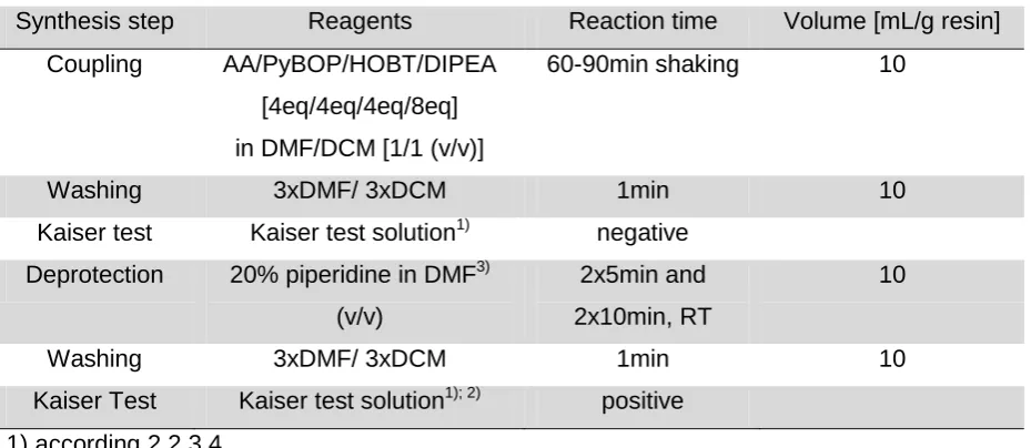
SPPS was carried out according to standard Fmoc/tBu protecting group strategy first published by Merrifield et al. [109]. Synthesis was performed either by an automated microwave-based system (Biotage AB, Uppsala, Sweden) or manually. For automated synthesis the pre-loaded resin was pre-swollen in a syringe reactor for 20min in NMP and for manual synthesis in DCM. The general synthesis procedure was performed as described in Table 2.5. (automated synthesis) and Table 2.6. (manual synthesis). After reaction and washing steps, solvents and non-reacted reagents were removed.
Table 2.5. General protocol for automated synthesis.
Synthesis step Reagents Reaction time Volume [mL/g resin] Coupling AA/PyBOP/HBTU/DIPEA
[4eq/4eq/4eq/8eq] in DMF/NMP [1/1 (v/v)]
3x/5x alternating 10min, 60°C and
5min, RT
10
Washing 3xDMF 1min 10
Deprotection 20% piperidine in DMF3) (v/v) 5x5min, RT 10
Washing 3xDMF 1min 10
35
Table 2.6. General protocol for manual synthesis.
Synthesis step Reagents Reaction time Volume [mL/g resin] Coupling AA/PyBOP/HOBT/DIPEA
[4eq/4eq/4eq/8eq] in DMF/DCM [1/1 (v/v)]
60-90min shaking 10
Washing 3xDMF/ 3xDCM 1min 10
Kaiser test Kaiser test solution1) negative Deprotection 20% piperidine in DMF3)
(v/v)
2x5min and 2x10min, RT
10
Washing 3xDMF/ 3xDCM 1min 10
Kaiser Test Kaiser test solution1); 2) positive 1) according 2.2.3.4.
2) not during synthesis, only last step
3) if needed addition of 2% 1,8-diazabicyclo[5.4.0] undec-7-en and 1% Triton
2.2.3.3. Cleavage of oligomers and targeting ligand
The oligomer was cleaved off the resin by treatment with a cleavage solution containing TFA/TIS/H2O [95/2.5/2.5 (v/v)] for 1.5h. After washing the resin two times
with trifluoroacetic acid (TFA) and DCM all solutions were combined, concentrated and precipitated by dropwise addition to a mixture of ice-cold MTBE/n-hexane [1/1 (v/v)] and cooled to -20°C. The resulting precipitate was centrifuged (10min; 4000rpm) and dried under vacuum. The resulting precipitate of the targeting ligand was dissolved in 50% (v/v) acetonitrile in water and lyophilized. In case of an oligomer precipitate, the pellet was re-suspended in size exclusion buffer [10mM HCl, 30% ACN, 70% H2O (v/v)]. The oligomer was purified by size exclusion
36
2.2.3.4. Kaiser Test
Kaiser test was used as a control to monitor the coupling and deprotection efficiency by detection of free amino groups. Due to the linkage of the chain to the resin via its C-terminus, the N-terminus is extending off. Ninhydrin is used to detect free amines (in case of deprotected resin) by color change. In case of a protected N-terminus the test gives a yellow result. The test was carried out after each coupling and deprotection step during manual synthesis. In case of automated synthesis, Kaiser test was just performed before manual coupling of Boc-Cys(Trt)-OH or after coupling of N10-(Trifluoroacetyl)pteroic acid.
A small amount of beats were transferred to an Eppendorf tube. One drop of each solution [80% (w/v) phenol in EtOH, 5% (w/v) ninhydrin in EtOH, 0.02mM potassium cyanide in pyridine] was added to the resin, vortexed and heated to 99°C for 4min.
2.2.4.Synthesis of shielding polymers
2.2.4.1. Synthesis of monovalent AzMMMan-PEG(5000)-MeO
Monovalent AzMMMan-PEG(5000)-MeO was synthesized by a one-step modification of DBCO-PEG(5000)-MeO (10mg, 2µmol) with AzMMMan (1.34mg, 8µmol). Therefore, all reagents were dissolved in MeOH (500µL) and the reaction was performed for 3h. The excess of AzMMMan was removed by precipitation of AzMMMan-PEG(5000)-MeO in 50mL diethyl ether (yield 90-95%).
2.2.4.2. Synthesis of bifunctional FolA-PEG(5000)-AzMMMan and bifunctional FolA-PEG(5000)-NHS via FolA-PEG6-cysteamine AzMMMan-modified bifunctional FolA-PEG(5000) was synthesized by a three-step reaction. Firstly, OPSS-PEG(5000)-NHS (10mg, 2µmol) was modified with DBCO-PEG4-NH2 (4.2mg, 8µmol). Therefore, OPSS-PEG(5000)-NHS was dissolved in
450µL MeOH and DBCO-PEG4-NH2 in 50µL DMSO. Afterwards, reagents were
mixed and reaction was performed for 3h at 25°C. The excess of DBCO-PEG4-NH2
37 dissolving 1eq OPSS-PEG(5000)-DBCO (10mg, 2µmol) in 450µL MeOH and 4eq FolA-PEG6-cysteamine (6.6mg, 8µmol) in 50µL DMF. The reaction was performed for
2h and purified by SEC using a PD Midi Trap self-packed with Sephadex LH 20 and MeOH as an eluent. The first fractions were collected and solvent was evaporated. Lastly, 1eq FolA-PEG(5000)-DBCO (10mg, 2µmol) was modified with 4q AzMMMan (1.34mg, 8µmol) accordingly the monovalent AzMMMan-PEG-MeO as stated above.
N-Hydroxysuccinimide (NHS)-modified bifunctional FolA-PEG(5000) was synthesized by dissolving 1eq OPSS-PEG(5000)-NHS (10mg, 2µmol) in 450µL MeOH and 4eq FolA-PEG6-cysteamine (6.6mg, 8µmol) in 50µL DMF. The reaction was performed for
2h and purified by SEC using a PD Midi Trap self-packed with Sephadex LH 20 and MeOH as an eluent. The first fraction was collected and solvent was evaporated.
2.2.4.3. Synthesis of bifunctional FolA-PEG(5000)-AzMMMan and bifunctional FolA-PEG(5000)-NHS via NHS-FolA
In a different approach FolA-PEG(5000)-AzMMMan and FolA-PEG(5000)-NHS was synthesized via NHS-FolA. In the first step NHS-FolA was synthesized according Liu et al. [110]. After removing the insoluble byproduct dicyclohexylurea by filtration, the FolA-NHS solution was precipitated in ACN and solvent was removed by evaporation. In the next step, 4eq DBCO-PEG4-Mal (5.4mg, 8µmol) was coupled to
1eq NH2-PEG(5000)-SH (10mg, 2µmol). In this regard, NH2-PEG(5000)-SH was
dissolved in 450µL MeOH and DBCO-PEG4-Mal in 50µL DMSO. Coupling was
performed for 3h and the excess of DBCO-PEG4-Mal was removed by SEC using a
PD MidiTrap column self-packed with Sephadex LH20 and MeOH as an eluent. The first fraction was collected and the solvent was evaporated. Afterwards, the targeting ligand FolA was introduced by dissolving NH2-PEG(5000)-DBCO (10mg, 2µmol) in
450µL MeOH/DMF [1/1 (v/v)] and NHS-FolA (4.2mg, 8µmol) in 50µL DMF. Reaction was performed for 4h under the atmosphere of an inert gas at RT. The product was purified by SEC using a PD MidiTrap column self-packed with Sephadex LH20 and DMF as an eluent. Again, the first fractions were collected and the solvent was evaporated. In the last step, 1eq FolA-PEG(5000)-DBCO (10mg, 2µmol) was modified either with 4eq AzMMMan (1.34mg, 8µmol) or NHS-C3-Azide (1.8mg,
38 dissolved in 500µL MeOH and reaction was performed for 4h at RT. The product was purified by precipitation in 50mL diethyl ether.
Note: These syntheses were unsuccessful according biological characterization section 3.3.1.
2.2.4.4. Synthesis of monovalent pHPMA-AzMMMan and monovalent pHPMA-TT
Monovalent polyN-(2-Hydroxypropyl)methacrylamide (pHPMA) polymers were synthesized, characterized and kindly provided by Dr. Libor Kostka.
2.2.4.5. Synthesis of multivalent pHPMA-AzMMMan and multivalent pHPMA-TT
Multivalent pHPMA polymers were synthesized, characterized and kindly provided by Dr. Libor Kostka.
2.2.4.6. Polyplex formation
39
2.2.4.7. Measurement of particle size and zeta potential
Polyplex formation as well as shielding and deshielding of polyplexes was monitored via particle size and zeta potential measurements by dynamic and electrophoretic light scattering (DLS) using a Zetasizer Nano ZS (Malvern Instruments, Worcestershire, U.K.) with following parameters: equilibration time 0min, viscosity 0,8872cP, dielectric constant 78.5, temp 25°C, F(Ka) 1.5 (Smoluchowski), measurement point 2.00mm, attenuator 11.
2.2.4.8. Agarose gel shift assay
To study pCMVL condensation as a function of N/P ratio, an agarose gel shift assay was performed. To visualize pCMVL, 120µL GelRed was added to the 1% agarose gel. Polyplexes were formed as stated above and 4µL of DNA loading buffer was added prior to loading to the gel. Electrophoresis was performed at a constant current of 120V for 80min. Thereafter, fluorescence of incorporated GelRed was detected using a transilluminator (Biostep, Jahnsdorf, Germany).
2.2.4.9. Ethidium bromide exclusion assay (EtBr assay)
Ethidium bromide exclusion assay was performed to study pCMVL condensation ability using a Cary Eclipse spectrophotometer (Varian, Bergisch Gladbach, Germany), Exc: 510nm Em: 590nm. The effect of polyplex stability was assessed by stepwise addition of oligomer solution to 10µg pCMVL in 1mL HBG containing 0.4µg EtBr. Free pCMVL and EtBr solution were set as 100% and an EtBr solution was set as a background value. Fluorescence intensity was measured 30s after each addition of oligomer aliquot.
2.2.4.10. Oligomer buffering capacity
40 reached. A control titration was performed with 3.5mL 50mM NaCl solution and 0.05M NaOH. The percentage of buffer capacity in a certain pH range (x-y) was calculated according to equation (1). The volume consumption of NaOH in the considered pH range is represented by ΔV.
2.2.4.11. Cell culture
Mouse neuroblastoma cells (Neuro-2a) were grown in Dulbecco’s Modified Eagle’s Medium- low glucose (DMEM) and hepatocarcinoma cells (HUH7) in a 50:50 mixture of Dulbecco’s Modified Eagle’s medium and Ham's F12 medium. All cell lines were
cultured at 37°C in a humidified atmosphere of 5% CO2 and 95% air. All media were
supplemented with 10% fetal calf serum (FCS), 100U/mL penicillin and 100μg/mL
streptomycin. Cells were harvested by treatment with trypsin/EDTA solution at 37°C for 5 min. Cells were mycoplasma free as tested by MycoAlertTM mycoplasma detection kit (Lonza, Köln, Germany).
2.2.4.12. Luciferase gene expression
In vitro experiments were performed in 96-well plates with 104 seeded Neuro-2a cells
41
2.2.4.13. Cell viability assay (MTT)
Viability of HUH7 and Neuro-2a cells was evaluated subsequently to the transfection experiments. Transfections were performed similar to luciferase gene expression studies. At indicated post-transfection time, 10µL 3-(4,5-dimethyl-2-thiazolyl)-2,5-diphenyl-2H-tetrazolium bromide (MTT) was added to each well reaching a final concentration of 0.5mg MTT/mL. After 2h of incubation, unreacted dye and medium were removed and the 96-well plates were stored at -80°C for one hour. The purple formazan product was dissolved in 100μL/well DMSO and quantified by a microplate
reader (Tecan, Männedorf, Switzerland) at 530nm with background correction at 630nm. The relative cell viability (%) related to control wells containing cell culture medium with 20μL HBG was calculated by [A]test/[A]control×100.
2.2.4.14. Cellular uptake: flow cytometry studies
Cellular internalization of the polyplexes was assessed by flow cytometry of Cy5-labeled polyplexes (Exc:636nm, Em:665nm) in a Cyan™ ADP flow Cytometer (Dako, Hamburg, Germany) equipped with Summit™ acquisition software (Summit,
Jamesville, USA). Data were analyzed by FlowJo® 7.6.5 flow cytometric analysis software. Cells were appropriately gated by forward/sideward scatter and pulse width for exclusion of doublets. All experiments were performed in triplicates.
42
2.2.4.15. Intracellular localization: laser scanning microscopy (LSM)
Neuro-2a cells (3x10⁴ per well) were seeded 24h prior to transfection in NuncTMLabtekTM chamber slides (Sigma-Aldrich). Before transfection cell medium was replaced with 80µL fresh medium. Polyplexes solution (20µL per well) was added and incubated for 24h at 37°C. Cells were washed three times with PBS and fixed with 4% paraformaldehyde for 30min. Staining was performed using DAPI and rhodamine phalloidin. Results were analyzed in a Zeiss Laser Scanning Microscope LSM 510 Meta and Axiovert 200 software (Zeiss).
2.2.4.16. In vivo delivery and expression
All animal experiments were performed according to the guidelines of the German law for the protection of animal life and were approved by the local animal ethics committee. Six weeks old female Rj:NMRI nu (nu/nu) (Janvier, Le-Genest-St-Isle, France) mice were housed in isolated ventilated cages with a 12h day/night interval and food and water ad libitum.
Neuro-2a cells (5x10⁶)suspended in 150µL PBS were injected subcutaneously into the left flank. On day 12 and 13 after tumor cell inoculation, 250µL polyplex solution containing 60µg pCMVL was administered via tail vein injection. Mice were euthanized on day 15 and tumor/organs were collected to assess luciferase activity
via ex vivo luciferase assay. Tumor and organs were homogenized in 500µL cell lysis
buffer using a tissue and cell homogenizer (FastPrep®-24). To separate insoluble cell components, the samples were centrifuged at 3000g at 4°C for 10min. Luciferase activity was measured in the supernatant using a Centro LB 960 luminometer.
2.2.4.17. Statistical analysis
43
3. Results
3.1. Designing new sequence-defined oligomers: evaluation of stabilizing effects of lysine residues on DNA polyplexes
44
Figure 3.1. Illustration of repeating cycles of deprotection-washing-coupling-washing steps characterizing SPPS. Firstly, the C-terminal amino acid is coupled to the linker, which is attached to the resin. By repeating a cycle of deprotection, washing, coupling and washing steps the peptide chain is elongated. At the end of the synthesis, the final peptide is obtained after the cleavage from the resin and removal of permanent protecting groups.
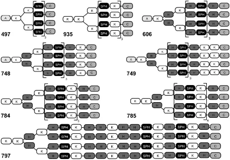
45 Based on this strategy, new sequence-defined DNA carriers were designed and synthesized. These carriers represent the third generation of previously reported oligomers with four-arm topology. In the first generation, the methods of four-arm oligomer syntheses [58], the discovery of Sph as particularly effective building block, and the polyplex-stabilizing properties of terminal cysteines [55] was established. In the previous second generation, incorporation of histidine residues for fine-tuning the proton sponge activity resulted in oligomers such as AK[HK[(H-Sph)3C]2]2 (compound
ID: 606) which already showed high endosomal escape and DNA transfer efficacy both in vitro and in vivo [56]. The current third-generation oligomers contain additional lysine residues to provide primary amino groups facilitating conjugation of amine-reactive shielding agents to the surface of polyplexes. Furthermore, an increased binding ability toward the negatively charged phosphates of DNA was hypothesized. Oligolysines have been extensively studied before, and are today well known for their ability to form stable particles [121-123]. Figure 3.3. and Table 3.1. give an overview over the synthesized oligomers.
46
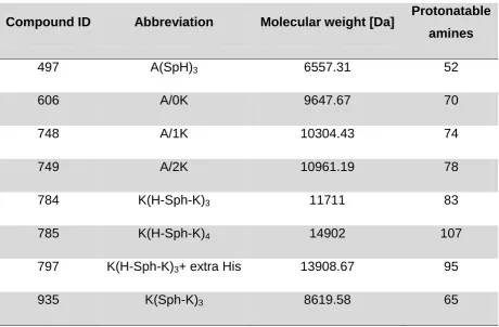
Table 3.1. Compound ID, abbreviation, molecular weight and amount of protonatable amines of all investigated oligomers. Oligomer 497 was synthesized by Dr. Edith Salcher (Pharmaceutical Biotechnology, LMU).
Compound ID Abbreviation Molecular weight [Da] Protonatable amines
497 A(SpH)3 6557.31 52
606 A/0K 9647.67 70
748 A/1K 10304.43 74
749 A/2K 10961.19 78
784 K(H-Sph-K)3 11711 83
785 K(H-Sph-K)4 14902 107
797 K(H-Sph-K)3+ extra His 13908.67 95
935 K(Sph-K)3 8619.58 65
47
3.1.1.Biophysical characterization of oligomers concerning DNA binding ability, particle size, zeta potential and buffering capacity
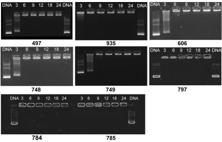
The binding ability of the various synthesized oligomers is expected to increase by the introduction of lysine residues and was therefore evaluated by agarose gel shift assays (Fig. 3.4.). In this assay, free pCMVL is migration without hindrance in the gel, whereas sufficient polyplex formation is resulting in a complete loss of electrophoretic mobility indicated by a sharp band at the position of the samples' well. In case of lysine-rich oligomers complete pCMVL binding, even at low nitrogen/phosphate ratio (N/P ratio) of 3, was observed with the exception of oligomer 748 and 749. Those oligomers displayed similar to the control oligomer 497 and 606, which are comprised of no additional stabilizing lysine residues, no sufficient pCMVL binding at N/P 3, suggesting a lower pCMVL binding affinity.
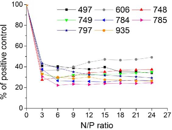
48 To confirm these findings, ethidium bromide (EtBr) assays were performed. In this assay, the pDNA binding ability of the oligomer correlates with a decreasing in fluorescence activity of ethidium bromide, which was monitored after stepwise addition of an oligomer solution to a solution containing free pCMVL. In accordance to the gel shift assays, results revealed that in presence of lysine-rich oligomers 784,
785, 797 and 935, less pCMVL is available to bind EtBr as compared to the
lysine-free counterpart structures 497 and 606. Figure 3.5. suggests that the pCMVL binding affinity of the lysine-rich oligomers at N/P 12 correlates to the amount of lysine residues present in the oligomeric structure. Oligomer 785 with the highest amount of lysine residues within the sequence showed least pCMVL availability to bind EtBr compared to all other oligomers. In comparison oligomer 748 and 749
comprising of one or two terminal lysine residue(s) mediated only a slight increased pCMVL binding affinity compared to the lysine-free counterparts. These findings confirm the well-know ability of lysines to form stable particle and further support the inclusion within the oligomeric structure to improve polyplex stability.
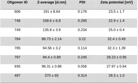
49 For a better understanding of these pCMVL particles, size (Z-average) and zeta potential of the polyplexes formed at N/P 12 was assessed (Table 3.2.). No significant differences were observed between the zeta potential of pCMVL polyplexes formed by lysine-rich and lysine-free oligomers. The observed zeta potentials were in the range between 22.9-32.4mV. However, polyplex sizes received by DLS measurements were in the range between 86.73 nm and 370nm. Polyplexes formed by lysine-rich oligomers 784, 785, 797 and 935 were of significantly smaller size (below 100nm) compared to those formed by lysine-free oligomers 606 and 497
(191-370nm) at the same N/P ratio. The incorporation of one (oligomer ID 748) or two terminal lysine residue(s) (oligomer ID 749) within the oligomer structure showed only a slight reduction of particle size compared to the 606 counterpart structure. Due to the fact that polyplex size is a critical parameter for successful gene delivery and bigger particle sizes are often connected to lower stability, those oligomers were not further evaluated.
Table 3.2 Particle size (Z-average), polydispersity index (PDI) and zeta potential of four-arm structured polyplexes formed at N/P 12 (n=3).
Oligomer ID Z-average [d.nm] PDI Zeta potential [mV]
606 191 ± 8.64 0.176 23,5 ± 1.7
748 168.6 ± 6.8 0.285 22.9 ± 1.4
749 135.8 ± 3.9 0.234 25.0 ± 0.4
784 86.73 ± 2.14 0.22 32.4 ± 0.49
785 94.56 ± 3.2 0.114 32.3 ± 1.39
797 94.4 ± 0.89 0.245 28.23 ± 0.95
935 96.31 ± 0.86 0.056 27.97 ± 0.64
50 In order to determine the most suitable excess of oligomer over pCMVL indicated by N/P ratio DLS was re-measured for polyplexes formed at N/P ratio 9, 12 and 18. Again, lysine-rich oligomers formed polyplexes of significant smaller sizes and slightly higher zeta potential compared to the lysine-free benchmark oligomer 606 (Fig. 3.6.). The formation of particles in case of lysine-rich oligomers seemed to be N/P-independent. Small and stable polyplexes were observed even at low N/P ratio of 9 and not further improved due to a higher N/P ratio. Within the lysine-rich oligomers, no differences in size or zeta potential were observed.
51 Since the endolysosomal pathway presents a threat to the nucleic acids' integrity, escaping from the endosome and buffering its pH has proven essential requirements for pCMVL polyplexes [124, 125]. For this reason the buffer capacity of the oligomers was assessed via acidimetric back titration in order to evaluate the effect of lysine residues present in the oligomeric structure on buffer capacity respectively endosomal escape. Figure 3.7. shows the titration curves between the endosomal pH of 5.0 and the physiological pH of 7.4, which has to be covered by the oligomer in order to mediate sufficient endosomal release. The results indicate a decreased buffer capacity for lysine-containing oligomers in comparison to the counterpart structure 606. However, due to the introduction of a histidine-rich moiety within the H-Sph-K repeating pattern of oligomer 797 buffer capacity is increased by 24% compared to oligomer 606. Within the whole set, the flattest slope was found for oligomer 797 indentifying the highest endosomal buffer capacity of all evaluated oligomers. In contrast, oligomer 935 comprising of a Sph-K repeating sequence pattern, exhibit the lowest buffering within the endosomal pH range, supporting the incorporation of histidine residues within the oligomer structure.