ABSTRACT
DAN, CHEN. Ligand Dynamics, Recognition and Structure/Function Relationships of Bio-nanoparticles. (Under the direction of Dr. Alex I. Smirnov).
Surface functionalized gold nanoparticles have been actively researched for many applications
in life sciences and biomedicine over the last decade. Polyfunctional water soluble Au
nanoparticles (Au NPs) demonstrated their potential in the imaging of biomolecular processes,
biosensing, drug delivery and development of new bio-inspired materials. 12 It is now well
documented that biorecognition properties of Au NPs could be tuned by choosing proper
monolayer terminal groups, opening new opportunities in biological and biomedical
applications. 3
However, detailed understanding of structure and dynamics of the nanoparticles’ ligand layer
– an essential pre-requisite for further development of Au NPs applications - is still missing in
the literature. Specifically, there is no full understanding of the dynamics of self-assembled
monolayers and the ns scale fluctuations of individual molecules; the local electrostatics and
hydrogen bonding at the bionanoparticle interface are also scarcely reported. The main
research aims of this project were to combine the advantages of the synthesis of novel spin
labels and probes and methods of spin labeling EPR with development of EPR spectroscopic
methods for assessing dynamics, structure, and electrostatic properties of bio-nanoparticle
interfaces.
In this project, we synthesized small water-soluble Au NPs protected by ligands bearing
as the pH of the medium changed. Disulfide and thioacetate ligands modified with both
ionizable and non-ionizable nitroxides were synthesized and employed for spin-labeling of Au
NPs through ligand exchange reactions. Basic functionalities of ionizable nitroxide participate
in proton-exchange reactions and report on the pH of the environment through changes in the
EPR spectral parameters, e.g., nitrogen hyperfine coupling constants and electronic g factors, as well as through the variations in order parameters. pKa’s of the ligands modified with pH-sensitive nitroxide were experimentally determined both in solution and at the nanoparticle
interface and these ligands were proposed to be useful for EPR studies of the electrostatics and
© Copyright 2014 by Dan Chen
Ligand Dynamics, Recognition and Structure/Function Relationships of Bio-nanoparticles
by Dan Chen
A thesis submitted to the Graduate Faculty of North Carolina State University
in partial fulfillment of the requirements for the degree of
Master of Science
Chemistry
Raleigh, North Carolina
2014
APPROVED BY:
_______________________________ ______________________________
Dr. Alex I. Smirnov Dr. Yaroslava G. Yingling Committee Chair
BIOGRAPHY
Dan Chen was born on Sept. 30th, 1987 in Mianyang, China, where she spent her first 18
years of the life and found herself interested in science. Then she followed her heart to learn
more about physics and chemistry in the University of Science and Technology of China and
got her B.S. degree there. After that, Dan continued her research in Physical Chemistry under
ACKNOWLEDGMENTS
There are so many people I would love to acknowledge. I would like to express my deepest
appreciation to my thesis advisor Prof. Alex I. Smirnov, you have backed me up through my
graduate life and my future career. Without your encouragement and support on my research
this thesis would not have been possible. I would also like to thank Prof. Gufeng Wang and
Prof. Yaroslava G. Yingling for serving as my committee members.
My sincere thanks also go to Dr. Maxim A. Voinov in our group for all the advices and help
on my research. Thank you for your patience and guidance on all the synthesis skills.
Thank everyone in Smirnov and Smirnov groups for all the days we worked together and all
TABLE OF CONTENTS
LIST OF TABLES ... vii
LIST OF FIGURES ... viii
LIST OF ABBREVIATIONS ... viii
CHAPTER 1 INTRODUCTION ... 1
1.1 Electron Spin Resonance ... 1
1.2 Nitroxide Radicals ... 5
1.3 Gold Nanoparticles (Au NPs) ... 8
CHAPTER 2 RESULTS AND DISCUSSION... 10
2.1 Ligands ... 10
2.1.1 Octanethiol(4) ... 10
2.1.2 11-(dimethylamino)undecane-1-thiol hydrochloride (5) ... 11
2.1.3 11-Mercapto-1-undecanol (6) ... 11
2.1.4 11, 11’-Dithiodiundecanol (7) ... 12
2.1.5 12-(Acetylthio)dodecanol(8) ... 13
2.1.6 10-(Acetylthio)decanoic acid (9) ... 13
2.2 Nitroxide Radicals ... 14
2.2.1 3-Carboxy-2,2,5,5-tetramethyl-3-pyrroline-1-oxyl(1) ... 14
2.2.2 2-(2-Carboxyethyl)-2,3,4,5,5-pentamethylimidazolidine-1-oxyl (2) ... 14
2.2.3 4-(2-Aminoethylamino)-1-oxyl-2,2,5,5-tetramethyl-2,5-dihydro-1H-imidazole(3) ... 16
2.3 Spin-labeled Ligands ... 17
2.3.1 1,2-bis(undecyl 1-oxyl-2,2,5,5-tetramethyl-2,5-dihydro-1H-pyrrole-3-carboxylate) disulfide (15) ... 18
2.3.2 12-(acetylthio)dodecyl 1-oxyl-2,2,5,5-tetramethyl-2,5-dihydro-1H-pyrrole-3-carboxylate (16) ... 18
2.3.3 12-(acetylthio)dodecyl 3-(3-oxyl-1,2,4,4,5-pentamethylimidazolidin-2-yl)propanoate (17) ... 19
2.3.4 S-10-oxo-10-(2-(1-oxyl-2,2,5,5-tetramethyl-2,5-dihydro-1H-imidazol-4-ylamino)ethylamino)decyl ethanethioate(18) ... 20
2.4 Au NPs ... 21
2.4.2 1,2-bis(undecyl 1-oxyl-2,2,5,5-tetramethyl-2,5-dihydro-1H-pyrrole-3-carboxylate)
disulfide-modified Au NPs(20) ... 27
2.4.3 12-(acetylthio)dodecyl 1-oxyl-2,2,5,5-tetramethyl-2,5-dihydro-1H-pyrrole-3-carboxylate-modified Au NPs(21) ... 29
2.4.4 12-(acetylthio)dodecyl 3-(3-oxyl-1,2,4,4,5-pentamethylimidazolidin-2-yl)propanoate-modified Au NPs(22) ... 31
2.4.5 S-10-oxo-10-(2-(1-oxyl-2,2,5,5-tetramethyl-2,5-dihydro-1H-imidazol-4-ylamino)ethylamino)decyl ethanethioate-modified Au NP(23) ... 32
2.5 Titration of pH Sensitive Spin-labeled Ligands ... 34
2.5.1 12-(acetylthio)dodecyl 3-(3-oxyl-1,2,4,4,5-pentamethylimidazolidin-2-yl)propanoate(17) ... 35
2.5.2 S-10-oxo-10-(2-(1-oxyl-2,2,5,5-tetramethyl-2,5-dihydro-1H-imidazol-4-ylamino)ethylamino)decyl ethanethioate (18) ... 38
2.6 Titration of S-10-oxo-10-(2-(1-oxyl-2,2,5,5-tetramethyl-2,5-dihydro-1H-imidazol-4-ylamino)ethylamino)decyl ethanethioate-modified Au NPs (23) ... 41
2.7 Conclusions ... 44
CHAPTER 3 EXPERIMENTAL PART ... 46
3.1 Instruments and Materials ... 46
3.2 Synthesis of Ligands ... 47
3.2.1 Synthesis of 11-(dimethylamino)undecane-1-thiol hydrochloride (5) ... 47
3.2.2 Synthesis of 11, 11’-Dithiodiundecanol (7). ... 51
3.2.3 Synthesis of 12-(Acetylthio)dodecanol(8). ... 53
3.2.4 Synthesis of 10-(Acetylthio)decanoic acid (9). ... 54
3.3 Synthesis of Nitroxide Radicals ... 56
3.3.1 Synthesis of 1-hydroxy-3-imidazoline derivative(11). ... 56
3.3.2 Synthesis of corresponding radical of 1-hydroxy-3-imidazoline derivative (12). ... 56
3.3.3 Synthesis of2-(2-methoxycarbonylethyl)-2,3,4,5,5-pentamethyl-3-imidazolinium 1-oxyl methylsulfate (13). ... 57
3.3.4 Synthesis of2-(2-methoxycarbonylethyl)-2,3,4,5,5-pentamethylimidazolidine 1-oxyl (14). ... 58
3.3.5 Synthesis of 2-(2-Carboxyethyl)-2,3,4,5,5-pentamethylimidazolidine-1-oxyl (2)... 61
3.4.1 Synthesis of1,2-bis(undecyl
1-oxyl-2,2,5,5-tetramethyl-2,5-dihydro-1H-pyrrole-3-carboxylate) disulfide (15) ... 62 3.4.2 Synthesis of 12-(acetylthio)dodecyl 1-oxyl-2,2,5,5-tetramethyl-2,5-dihydro-1H-pyrrole-3-carboxylate (16) ... 63 3.4.3 Synthesis of 12-(acetylthio)dodecyl 3-(3-oxyl-1,2,4,4,5-pentamethylimidazolidin-2-yl)propanoate(17) ... 65 3.4.4 Synthesis of S-10-oxo-10-(2-(1-oxyl-2,2,5,5-tetramethyl-2,5-dihydro-1H-imidazol-4-ylamino)ethylamino)decyl ethanethioate (18) ... 66 3.5 Synthesis of Functionalized Au NPs ... 68
3.5.1 Synthesis of 11-(dimethylamino)undecane-1-thiol hydrochloride-coated water soluble Au NP (19) ... 68 3.5.2 Synthesis of 1,2-bis(undecyl
1-oxyl-2,2,5,5-tetramethyl-2,5-dihydro-1H-pyrrole-3-carboxylate) disulfide-modified Au NPs (20) ... 69 3.5.3 Synthesis of 12-(acetylthio)dodecyl 1-oxyl-2,2,5,5-tetramethyl-2,5-dihydro-1H-pyrrole-3-carboxylate-modified Au NPs(21). ... 70 3.5.4 Synthesis of 12-(acetylthio)dodecyl
LIST OF TABLES
Table 1.1 Spin Numbers and Hyperfine Splitting Constants ... 5
Table 2.1 Statistics of DLS data for size measurement of 11-(dimethylamino)undecane-1-thiol hydrochloride-coated water soluble Au NP from the same freshly prepared sample ... 22
Table 2.2 Titration Data for 12-(acetylthio)dodecyl 3-(3-oxyl-1,2,4,4,5-pentamethylimidazolidin-2-yl)propanoate in Buffer/Isopropanol Solutions of Various Compositions ... 36
LIST OF FIGURES
Fig. 1.1 Energy levels of s=1/2 spin in presence and absence of magnetic field and the resonance
conditions ... 2
Fig. 1.2 The layout of a continuous-wave EPR spectrometer ... 3
Fig. 1.3 A typical EPR spectrum for free nitroxide radicals. ... 4
Fig. 1.4 Hyperfine interaction between an electron and a 14 N nucleus. ... 5
Fig. 1.5. Chemical structures of the nitroxides used in this project. ... 6
Fig. 1.6 Representative room-temperature X-band EPR spectra of 2-(2-Carboxyethyl)-2,3,4,5,5-pentamethylimidazolidine-1-oxyl-modified ligand in a series of buffer solutions containing 40% of isopropanol. ... 7
Fig. 1.7 Scheme of the synthesis of 11-(dimethylamino)undecane-1-thiol hydrochloride-coated water soluble Au NPs. ... 9
Fig. 2.1 Scheme of the structure of Octanethiol (4). ... 10
Fig. 2.2 Scheme of structure of 11-(dimethylamino)undecane-1-thiol hydrochloride (5). ... 11
Fig. 2.3 Scheme of structure of 11-Mercapto-1-undecanol (6). ... 11
Fig. 2.4 Scheme of structure of11, 11’-Dithiodiundecanol (7). ... 12
Fig. 2.5 Scheme of structure of 12-(Acetylthio)dodecanol (8). ... 13
Fig. 2.6 Scheme of the structure of 10-(Acetylthio)decanoic acid (9). ... 13
Fig. 2.7 Scheme of the structure of 3-Carboxy-2,2,5,5-tetramethyl-3-pyrroline-1-oxyl (1). ... 14
Fig. 2.8 Scheme of the structure of 2-(2-Carboxyethyl)-2,3,4,5,5-pentamethylimidazolidine-1-oxyl (2) ... 14
Fig. 2.9 Scheme of synthesis of 2-(2-Carboxyethyl)-2,3,4,5,5-pentamethylimidazolidine-1-oxyl (2) . 15 Fig. 2.10 Scheme of structure of 4-(2-Aminoethylamino)-1-oxyl-2,2,5,5-tetramethyl-2,5-dihydro-1H-imidazole (3). ... 16
Fig. 2.11 Scheme of chemical structures of spin-labeled ligands used in this project. ... 17
Fig. 2.12 Scheme of the synthesis of 1,2-bis(undecyl 1-oxyl-2,2,5,5-tetramethyl-2,5-dihydro-1H-pyrrole-3-carboxylate) disulfide (15). ... 18
Fig. 2.13 Scheme of the synthesis of 12-(acetylthio)dodecyl 1-oxyl-2,2,5,5-tetramethyl-2,5-dihydro-1H-pyrrole-3-carboxylate (16). ... 19
Fig. 2.14 Scheme of the synthesis of 12-(acetylthio)dodecyl 3-(3-oxyl-1,2,4,4,5-pentamethylimidazolidin-2-yl)propanoate (17). ... 19
Fig. 2.16 Scheme of the synthesis of 11-(dimethylamino)undecane-1-thiol hydrochloride-coated water soluble Au NP (19). ... 21
Fig. 2.17 TEM images of Au NPs. ... 22
Fig. 2.18 DLS analysis of the nanoparticle solution from the same sample of
11-(dimethylamino)undecane-1-thiol hydrochloride-coated water soluble Au NP (19) freshly prepared ... 23
Fig. 2.19 Results of the TGA analysis of 11-(dimethylamino)undecane-1-thiol hydrochloride-coated water soluble Au NP (19) ... 25
Fig. 2.20 Zeta potential of 11-(dimethylamino)undecane-1-thiol hydrochloride-coated water soluble Au NP (19) in ultra-pure water ... 27
Fig. 2.21 Scheme of the synthesis of 1,2-bis(undecyl 1-oxyl-2,2,5,5-tetramethyl-2,5-dihydro-1H-pyrrole-3-carboxylate) disulfide-modified Au NPs (20)... 28
Fig. 2.22 EPR spectra acquired from samples of 1,2-bis(undecyl 1-oxyl-2,2,5,5-tetramethyl-2,5-dihydro-1H-pyrrole-3-carboxylate) disulfide-modified Au NPs ligand exchanged at various 15/NP ratios ... 28
Fig. 2.23 Scheme of the synthesis of 12-(acetylthio)dodecyl 1-oxyl-2,2,5,5-tetramethyl-2,5-dihydro-1H-pyrrole-3-carboxylate-modified Au NPs (21) ... 29
Fig. 2.24 EPR spectra acquired from 12-(acetylthio)dodecyl 1-oxyl-2,2,5,5-tetramethyl-2,5-dihydro-1H-pyrrole-3-carboxylate-modified Au NPs ligand exchanged at various 16/NP ratios ... 30
Fig. 2.25 Scheme of the synthesis of 12-(acetylthio)dodecyl
3-(3-oxyl-1,2,4,4,5-pentamethylimidazolidin-2-yl)propanoate-modified Au NPs (22) ... 31
Fig. 2.26 EPR spectra of 12-(acetylthio)dodecyl 3-(3-oxyl-1,2,4,4,5-pentamethylimidazolidin-2-yl)propanoate-modified Au NPs under different pH conditions ... 32
Fig. 2.27 Scheme of the synthesis of S-10-oxo-10-(2-(1-oxyl-2,2,5,5-tetramethyl-2,5-dihydro-1H-imidazol-4-ylamino)ethylamino)decyl ethanethioate-modified Au NP ... 33
Fig. 2.28. Representative room-temperature X-band EPR spectra of S-10-oxo-10-(2-(1-oxyl-2,2,5,5-tetramethyl-2,5-dihydro-1H-imidazol-4-ylamino)ethylamino)decyl ethanethioate-modified Au NP taken at various pH in a series of aqueous solutions in the presence of SDS ... 33
Fig. 2.29 Representative room-temperature X-band EPR spectra of 12-(acetylthio)dodecyl 3-(3-oxyl-1,2,4,4,5-pentamethylimidazolidin-2-yl)propanoate (17) in a series of buffer solutions containing 40% of isopropanol ... 35
Fig. 2.30 Experimental X-band EPR titration data for 12-(acetylthio)dodecyl 3-(3-oxyl-1,2,4,4,5-pentamethylimidazolidin-2-yl)propanoate (17) measured at room-temperature in
buffer/isopropanol solutions... 37
Fig. 2.31 Linear regression of pKa of 12-(acetylthio)dodecyl
Fig. 2.32 Representative room-temperature X-band EPR spectra of S-10-oxo-10-(2-(1-oxyl-2,2,5,5-tetramethyl-2,5-dihydro-1H-imidazol-4-ylamino)ethylamino)decyl ethanethioate (18) in a series of
40% isopropanol buffer solutions of different pH ... 39
Fig. 2.33 Experimental X-band EPR titration data for S-10-oxo-10-(2-(1-oxyl-2,2,5,5-tetramethyl-2,5-dihydro-1H-imidazol-4-ylamino)ethylamino)decyl ethanethioate (18) measured at room-temperature in buffer/isopropanol solutions ... 40
Fig. 2.34 Linear regression of pKa of S-10-oxo-10-(2-(1-oxyl-2,2,5,5-tetramethyl-2,5-dihydro-1H-imidazol-4-ylamino)ethylamino)decyl ethanethioate (18) vs. percentage of bulk water ... 40
Fig. 2.35 (a) Representative room-temperature X-band EPR spectra of S-10-oxo-10-(2-(1-oxyl-2,2,5,5-tetramethyl-2,5-dihydro-1H-imidazol-4-ylamino)ethylamino)decyl ethanethioate-modified Au NPs (23) taken at various pH in a series of aqueous solutions in the presence of SDS and (b) EPR spectra comparison of 23-modified Au NPs in completely protonated and free forms ... 42
Fig. 2.36 EPR titration data for S-10-oxo-10-(2-(1-oxyl-2,2,5,5-tetramethyl-2,5-dihydro-1H-imidazol-4-ylamino)ethylamino)decyl ethanethioate-modified Au NPs (23) ... 44
Fig. 3.1 Scheme of synthesis of thioacetic acid-S-(11-bromoundecyl) ester ... 47
Fig. 3.2 The 1H NMR spectrum of thioacetic acid-S-(11-bromoundecyl) ester ... 48
Fig. 3.3 Scheme of the synthesis of 11-(dimethylamino)undecane-1-thiol hydrochloride (5) ... 48
Fig. 3.4 1H NMR spectrum of 11-(dimethylamino)undecane-1-thiol hydrochloride (5). ... 50
Fig. 3.5. IR spectrum of 11-(dimethylamino)undecane-1-thiol hydrochloride (5) ... 51
Fig. 3.6 Scheme of the synthesis of 11, 11’-Dithiodiundecanol (7) ... 51
Fig. 3.7 Scheme of the synthesis of 11, 11’-Dithiodiundecanol (7). ... 52
Fig. 3.8 1H NMR spectrum of 11, 11’-Dithiodiundecanol (7) ... 53
Fig. 3.9 Scheme of the synthesis of 8 ... 53
Fig. 3.10 1H NMR spectrum of 12-(Acetylthio)dodecanol (8) ... 54
Fig. 3.11 Scheme of the synthesis 10-(Acetylthio)decanoic acid (9) ... 54
Fig. 3.12 1H NMR spectrum of 10-(Acetylthio)decanoic acid (9) ... 55
Fig. 3.13 Scheme of the synthesis of 1-hydroxy-3-imidazoline derivative (11) ... 56
Fig. 3.14 Scheme of the synthesis of corresponding radical of 1-hydroxy-3-imidazoline derivative (12) ... 56
Fig. 3.15 Scheme of the synthesis of 2-(2-methoxycarbonylethyl)-2,3,4,5,5-pentamethyl-3-imidazolinium 1-oxyl methylsulfate (13) ... 57
Fig. 3.17 IR spectra of the diastereomers of
2-(2-methoxycarbonylethyl)-2,3,4,5,5-pentamethylimidazolidine 1-oxyl (14). ... 59
Fig. 3.18 Mass spectra of the diastereomers of
2-(2-methoxycarbonylethyl)-2,3,4,5,5-pentamethylimidazolidine 1-oxyl (14). ... 60
Fig. 3.19 Scheme of the synthesis of 2-(2-Carboxyethyl)-2,3,4,5,5-pentamethylimidazolidine-1-oxyl (2). ... 61
Fig. 3.20 IR spectrum of 2-(2-Carboxyethyl)-2,3,4,5,5-pentamethylimidazolidine-1-oxyl (2). ... 62
Fig. 3.21 Scheme of the synthesis of 1,2-bis(undecyl 1-oxyl-2,2,5,5-tetramethyl-2,5-dihydro-1H-pyrrole-3-carboxylate) disulfide (15). ... 62
Fig. 3.22 IR spectrum of 1,2-bis(undecyl 1-oxyl-2,2,5,5-tetramethyl-2,5-dihydro-1H-pyrrole-3-carboxylate) disulfide (15) ... 63
Fig. 3.23 Scheme of the synthesis of 12-(acetylthio)dodecyl 1-oxyl-2,2,5,5-tetramethyl-2,5-dihydro-1H-pyrrole-3-carboxylate (16). ... 63
Fig. 3.24 IR spectrum of 12-(acetylthio)dodecyl 1-oxyl-2,2,5,5-tetramethyl-2,5-dihydro-1H-pyrrole-3-carboxylate (16) ... 64
Fig. 3.25 Scheme of the synthesis of 12-(acetylthio)dodecyl
3-(3-oxyl-1,2,4,4,5-pentamethylimidazolidin-2-yl)propanoate (17). ... 65
Fig. 3.26 IR spectrum of 12-(acetylthio)dodecyl 3-(3-oxyl-1,2,4,4,5-pentamethylimidazolidin-2-yl)propanoate (17) ... 65
Fig. 3.27 The mass spectra of 12-(acetylthio)dodecyl 3-(3-oxyl-1,2,4,4,5-pentamethylimidazolidin-2-yl)propanoate (17) ... 66
Fig. 3.28 Scheme of the synthesis of S-10-oxo-10-(2-(1-oxyl-2,2,5,5-tetramethyl-2,5-dihydro-1H-imidazol-4-ylamino)ethylamino)decyl ethanethioate (18) ... 66
Fig. 3.29 IR spectrum of S-10-oxo-10-(2-(1-oxyl-2,2,5,5-tetramethyl-2,5-dihydro-1H-imidazol-4-ylamino)ethylamino)decyl ethanethioate (18) ... 67
Fig. 3.30 Mass spectrum of S-10-oxo-10-(2-(1-oxyl-2,2,5,5-tetramethyl-2,5-dihydro-1H-imidazol-4-ylamino)ethylamino)decyl ethanethioate (18) ... 68
Fig. 3.31 Scheme of the synthesis of 11-(dimethylamino)undecane-1-thiol hydrochloride-coated water soluble Au NP (19). ... 68
Fig. 3.32 IR spectra of (a) 5 and (b) of 11-(dimethylamino)undecane-1-thiol hydrochloride-coated water soluble Au NP (19) ... 69
Fig. 3.33 Scheme of the synthesis of 1,2-bis(undecyl 1-oxyl-2,2,5,5-tetramethyl-2,5-dihydro-1H-pyrrole-3-carboxylate) disulfide-modified Au NPs (20)……….66
Fig. 3.35Scheme of synthesis of 12-(acetylthio)dodecyl 3-(3-oxyl-1,2,4,4,5-pentamethylimidazolidin-2-yl)propanoate-modified Au NPs (22)……….68
Fig. 3.36Scheme of synthesis of
LIST OF ABBREVIATIONS
AIBN Azobisisobutyronitrile
DCC N,N'-Dicyclohexylcarbodiimide
DMAP 4-Dimethylaminopyridine
DLS Dynamic light scattering
DMSO Dimethyl sulfoxide
DMF Dimethylformamide
EPR Electron paramagnetic resonance
IR Infrared spectroscopy
NMR Nuclear magnetic resonance
THF Tetrahydrofuran
TLC Thin layer chromatography
CHAPTER 1 INTRODUCTION
1.1 Electron Spin Resonance
In 1922, two German physicists, Otto Stern and Walther Gerlach, carried out one of
the most important and fundamental experiment in quantum mechanics. In
Stern-Gerlach experiment, atoms could only have certain discrete orientations when passing
through an inhomogeneous magnetic field, which indicated the existence of electron
spin. This experiment led to development of the concept of spin in quantum
mechanics, and, ultimately, several decades later, to discovery of magnetic resonance – both nuclear magnetic resonance (NMR) and electron paramagnetic resonance
(EPR).
When a magnetic moment is exposed to a magnetic field , it has energy
cos( , )
E B B B (1.1)
n quantum mechanics, this equation can also be expressed as
H B
(1.2)
According to Dirac’s formulation of quantum mechanics, 𝐻∧ is the Hamiltonian operator,
corresponding to the total energy of the system. 𝜇∧, the magnetic momentum operator,
is proportional to the spin angular momentum 𝑠∧,
e g s
(1.3)
𝛾𝑒 is called the magnetogyric ratio,
2 e
e e m
(1.4)
And 𝑔 is called the 𝑔 − 𝑓𝑎𝑐𝑡𝑜𝑟 of electron, which is very close to 2. The exact value
of g-factor reflects some intrinsic properties (structure) of the radicals. For free
electrons,𝑔𝑒 = 2.0023.
In magnetic resonance magnetic field is always taken along z-direction. The
z z g es
(1.5)
thus
z e z
H B g s B
(1.6)
For spin s=1/2, the eigenvalue of 𝑠∧𝑧 is 𝑚𝑠ℏ with 𝑚𝑠 = ±12, and, accordingly, the total
energy of an electron in magnetic field is
s
m e s B s
E g m B g m B (1.7)
The Bohr magneton, 𝜇𝐵, is defined as
24 1 9.724 10 2 B e e e JT m
(1.8)
As a result, states of different 𝑚𝑠 values would be degenerate in the absence of
magnetic field and the degeneracy will be removed by applying magnetic field Two
energy levels are formed with the gap given as (see also Figure 1) :
B E E E g B
(1.9)
The existence of these energy levels can be probed by applying a radiofrequency field
with frequency 𝜈 to cause 𝛼 → 𝛽 transition at the resonance conditions:
B
h g B (1.10)
Fig. 1.1 Energy levels of s=1/2 spin in presence and absence of magnetic field and the
Electron spin resonance is a spectroscopic technique studying molecular species with
unpaired electrons by analyzing the magnetic fields that in resonance with
monochromatic (microwave or millimeter wave) radiation. The magnetic field applied
in a typical commercial EPR spectrometer is around 0.3 T, thus, the resonating
frequency of electromagnetic field is around 10 GHz, corresponding to wavelength
within microwave range (wavelength of about 2 cm).
Fig. 1.2 The layout of a continuous-wave EPR spectrometer (redrawn from reference 4)
In continuous wave (CW) EPR experiment the EPR spectrm is detected using
phase-sensitive detection when magnetic field is modulated at ca. 100 kHz. Then the
detected EPR spectrum is the first-derivative of absorption. A typical spectrum of free
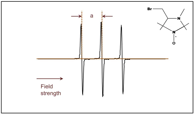
Fig. 1.3 A typical EPR spectrum for freenitroxide radicals.
𝒂is the nitrogen hyperfine splitting of the spectrum. The position of the central line is determined by equation 𝑩 = 𝒉𝝂
𝒈𝝁𝑩.
The hyperfine splitting is a very important property of EPR. It arises from two aspects.
Firstly, for p-orbital electrons, the hyperfine splitting is caused by magnetic
dipole-dipole interaction between electron spin and the nuclear spin present in vicinity. This
dipolar interaction is anisotropic and can be averaged out by molecular motion.
Secondly, for s-orbital electrons, there is an isotropic Fermi contact interaction. Due
to these interactions, local fields are formed and vectorially added to the external field.
As the consequence, the effective field is
eff local I
H HH Ham (1.11)
𝑎 is the hyperfine splitting constant, which has different values for different nuclei, and
Table 1.1 Spin Numbers and Hyperfine Splitting Constants 4
As a result, the energy levels will be further split by this electron-nucleus interaction,
as shown in Fig. 1.4.
Fig. 1.4 Hyperfine interaction between an electron and a 14
N nucleus.
The resulting EPR spectrum is shown in Fig. 1.3. The intensities of every peak are the same (redrawn from reference 4).
1.2 Nitroxide Radicals
Nitroxide radicals is a family of organic persistent free radicals containing an unpaired
electron delocalized between nitrogen and oxygen atoms of the nitroxide group
group is 0, so it has only one allowed nuclear spin state which results in no hyperfine
interactions with the unpaired electron. However, the spin quantum number of nearby
nitrogen nucleus (14N) could be 0 or ±1 (table 1.1). Because of the magnetic
dipole-dipole interaction between electronic spin and the nitrogen nuclear spin, hyperfine
structure of the three absorption lines is observed in the EPR spectra of nitroxide
radicals (Fig. 1.3). Chemical structures of representative nitroxides are shown in Fig.
1.5.
Nitroxide radicals attached to other molecules or biopolymer chains act as spin probes.
When tumbling freely, spin probes give an EPR spectrum similar to that shown in Fig.
1.3. However, once the local enviroment changes, their movements could be
restricted, and the lineshapes would be dramatically affected, providing information
on dynamic properties of spin probes and their immediate molecular enviroment.
In this project, the following nitroxide radicals (Fig. 1.5) were employed.
3-Carboxy-2,2,5,5-tetramethyl-3-pyrroline-1-oxyl (1, Fig. 1.5) is used to study the dynamics of the
self-assembled monolayers. 1 is not intrinsically sensitive to the pH of the medium
since it does not contain an ionizable group in the vicinity of the NO● group (note:
COOH group is too far from the NO● group to noticeably affect its magnetic
parameters).
2-(2-Carboxyethyl)-2,3,4,5,5-pentamethylimidazolidine-1-oxyl (2, Fig. 1.5) and
4-(2-Aminoethylamino)-1-oxyl-2,2,5,5-tetramethyl-2,5-dihydro-1H-imidazole (3, Fig. 1.5)
are pH-sensitive nitroxide radicals. Nitroxide 2 has a tertiary amino group at position
3 of the imidazolidine heterocycle, which could protonated or nonprotonated in
different environment. Nitroxide 3 has a protonatable amidino group as a part of the
heterocycle that can report on its ionization state of the molecule through the changes
in magnetic parameters (e.g., isotropic nitrogen hyperfine coupling constant, Aiso, or
electronic g-factor), or order parameters. Functional groups in the side chains of the
nitroxides 1-3 can be used as attachment sites for molecules of interest.
When attached to the ligands, nitroxides 2 and 3 act as spin probes and parameters
of their EPR spectra (e.g., Aiso) report on the ionization state of corresponding
spin-labeled ligands. Monitoring how Aiso changes with pH allows one to determine the pKa
of the ionizable group of the nitroxide in the spin-labeled ligand. Fig. 1.6 shows an
example how the X-band (9.5 GHz) EPR spectra of the ligand modified with the
nitroxide 2 change with pH in buffer solutions containing 40 v/v% of isopropanol.
Fig. 1.6 Representative room-temperature X-band EPR spectra of
2-(2-Carboxyethyl)-2,3,4,5,5-pentamethylimidazolidine-1-oxyl-modified ligand in a series of buffer solutions containing
Magnetic Field, G
3480 3490 3500 3510 3520 3530
pH=0.63
pH=1.72
pH=2.84
pH=3.52
pH=4.64
pH=5.65
40% of isopropanol. pH values of the solutions are indicated next to the corresponding
spectra. Dashed lines mark approximate positions of the high-field nitrogen hyperfine
coupling components corresponding to protonated and nonprotonated forms of the nitroxide.
1.3 Gold Nanoparticles (Au NPs)
Gold is one of the most widely studied metals in the world. Besides its aesthetic and
collectible values, it also plays an important role in scientific research.
During the past decade polyfunctional water soluble Au NPs have found vast
applications in biomedical imaging, biomolecular processes, biosensing, and drug
delivery, as well as for fabricating new bio-inspired materials. These applications hinge
on the electrostatic and/or hydrophobic interactions of properly decorated Au NPs with
biological molecules. 1-2
Though the molecular recognition property of Au NPs could be tuned by choosing
proper monolayer terminal groups, 3 one could find very few literature reports
describing molecular level properties of the NPs’ interfacial region, such as surface
electrostatics and a gradient of local dielectric constant across the interface. Measuring and understanding these fundamental biophysical parameters at the NPs’
interface will open new opportunities in biological and biomedical applications. The
goal of our research is to combine advances of modern nanotechnology, organic
synthesis and EPR spectroscopy and to develop spin probe EPR methods to assess
dynamics and electrostatic and dielectric properties of the nanoparticle-biomolecule
interfaces. The first step in this project is a preparation of small Au NPs coated with
ionizable functional groups so the electrostatic properties of NPs can be adjusted and
studied as a function of pH.
Two methods were employed to synthesize water-soluble Au NPs. First, we tried the
two-phase synthesis method, which was introduced by Mathias Brust in 1994.5 In this
method, a solution of octanethiol (4) in toluene was added to aqueous
interface. The intermediate was reduced by sodium borohydride to yield the Au NPs.
Using this method we successfully synthesized 4-coated Au NPs with diameters
around 0.5 nm. 4-coated Au NPs are hydrophobic, but we wanted our Au NPs to be
water-soluble. To prepare water-soluble NPs, we synthesized a ligand with charged
terminal amino groups - 11-(dimethylamino)undecane-1-thiol hydrochloride (5) 6 and
tried to replace 4 with 5 through the ligand-exchange reaction.78 However, due to the
low ligand exchange efficiency and nanoparticles aggregations caused by the long
chain of ligands during the reaction,9 we had to give up this method and tried to find
a way to directly coat 5 onto Au NPs.
The second approach we applied is based on the one-pot synthesis method,10 11
which is widely used in recent years. We utilized a mixture of THF and acetic acid as
the solvent instead of pure THF mentioned in literature.
Fig. 1.7 Scheme of the synthesis of 11-(dimethylamino)undecane-1-thiol hydrochloride-coated
water soluble Au NPs.
5 and hydrochloroauric acid were co-dissolved in this solvent to form an intermediate.
Then the intermediate was reduced by sodium borohydride and developed into
water-soluble Au NPs.12 The average diameter of these Au NPs were found to be around
1.5 nm by DLS and TEM. NP diameters from 0.5 to 2.0 nm are considered to be
CHAPTER 2 RESULTS AND DISCUSSION
2.1 Ligands
Ligands with long hydrophobic carbon chains were synthesized and employed in our
project. Some of them were used in the synthesis of Au NPs in order to make NPs
ionizable so that NPs would be stable and monodispersed in different pH conditions.
Other ligands were connected to nitroxides before coating to Au NPs. Such
spin-labeled ligands are EPR active and could act as probes to detect local electrostatic
properties of Au NPs.
2.1.1 Octanethiol(4)
Octanethiol is commercially available.
Octanethiol-coated Au NPs have hydrophobic properties, and are able to act as
precursor towards water soluble Au NPs through ligand exchange reaction. The
structure of 4 is shown below in Fig. 2.1.
Fig. 2.1 Scheme of the structure ofOctanethiol (4).
Octanethiol was used as a ligand to synthesize some hydrophobic Au NPs through
two phase method.5 This method was chosen because it is very mature and yielded
highly stable Octanethiol-coated Au NPs with small (around 0.5 nm) diameter.
Our initial approach was to obtain some water soluble Au NPs by conducting ligand
exchange experiment 7 8 using Octanethiol-coated Au NPs and another ligand
containing ionizable terminal group. Even though we did obtain some water soluble
process of the ligand exchange.9Thus, we had no choice but abandon this approach
and tried an alternative procedure to directly synthesize water soluble Au NPs.
2.1.2 11-(dimethylamino)undecane-1-thiol hydrochloride (5)
11-(dimethylamino)undecane-1-thiol hydrochloride contains an ionizable functional
group so once decorated on Au NPs, it can make Au NPs more hydrophilic, and the
electrostatic properties of NPs can be adjusted and studied as a function of pH. The
structure of the ligand is shown below in Fig. 2.2.
Fig. 2.2 Scheme of structure of 11-(dimethylamino)undecane-1-thiol hydrochloride (5).
5 was synthesized in three steps according to the literature protocol.6 Firstly,
11-bromo-1-undecene was reacted with thioacetic acid in the presence of AIBN, resulting
in thioacetic acid-S-(11-bromoundecyl) ester. 6a We verified thioacetic
acid-S-(11-bromoundecyl) ester by 1H NMR (400 MHz, CDCl
3, δ). Secondly, dimethylamine
reacted with thioacetic acid-S-(11-bromoundecyl) ester, 6b and thirdly the resulting
product was taken up in 20% solution of gaseous HCl in THF to form 5.6b 1H NMR
(400 MHz, CD3OD, δ) and IR were applied to characterize 5.
2.1.3 11-Mercapto-1-undecanol (6)
6 was commercially purchased. The structure of 6 is shown in Fig. 2.3.
Compound 6 was used in two ways. Firstly, it was employed in ligand-exchange
reaction in order to attach radicals to Au NPs. We tried to synthesize some Au NPs
coated with both 5 and 6, and then attach nitroxide radicals to 6 by a coupling reaction
of OH group on 6 and COOH group on nitroxide radicals. However, the resulting Au
NPs were EPR silent. This could be attributed to the low coverage of 6 on Au NPs.
Alternatively, the long chain of 5 could produce steric problem for nitroxide radicals to
get close to the OH group of 6.
Secondly, compound 6 could also acts as a precursor of a disufide derivative, which
will be described in 2.1.4 and later chapters.
2.1.4 11, 11’-Dithiodiundecanol (7)
The structure of this compound is shown in Fig. 2.4.
Fig. 2.4 Scheme of structure of11, 11’-Dithiodiundecanol (7).
7 was synthesized from 6 by an oxidation reaction with DMSO 13 or I
2, 14 and
characterized by 1H NMR (400 MHz, CDCl 3, δ).
We were able to attach nitroxide radical to 7 through DCC-mediated coupling reaction
15 of its OH groups and COOH group of nitroxide radical, as a result, we obtained a
spin-labeled ligand. Then, the spin-labeled ligand was employed in ligand exchange
experiments to obtain spin-labeled Au NPs.
2.1.5 12-(Acetylthio)dodecanol(8)
The structure of 8 is shown in Fig. 2.5.
Fig. 2.5 Scheme of structure of 12-(Acetylthio)dodecanol (8).
8 was synthesized by a ssubstitution reaction of 11-Mercapto-1-dodecanol and
Potassium thioacetate, 16 and characterized by 1H NMR (400 MHz, CDCl3, δ). We
were able to attach nitroxide radical to this compound through coupling reaction to
obtain a spin-labeled ligand. Then, the spin-labeled ligand was employed in ligand
exchange to obtain spin-labeled Au NPs.
Details of these experiments will be provided in later chapters.
2.1.6 10-(Acetylthio)decanoic acid (9)
The structure of ths compound is shown in Fig. 2.6
Fig. 2.6 Scheme of the structure of 10-(Acetylthio)decanoic acid (9).
This ligand was synthesized by a ssubstitution reaction of 10-bromodecanoic acid and
Potassium thioacetate, 16 and verified by 1H NMR (300 MHz, CDCl
3, δ). It was
successfully coupled with nitroxide radical to form a spin-labeled ligand and then
Details of these experiments will be provided in later chapters.
2.2 Nitroxide Radicals
2.2.1 3-Carboxy-2,2,5,5-tetramethyl-3-pyrroline-1-oxyl(1)
Fig. 2.7 Scheme of the structure of 3-Carboxy-2,2,5,5-tetramethyl-3-pyrroline-1-oxyl (1).
1 was purchased from a commercial source. This radical was employed to study the
dynamics of the self-assembled monolayers. We were able to attach OH group
containing ligands to 1 to form spin-labeled ligands. 1 is not intrinsically sensitive to
the pH of the medium since it does not contain an ionizable group.
2.2.2 2-(2-Carboxyethyl)-2,3,4,5,5-pentamethylimidazolidine-1-oxyl (2)
Fig. 2.8 Scheme of the structure of
2-(2-Carboxyethyl)-2,3,4,5,5-pentamethylimidazolidine-1-oxyl (2)
2 is pH-sensitive nitroxide since it has a nitrogen atom at position 3, which could be
protonated or nonprotonated in different environment. When attached to other ligands,
expected to have different isotropic nitrogen hyperfine splittings that could be
measured from EPR spectra. 17Based on the change in isotropic hyperfine splittings
we were able to determine the pKa of these spin-labeled ligands. After coating such
spin-labeled ligands onto Au NPs, interfacial pKa of the resulting spin-labeled Au NPs
could be measured.
2 was synthesized through five steps, 18as shown in Fig. 2.9.
Fig. 2.9 Scheme of synthesis of 2-(2-Carboxyethyl)-2,3,4,5,5-pentamethylimidazolidine-1-oxyl
(2)
Firstly, the corresponding 1-hydroxy-3-imidazoline derivative (11, Fig. 2.3) was formed
by slowly refluxing hydroxyamino ketone (10, Fig. 2.3) in methanol in the presence of
ammonium acetate. Secondly, 11 was oxidized by MnO2 in chloroform to form the
corresponding radical (12, Fig. 2.3). Thirdly,
2-(2-methoxycarbonylethyl)-2,3,4,5,5-pentamethyl-3-imidazolinium 1-oxyl methylsulfate (13, Fig. 2.3) was formed by mixing
12 and dimethyl sulfate in try ether. Fourthly, 13 was reduced by sodium borohydride
to form 2-(2-methoxycarbonylethyl)-2,3,4,5,5-pentamethylimidazolidine 1-oxyl (14,
The success of this 5-step process was verified by characterization 14 with IR and
Mass Spec and 2 with IR.
2.2.3 4-(2-Aminoethylamino)-1-oxyl-2,2,5,5-tetramethyl-2,5-dihydro-1H-imidazole
(3)
Fig. 2.10 Scheme of structure of
4-(2-Aminoethylamino)-1-oxyl-2,2,5,5-tetramethyl-2,5-dihydro-1H-imidazole (3).
3 was provided by Dr. Maxim A. Voinov in our group.
3 has a nitrogen atom at position 3 as well as two amino groups on its side chain. As
a result, the nitrogen atom at position 3 and the two amino groups on its side chain
are all ionizable and could be protonated or nonprotonated in different environment.
3 could act as a pH-sensitive spin probe: its protonated and non-protonated forms are
expected to have different isotropic nitrogen hyperfine splittings that could be
measured from EPR spectra. Based on the change in isotropic hyperfine splittings we
were able to determine the pKa of these labeled ligands. After coating such
spin-labeled ligands onto Au NPs, interfacial pKa of the resulting spin-labeled Au NPs could
2.3 Spin-labeled Ligands
Spin-labeled ligands were obtained through DCC-mediated coupling reactions15 of
ligands and nitroxide radicals. The ligands were introduced into the ligand layer of Au
NPs through ligand exchange experiments, so that they could act as probes to detect
local environment of Au NPs.
Structures of nitroxide-modified disulfide and thioacetyl ligands used in this project are
shown in the Fig. 2.11. The details of the synthesis of the nitroxides and the
spin-labeled ligands are discussed further in this Chapter and in the Experimental Part.
Fig. 2.11 Scheme of chemical structures of spin-labeled ligands used in this project. 15:
1,2-bis(undecyl 1-oxyl-2,2,5,5-tetramethyl-2,5-dihydro-1H-pyrrole-3-carboxylate)disulfide, 16:
(acetylthio)dodecyl 1-oxyl-2,2,5,5-tetramethyl-2,5-dihydro-1H-pyrrole-3-carboxylate, 17:
12-(acetylthio)dodecyl 3-(3-oxyl-1,2,4,4,5-pentamethylimidazolidin-2-yl)propanoate, 18:
S-10-oxo-10-(2-(1-oxyl-2,2,5,5-tetramethyl-2,5-dihydro-1H-imidazol-4-ylamino)ethylamino)decyl
2.3.1 1,2-bis(undecyl
1-oxyl-2,2,5,5-tetramethyl-2,5-dihydro-1H-pyrrole-3-carboxylate) disulfide (15)
15 was synthesized through coupling reaction of 7 and 1. 15The reaction formula is
shown in Fig. 2.12.
Fig. 2.12 Scheme of the synthesis of 1,2-bis(undecyl
1-oxyl-2,2,5,5-tetramethyl-2,5-dihydro-1H-pyrrole-3-carboxylate) disulfide (15).
15 was verified by IR spectrum. 15 could be coated to Au NPs through ligand
exchange experiment with 5-coated Au NPs, resulting in Au NPs partially coated by
15. Such 15-coated Au NPs were EPR active. We chose to use 15 because it was
reported that disulfide nitroxides could be attached to Au NPs through ligand
exchange experiment. 19 However, due to the steric problem caused by the long
carbon chain of 5, the efficiency of ligand exchange experiment was rather low.
Although we were able to collect some EPR spectra, the peak intensities were
extremely low. We will follow 15-coated Au NPs later in this chapter as well as in other
chapters.
2.3.2 12-(acetylthio)dodecyl
1-oxyl-2,2,5,5-tetramethyl-2,5-dihydro-1H-pyrrole-3-carboxylate(16)
16 was synthesized through coupling reaction of 8 and 1.15 The reaction formula is
Fig. 2.13 Scheme of the synthesis of 12-(acetylthio)dodecyl
1-oxyl-2,2,5,5-tetramethyl-2,5-dihydro-1H-pyrrole-3-carboxylate (16).
16 was verified by IR spectrum. 16 was coated to Au NPs through ligand exchange
experiment with 5-coated Au NPs, resulting in Au NPs partially coated by 16. Since 8
has a longer carbon chain than 5, the radical group of 16 was able to stretch out and
was detected by EPR. Besides, 16 is more sterically favorable than 15 in the ligand
exchange experiment. As a result, 16-coated Au NPs produced informative EPR
spectra that will be described in details later in this chapter.
2.3.3 12-(acetylthio)dodecyl
3-(3-oxyl-1,2,4,4,5-pentamethylimidazolidin-2-yl)propanoate (17)
17 was synthesized through coupling reaction of 8 and 2.15 The reaction formula is
shown in Fig. 2.14.
Fig. 2.14 Scheme of the synthesis of 12-(acetylthio)dodecyl
17 was verified by IR spectrum and Mass Spectroscopy. 17 was coated to Au NPs
through ligand exchange experiment with 5-coated Au NPs, resulting in Au NPs
partially coated by 17. Since 17 is similar to 16 ligand, 17 also yielded informative
EPR spectra. It os important to note here is that 17 is pH-sensitive due to ionizable
property of 2, thus, 17-coated Au NPs were also pH-sensitive and were able to act as
probes to detect local electrostatic environment.
However, according to our pH-titrating result (later in this chapter), pKa of 17 in bulk
water is 3.28. Our aim is to apply spin-labeled Au NPs in biosystems, which have
relatively neutral pH. In such neutral environment 17-labeled Au NPs would be
completely nonprotonated, making it silent to any slight change in local electrostatic
environment which we are interested in. So we had to find out another spin-labeled
ligand with higher pKa value.
2.3.4
S-10-oxo-10-(2-(1-oxyl-2,2,5,5-tetramethyl-2,5-dihydro-1H-imidazol-4-ylamino)ethylamino)decyl ethanethioate(18)
18 was synthesized through coupling reaction of 9 and 3. 15The reaction formula is
shown in Fig. 2.15.
Fig. 2.15 Scheme of the synthesis of
18 was verified by IR spectrum and Mass Spectroscopy. 18 was coated to Au NPs
through ligand exchange experiment with 5-coated Au NPs, resulting in Au NPs
partially coated by 18. Since 18 also contains a long chain, it also produced
informative EPR spectra. More importantly, due to the ionizable properties of nitrogen
atom at position 3 of 3 and the two amino groups on its side chain, 18 was expected
to be of a higher pKa than 17. This assumption was verified in pH-titration experiment
using compand 18 that will be described later in this chapter.
2.4 Au NPs
2.4.1 11-(dimethylamino)undecane-1-thiol hydrochloride-coated water soluble Au
NP(19)
19 was synthesized using a slightly modified procedure of Dr. Victor Chechik (Fig.
2.16).20 Tetrachloroaurate and 5 were vigorously stirred in a mixture of THF and acetic
acid (THF/acetic acid = 6) at room temperature overnight to form an intermediate and
then cooled to 0 0C. Ice-cold sodium borohydride aqueous solution was added quickly
to the ice-cold reaction mixture and vigorously stirred at 0 oC for another 24 hrs to form
stable Au NPs of small sizes.
Fig. 2.16 Scheme of the synthesis of 11-(dimethylamino)undecane-1-thiol
hydrochloride-coated water soluble Au NP (19).
The average diameter of 19 was measured by TEM and DLS, as shown in Fig. 2.17
Fig. 2.17 TEM images of Au NPs.
Both of them came from the same freshly synthesized 19. The one on the left has a ruler of
2nm while the right one has a ruler of 10nm.
Table 2.1 Statistics of DLS data for size measurement of 11-(dimethylamino)undecane-1-thiol
hydrochloride-coated water soluble Au NP from the same freshly prepared sample
Size Mean Std Dev Size Mean Std Dev Size Mean Std Dev Size Mean Std Dev d.nm Number %Number % d.nm Number %Number % d.nm Number %Number % d.nm Number %Number %
0.4 0 5.615 0 78.82 0 1106 0
0.4632 0 6.503 0 91.28 0 1281 0
0.5365 0 7.531 0 105.7 0 1484 0
0.6213 0 8.721 0 122.4 0 1718 0
0.7195 0 10.1 0 141.8 0 1990 0
0.8332 0 11.7 0 164.2 0 2305 0
0.9649 4 3.8596 13.54 0 190.1 0 2669 0
1.117 15.9 17.7603 15.69 0 220.2 0 3091 0
1.294 26.3 34.0322 18.17 0 255 0 3580 0
1.499 24.8 37.1752 21.04 0 295.3 0 4145 0
1.736 16 27.776 24.36 0 342 0 4801 0
2.01 8.1 16.281 28.21 0 396.1 0 5560 0
2.328 3.4 7.9152 32.67 0 458.7 0 6439 0
2.696 1.2 3.2352 37.84 0 531.2 0 7456 0
3.122 0.4 1.2488 43.82 0 615.1 0 8635 0
3.615 0.1 0.3615 50.75 0 712.4 0 1.00E+04 0
4.187 0 58.77 0 825 0
Table 2.1 contitued
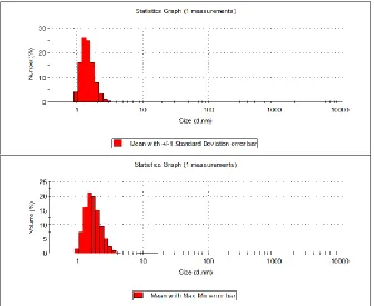
Fig. 2.18 DLS analysis of the nanoparticle solution from the same sample of
11-(dimethylamino)undecane-1-thiol hydrochloride-coated water soluble Au NP (19) freshly
prepared. These graphs were generated based on the corresponding data from table 2.1.
Size Mean Std Dev Size Mean Std Dev Size Mean Std Dev Size Mean Std Dev d.nm Volume %Volume % d.nm Volume %Volume % d.nm Volume %Volume % d.nm Volume %Volume %
0.4 0 5.615 0 78.82 0 1106 0
0.4632 0 6.503 0 91.28 0 1281 0
0.5365 0 7.531 0.1 105.7 0 1484 0
0.6213 0 8.721 0.1 122.4 0 1718 0
0.7195 0 10.1 0.1 141.8 0 1990 0
0.8332 0 11.7 0.1 164.2 0 2305 0
0.9649 1.5 13.54 0.1 190.1 0 2669 0
1.117 7.4 15.69 0.1 220.2 0 3091 0
1.294 16.2 18.17 0.1 255 0 3580 0
1.499 21.2 21.04 0.1 295.3 0 4145 0
1.736 20 24.36 0.1 342 0 4801 0
2.01 15 28.21 0 396.1 0 5560 0
2.328 9.4 32.67 0 458.7 0 6439 0
2.696 5 37.84 0 531.2 0 7456 0
3.122 2.2 43.82 0 615.1 0 8635 0
3.615 0.8 50.75 0 712.4 0 1.00E+04 0
4.187 0.2 58.77 0 825 0
From TEM and DLS results we can conclude that the average diameter of Au NPs is
1.49 nm. From TEM, we can visually see that NPs were roughly of similar sizes around
2 nm (Fig. 2.17). And DLS (Fig. 2.18) provides statistics data (Table 2.1) from which
we were able to calculate the average size of Au NPs to be 1.49 nm:
1 % 100%
n i i i Size MeanNumberSize (2.1)
We did not use the mean volume to do the calculation because there were some large
sizes of pretty small percentages in its statistics data. These data may be attributed
to dusts in solution since there sizes were not close to that of Au NPs if we compare
the TEM images and DLS graphs. Besides, from DLS graph based on mean number%
we can also see that the number of such dusts was too low to be counted in.
The extent of the nanoparticle surface coating was determined using
thermogravimetric analysis (TGA) (Fig. 2.19). In the TGA graph, 19 were stable when
heated from room temperature to 200 0C, followed by mass loss from 200 0C to 500
0C and then the weight of residue was stable with increasing temperature. We
attributed the mass loss between 2000C and 5000C to the loss of 5, so this mass loss
should be the mass of 5 coating on Au NPs. The weight of residue was the mass of
Au cores (table in Fig. 2.19). As a result, we could calculate the moles of 5 and Au
Fig. 2.19 Results of the TGA analysis of 11-(dimethylamino)undecane-1-thiol
hydrochloride-coated water soluble Au NP (19).
Using a combination of the results from the nanoparticle size measurement and the
thermogravimetric analysis, the composition of 19 was estimated as follows.
Given that the diameter of Au NPs is 1.49 nm (based on DLS analysis), the volume of
each Au nanocore is
VNP =43πr3 =4 3π(
14.9
2 )3 = 173 Å3 = 1.73 × 10⁻²⁷𝑚3 = 1.73 × 10⁻²¹𝑐𝑚3 (2.2)
The volume of Au cubic unit cell is
VAu cubic unit= 𝑙3 = (4.0786Å)3 = 67.847Å3 (2.3)
The density of gold is 19.3 g/cm3, so mass of gold nanocore = 3.34× 10⁻²⁰g.
So number of gold atoms in nanoparticle is
mass
197g m⁄ ol× NA=
3.34×10⁻²⁰g
197g m⁄ ol × 6.022 × 10²³⁄ ol = 102m atoms per nanoparticle (2.4)
Surface area of NP sphere
S=4πr2 = 4π (14.9A
∘
2 ) 2
According to ref 21, the average length of Au-Au bond on surface of Au NP is 2.93 A∘ .
If we use a lattice model to mimic the gold arrangement on surface, the number of
Au atom in each cell is 1. Number of cells on the surface= 2.93𝑆 2≈ 81 cells. As a result, 21 atoms are in the core while 81 atoms on the surface.
From TGA data, sample containing 1.0358 × 10⁻⁵ mol Au atoms also contains 6.48 ×
10-6 mol ligands (266.5 g/mol) (We consider the weight loss from 200℃ to 293℃ as
the loss of ligands). So the amount of Au NPs in TGA sample is
Au NPs =1.0358×10102 −5 = 1.02 × 10⁻⁷mol (2.6)
The amount of ligands in each Au NP is
Ligand
NPs = (6.48 × 10⁻⁶mol )/(1.02 × 10−7mol) = 63 𝑙igands/NP (2.7)
Thus, the composition of 19 we have synthesized is estimated to be Au102ligand63.
This estimated composition is very close to the reported Au NP composition 22,
Au144ligand60, the size of which is also close to the NPs synthesized by us. Besides,
the model applied in our estimation results in less Au atoms in the nanocore, since Au
atoms are reported 23 to pack more tightly than natural Au which has a density of 19.3
g/cm3. So it is reasonable to think that there should be more than 102 Au atoms per NP. As a result, we assume that the composition of Au NPs we obtained is
Au144ligand60.
The stability of 19 was characterized by zeta potential as a function of pH,24 which is
Fig. 2.20 Zeta potential of 11-(dimethylamino)undecane-1-thiol hydrochloride-coated water
soluble Au NP (19) in ultra-pure water. The pH of each data point was adjusted by 0.02 M HCl
solution and 0.02 M NaOH solution.
When the pH goes up, the zeta potential goes down. This demonstrates that 19 is
most stable under acetic conditions, which coincides with our expectation, since the
amino groups would better stabilize NPs by repulsion while more of them are positively
charged.25
2.4.2 1,2-bis(undecyl
1-oxyl-2,2,5,5-tetramethyl-2,5-dihydro-1H-pyrrole-3-carboxylate) disulfide-modified Au NPs (20)
Fig. 2.21 Scheme of the synthesis of 1,2-bis(undecyl
1-oxyl-2,2,5,5-tetramethyl-2,5-dihydro-1H-pyrrole-3-carboxylate) disulfide-modified Au NPs (20).
Ligand exchange experiments were conducted with 15 : Au NP =5:1,10:1, and 20:1.
We purified each 15-modified NPs sample and collected X-band (9.5 GHz) EPR
spectra as shown in Fig. 2.22.
Magnetic Field, G
3440 3460 3480 3500 3520 3540 3560 3580
15 : Au NP=5:1 15 : Au Np=10:1 15 : Au Np=20:1 Free 15
Fig. 2.22 EPR spectra acquired from samples of 1,2-bis(undecyl
1-oxyl-2,2,5,5-tetramethyl-2,5-dihydro-1H-pyrrole-3-carboxylate) disulfide-modified Au NPsligand exchanged at various
As is shown above, the EPR signals of 20 are weak. Only those ligand exchanged
with highest 15/NP ratio (15 : NP = 20:1) have relatively strong EPR signal with low
noise. However, NPs of this 15/NP ratio were easily aggregated in water since the
high percentage of 15 coating on NP surface highly weakened the repulsion created
by 5 between NPs. As a result, we were in need of a different kind of spin-labeled
ligand which has longer chain than 5 and better accessibility to surface of NP than 15
so that nitroxide radical on the tail of 15 could be easily detected by EPR other than
buried by 5.
2.4.3 12-(acetylthio)dodecyl
1-oxyl-2,2,5,5-tetramethyl-2,5-dihydro-1H-pyrrole-3-carboxylate-modified Au NPs(21)
16 was attached to Au NPs surface through ligand exchange experiment (Fig. 2.13).
Fig. 2.23 Scheme of the synthesis of 12-(acetylthio)dodecyl
1-oxyl-2,2,5,5-tetramethyl-2,5-dihydro-1H-pyrrole-3-carboxylate-modified Au NPs (21).
Ligand exchange experiments were conducted with 16 : Au NP =10:1, 20:1, and 40:1.
We purified each 21 sample and collected X-band (9.5 GHz) EPR spectra as shown
Magnetic Field, G
3440 3460 3480 3500 3520 3540 3560 3580
16 : Au NP = 40:1 16 : Au NP = 20:1 16 : Au NP = 10:1 Free 16
Fig. 2.24 EPR spectra acquired from 12-(acetylthio)dodecyl
1-oxyl-2,2,5,5-tetramethyl-2,5-dihydro-1H-pyrrole-3-carboxylate-modified Au NPs ligand exchanged at various 16/NP ratios.
As is shown above, the EPR signals of 21 have reasonable intensity with a low noise.
When 16/NP = 10:1, Au NPs have relatively noticeable EPR signal with low noise and
little aggregation, while higher spin-labeled ligand ratios caused aggregation of NPs.
So the proper length of spin-labeled ligand should be a little bit longer than 5 in order
to obtain satisfying EPR signals. So pH-sensitive spin-labeled ligands with proper
ligand lengths could be used to coat NPs so that the electronic properties of local
environment of Au NPs could be detected by EPR once NPs are coated by this kind
2.4.4 12-(acetylthio)dodecyl
3-(3-oxyl-1,2,4,4,5-pentamethylimidazolidin-2-yl)propanoate-modified Au NPs(22)
17 was attached to Au NPs surface through ligand exchange experiment (Fig. 2.25).
20
Fig. 2.25 Scheme of the synthesis of 12-(acetylthio)dodecyl
3-(3-oxyl-1,2,4,4,5-pentamethylimidazolidin-2-yl)propanoate-modified Au NPs (22).
Ligand exchange experiments were conducted with 17 : Au NP = 10:1. We purified 22
samples and collected X-band (9.5 GHz) EPR spectra in buffer solutions of different
Magnetic Field, G
3440 3460 3480 3500 3520 3540 3560 3580 3600
17 : Au NP=10:1, pH=2.79 17 : Au NP=10:1, pH=7.02
Fig. 2.26 EPR spectra of 12-(acetylthio)dodecyl
3-(3-oxyl-1,2,4,4,5-pentamethylimidazolidin-2-yl)propanoate-modified Au NPs under different pH conditions.
As shown above, the EPR spectra of Au NPs under different pH conditions are very
similar in shape with each other, indicating that the local electro-dynamic environment
does not change much under these pH conditions. This is because that the pKa (3.28)
of 22 is even lower after attached to Au NPs so that even at acetic condition (pH=2.79)
it is still nonprotonated.
2.4.5
S-10-oxo-10-(2-(1-oxyl-2,2,5,5-tetramethyl-2,5-dihydro-1H-imidazol-4-ylamino)ethylamino)decyl ethanethioate-modified Au NP (23)
18 was attached to Au NPs surface through ligand exchange experiment (Fig. 2.27).
Fig. 2.27 Scheme of the synthesis of
S-10-oxo-10-(2-(1-oxyl-2,2,5,5-tetramethyl-2,5-dihydro-1H-imidazol-4-ylamino)ethylamino)decyl ethanethioate-modified Au NP.
Ligand exchange experiments were conducted with 18 : Au NP = 40:1. We purified 23
samples and collected X-band (9.5 GHz) EPR spectra in SDS solutions (SDS/5, 4:1)
of different pH as shown in Fig. 2.28.
Magnetic Field, G
3460 3480 3500 3520 3540
pH=0.96
pH=3.33
pH=5.28
pH=6.93
pH=7.91
pH=9.21
Fig. 2.28. Representative room-temperature X-band EPR spectra of
S-10-oxo-10-(2-(1-oxyl-2,2,5,5-tetramethyl-2,5-dihydro-1H-imidazol-4-ylamino)ethylamino)decyl
ethanethioate-modified Au NP taken at various pH in a series of aqueous solutions in the presence of SDS
As shown above, shapes of EPR spectra of 23 changed according to pH,
demonstrating the pH sensitivity of 23. Details of this change will be further discussed
in the titration part of this chapter.
SDS was used as charge compensation which has a relevance to biological
application of NPs, because an interaction of NPs with such negatively charged
polyelectrolytes as nucleic acids would result in a similar effect. SDS is mimicking the
electrostatic properties of nucleic acids, providing a model environment for
biological/biophysical applications of spin-labeled Au NPs. More advantages of
applying SDS will be discussed in the titration part of this chapter.
As a result, 23 was successfully synthesized as pH sensitive Au NPs that has the
potential to be applied to biosystems.
2.5 Titration of pH Sensitive Spin-labeled Ligands
It is expected that the intrinsic pKa of the reported nitroxides will change for the terms
corresponding to electrostatic and polarity contribution after the nitroxide-modified
ligands were incorporated into the charged non-polar ensemble of NP-coating ligands:
, p p
p
p 0 apol
el a a
i
a K K K
K where 0
pKa is an intrinsic pKa of the probe observed in
pure water, and p pol a K
and el
a K p
are electrostatic and polarity contributions,
respectively.
Thus, to characterize the nitroxide-modified ligands we have synthesized, their
intrinsic pKa’s (pKa’s in aqueous solution) have to be determined. However, these
spin-labeled ligands are very hydrophobic and insoluble in water. In order to determine
their pKa’s in bulk water, we pH-titrated these ligands in a series of buffer
solution-isopropanol mixtures varying the concentration of solution-isopropanol.26,27 The experimental
2.5.1 12-(acetylthio)dodecyl
3-(3-oxyl-1,2,4,4,5-pentamethylimidazolidin-2-yl)propanoate(17).
An experimental X-band (9.5 GHz) EPR spectra of the nitroxide-modified ligand 17
taken in buffer solution-isopropanol mixtures at various pH are shown in Fig. 2.29.
Fig. 2.29 Representative room-temperature X-band EPR spectra of 12-(acetylthio)dodecyl
3-(3-oxyl-1,2,4,4,5-pentamethylimidazolidin-2-yl)propanoate (17) in a series of buffer solutions
containing 40% of isopropanol. pH of the solutions are indicated next to the corresponding
spectra. Dashed lines mark approximate positions of the high-field nitrogen hyperfine
coupling components corresponding to protonated and nonprotonated forms of the nitroxide.
It is clearly seen that that isotropic nitrogen hyperfine splitting constant Aiso decreases
as pH decreases. Aiso at intermediate pH were calculated as weighted averages of the
isotropic nitrogen hyperfine couplig constants of the protonated and non-protonated
were obtained (Table 2.2) by fitting the experimental data into the modified
Henderson-Hasselbalch equation:
(pH pK)
N NH
iso (pH pK)
A 10 A
A
1 10
(2.8)
where AN is the isotropic nitrogen hyperfine splitting constant of EPR spectrum of
nonprotonated form of the spin-labeled ligand, and ANH+ is the hyperfine splitting
constant of EPR spectrum of completely protonated form. Experimental EPR titration
curves for the spin-labeled ligand 17 are shown in Fig. 2.30. A linear regression of the
experimental pKa to 100% water is shown in Fig. 2.31. Thus, the intrinsic pKa of 17
was determined to be pKa = 3.28±0.33. It should be noted that the titration curves in
Fig 2.30 are shifted with respect to each other not only along the X-axis (difference in
the pKa values), but also along the Y-axis (difference in the Aiso). The latter effect is
explained by the explicit dependence of Aiso on the polarity of the environment.
Table 2.2 Titration Data for 12-(acetylthio)dodecyl
pH
0 1 2 3 4 5 6
A iso, G 13.8 14.0 14.2 14.4 14.6 14.8 15.0 15.2 15.4 15.6
Fig. 2.30 Experimental X-band EPR titration data for 12-(acetylthio)dodecyl
3-(3-oxyl-1,2,4,4,5-pentamethylimidazolidin-2-yl)propanoate (17) measured at room temperature in buffer/isopropanol solutions of the following ratios (v/v): (●) 80:20, (▲) 60:40, (x) 40:60.
%Water
30 40 50 60 70 80 90 100 110
pKa 1.6 1.8 2.0 2.2 2.4 2.6 2.8 3.0 3.2 3.4
Fig. 2.31 Linear regression of pKa of 12-(acetylthio)dodecyl
3-(3-oxyl-1,2,4,4,5-pentamethylimidazolidin-2-yl)propanoate (17) vs. percentage of bulk water. pKa value in bulk
2.5.2
S-10-oxo-10-(2-(1-oxyl-2,2,5,5-tetramethyl-2,5-dihydro-1H-imidazol-4-ylamino)ethylamino)decyl ethanethioate (18)
The intrinsic pKa of the nitroxide-modified ligand 18 was determined in a similar way.
A series of representative X-band EPR spectra of 18 taken in buffer
solution-isopropanol mixtures at various pH is shown in Fig. 2.32. Experimental X-band EPR
titration data with the least-squares Henderson-Hasselbalch titration curves are
shown in Fig. 2.33. A linear regression of the experimental pKa to 100% water is shown
in Fig. 2.34. The intrinsic pKa of 18 in 100% water was determined to be pKa =
5.99±0.33. The results of the EPR titration are summarized in Table 2.3.
Table 2.3 Titration Data for
S-10-oxo-10-(2-(1-oxyl-2,2,5,5-tetramethyl-2,5-dihydro-1H-imidazol-4-ylamino)ethylamino)decyl ethanethioate 18 in Buffer/Isopropanol Solutions of Various
Fig. 2.32 Representative room-temperature X-band EPR spectra of
S-10-oxo-10-(2-(1-oxyl-2,2,5,5-tetramethyl-2,5-dihydro-1H-imidazol-4-ylamino)ethylamino)decyl ethanethioate (18) in a
series of 40% isopropanol buffer solutions of different pH indicated next to the spectra.
Dashed lines mark approximate positions of the high-field nitrogen hyperfine coupling
pH
0 2 4 6 8 10
A iso, G 14.4 14.6 14.8 15.0 15.2 15.4 15.6 15.8
Fig. 2.33 Experimental X-band EPR titration data for
S-10-oxo-10-(2-(1-oxyl-2,2,5,5-tetramethyl-2,5-dihydro-1H-imidazol-4-ylamino)ethylamino)decyl ethanethioate (18) measured at room-temperature in buffer/isopropanol solutions of the following ratios (v/v): (●) 80:20, (▲) 60:40,
(x) 40:60.
%Water
30 40 50 60 70 80 90 100 110
pKa 4.0 4.2 4.4 4.6 4.8 5.0 5.2 5.4 5.6 5.8 6.0 6.2
Fig. 2.34 Linear regression of pKa of
S-10-oxo-10-(2-(1-oxyl-2,2,5,5-tetramethyl-2,5-dihydro-1H-imidazol-4-ylamino)ethylamino)decyl ethanethioate (18) vs. percentage of bulk water. pKa