A Survey on Applications of Digital Image
Processing in Biomedical Field
S.S.Sudha, S.Ranjini
Assistant Professor, Dept. of CS, PSG College of Arts & Science, Coimbatore, Tamilnadu, India
Research Scholar, Dept. of CS, PSG College of Arts & Science, Coimbatore, Tamilnadu, India
ABSTRACT: This paper gives the details about the methods of biomedical image processing and after that it also
describe about medical imaging modalities. Some of the medical imaging modalities are described in this paper like X-ray imaging, CT, MRI, and ultrasound. The optical modalities like endoscopy, photography and microscopy are also more important in this field. The following steps of image analysis are explained in this paper, feature extraction, segmentation, classification, quantitative measurements and interpretation. It mainly focuses on segmentation of biomedical images, because of its high relevance. Special segmentation methods and techniques have been developed in the medical field.
KEYWORDS: Medical imaging modalities, optical modalities, image analysis, segmentation
I. INTRODUCTION
The uses of digital imaging systems are nowadays increased in medical diagnostics. So that digital image processing becomes more and more important in health centre. Digital methods Computed Tomography (CT) or Magnetic Resonance Imaging (MRI), the analogue imaging modalities such as endoscopy or microscopy are nowadays equipped with digital sensors. Normally, digital image means collection of individual pixels (which stands for the word “picture” and “element”), to which discrete brightness or colour values are assigned. The entire spectrum of image processing is now used in medical field, based on the techniques used in digital imaging.
II. STEPS OF IMAGE PROCESSING
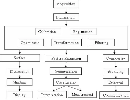
The term biomedical image processing means the provision of digital image processing for biomedical sciences. Four major areas of image processing are described below (Fig. 1.1):
A. Image formation
In this the step includes from capturing the image to forming a digital image matrix.
B. Image visualization
It refers to all types of manipulation of this matrix, resulting in an optimized output of the image.
C. Image analysis
This step includes all the steps of processing, which are used for quantitative measurements and abstract interpretations of biomedical images. These steps require a priori knowledge on the nature and content of the images, which must be incorporated into the algorithms on a high level of concept.
D. Image management
It sums up all techniques that provide the communication, efficient storage, transmission, access and archiving of image data. In the image management, the methods of telemedicine also one parts.
III. IMAGING MODALITIES
The medical imaging modalities and optical modalities are described below:
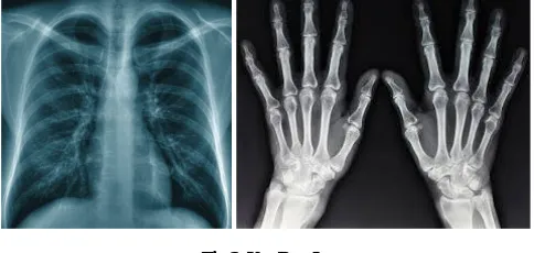
A. X-Ray Imaging
Commonly there are two radiographic images used in medical field imaging, projection radiography and fluoroscopy. Although the 3D tomography is available, these 2D techniques are in wide use because of its high resolution, low cost and depending on application, lower radiation dosages. For imaging acquisition this two imaging modality utilizes a wide beam of x rays. In this large modern medicine field this is the first imaging technique.
Fig 2. X – Ray Images
B. Fluoroscopy
C. Projection Radiography
It is another most commonly used x-rays. This type of x-rays used to find the type and extent of a fracture and also used for detecting pathological changes in the lungs. With the help of barium which is the radio-opaque contrast media can visualize the structure of the stomach and intestines.
Fig 4. Projection Radiography Images
D. Tomography
It is a method of imaging a single plane, or slice of an object resulting in a tomogram. Generally there are two methods of such images, namely conventional tomography and computer-assisted tomography.
a. Conventional Tomography
It always uses mechanical movement to record an image which goes directly onto X-ray film. This type of tomography contains various techniques as follows:
1. Linear Tomography 2. Poly Tomography 3. Zonography
4. Panoramic radiography
b. Computer Assisted Tomography
In This type of tomography, a computer processes data which received form radiation detectors and computationally constructs the scanned image of the structures. This type of imaging technique is better than conventional tomography as they can easily image both hard and soft tissues. But in this case it is complex to conventional tomography at imaging soft issues. The following techniques are exist:
E. X-ray computed tomography (CT)
This is a helical tomography technique. First it gives a 2D image of the structures of the body. By using CT, a beam of X-rays spins around an object being examined. After that the examined image is chosen by sensitive radiation detectors after having penetrated the object from multiple angles. Then finally a computer analyses the data received form the radiation detectors and then creates a detailed image of the object and its contents using the mathematical principles laid out in the Radon transform.
Fig 5. X-ray computed tomography Images
F. PET imaging
spread away from the annihilation site in opposite directions. The patient is enclosed by multiple rings of gamma photon detectors, so that no detector rotation is required.
Fig 6. PET Images
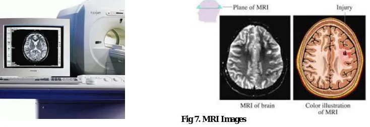
G. Magnetic Resonance Imaging (MRI)
Magnetic resonance imaging is a non-ionizing technique that uses radio frequency electromagnetic radiation and large magnetic fields. The large magnetic fields are produced by superconducting magnets, in which current is passed through coils of superconducting wire whose electrical resistance is virtually zero. A magnetic resonance imaging (MRI) scanner or nuclear magnetic resonance (NMR) imaging scanner uses great magnets to polarize and excite hydrogen nuclei of water molecules in human tissue, producing a detectable signal which is spatially encoded, resulting in images of the body. Like CT, MRI also creates a 2D image of a thin slice of the body and is so considered a tomography imaging technique. Nowadays MRI imaging is producing images in the form of 3D blocks.
Fig 7. MRI Images
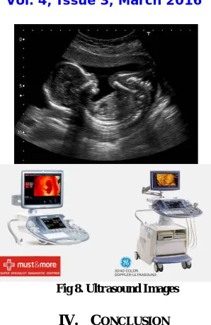
H. Ultrasound
Fig 8. Ultrasound Images
IV. CONCLUSION
The various image modalities are available to imagine internal parts of human organs. Magnetic Resonance Imaging (MRI) is an innovator non-invasive imaging technique. Image processing and analysis tools support quantities medical diagnosis. Digital image analysis can give to enlarged accuracy and impartiality of medical diagnosis. Image processing/analysis is not only consider those issues described in this paper, it also considered such as image registration to find many other issues available in medical field.
REFERENCES
1. R.A. Gonzalez, R.E. Woods, ‘DigitalImage Processing’ Second Edition. PrenticeHall 2002.
2. R.A. Gonzalez, R.E. Woods, S.L. Eddins, ‘DigitalImage Processing Using Matlab’. PrenticeHall 2004.
3. J.K. Udupa, G.T. Herman, ‘3D Imaging in Medicine’ CRC Press 2000.
4. Dougherty D. ‘Digital Image Processing for Medical Applications’ Cambridge: Cambridge University Press; 2009.
5. Guy C, Ffytche D. ‘Introduction to the Principles of Medical Imaging’. London: Imperial College Press; 2005.
6. Kim Y, Horii SC (eds). ‘Handbook of Medical Imaging’. Vol. 3: Display and PACS. Bellingham: SPIE Press; 2000.
7. Rangayyan RM. ‘Biomedical Image Analysis’. New York: CRC Press; 2005.
8. Suetens P. ‘Fundamentals of Medical Imaging’. Cambridge: Cambridge University Press; 2002.
9. Dougherty ER (ed). ‘Digital Image Processing Methods’. New York: CRC Press; 1994.
10. A.Jain, ‘Fundamentals of Digital Image Processing’, Prentice Hall of India.
BIOGRAPHY
Mrs. S. S. Sudha received her M.Sc., (Computer Science) degree from Madurai Kamarajar University, and M.Phil.,
degree in Computer Science from Madurai Kamarajar University. She has Presented Papers in National Conferences. She has 8 and a half years of Teaching Experience and she is currently working in PSG College of Arts & Science, Coimbatore. Her Research interests include Digital Watermarking and Image Data Hiding.
S. Ranjini received her B.Sc., (Computer Science) degree in 2010 from Periyar University, Salem, M.Sc., (Computer