ISSN(Online): 2320-9801 ISSN (Print): 2320-9798
I
nternational
J
ournal of
I
nnovative
R
esearch in
C
omputer
and
C
ommunication
E
ngineering
(A High Impact Factor, Monthly, Peer Reviewed Journal) Website: www.ijircce.com
Vol. 7, Issue 10, October 2019
Abnormalities of Fetal Brain Classification
Using Deep Learning Techniques
Sangeetha.K
1, Venipriya.T
2P.G. Student, Department of Computer Engineering, Arasu Engineering College, Kumbakonam, India1
Associate Professor, Department of Computer Engineering, Arasu Engineering College, Kumbakonam, India2
ABSTRACT: Magnetic Resonance Imaging (MRI) is a primary tool for clinical investigation of the brain and for neuroscience. Fetal Magnetic Resonance Imaging (MRI) in early phases of the cerebral development during gestation offers insights into the emergence of brain structures, their characteristics and variability across the population. To collect substantial bodies of observations automatic analysis of these data is necessary. However, automatic segmentation and classification are challenging due to image quality, low contrast between brain tissues, and the rapid development at this early age. Existing machine learning based segmentation approaches perform well in the adult population, but they are unable to cover the rapid changes during early development phases. In this project, we introduce a spatio-temporal group-wise segmentation of fetal brain structures given a single annotated example. The method is based on an emerging spatio-temporal latent atlas that captures the age dependent characteristics in the training population, and supports the segmentation of brain structures. So we can implement the proposed system which includes active contour method and deep learning methods. The proposed deep learning approaches of subcortical structures possible by integrating information across a large number of subjects. It encodes the average development and its variability, which is ultimately relevant for diagnosis. The deep learning method includes convolutional neural network algorithm to improve the accuracy in abnormality prediction. Experimental results shows that the proposed segmentation and classification algorithm can be provide less number of errors in abnormal tissue prediction.
KEYWORDS: Magenetic resonance imaging ,fetal brain classification, fetal brain segmentation, active countor, CNN
I. INTRODUCTION
1.1 IMAGE PROCESSING
In imaging science, image processing is processing of images using mathematical operations by using any form of signal processing for which the input is an image, a series of images, or a video, such as a photograph or video frame the output of image processing may be either an image or a set of characteristics or parameters related to the image. Most image-processing techniques involve treating the image as a two-dimensional signal and applying standard signal-processing techniques to it. Images are also processed as three-dimensional signals with the third-dimension being time or the z-axis. Image processing usually refers to digital image processing, but optical and analog image processing also are possible. This article is about general techniques that apply to all of them. The acquisition of images (producing the input image in the first place) is referred to as imaging.
1.2 STEPS OF IMAGE PROCESSING Image Acquisition
ISSN(Online): 2320-9801 ISSN (Print): 2320-9798
I
nternational
J
ournal of
I
nnovative
R
esearch in
C
omputer
and
C
ommunication
E
ngineering
(A High Impact Factor, Monthly, Peer Reviewed Journal) Website: www.ijircce.com
Vol. 7, Issue 10, October 2019
Image Enhancement
Image enhancement is among the simplest and most appealing areas of digital image processing. Basically, the idea behind enhancement techniques is to bring out detail that is obscured, or simply to highlight certain features of interest in an image. Such as, changing brightness & contrast etc.
Image Restoration
Image restoration is an area that also deals with improving the appearance of an image. However, unlike enhancement, which is subjective, image restoration is objective, in the sense that restoration techniques tend to be based on mathematical or probabilistic models of image degradation.
Color Image Processing
Color image processing is an area that has been gaining its importance because of the significant increase in the use of digital images over the Internet. This may include color modeling and processing in a digital domain etc.
Wavelets and Multi-resolution Processing
Wavelets are the foundation for representing images in various degrees of resolution. Images subdivision successively into smaller regions for data compression and for pyramidal representation.
Compression
Compression deals with techniques for reducing the storage required to save an image or the bandwidth to transmit it. Particularly in the uses of internet it is very much necessary to compress data.
Morphological Processing
Morphological processing deals with tools for extracting image components that are useful in the representation and description of shape.
1.3 SEGMENTATION
Segmentation procedures partition an image into its constituent parts or objects. In general, autonomous segmentation is one of the most difficult tasks in digital image processing. A rugged segmentation procedure brings the process a long way toward successful solution of imaging problems that require objects to be identified individually. Segmentation is a classifier which helps to fragment each character from a word present in a given image or page. The objective of the segmentation is to extract each character from the text present in the image A method stemming from concepts used in machine vision for recognition of occluded objects. A holistic process recognizes an entire word as a unit.
Representation and Description:
Representation and description almost always follow the output of a segmentation stage, which usually is raw pixel data, constituting either the boundary of a region or all the points in the region itself. Choosing a representation is only part of the solution for transforming raw data into a form suitable for subsequent computer processing. Description deals with extracting attributes that result in some quantitative information of interest or are basic for differentiating one class of objects from another.
Object recognition: Recognition is the process that assigns a label, such as, “vehicle” to an object based on its descriptors.
ISSN(Online): 2320-9801 ISSN (Print): 2320-9798
I
nternational
J
ournal of
I
nnovative
R
esearch in
C
omputer
and
C
ommunication
E
ngineering
(A High Impact Factor, Monthly, Peer Reviewed Journal) Website: www.ijircce.com
Vol. 7, Issue 10, October 2019
Color based image segmentation:
Color segmentation may be more accurate because of more information at the pixel level comparing to gray scale images. The standard Red-Green-Blue (RGB) color representation has strongly interrelated color components, and a number of other color systems have been designed in order to exclude redundancy, determine actual object / background colors irrespectively of illumination, and obtain more stable segmentation. Choosing a proper color space is a very important issue for color image segmentation process. Generally L*A*B* and HSV are the two frequently chosen color spaces. In this paper a comparative analysis is performed between these two color spaces with respect to color image segmentation. For measuring their performance, we consider the parameters: mse and psnr. It is found that HSV color space is performing better than L*A*B*.
Texture based segmentation:
Image segmentation methods can be subdivided into region based vs. edge-based methods. In order to identify possibilities for acquisition of scene information by digital images an analysis of the principle features of these images is required. In this regard, textures are the only possibility to derive information from imagery, besides the grey or color values and structural features and texture-based segmentation seems to be an adequate approach, because of the panchromatic images. To extract boundaries between major texture regions. Texture is a difficult concept to represent. The identification of specific textures in an image is achieved primarily by modeling texture as a two-dimensional gray level variation. The relative brightness of pairs of pixels is computed such that degree of contrast, regularity, coarseness and directionality.
II. RELATED WORK
“AUTOMATIC BRAIN LOCALIZATION IN FETAL MRI USING SUPERPIXEL GRAPH” by
AMIR ALANSARY , 2015 the papper contributes Fetal magnetic resonance imaging (MRI) has significantly improved in the last two decades, and is emerging as a novel, non-invasive tool for diagnosis and planing of surgical interventions. It provides higher contrast and larger fieldof-view than ultrasound. Thus, it provides better structural information of the different fetal organs such as the brain, spine and body. Fetal brain localization is important for assessing the fetal brain development and maturation.. Therefore, we have developed a new superpixel graphical model based on both spatial and intensity distances in 3D.Then each superpixel’s histogram is normalized with its neighbors in the graphical model. During the fourth step, we use a random forest to generate a probability map of the brain for every superpixel. Finally, this probability map is refined using another auto-context classifier followed by selecting the largest 3D component.
“A REVIEW ON AUTOMATIC FETAL AND NEONATAL BRAIN MRI SEGMENTATION”by ANTONIOS MAKROPOULOS, 2017the papper contributes Automated morphometric analysis of the perinatal brain is essential to quantitatively assess normal brain development and investigate the neuroanatomical correlates of cognitive impairments. Several neurological deficits have been associated with abnormalities in the developing brain, presenting a window for therapeutic intervention. In recent years, a variety of segmentation methods have been proposed for automatic delineation of the fetal and neonatal brain MRI. Challenges relating to the image acquisition, the rapid brain developments as well as the limited availability of imaging data however hinder this segmentation task. In this paper, we review methods adopted for the perinatal brain and categorise them according to the target population, structures segmented and methodology. We outline different methods proposed in the literature and discuss their major contributions. Different approaches for the evaluation of the segmentation accuracy and benchmarks used for the segmentation quality are presented. We conclude this review with a discussion on shortcomings in the perinatal domain and possible future directions.
III.PROPOSED ALGORITHM
3.1 PROPOSED SYSTEM
ISSN(Online): 2320-9801 ISSN (Print): 2320-9798
I
nternational
J
ournal of
I
nnovative
R
esearch in
C
omputer
and
C
ommunication
E
ngineering
(A High Impact Factor, Monthly, Peer Reviewed Journal) Website: www.ijircce.com
Vol. 7, Issue 10, October 2019
fetal life, the germinal matrix is the site of production of both neurons and glial cells which then migrate out to their final location. The proposed approach for the automatic localization of the fetal brain consists of three steps such as preprocessing, segmentation and classification. In this project we propose a fully-automated framework for localizing the fetal brain in fetal MRI scans. Brain extraction, as an initial preprocessing step for many brain MRI-based processing methods, is an important basis for accurate fetal MRI analysis. However, it is very challenging to automatically extract fetal brains from fetal MRI due to the large variation in fetal brains across different gestational weeks and complex maternal tissues surrounding the fetal brains. Rather than working on individual pixels we make use of contour detection for a faster and more efficient detection using Guided Active Contour algorithm. Because of the nature of boundary that most likely represents the rigid regions in the image, using contours neighbors instead of pixel neighbors can reduce the effect of pixel artifacts. And implement deep learning approach to classify the brain tissues for abnormality detection. In deep learning we use convolutional neural network algorithm to improve the accuracy in classification. A Convolutional Neural Network (ConvNet/CNN) is a Deep Learning algorithm which can take in an input image, assign importance (learnable weights and biases) to various aspects/objects in the image and be able to differentiate one from the other. The pre-processing required in a ConvNet is much lower as compared to other classification algorithms.
3.1.1 ADVANTAGES
Parallel processing in deep learning
Earlier prediction of abnormalities
Eliminates outliers in segmentation stage
Improve the accuracy in classification in different fetal gestational age.
3.2MORPHOLOGICAL OPERATIONS
Morphological image processing is a collection of non-linear operations related to the shape or morphology of features in an image. Morphological operations can also be applied to greyscale images such that their light transfer functions are unknown and therefore their absolute pixel values are of no or minor interestThe structuring element is positioned at all possible locations in the image and it is compared with the corresponding neighbourhood of pixels. Opening and closing are two important operators from mathematical morphology. They are both derived from the fundamental operations of erosion and dilation. Like those operators they are normally applied to binary images, although there are also graylevel versions. The basic effect of an opening is somewhat like erosion in that it tends to remove some of the foreground (bright) pixels from the edges of regions of foreground pixels
OPENING OPERATION
In mathematical morphology, opening is the dilation of the erosion of a set A by a structuring element B:
A ∘ B = (A ⊖ B) ⊕ B
Where ⊖ and ⊕, erosion and dilation operations.
CLOSING OPERATION
In mathematical morphology, the closing of a set (binary image) A by a structuring element B is the erosion of the dilation of that set,
A ∘ B = (A ⊕ B) ⊖ B
Where ⊖ and ⊕, erosion and dilation operations
3.3 ACTIVE CONTOUR SEGMENTATION
Active contour model, also called snakes, is a framework in computer vision introduced by Michael Kass, Andrew Witkin and Demetri Terzopoulos for delineating an object outline from a possibly noisy 2D image. The snakes model is popular in computer vision, and snakes are widely used in applications like object tracking, shape recognition, segmentation, edge detection and stereo matching.
ISSN(Online): 2320-9801 ISSN (Print): 2320-9798
I
nternational
J
ournal of
I
nnovative
R
esearch in
C
omputer
and
C
ommunication
E
ngineering
(A High Impact Factor, Monthly, Peer Reviewed Journal) Website: www.ijircce.com
Vol. 7, Issue 10, October 2019
They autonomously and adaptively search for the minimum state.
External image forces act upon the snake in an intuitive manner.
Incorporating Gaussian smoothing in the image energy function introduces scale sensitivity.
They can be used to track dynamic objects.
IV.SIMULATIONRESULTS
Description
The proposed deep learning approaches of subcortical structures possible by integrating information across a large number of subjects. It encodes the average development and its variability, which is ultimately relevant for diagnosis. The deep learning method includes convolutional neural network algorithm to improve the accuracy in abnormality prediction. Experimental results shows that the proposed segmentation and classification algorithm can be provide less number of errors in abnormal tissue prediction.Thus the work based on image processing has achieved 90% of accuracy in fetal brain
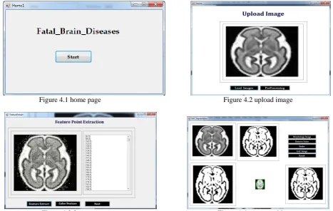
Figure 4.1 home page Figure 4.2 upload image
ISSN(Online): 2320-9801 ISSN (Print): 2320-9798
I
nternational
J
ournal of
I
nnovative
R
esearch in
C
omputer
and
C
ommunication
E
ngineering
(A High Impact Factor, Monthly, Peer Reviewed Journal) Website: www.ijircce.com
Vol. 7, Issue 10, October 2019
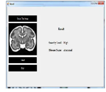
Figure 4.5 resulted image Figure 4.6 resulted image
V. CONCLUSION AND FUTURE WORK
CONCLUSION
Segmentation and classification of the fetal and neonatal brain is increasingly gaining interest with the acquisition of better quality images and the increased focus on fetal and neonatal development. We presented an automatic method for brain tissue segmentation in fetal MRI into seven tissue classes using convolutional neural networks. We demonstrated that the proposed method learns to cope with intensity inhomogeneity artifacts by augmenting the training data with synthesized intensity in homogeneity artifacts. To better understand the relevance of our predictive CNN to fetal development, we use sensitivity analysis to isolate regions critical for CNN performance, and discovered that our most sensitive regions were regions that are high in metabolic activity in early human brain development. In this proposed system we can implement morphological operations, guided active contour method and CNN based classification. Future work explores extracting diagnostic features in the segmented brain regions of the MRI image and integrates these methods in the developed CAD system for autism. In order to enhance the diagnostic accuracy, future work will investigate integrating other diagnostic features that will be extracted from other brain structures.
REFERENCES
[1] M. Havaei et al., “Brain tumor segmentation with Deep Neural Networks,” Med. Image Anal., vol. 35, pp. 18–31, Jan. 2017.
[2] A. Alansary et al., “Automatic Brain Localization in Fetal MRI Using Superpixel Graphs,” in Machine Learning Meets Medical Imaging, 2015, pp. 13–22.
[3] A. Makropoulos, S. J. Counsell, and D. Rueckert, “A review on automatic fetal and neonatal brain MRI segmentation,” NeuroImage, Jun. 2017. [4] J. Levman and E. Takahashi, “Multivariate analyses applied to fetal, neonatal and pediatric MRI of neurodevelopmental disorders,” NeuroImage Clin., vol. 9, pp. 532–544, Jan. 2015
[5] M. Sanz-Cortes et al., “Automatic Quantitative MRI Texture Analysis in Small-for-Gestational-Age Fetuses Discriminates Abnormal Neonatal Neurobehavior,” PLOS ONE, vol. 8, no. 7, p. e69595, Jul. 2013.
[6] M. Sanz-Cortés et al., “Fetal brain MRI texture analysis identifies different microstructural patterns in adequate and small for gestational age fetuses at term,” Fetal Diagn. Ther., vol. 33, no. 2, pp. 122–129, 2013.
[7] G. Ball et al., “Machine-learning to characterise neonatal functional connectivity in the preterm brain,” Neuroimage, vol. 124, no. Pt A, pp. 267– 275, Jan. 2016.
[8] C. D. Smyser et al., “Prediction of brain maturity in infants using machine-learning algorithms,” NeuroImage, vol. 136, p. 1, Aug. 2016. [9] “Early prediction of cognitive deficits in very preterm infants using functional connectome data in an artificial neural network framework,” NeuroImage Clin., vol. 18, pp. 290–297, Jan. 2018.
[10] Y. Jin et al., “Identification of Infants at High-Risk for Autism Spectrum Disorder Using Multiparameter Multiscale White Matter Connectivity Networks,” Hum. Brain Mapp., vol. 36, no. 12, pp. 4880–4896, Dec. 2015.
[11] Rossi AC Prefumo F “Additional value of fetal magnetic resonance imaging in the prenatal diagnosis of central nervous system anomalies,” Vol 389 February 4, 2017.