Address for correspondence Dr. Tasleem Arif
Department of Dermatology, STD & Leprosy, Government Medical College,
Srinagar, Jammu and Kashmir, India. Phone: 9557480345
Email: [email protected]
Original Article
Upper gastrointestinal endoscopy in systemic
sclerosis: A cross sectional study
Introduction
Systemic sclerosis is an autoimmune connective tissue disease characterized by vasomotor
abnormalities, fibrosis and associated
inflammatory manifestations.1 The disease involves the skin and other internal organs. Gastrointestinal involvement is the third most common manifestation of systemic sclerosis after cutaneous abnormalities and Raynaud’s phenomenon.2 It is seen in 9 out of 10 patients of systemic sclerosis and occurs due to myopathic and neuropathic abnormalities.3 The esophagus is involved in most cases, with esophageal smooth muscle atrophy and replacement by fibrous tissue leading to dysmotility.4 Dysphagia
Tasleem Arif, Mohammad Adil*, Jaswinder Singh Sodhi**
Department of Dermatology, STD & Leprosy, Government Medical College, Srinagar, Jammu and Kashmir, India.
* Department of Dermatology, STDs and Leprosy, Jawaharlal Nehru Medical College (JNMC), Aligarh Muslim University (AMU), Aligarh, India.
** Department of Gastroenterology, SKIMS, Soura, Srinagar, Kashmir, India.
Abstract
Background and Aims Involvement of the gastrointestinal tract is common in systemic sclerosis. The aim of this study was to identify the frequency and severity of dyspeptic symptoms in systemic sclerosis patients and to identify the common patterns of upper gastrointestinal endoscopic pathology from Kashmir valley, the study being first of its kind in the region.Methods Thirty seven sequential, newly and already diagnosed cases of systemic sclerosis were taken up for the study. This included 28 patients with limited systemic sclerosis and 9 patients with diffuse disease. They were enquired about the frequency and severity of symptoms of dysphagia, acid regurgitation and heartburn. Patients were then subjected to upper gastrointestinal endoscopy to identify abnormal findings.
Results Esophageal symptoms were present in 26 (70.3%) patients. 17 (60.7%) patients with limited disease showed esophageal symptoms while all 9 (100%) patients with diffuse disease showed these symptoms (p=0.025). Upper gastrointestinal endoscopy showed abnormal findings in 23 (62.2%) patients. Esophagus was most frequently involved organ with abnormalities seen in 15 (40.5%) patients, followed by involvement of the stomach in 13 (35.1%) patients and the duodenum in 5 (13.5%) patients. Reflux esophagitis was the commonest abnormality, seen in 12 (32.4%).
Conclusion Esophageal symptoms are a common occurrence in systemic sclerosis patients and seem to occur with a greater frequency in the diffuse disease subset. There seems to be no statistical difference between the two disease subsets in terms of the severity of the symptoms. Esophagitis appears to be the most common finding on upper gastrointestinal endoscopy.
Key words
and gastroesophageal reflux occurs due to loss of peristalsis and low esophageal sphincter pressures.5 The stomach may show low acid secretion, impaired motility and muscular atrophy.6 Consequently, patients of systemic sclerosis are at an increased risk of peptic stricture, esophageal stenosis, Barret’s esophagus and esophageal adenocarcinoma.7,8 This study aims to find the severity and frequency of upper gastrointestinal symptoms in patients of systemic sclerosis and to identify the upper gastrointestinal endoscopy findings in these patients. It also compares these findings between limited and diffuse types of systemic scleroderma patients. This is the first study from Kashmir valley, a different geographical area, in which all the three parts of upper gastrointestinal tract viz., esophagus, stomach and duodenum of systemic sclerosis patients have been studied.
Materials and Methods
This cross-sectional study was carried out in the
Department of Dermatology, Sexually
Transmitted Diseases and Leprosy, Sri Maharaja Hari Singh (SMHS) Hospital of Government Medical College, Srinagar and Department of Gastroenterology, Sher-i-Kashmir Institute of Medical Sciences, Soura, Srinagar during the period January 2012 to December 2013. Clearance from the ethical committees of the two institutions was obtained for the study viz., Institutional Ethics Committee (IEC), SKIMS and Ethical Committee, Government Medical College (EC-GMC) Srinagar. All sequential, new and previously diagnosed cases of systemic sclerosis attending the outpatient department of dermatology, SMHS hospital were included in
the study. The American Rheumatology
Association (ARA) criteria were used to make the diagnosis of systemic sclerosis.9 The inclusion criteria for the study was all new and previously diagnosed patients of systemic sclerosis, patients of both sexes and patients
aged more than 18 years. Patients less than 18 years of age, those who were pregnant at the time of study or in the previous 6 months of presentation, patients with features of other connective tissue diseases, mixed connective tissue disease or who were suffering from diabetes mellitus were excluded from the study.
Informed consent for the study, including collection, recording and publishing of personal details and clinical photographs was given by the patients. The parameters recorded for the study were age, sex, age at onset of disease, duration of disease and type of disease (limited or diffuse as per the Le Roy classification.10 Data was also recorded for the presence or absence of heartburn, acid regurgitation and dysphagia, and each of these were graded on the basis of their severity on a numeric scale of 0 to 3 with 0=absent symptoms, 1= mild symptoms, could be ignored by the patient, 2= moderate symptoms which could not be ignored but having no effect on daily life activities and 3= severe or incapacitating with impairment of day to day activities of the patient. The frequency of each of these symptoms, namely heartburn, acid regurgitation and dysphagia, were graded from 0 to 3 as follows: 0= no symptoms or less than once a month, 1= less than once per week, 2= several times per week and 3= every day.11 Total of these scores was done to provide a symptom score. The severity of the disease was graded on the basis of the cumulative scores obtained as mild (<6 or =6), moderate (7-12) and severe (>12 or if one of the three symptoms was considered incapacitating every day, giving a score of 9). Proton Pump inhibitors and H2 receptor blockers (which block gastric acid secretion), anticholinergics, antianginals and antihypertensives were discontinued 2 weeks before inclusion.
Each patient was subjected to upper
video-endoscope (Fujinon EG-201FP, Japan) was used for this purpose. Each patient was advised to avoid taking any medication that may interfere with motility of the gastrointestinal tract 2 weeks prior to the procedure. Overnight fasting was advised. Each patient was put in left lateral position for the procedure with the head under a pillow. Any tight object around the neck, such as jewellery or scarf were removed. Topical anaesthesia of the oropharynx was achieved by the help of a lidocaine spray. A mouth guard was placed to avoid the patient from biting the tube. The endoscope was lubricated and passed over the tongue, through the oropharynx and into the oesophagus. It was then advanced into the stomach and through the pylorus to examine the duodenum. Any abnormality of the esophagus, stomach or duodenum was looked for and photographed. Reflux esophagitis, if present, was graded on the basis of Los Angeles classification.12,13 The data was collected and analyzed by using statistical package for social sciences (SPSS) software version 16.0. A ‘p’ value of less than 0.05 was considered as significant for all tests.
Observations
A total of 37 sequential patients of systemic sclerosis were recruited for the study. The general characteristics of the patients have been given in Table 1 & 2.
Only 2 (5.4%) patients out of 37 were males, rest 35 (94.6%) were females. The mean age of these patients was 44.7 years with a standard deviation of 13.8 years. The youngest was aged 20 years while the oldest was aged 75 years. The mean age of onset of the disease was 35.8 years with a standard deviation of 12.4 years. The mean duration of disease at the time of presentation was 8.9 years with a standard deviation of 8.1 years. Maximum number of patients 10 (27.0%) were in the 40-49 year age
group, followed by 9 (24.3%) patients in the 50-59 year age group and 7 (18.9%) patients in the 30-39 year age group. 5 (13.5%) patients were in the 20-29 year age group while 6 (16.3%) patients were more than 60 years of age at the time of presentation. Of the total, 28 (75.7%) patients had the limited form of the disease while only 9 (24.3%) had the diffuse type of disease.
Esophageal symptoms were present in 26 (70.3%) patients. This included 17 of the 28 (60.7%) patients with limited disease and all 9 (100.0%) patients with diffuse systemic sclerosis. The difference in the fraction of patients showing esophageal symptoms in the two groups was found to be statistically significant (p<0.05). Heartburn and acid regurgitation were seen in 24 (64.9%) patients of the total 37 patients. The symptoms were present in 15 (64.9%) and 9 (100.0%) patients in the limited and diffuse type of disease respectively.
The difference in heartburn and acid
regurgitation in the two groups was statistically significant. Dysphagia was seen in 21 (56.8%) patients. This number includes 14 of the 28 (50.0%) patients with limited disease and 7 of the 9 (77.8%) patients with diffuse disease. The difference between the two was not found to be statistically significant (p>0.05).
Table 1 Basic parameters of the study Total number of patients 37
Males 2
Females 35
Mean Age 44.7 ± 13.8 years
Mean Age at onset of disease 35.8 +- 12.4 years Mean duration of disease 8.9 +- 8.1 years Patients with limited disease 28
Patients with diffuse disease 9 Table 2 Age distribution of the patients
Age group (years) Number Percentage (%)
20-29 5 13.5
30-39 7 18.9
40-49 10 27.0
50-59 9 24.3
>60 6 16.3
Table 3 Esophageal symptoms in patients of systemic sclerosis
Parameter Total
Number (percent)
Limited Number (percent)
Diffuse
Number (percent) p value
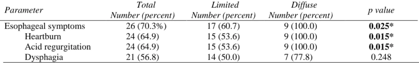
Esophageal symptoms 26 (70.3%) 17 (60.7) 9 (100.0) 0.025*
Heartburn 24 (64.9) 15 (53.6) 9 (100.0) 0.015*
Acid regurgitation 24 (64.9) 15 (53.6) 9 (100.0) 0.015*
Dysphagia 21 (56.8) 14 (50.0) 7 (77.8) 0.248
* : statistically significant at p<0.05
Table 4 Severity of esophageal symptoms in systemic sclerosis
Parameter Total
Number (percent)
Limited Number (percent)
Diffuse
Number (percent) p value
Esophageal symptoms 26 (70.3) 17 (60.7) 9 (100.0) 0.025*
Mild 16 (43.2) 11 (39.3) 5 (55.6)
0.852
Moderate 8 (21.6) 5 (17.9) 3 (33.3)
Severe 2 (5.4) 1 (3.6) 1 (11.1)
* : statistically significant at p<0.05
Table 5 Upper Gastrointestinal endoscopy findings in systemic sclerosis Part of GI
tract Findings
Total Number (percent)
Limited Number (percent)
Diffuse
Number (percent) p value
Total All findings 23 (62.2) 16 (57.1) 7 (77.8) 0.266
Esophagus 15 (40.5) 11 (39.3) 4 (44.4) 0.784
Esophagitis 12 (32.4) 8 (28.6) 4 (44.4)
Polyp 2 (5.4) 2 (7.1) 0 (0)
Patulous LES 2 (5.4) 2 (7.1) 0 (0)
Telangiectasia 1 (2.7) 1 (3.5) 0 (0)
Stricture 1 (2.7) 0 (0) 1 (11.1)
Stomach 13 (35.1) 10 (35.7) 3 (33.3) 0.896
Antral gastritis 8 (21.6) 6 (21.4) 3 (33.3)
Gastritis of other parts 2 (5.4) 2 (7.1) 0 (0)
Antral angioma 1 (2.7) 1 (3.6) 0 (0)
Erosions 1 (2.7) 1 (3.6) 0 (0)
Bile in stomach 1 (2.7) 0 (0) 1 (11.1)
Duodenum 5 (13.5) 4 (14.3) 1 (11.1) 0.081
Duodenitis 1 (2.7) 1 (3.6) 0 (0)
Ulcer/erosion 3 (8.1) 2 (7.1) 1 (11.1)
Healed ulcer 1 (2.7) 1 (3.6) 0 (0)
Table 3 depicts the esophageal symptoms in the patients. Table 4 shows the severity of esophageal symptoms in the patients. Out of the total patients, 16 (43.2%) patients reported mild symptoms, 8 (21.6%) had moderate symptoms and only 2 (5.4%) had severe symptoms. 11 (39.3%) patients with limited systemic disease had mild symptoms, 5 (17.9%) had moderate symptoms and 1 (3.6%) patient had severe symptoms. 5 (55.6%) patients had mild symptoms, 3 (33.3%) patients had moderate symptoms and 1 (11.1%) patients had severe
symptoms in the group with diffuse systemic sclerosis. The difference in severity of symptoms in the two groups was not significant statistically. Upper gastrointestinal (GI) endoscopy findings were seen in 23 (62.2%) patients out of the total 37 patients and have been summarized in Table 5.
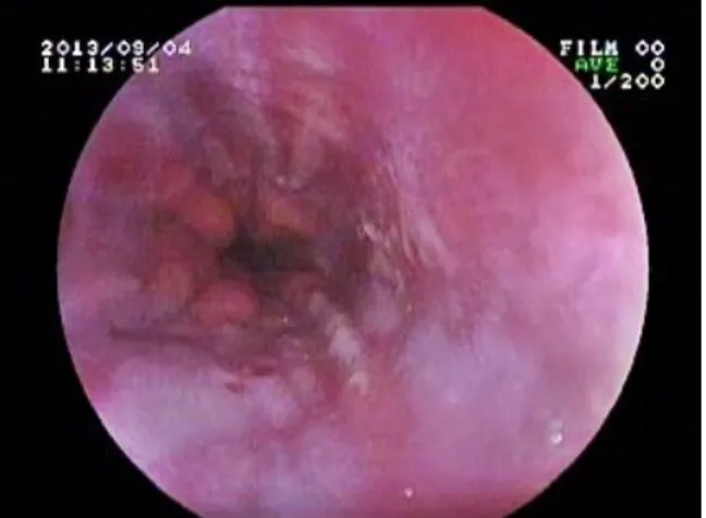
Figure 1 Grade 4 esophagitis in SSC involving almost the entire circumference of esophagus
Figure 2 Esophageal polyp in SSC
This included 11 (39.3%) patients in the limited group and 4 (44.4%) patients in the diffuse group. This was no significant difference in the esophageal abnormalities in patients of limited and diffuse subsets (p<0.05). Esophagitis was most commonly seen abnormality and was present in 12 (32.4%) patients. Grade 1 esophagitis was seen in 5 (13.5%) cases, grade 2 in 5 (13.5%) cases, grade 3 and grade 4 esophagitis (Figure 1) were present in 1 (2.7%) patient each. Esophageal polyps (Figure 2) were present in 2 (5.4%) patients, patulous lower esophageal sphincter was seen in 2 (5.4%) patients, esophageal telangiectasia in 1 (2.7%) patient and esophageal stricture in 1 (2.7%) patient. Abnormalities of the stomach were seen in 10 (35.7%) patients of limited disease and 3 (33.3%) patients of diffuse systemic sclerosis, a
statistically insignificant difference. Antral gastritis was the most common abnormality seen in 8 (21.6%) patients, gastritis of the other parts of the stomach was seen in 2 (5.4%) patients. Antral angiomas, gastric erosions and excessive bile in the stomach was seen in 1 (2.7%) patient each. Duodenal abnormalities were seen in 4 (14.3%) patients with limited systemic sclerosis and 1 (1.1%) patient with diffuse systemic sclerosis. The inflammation of the duodenum was the most common abnormality and was seen in 3 (8.1%) patients. One (2.7%) patient showed duodenal inflammation and 1 (2.7%) had a healed duodenal ulcer.
Discussion
Only two of our 37 patients were males. Systemic sclerosis is well known for its female predisposition, as documented by various studies showing a female to male ratio of as high as 10:1.14-17 The mean age of our patients was 44.7 years. The age of onset of the disease was 35.8 years and patients took an average of 8.9 years for presentation to our department. Our findings are in accordance to other studies that report systemic sclerosis to begin in the fourth decade of life.14,17-20 The long duration of disease before presentation, as seen in our case, is explained by the comparatively less symptomatic disease in patients who present to the dermatology department.17 Most of our patients were in the 20-50 year age bracket. This is in accordance with other studies showing about four-fifths of all cases of systemic sclerosis in the 20-60 year age group.21 28 of the 37 patients or 75.7% of the patients in our study had limited systemic sclerosis and rest 24.3% suffered from diffuse systemic sclerosis. This observation is consistent with previous studies showing a predominance of patients with limited disease.22,23
42% to 79%.24-30 Our study pinned the number to 70.3%, a finding in accordance to other studies. The difference in severity of esophageal symptoms between limited and diffuse form of the disease was found to be statistically significant in our study. The symptoms were mild in 43.2% patients, moderate in 21.6% and severe in intensity in 5.4% of the patients. There was no difference between the two groups in the grade of severity of the symptoms. Thus, our study infers that the patients with the diffuse type of SSc have greater chance of having symptomatic disease but the severity of esophageal symptoms between diffuse and limited SSc is not significantly different. Information about the difference in esophageal symptoms in the two subsets of SSc is scarce, but studies have pointed to a slight increase in the frequency and severity of esophageal symptoms in the diffuse subset.22,31 Ostojić et al. in 2006 had evaluated 50 patients of limited SSc and 55 patients of diffuse SSc and found esophageal dysmotility to be more severe statistically in the latter form of the disease.32 Though there is no direct correlation between esophageal dysmotility and symptoms, a link between the two has been proposed.33,34 However, other studies found no association between subset of SSc and symptoms.35 Acid regurgitation and heartburn was present in 64.9% patients in our study and dysphagia was present in 56.8%. The difference between the two disease subsets was statistically significant as far as heartburn and acid regurgitation was concerned. There was considerable difference between the two groups for dysphagia, though statistically significant levels were not reached. Other studies have also documented dysphagia, heartburn and acid regurgitation to be present in 50-80% of the patients.36 Airo found difference between the two subsets to reach statistically significant levels for burning and dysphagia.31 Lahcene et al. found the esophageal symptoms to be mild in 38.1% cases, moderate in 13.9%
cases and severe in 8.7% cases, findings much similar to our study.22
The upper gastrointestinal endoscopy revealed abnormalities in 62.2% of our patients with no difference between the limited and diffuse forms of the disease. The esophagus was the most frequently involved organ (40.5%), followed by the stomach (35.1%) and the duodenum (13.5%). Among the esophageal findings, we found reflux esophagitis to be the most common (32.4%) finding. Most patients with esophagitis had grade 1 or grade 2 esophagitis. Prevalence of reflux esophagitis in systemic sclerosis has been variable in different studies ranging from 3.2-60%.37-40 The reports with patients having esophagitis on the higher side can be explained by the fact that the patients included in the study had a large intake of non-steroidal anti-inflammatory drugs. Complicated forms of
esophageal inflammatory changes like
esophageal stenosis and Barret’s esophagus are also more common in SSc. Both have been found in 3-40% cases in various studies.38,39,41,42 We found just 1 case of esophageal stenosis, 2 cases of esophagus with a patulous sphincter and no cases with Barret’s esophagus, which is
lower than other studies. Esophageal
gastritis in 92% patients. Of these, 31% had erosive gastritis.45 Different studies have revealed that gastric antral vascular ectasia produces the ‘watermelon stomach’ that is a rare but important cause of bleeding in SSc patients.46 Sometimes this ectasia of vessels may be mistaken for antral gastritis, which needs biopsy for confirmation.47,48 Biopsy from the involved areas was not done in our cases. Gastric angioma has been described in case reports in systemic sclerosis and may rarely be
responsible for upper gastrointestinal
bleeding.49,50 One of our patients had bile filled stomach. This is significant in systemic sclerosis as it may indicate reflux of intestinal contents into the stomach. We could not find any such report in the literature. The gastric pathology in patients of systemic sclerosis is more commonly electrophysiological in nature. It leads to delayed gastric emptying and this contributes to the gastro-oesophageal reflux in the patients. The symptoms of gastric disturbance produce a significant morbidity and mortality in patients.51 Inflammation of the duodenum was seen in one patient while duodenal ulcer was seen in three of our cases and one had a healed ulcer. Involvement of the small intestine usually occurs in the form of hypomotility and as a result bacterial overgrowth and malabsorption occurs in patients. Although duodenum is believed to be the most involved part of the small intestine and presents with delayed transit time52, duodenal ulcers seen in few of our cases can best be described as an incidental finding. This might be due to high prevalence of
Helicobacter pylori infection in the general
population in Kashmir valley of India.53 We did not perform studies for the presence of this bacteria in our study.
Our study identifies the upper gastrointestinal findings in patients of systemic sclerosis, with comparison of the limited and diffuse subsets based on subjective and objective criteria. Also
this study adds duodenal findings in systemic sclerosis which needs further research. Presence of bile filled stomach in one of our cases also adds rarity to our study. However, there were no controls in our study and it was carried out on a smaller number of patients. The absence of biopsy in every case from the pathologic site and tests for Helicobacter pylori are other limitations of our study.
Conclusion
Esophageal symptoms are very common in patients of systemic sclerosis. Most of the patients have oesophageal symptoms in the form of heartburn, acid regurgitation or dysphagia. The diffuse subset of systemic sclerosis has presented with a significant greater frequency of esophageal symptoms than the limited subset, though the severity of these symptoms are statistically equally severe in both the groups.
Upper gastrointestinal endoscopy shows
abnormal findings in a majority of the patients. The esophagus is the most frequently involved site. Endoscopy shows that esophagitis and gastritis are the commonest abnormalities. The frequency of abnormal findings has no difference between the limited and diffuse subsets of systemic sclerosis. The occurrence of antral gastritis and bile filled stomach found in our study needs to be studied further. Thus, all patients with systemic sclerosis need evaluation for upper gastrointestinal abnormalities and endoscopic evaluation even if they are asymptomatic or from any disease subset. Early identification of upper gastrointestinal pathology shall help in preventing complications of reflux esophagitis and bleeding, leading to a healthier life to patients of systemic sclerosis.
References
textbook of dermatology. 9th ed. West Sussex (UK);Wiley Blackwell: 2016.p.56.1-23.
2. Hansi N, Thoua N, Carulli M, et al. Consensus best practice pathway of the UK scleroderma study group: gastrointestinal manifestations of systemic sclerosis. Clin Exp Rheumatol. 2014; 32(6 Suppl 86): S214-21.
3. Rose S, Young MA, Reynolds JC. Gastrointestinal manifestations of scleroderma. Gastroenterol Clin North Am. 1998; 27: 563-94.
4. Poirier TJ, Rankin GB. Gastrointestinal manifestations of progressive systemic scleroderma based on a review of 364 cases. Am J Gastroenterol. 1972; 58: 30-44. 5. Orringer MB, Dabich L, Zarafonetis CJ,
Sloan H. Gastroesophageal reflux in esophageal scleroderma: diagnosis and implications. Ann Thorac Surg. 1976; 22: 120-30.
6. Sjogren RW. Gastrointestinal motility disorders in scleroderma. Arthritis Rheum. 1994; 37: 1265-82.
7. Ebert EC. Esophageal disease in scleroderma. J Clin Gastroenterol. 2006; 40: 769–775.DOI:
10.1097/01.mcg.0000225549.19127.90. 8. Ntoumazios SK, Voulgari PV, Potsis K,
Koutis E, Tsifetaki N, Assimakopoulos DA. Esophageal involvement in scleroderma: gastroesophageal reflux, the common problem. Semin Arthritis Rheum. 2006; 36: 173-81.
doi: 10.1016/j.semarthrit.2006.08.002. 9. Subcommittee for Scleroderma Criteria of
the American Rheumatism Association Diagnostic and Therapeutic Criteria Committee. Preliminary criteria for the classification of systemic sclerosis (scleroderma). Arthritis Rheum. 1980; 23: 58-90.
10. Le Roy EC, Black C, Fleischmajer R. Scleroderma (systemic sclerosis): classification, subsets and pathogenesis. J Rheumatol. 1988; 15: 202-5.
11. Dombal FT, Hall R. The evaluation of medical care from the clinician’s point of view, what should we measure, and can we trust our measurements ? In evaluation of efficacy of medical action. Amsterdam: Elsevier-North Holland; 1979: 13-29. 12. Armstrong D, Bennett JR, Blum AL, et al.
The endoscopic assessment of esophagitis: a
progress report on observer agreement. Gastroenterol. 1996; 111: 85-92.
13. Lundell L, Dent J, Bennett J, et al. Endoscopic assessment of esophagitis: clinical and functional correlates and further validation of the Los Angeles classification. Gut. 1999; 45: 172-80.
14. Mayes MD. Scleroderma epidemiology. Rheum Dis Clin North Am 2003; 29: 139-54.
15. Silver RM. Clinical aspects of systemic sclerosis. Ann Rheum Dis. 1991; 50: 854-61.
16. Laing TJ, Gillespie BW, Toth MB, et al . Racial differences in scleroderma among women in Michigan. Arthritis Rheum. 1997; 40: 734-42.
17. Sharma VK, Trilokraj T, Khaitan BK, Krishna SM. Profile of systemic sclerosis in a tertiary care center in North India. Indian J Dermatol Venereol Leprol. 2006; 72: 416-20.
18. Medsger TA, Masi AT. Epidemiology of systemic sclerosis (scleroderma). Ann Intern Med. 1971; 74: 714-21.
19. Ruangjutipopan S, Kasitanon N, Louthrenoo W, Sukitawut W, Wichainun R. Causes of death and poor survival prognostic factors in Thai patients with systemic sclerosis. J Med Assoc Thai. 2002; 85: 1204-9.
20. Krishnamurthy V, Porkodi R, Ramakrishnan S, et al . Progressive systemic sclerosis in south India. J Assoc Physics India. 1991; 39: 254-7.
21. Christianson HB, Dorsey CS, Kierland RR, O’Leary PA. Localized scleroderma: a clinical study of 235 cases. Arch Dermatol. 1956; 74: 629-39.
22. Lahcene M, Oumnia N, Matougui N, Boudjella M, Tebaibia A, Touchene B. Esophageal involvement in scleroderma: Clinical, Endoscopic, and Manometric Features. ISRN Rheumatol. 2011; 2011: 1-5.
Article id: 325826.
doi:10.5402/2011/325826.
23. Kinuya K, Nakajima K, Kinuya S, Michigishi T, Tonami N, Takehara K. Esophageal hypomotility in systemic sclerosis: close relationship with pulmonary involvement. Ann Nuclear Med. 2001; 15: 97-101.
24. Tuffanelli DL, Winkelman RK. Systemic scleroderma: a clinical study of 727 cases. Arch Dermatol. 1961; 84: 359-67.
progressive systemic sclerosis. Am J Gastroenterol. 1997;92:763-71.
26. Rose S, Young MA, Reynolds JC. Gastrointestinal manifestations of scleroderma. Gastroenterol Clin North Am. 1998; 27: 563-94.
27. Abu-Shakra M, Guillemin F, Lee P. Gastro-intestinal manifestations of systemic sclerosis. Semin Arthritis Rheum. 1994; 24: 29-39.
28. Aubert A, Lazareth I, Vayssairat M, Fiessinger JN, Petite JP. L'oesophagite au cours de la sclérodermiesystémique. Prévalence-et-facteurs de survenue chez 46 patients. Gastroenterol Clin Biol. 1991; 15: 945-9.
29. Turner R, Lipshutz W, Miller W, Rittenberg G, Schumacher HR, Cohen S. Esophageal dysfunction in colllagen disease. Am J Med Sci. 1973; 265: 191-9.
30. Poirier TJ, Ranklin GB. Gastrointestinal manifestation of progressive systemic scleroderma based on a review of 364 cases. Am J Gastroenterol. 1972; 58: 30-44. 31. Airo P, Case DD, Danieli E, Missale G,
Cattaneo R, Cestari R. Oesophageal manometry in early and definite systemic sclerosis. Clin Rheumatol. 2005; 24: 370-6. 32. Ostojić P, Damjanov N. Different clinical
features in patients with limited and diffuse systemic sclerosis. Clin Rheumatol. 2006; 25: 453-7.
33. Bassotti G, Battaglia E, Debernardi V, et al. Esophageal dysfunction in scleroderma. Relationship with disease subsets. Arthritis Rheum. 1997; 40: 2252-9.
34. Wegener M, Adamek RJ, Wedmann B, Jergas M, Altmeyer P. Gastrointestinal transit through esophagus, stomach, small and large intestine in patients with progressive systemic sclerosis. Dig Dis Sci. 1994; 39: 2209-15.
35. Koshino Y, Takai T, Kato T, et al. Manometric evaluation of esophageal function in progressive systemic sclerosis with special regard to the disease severity. Gastroenterol Jpn. 1991; 26: 575-81. 36. Calderaro DC, de carvalho MAP,
Moretzsohn LD. Esophageal manometry in 28 systemic sclerosis Brazilian patients: findings and correlations. Diseases of the Esophagus. 2009; 22: 700-4.
37. Arif T, Masood Q, Singh J, Hassan I. Assessment of esophageal involvement in systemic sclerosis and morphea (localized scleroderma) by clinical, endoscopic,
manometric and pH metric features: a prospective comparative hospital based study. BMC Gastroenterol. 2015; 15:24. 38. Marie I, Ducrotté P, Denis P, Hellot MF,
Levesque H. Oesophageal mucosal involvement in patients with systemic sclerosis receiving proton pump inhibitor therapy. Aliment Pharmacol Ther. 2006; 24: 1593-601.
39. Poirer TJ, Rankin GB. gastrointestinal manifestations of progressive systemic scleroderma based on a review of 364 cases. Am J Gastroenterol. 1972; 58: 30-44. 40. Hendel L. Esophageal and small intestinal
manifestations of progressive systemic sclerosis. Danish Medical Bulletin. 1994; 41: 371-85.
41. Wipff J, Allanore Y, Soussi F, et al. Prevalence of Barret’s esophagus in systemic sclerosis. Arthritis Rheum. 2005; 52: 2882-8.
42. Ebert EC. Esophageal disease in scleroderma. J Clin Gastroenterol. 2006; 40: 769-75.
43. Segel MC, Campbell WL, Medsger TA, Roumm AD. Systemic sclerosis (scleroderma) and esophageal carcinoma: is increased patient screening necessary. Gastroenterol. 1985; 89: 485-8.
44. Duchini A, Sessoms SL. Gastrointestinal hemorrhage in patients with systemic sclerosis and CREST syndrome. Am J Gastroenterol. 1998; 93: 1453-6.
45. Thonhofer R, Siegel C, Trummer M, Graninger W. Early endoscopy in systemic sclerosis without gastrointestinal symptoms. Rheumatol International. 2012; 32: 165-8. 46. Parrado RH, Lemus HN, Coral-Alvarado
PX, Quintana G. Gastric antral vascular ectasia in systemic sclerosis: current concepts. Int J Rheumatol. 2015; 2015: 762546. doi:10.1155/2015/762546.
47. Marie I, Ducrotte P, Antonietti M, Herve S, Levesque H. Watermelon stomach in systemic sclerosis: its incidence and management. Aliment Pharmacol Ther. 2008; 28: 412-21.
48. Selinger CP, Ang YS. Gastric antral vascular ectasia (GAVE): an update on clinical presentation, pathophysiology and treatment. Digestion. 2008; 77: 131-7. 49. Dalle I, Giboes K. Vascular lesions of the
gastrointestinal tract. Acta Gastroenterol Belg 2002;65:213-9.
telangiectasis: a rare cause of severe blood loss in CREST syndrome. Postgrad Med J 1994; 70: 302-4.
51. Forbes A, Marie I. Gastrointestinal complications: the most frequent internal complications of systemic sclerosis. Rheumatology 2009 (Suppl 3); 48:iii36-9. 52. Sallam H, McNearny TA, Chen JZ.
Systematic review: pathophysiology and management of gastrointestinal dysmotility
in systemic sclerosis (scleroderma). Aliment Pharmacol Ther. 2006; 23: 691-712.