C H A P T E R
8
From DNA to
Proteins
8.1 Identifying DNA as the Genetic Material
DNA was identified as the genetic material through a series of experiments.8.2 Structure of DNA
DNA structure is the same in all organisms.
8.3 DNA Replication
DNA replication copies the genetic information of a cell.
8.4 Transcription
Transcription converts a gene into a single-stranded RNA molecule.
8.5 Translation
Translation converts an mRNA message into a polypeptide, or protein.
8.6 Gene Expression and Regulation
Gene expression is carefully regulated in both prokaryotic and eukaryotic cells.
8.7 Mutations
Mutations are changes in DNA that may or may not affect phenotype.
K E Y CO N C E P T S
BIOLOGY RESOURCE CENTER
B I O LO GY C L A SS Z O N E .CO M
View animated chapter concepts.
• DNA Replication • Build a Protein
Keep current with biology news. • Featured stories
• News feeds • Strange Biology
Get more information on • DNA
• RNA • Mutations
Connecting
CO N C E P TSWhy is this
mouse glowing?
T
his mouse’s eerie green glow comes
from green fluorescent protein (GFP),
which glows under ultraviolet light.
Scien-tists put a gene from a glowing jellyfish
into a virus that was allowed to infect a
mouse egg. The jellyfish gene became part
of the mouse’s genes. As a result, the mouse
cells produce the same protein. Researchers
hope to track cancer cells using GFP.
Translation This computer model of GFP shows the amino acids (purple) in the center of the protein that make the protein glow. The genetic code is universal, which means that a gene from one organism can be correctly translated into a protein in another organism. Although the gene for GFP comes from a jellyfish, GFP has been made in bacteria, yeast, slime mold, plants, fruit flies, zebrafish, and mammals.
a^kZ HWVXiZg^V a^kZ GWVXiZg^V ]ZVi"`^aaZY HWVXiZg^V ]ZVi"`^aaZYHWVXiZg^V a^kZGWVXiZg^V
YZVYbdjhZ a^kZbdjhZ a^kZbdjhZ YZVYbdjhZ
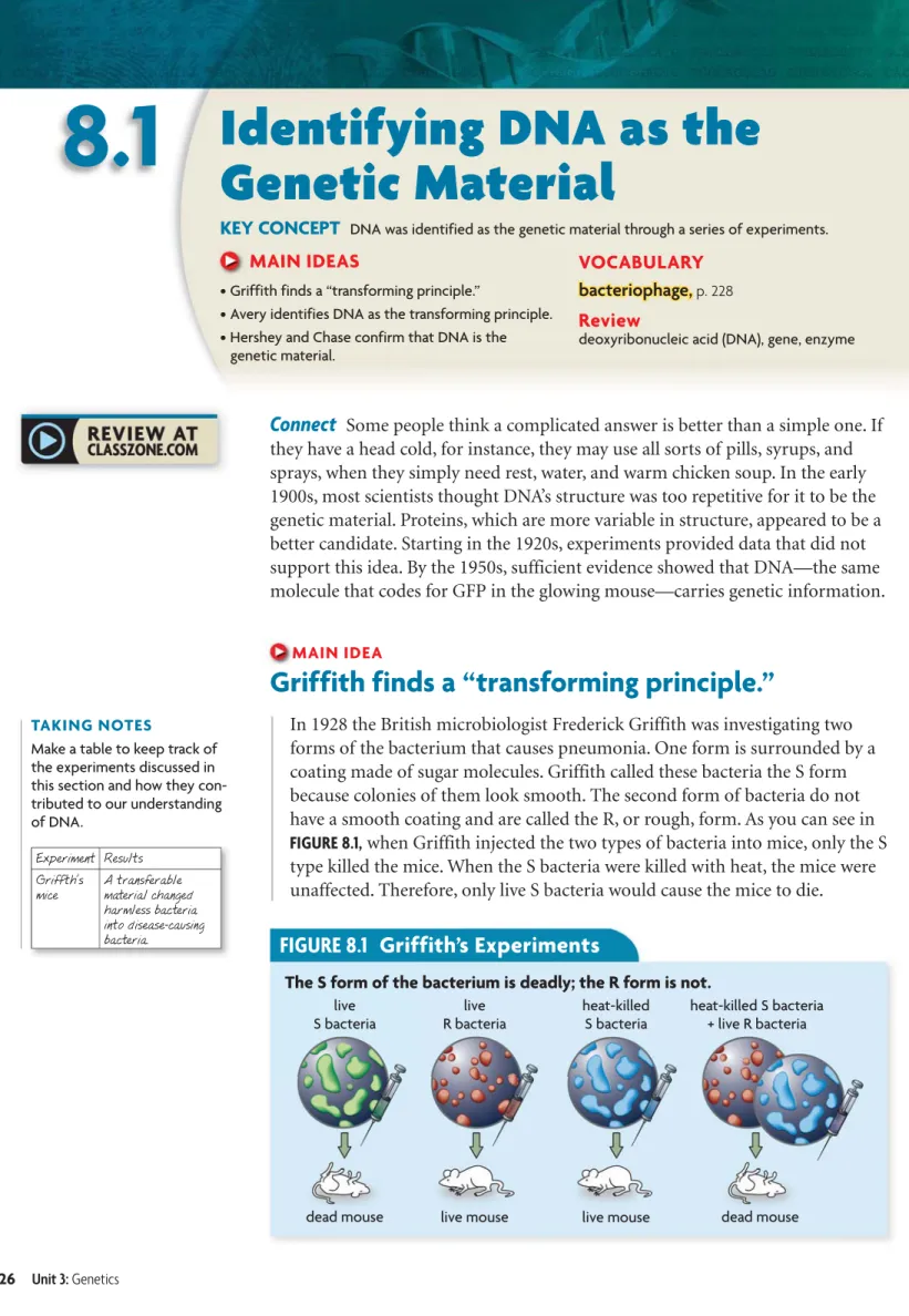
FIGURE 8.1
Griffith’s Experiments
The S form of the bacterium is deadly; the R form is not.
TAKING NOTES
Make a table to keep track of the experiments discussed in this section and how they con-tributed to our understanding of DNA. Experiment Results Griffth’s mice A transferable material changed harmless bacteria into disease-causing bacteria.
8.1
Identifying DNA as the
Genetic Material
KEY CONCEPT DNA was identified as the genetic material through a series of experiments. MAIN IDEAS
• Griffith finds a “transforming principle.”
• Avery identifies DNA as the transforming principle. • Hershey and Chase confirm that DNA is the
genetic material.
VOCABULARY bacteriophage, bacteriophage, p. 228
Review
deoxyribonucleic acid (DNA), gene, enzyme
Connect Some people think a complicated answer is better than a simple one. If
they have a head cold, for instance, they may use all sorts of pills, syrups, and sprays, when they simply need rest, water, and warm chicken soup. In the early 1900s, most scientists thought DNA’s structure was too repetitive for it to be the genetic material. Proteins, which are more variable in structure, appeared to be a better candidate. Starting in the 1920s, experiments provided data that did not support this idea. By the 1950s, sufficient evidence showed that DNA—the same molecule that codes for GFP in the glowing mouse—carries genetic information.MAIN IDEA
Griffith finds a “transforming principle.”
In 1928 the British microbiologist Frederick Griffith was investigating two forms of the bacterium that causes pneumonia. One form is surrounded by a coating made of sugar molecules. Griffith called these bacteria the S form because colonies of them look smooth. The second form of bacteria do not have a smooth coating and are called the R, or rough, form. As you can see in FIGURE 8.1, when Griffith injected the two types of bacteria into mice, only the S type killed the mice. When the S bacteria were killed with heat, the mice were unaffected. Therefore, only live S bacteria would cause the mice to die.
FIGURE 8.2
Avery’s Discoveries
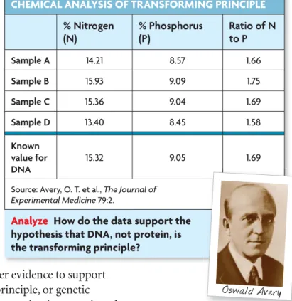
CHEMICAL ANALYSIS OF TRANSFORMING PRINCIPLE
% Nitrogen (N) % Phosphorus (P) Ratio of N to P Sample A 14.21 8.57 1.66 Sample B 15.93 9.09 1.75 Sample C 15.36 9.04 1.69 Sample D 13.40 8.45 1.58 Known value for DNA 15.32 9.05 1.69
Source: Avery, O. T. et al.,The Journal of Experimental Medicine 79:2.
Analyze How do the data support the hypothesis that DNA, not protein, is the transforming principle?
Oswald Avery
Connecting
Microbiology Much of our knowledge of the chemical basis of genetics has come from the study of bacteria. You will learn much more about bacteria in Chapter 18.
CO N C E P TS
Griffith next injected mice with a combination of heat-killed S bacteria and live R bacteria. To his surprise, the mice died. Even more surprising, he found live S bacteria in blood samples from the dead mice. Griffith concluded that some material must have been transferred from the heat-killed S bacteria to the live R bacteria. Whatever that material was, it contained information that changed harmless R bacteria into disease-causing S bacteria. Griffith called this mystery material the “transforming principle.”
Infer What evidence suggested that there was a transforming principle?
MAIN IDEA
Avery identifies DNA as the transforming principle.
What exactly is the transforming principle that Griffith discovered? That question puzzled Oswald Avery and his fellow biologists. They worked for more than ten years to find the answer. Avery’s team began by combining living R bacteria with an extract made from S bacteria. This procedure allowed them to directly observe the transformation of R bacteria into S bacteria in a petri dish.
Avery’s group next developed a process to purify their extract. They then performed a series of tests to find out if the transforming principle was DNA or protein.
• Qualitative tests Standard chemical tests showed that no protein was present. In contrast, tests revealed that DNA was present.
• Chemical analysis As you can see inFIGURE 8.2, the proportions of elements in the extract closely matched those found in DNA. Proteins contain almost no phosphorus.
• Enzyme tests When the team added to the extract enzymes known to break down proteins, the extract still transformed the R bacteria to the S form. Also, transformation occurred when researchers added an enzyme that breaks down RNA (another nucleic acid). Transformation failed to occur only when an enzyme was added to destroy DNA.
In 1944 Avery and his group presented this and other evidence to support their conclusion that DNA must be the transforming principle, or genetic material. The results created great interest. However, some scientists questioned whether the genetic material in bacteria was the same as that in other organ-isms. Despite Avery’s evidence, some scientists insisted that his extract must have contained protein.
Summarize List the key steps in the process that Avery’s team used to identify the transforming principle.
8.1
A SS E SS M E N T
Connecting
CO N C E P TSONLINE QUIZ
ClassZone.com
FIGURE 8.3 This micrograph
shows the protein coat of a bacteriophage (orange) after it has injected its DNA into an
E. coli bacterium (blue). (colored
TEM; magnification 115,000⫻)
MAIN IDEA
Hershey and Chase confirm that DNA is the
genetic material.
Conclusive evidence for DNA as the genetic material came in 1952 from two American biologists, Alfred Hershey and Martha Chase. Hershey and
Chase were studying viruses that infect bacteria. This type of virus, called a
bacteriophage
bacteriophage (bak-TEER-ee-uh-FAYJ), or “phage” for short, takes over a
bacterium’s genetic machinery and directs it to make more viruses.
Phages like the ones Hershey and Chase studied are relatively simple—little more than a DNA molecule surrounded by a protein coat. This two-part structure of phages offered a perfect opportunity to answer the question, Is the genetic material made of DNA or protein? By discovering which part of a phage (DNA or protein) actually entered a bacterium, as shown inFIGURE 8.3, they could answer this question once and for all.
Hershey and Chase thought up a clever procedure that made use of the chemical elements found in protein and DNA. Protein contains sulfur but very little phosphorus, while DNA contains phosphorus but no sulfur. The re-searchers grew phages in cultures that contained radioactive isotopes of sulfur or phosphorus. Hershey and Chase then used these radioactively tagged phages in two experiments.
• Experiment 1 In the first experiment, bacteria were infected with phages that had radioactive sulfur atoms in their protein molecules. Hershey and Chase then used an ordinary kitchen blender to separate the bacteria from the parts of the phages that remained outside the bacteria. When they examined the bacteria, they found no significant radioactivity.
• Experiment 2 Next, Hershey and Chase repeated the procedure with phages that had DNA tagged with radioactive phosphorus. This time, radioactivity was clearly present inside the bacteria.
From their results, Hershey and Chase concluded that the phages’ DNA had entered the bacteria, but the protein had not. Their findings finally convinced scientists that the genetic material is DNA and not protein.
Apply How did Hershey and Chase build upon Avery’s chemical analysis results?
REVIEWING MAIN IDEAS 1. What was “transformed” in Griffith’s
experiment?
2. How did Avery and his group identify the transforming principle?
3. Summarize how Hershey and Chase confirmed that DNA is the genetic material.
CRITICAL THINKING
4. Summarize Why was the bacterio- bacterio-phage
phage an excellent choice for research to determine whether genes are made of DNA or proteins?
5. Analyze Choose one experiment from this section and explain how the results support the conclusion.
6. Mendelian Genetics Describe how Mendel’s studies relate to the experiments discussed in this section.
CHAPTER 8
I N V E S T I G AT I O N
MATERIALS• balance
• 10 g raw wheat germ • laboratory spatula • test tube
• test tube rack • 10 mL warm distilled water • 2 eyedroppers • 4 10-mL graduated cylinders • 20 mL detergent solution • 3 g meat tenderizer • 20 mL salt solution • 10 mL cold isopropyl alcohol
• glass stirring rod
PROCESS SKILLS
• Observing • Analyzing
Extracting DNA
Oswald Avery wrote in a scientific article, “At a critical concentration . . . of alcohol the active material separates out in the form of fibrous strands that wind
themselves around the stirring rod.” In this lab, you can observe the same thing Avery observed as you extract DNA from wheat germ. This procedure is a simplified version of the one scientists commonly use to extract DNA today. P R O B L E M How do you extract the DNA from plant cells?
PROCEDURE
1. Place a small amount of wheat germ in a test tube. The wheat germ should be about 1 cm high in the test tube.
2. Add enough distilled water to wet and cover all of the wheat germ.
3. Add 25–30 drops of detergent solution to the test tube. For 3 minutes, gently swirl the test-tube contents. Avoid making bubbles.
4. Add 3 g of meat tenderizer.
5. Add 25–30 drops of salt solution to the test tube. Swirl for 1 minute.
6. Tilt the test tube at an angle as shown. Slowly add alcohol so that it runs down the inside of the test tube to form a separate layer on top of the mixture in the tube. Add enough alcohol to double the total volume in the tube. Let the test tube stand for 2 minutes.
7. Watch for stringy, cloudy material to rise from the bottom layer into the alcohol layer. This is the DNA.
8. Use the glass stirring rod to remove some DNA. Be careful to probe only the alcohol layer.
9. Draw in your lab report what the mixture and DNA looked like in steps 2–7. Be sure to include color, texture, and what happened after a new solution was added.
ANALYZE AND CONCLUDE
1. Connect Consider what you know about cell structure and the location of DNA. Suggest a reason for adding detergent solution to the test tube.
2. Predict What do you think might happen if the alcohol were added quickly and the two layers mixed?
3. Infer Meat tenderizer contains enzymes that break down proteins. What do you think is the purpose of adding meat tenderizer in this procedure?
4. Connect In what type of real-life situation would the extraction of DNA be useful?
EXTEND YOUR INVESTIGATION
Determine a method to calculate what percentage of the wheat germ consists of DNA.
step 8 step 6
The small units, or monomers, that make up a strand of DNA are called nucleotides.
nucleotides. Nucleotides have three parts. VISUAL VOCAB nitrogen-containing base phosphate group deoxyribose (sugar) Biochemistry The nucleotides in
a strand of DNA all line up in the same direction. As a result, DNA has chemical polarity, which means that the two ends of the DNA strand are different. The 5⬘ carbon is located at one end of the DNA strand, and the 3⬘ car-bon is located at the other end. When the two strands of DNA pair together, the 5⬘ end of one strand aligns with the 3⬘ end of the other strand.
Connecting
CO N C E P TS 6 < I I 8 6 @ @ @ @8.2
Structure of DNA
KEY CONCEPT DNA structure is the same in all organisms. MAIN IDEAS• DNA is composed of four types of nucleotides. • Watson and Crick developed an accurate
model of DNA’s three-dimensional structure. • Nucleotides always pair in the same way.
VOCABULARY nucleotide, nucleotide, p. 230
double helix, double helix, p. 232
base pairing rules, base pairing rules, p. 232
Review
covalent bond, hydrogen bond
Connect The experiments of Hershey and Chase confirmed that DNA carries
the genetic information, but they left other big questions unanswered: What exactly is this genetic information? How does DNA store this information? Scientists in the early 1950s still had a limited knowledge of the structure of DNA, but that was about to change dramatically.MAIN IDEA
DNA is composed of four types of nucleotides.
Since the 1920s, scientists have known that the DNA molecule is a very long polymer, or chain of repeating units. The small units, or monomers, that make up DNA are callednucleotidesnucleotides (NOO-klee-oh-TYDZ). Each nucleotide hasthree parts.
• A phosphate group (one phosphorus with four oxygens)
• A ring-shaped sugar called deoxyribose
• A nitrogen-containing base (a single or double ring built around nitrogen and carbon atoms) One molecule of human DNA contains billions of nucleotides, but there are only four types of nucleotides in DNA. These nucleotides differ only in their nitrogen-containing bases.
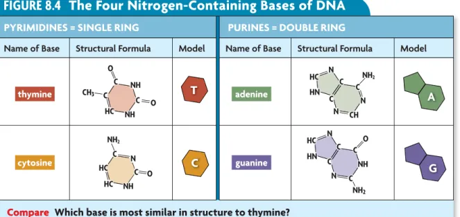
The four bases in DNA are shown inFIGURE 8.4. Notice that the bases cytosine (C) and thymine (T) have a single-ring structure. Adenine (A) and guanine (G) have a larger, double-ring structure. The letter abbreviations refer both to the bases and to the nucleotides that contain the bases.
For a long time, scientists hypothesized that DNA was made up of equal amounts of the four nucleotides, and so the DNA in all organisms was exactly the same. That hypothesis was a key reason that it was so hard to convince scientists that DNA was the genetic material. They reasoned that identical molecules could not carry different instructions across all organisms.
FIGURE 8.4
The Four Nitrogen-Containing Bases of DNA
PYRIMIDINES = SINGLE RING PURINES = DOUBLE RINGName of Base Structural Formula Model Name of Base Structural Formula Model
C= C= C= 8 8 C=C=C= 8 8 =8 =8 =8 8 8 8 8=( D D D D I 88 88 8 8 8= 8= 8= C C C C =C =C =C =8 =8 =8 CC C=C=C='' 6 C= C= C= 8 8 =8 =8 =8 =8 =8 =8 88 D D C C C= C=' C=' 8 C C C= C=' C= C= 8 8 8 =C =C =8 =8 C 8 D C C=' C= 8 8 8 =C =8 C 8 D <
Compare Which base is most similar in structure to thymine? thymine
cytosine
adenine
guanine
Rosalind Franklin FIGURE 8.5 Rosalind Franklin (above) produced x-ray photo-graphs of DNA that indicated it was a helix. Her coworker, Mau-rice Wilkins, showed the data without Franklin’s consent to Watson and Crick, which helped them discover DNA’s structure.
VOCABULARY
An amine is a molecule that contains nitrogen. Notice that the four DNA bases end in -ine and all contain nitrogen. By 1950 Erwin Chargaff changed the thinking about DNA by analyzing the
DNA of several different organisms. Chargaff found that the same four bases are found in the DNA of all organisms, but the proportion of the four bases differs somewhat from organism to organism. In the DNA of each organism, the amount of adenine approximately equals the amount of thymine. Simi-larly, the amount of cytosine roughly equals the amount of guanine. These A = T and C = G relationships became known as Chargaff ’s rules.
Summarize What is the only difference among the four DNA nucleotides?
MAIN IDEA
Watson and Crick developed an accurate
model of DNA’s three-dimensional structure.
The breakthrough in understanding the structure of DNA came in the early 1950s through the teamwork of American geneticist James Watson and British physicist Francis Crick. Watson and Crick were supposed to be study-ing the structure of proteins. Both men, however, were more fascinated by the challenge of figuring out DNA’s structure. Their interest was sparked not only by the findings of Hershey, Chase, and Chargaff but also by the work of the biochemist Linus Pauling. Pauling had found that the structure of some proteins was a helix, or spiral. Watson and Crick hypothesized that DNA might also be a helix.
X-Ray Evidence
At the same time, Rosalind Franklin, shown inFIGURE 8.5, and Maurice Wilkins were studying DNA using a technique called x-ray crystallography. When DNA is bombarded with x-rays, the atoms in DNA diffract the x-rays in a pattern that can be captured on film. Franklin’s x-ray photographs of DNA showed an X surrounded by a circle. Franklin’s data gave Watson and Crick the clues they needed. The patterns and angle of the X suggested that
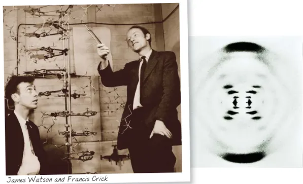
FIGURE 8.6 James Watson (left) and Francis Crick (right) used a model to fig-ure out DNA’s structfig-ure. Their model was influ-enced by data from other researchers, including an x-ray image (far right) taken by Rosalind Franklin. When x-rays bounce off vertically suspended DNA, they form this characteristic x-shaped pattern.
James Watson and Francis Crick
Chemical Bonds Recall from Chapter 2 that a covalent bond is a strong bond in which two atoms share one or more pairs of electrons. Hydrogen bonds are much weaker than covalent bonds and can easily be broken.
Connecting
CO N C E P TSThe Double Helix
Back in their own laboratory, Watson and Crick made models of metal and wood to figure out the structure of DNA. Their models placed the sugar-phosphate backbones on the outside and the bases on the inside. At first, Watson reasoned that A might pair with A, T with T, and so on. But the bases A and G are about twice as wide as C and T, so this produced a helix that varied in width. Finally, Watson and Crick found that if they paired double-ringed nucleotides with single-double-ringed nucleotides, the bases fit like a puzzle. In April 1953 Watson and Crick published their DNA model in a paper in the journalNature.FIGURE 8.6 shows theirdouble helixdouble helix (HEE-lihks) model, in which two strands of DNA wind around each other like a twisted ladder. The strands are complementary —they fit together and are the opposite of each other. That is, if one strand is ACACAC, the other strand is TGTGTG. The pairing of bases in their model finally explained Chargaff ’s rules.
Apply How did the Watson and Crick model explain Chargaff’s rules?
MAIN IDEA
Nucleotides always pair in the same way.
The DNA nucleotides of a single strand are joined together by covalent bonds that connect the sugar of one nucleotide to the phosphate of the next nucleo-tide. The alternating sugars and phosphates form the sides of a double helix, sort of like a twisted ladder. The DNA double helix is held together by hydro-gen bonds between the bases in the middle. Individually, each hydrohydro-gen bond is weak, but together, they maintain DNA structure.
As shown inFIGURE 8.7, the bases of the two DNA strands always pair up in the same way. This is summarized in the base pairing rules: base pairing rules: thymine (T) always pairs with adenine (A), and cytosine (C) always pairs with guanine (G). These pairings occur because of the sizes of the bases and the ability of the
8.2
A SS E SS M E N T
Connecting
CO N C E P TSONLINE QUIZ
ClassZone.com
FIGURE 8.7
Base Pairing Rules
The base pairing rules base pairing rules describe how nucleotidesnucleotides form pairs in DNA. T always pairs with A, and G always pairs with C.
This ribbonlike part represents the phosphate groupsand deoxyribose
sugar molecules that make up DNA’s “backbone.”
Synthesize Which base pairs do you think are held more tightly together? Why?
6 < I 8 I 8 6 < hydrogen bond G C A T G C T A The nitrogen-containing bases bond in the middle to form the rungs of the DNA ladder.
covalent bond
bases to form hydrogen bonds with each other. Due to the arrangement of their molecules, A can form unique hydrogen bonds with T, and C with G. Notice that A and T form two hydrogen bonds, whereas C and G form three.
You can remember the rules of base pairing by noticing that the letters C and G have a similar shape. Once you know that C and G pair together, you know that A and T pair together by default. If a sequence of bases on one strand of DNA is CTGCTA, you know the other DNA strand will be GACGAT.
Apply What sequence of bases would pair with the sequence TGACTA?
REVIEWING MAIN IDEAS 1. How many types of nucleotides nucleotides
are in DNA, and how do they differ?
2. How are the base pairing rules base pairing rules
related to Chargaff’s research on DNA?
3. Explain how the doubledoublehelixhelix
model of DNA built on the research of Rosalind Franklin.
CRITICAL THINKING
4.Infer Which part of a DNA mol-ecule carries the genetic instruc-tions that are unique for each individual: the sugar-phosphate backbone or the nitrogen-contain-ing bases? Explain.
5. Predict In a sample of yeast DNA, 31.5% of the bases are adenine (A). Predict the approximate percent-ages of C, G, and T. Explain.
6. Evolution The DNA of all organisms contains the same four bases (adenine, thymine, cytosine, and guanine). What might this similarity indicate about the origins of life on Earth?
DATA ANALYSIS
ClassZone.com
GRAPH 1. NOBEL PRIZE WINNERS BY AGE
CjbWZgd[ l^ccZgh 6\ZVii^bZd[l^cc^c\ % &% '% (% )% *% +% (%Ä(. )%Ä). *%Ä*. +%Ä+. ,%Ä,. -%Ä-.
DATA A N A LYS I S
I N T E R P R E T I N G
H I S T O G R A M S
Frequency Distributions
A histogram is a graph that shows the frequency distribution of a data set. First, a scientist collects data. Then, she groups the data values into equal intervals. The number of data values in each interval is the frequency of the interval. The intervals are shown along the x-axis of the histogram, and the frequencies are shown on the y-axis.
EXAMPLE
The histogram at right shows the frequency distribution of the ages of winners of the Nobel Prize in
Medicine at the time of winning. Francis Crick was 46 and James Watson was 34 when they were jointly awarded a Nobel Prize in Medicine in 1962.
According to the histogram, the most winners have been between 50 and 59 years old at the time of winning. Only five scientists have been between the ages of 80 and 89 at the time of winning a Nobel Prize in Medicine.
ANALYZE A HISTOGRAM
The histogram below categorizes data collected based on the number of genes in 11 species.
1. Identify How many species had between 10,001 and 15,000 genes?
2. Analyze Are the data in graph 2 sufficient to reveal a trend in the number of genes per species? Explain your reasoning.
GRAPH 2. NUMBER OF GENES IN SELECT SPECIES
CjbWZgd[heZX^Zh CjbWZgd[\ZcZh 1&%%%
<&
H <'
B
Cell Biology In Chapter 5 you learned that the cell cycle has four main stages. DNA is replicated during the S (synthesis) stage.
Connecting
CO N C E P TS8.3
DNA Replication
KEY CONCEPT DNA replication copies the genetic information of a cell. MAIN IDEAS• Replication copies the genetic information. • Proteins carry out the process of replication. • Replication is fast and accurate.
VOCABULARY replication, replication, p. 235 DNA polymerase, DNA polymerase, p. 236 Review
base pairing rules, S phase
Connect Do you know that some of your cells are dying right now? You may live
to the ripe old age of 100, but most of your cells will have been replaced thou-sands of times before you blow out the candles on that birthday cake. Every time that cells divide to produce new cells, DNA must first be copied in a remarkable process of unzipping and zipping by enzymes and other proteins. The next few pages will take you through that process.MAIN IDEA
Replication copies the genetic information.
One of the powerful features of the Watson and Crick model was that it suggested a way that DNA could be copied. In fact, Watson and Crick ended the journal article announcing their discovery with this sentence: “It has not escaped our notice that the specific pairing we have postulated immediately suggests a possible copying mechanism for the genetic material.”Recall that the bases that connect the strands of DNA will pair only in one way, according to the rules of base pairing. An A must bind with a T, and a C must bind with a G. If the base sequence of one strand of the DNA double helix is known, the sequence of the other strand is also known. Watson and Crick realized that a single DNA strand can serve as a template, or pattern, for a new strand. This process by which DNA is copied during the cell cycle is calledreplication.replication.
Suppose all of your classmates took off their shoes, placed their left shoe in a line, and tossed their right shoe into a pile. You could easily pick out the right shoes from the pile and place them with the matching left shoes. The order of the shoes would be preserved. Similarly, a new strand of DNA can be synthesized when the other strand is a template to guide the process. Every time, the order of the bases is preserved, and DNA can be accurately replicated over and over again.
Replication assures that every cell has a complete set of identical genetic information. Recall that your DNA is divided into 46 chromosomes that are replicated during the S phase of the cell cycle. So your DNA is copied once in each round of the cell cycle. As a result, every cell has a complete set of DNA.
DNA polymerases
DNA polymerases are enzymes that form bonds between nucleotides during replication.
VISUAL VOCAB
DNA
polymer
ase
The ending -ase signals that this is an enzyme.
This part of the name tells what the enzyme does—makes DNA polymers.
TAKING NOTES
Use a cycle diagram to take notes about processes such as replication. existing molecule unzipping two DNA molecules formed nucleotides added
Biochemistry You read in Chapter 2 that many proteins are enzymes that function as catalysts. Enzymes decrease the activation energy and increase the rate of chemical reactions. DNA polymerase catalyzes the reaction that bonds two nucleotides together.
Connecting
CO N C E P TSThe fact that cells throughout the body have complete sets of DNA is very useful for forensic scientists. They can identify someone from nearly any cell in the body. A few cells from a drop of blood or from saliva on a cigarette butt are all detectives need to produce a DNA “fingerprint” of a criminal suspect.
Apply How does replication ensure that cells have complete sets of DNA?
MAIN IDEA
Proteins carry out the process of replication.
Although people may say that DNA copies itself, the DNA itself does nothing more than store information. Enzymes and other proteins do the actual work of replication. For example, some enzymes start the process by unzipping the double helix to separate the strands of DNA. Other proteins hold the strands apart while the strands serve as
templates. Nucleotides that are floating free in the nucleus can then pair up with the nucleotides of the existing DNA strands. A group of enzymes calledDNA polymerasesDNA polymerases
(PAHL-uh-muh-rays) bond the new nucleotides together. When the process is finished, the result is two complete molecules of DNA, each exactly like the original double strand.
The Replication Process
The following information describes the process of DNA replication in eukaryotes, which is similar in prokaryotes. As you read, follow along with each step illustrated inFIGURE 8.8.
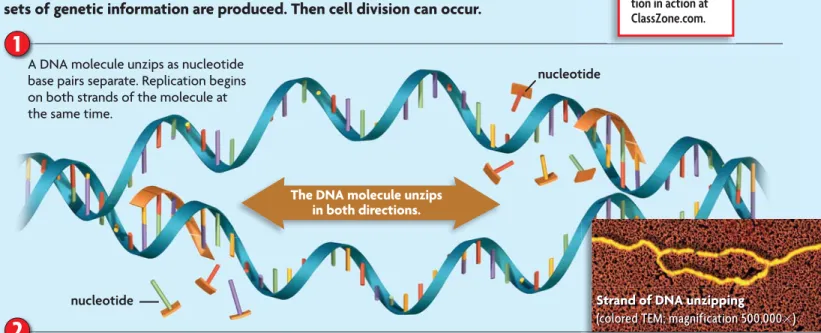
1
Enzymes begin to unzip the double helix at numerous places along the chromosome, called origins of replication. That is, the hydrogen bonds connecting base pairs are broken, the original molecule separates, and the bases on each strand are exposed. Unlike unzipping a jacket, this process proceeds in two directions at the same time.2
Free-floating nucleotides pair, one by one, with the bases on the template strands as they are exposed. DNA polymerases bond the nucleotides together to form new strands that are complementary to each template strand. DNA replication occurs in a smooth, continuous way on one of the strands. Due to the chemical nature of DNA polymerase, replication of the other strand is more complex. It involves the formation of many small DNA segments that are joined together. This more complex process is not shown or described in detail here.3
Two identical molecules of DNA result. Each new molecule has one strand from the original molecule and one new strand. As a result, DNA replication is called semiconservative because one old strand is con-served, and one complementary new strand is made.DNA polymerase DNA polymerase
nucleotide
Two molecules of DNA
Strand of DNA unzipping
(colored TEM; magnification 500,000⫻)
nucleotide
nucleotide
new strand
The DNA molecule unzips in both directions. new strand original strand
1
2
3
BIOLOGYSee DNA replica-tion in acreplica-tion at ClassZone.com.
Two identical double-stranded DNA molecules result from replication. DNA replication is semiconservative. That is, each DNA molecule contains an original strand and one new strand.
Each existing strand of the DNA molecule is a template for a new strand. Free-floating nucleotides pair up with the exposed bases on each template strand. DNA polymerases bond these nucleotides together to form the new strands. The arrows show the directions in which new strands form. A DNA molecule unzips as nucleotide
base pairs separate. Replication begins on both strands of the molecule at the same time.
FIGURE 8.8
Replication
When a cell’s DNA is copied, or replicated, two complete and identical sets of genetic information are produced. Then cell division can occur.
How is each new molecule of DNA related to the original molecule?
CRITICAL VIEWING
8.3
A SS E SS M E N T
Connecting
CO N C E P TS ONLINE QUIZ ClassZone.comQ U I C K L A B
Replication
Use two zipping plastic bags to model how complementary strands of DNA attach to template strands during replication.
PROCEDURE
1. Cut the sliding zippers off both bags. One zipper represents the template strands of a DNA molecule.
2. Cut the other zipper into four smaller pieces and unzip each of them. These represent free nucleotides. Don’t worry about which nucleotide is which in this activity. 3. Use the pieces to model replication as shown on page 237. ANALYZE AND CONCLUDE
Evaluate What are the limitations of this model?
MODE LING
MATERIALS
• 2 zipping bags
• scissors
FIGURE 8.9 Eukaryotic
chromo-somes have many origins of repli-cation. The DNA helix is unzipped at many points along each chro-mosome. The replication “bub-bles” grow larger as replication progresses in both directions, resulting in two complete copies.
6 7 8 9
MAIN IDEA
Replication is fast and accurate.
In every living thing, DNA replication happens over and over again, and it happens remarkably fast. In human cells, about 50 nucleotides are added every second to a new strand of DNA at an origin of replication. But even at this rate, it would take many days to replicate a molecule of DNA if the molecule were like a jacket zipper, unzipping one tooth at a time. Instead, replication proceeds from hundreds of origins of replication along the chromosome, as shown inFIGURE 8.9, so the process takes just a few hours.
Another amazing feature of replication is that it has a built-in “proofread-ing” function to correct errors. Occasionally, the wrong nucleotide is added to the new strand of DNA. However, DNA polymerase can detect the error, remove the incorrect nucleotide, and replace it with the correct one. In this way, errors in replication are limited to about one error per 1 billion nucleotides.
Replication is happening in your cells right now. Your DNA is replicated every time your cells turn over, or replicate themselves. Your DNA has repli-cated trillions of times since you grew from a single cell.
Infer Why does a cell need to replicate its DNA quickly?
REVIEWING MAIN IDEAS 1. Explain the function of replication.replication. 2. Explain how DNA serves as its own
template during replication.
3. How do cells help ensure that DNA replication is accurate?
CRITICAL THINKING 4. Summarize Describe two
major functions of DNA DNA polymerases.
polymerases.
5. Infer Why is it important that human chromosomes have many origins of replication?
6. Cell Biology DNA is replicated before both mitosis and meiosis. How does the amount of DNA produced in a cell during mitosis compare with that produced during meiosis?
FIGURE 8.10 The central dogma describes the flow of information from DNA to RNA to proteins. It involves three major processes, shown in a eukaryotic cell below.
cjXaZjh 9C6 9C6 GC6 GC6 EgdiZ^c EgdiZ^c 9C6 GC6 EgdiZ^c XnideaVhb Replication Transcription Translation
8.4
Transcription
KEY CONCEPT Transcription converts a gene into a single-stranded RNA molecule. MAIN IDEAS• RNA carries DNA’s instructions. • Transcription makes three
types of RNA.
• The transcription process is similar to replication. VOCABULARY central dogma, central dogma, p. 239 RNA, RNA, p. 239 transcription, transcription, p. 240 RNA polymerase, RNA polymerase, p. 240
messenger RNA (mRNA), messenger RNA (mRNA),
p. 240
ribosomal RNA (rRNA), ribosomal RNA (rRNA), p. 240
transfer RNA (tRNA), transfer RNA (tRNA), p. 240
Connect Suppose you want to play skeeball at a game center, but the skeeball
lane only takes tokens. You only have quarters. Do you go home in defeat? Stand idly by as someone else becomes high scorer? No, you exchange your quarters for tokens and then proceed to show the other players how it’s done. In a similar way, your cells cannot make proteins directly from DNA. They must convert the DNA into an intermediate molecule called RNA, or ribonucleic acid. That conversion process, called transcription, is the focus of this section.MAIN IDEA
RNA carries DNA’s instructions.
Soon after his discovery of DNA structure, Francis Crick defined the
central dogma
central dogma of molecular biology, which states that information flows in one direction, from DNA to RNA to proteins. The central dogma involves three processes, as shown inFIGURE 8.10.
• Replication, as you just learned, copies DNA (blue arrow).
• Transcription converts a DNA message into an intermediate molecule, called RNA (red arrow).
• Translation interprets an RNA message into a string of amino acids, called a polypeptide. Either a single polypeptide or many polypeptides working together make up a protein (green arrow).
In prokaryotic cells, replication, transcription, and translation all occur in the cytoplasm at approximately the same time. In eukaryotic cells, where DNA is located inside the nuclear membrane, these processes are separated both in location and time. Replication and transcription occur in the nucleus, while translation occurs in the cytoplasm. In addition, the RNA in eukaryotic cells goes through a processing step before it can be transported out of the nucleus. Unless otherwise stated, the rest of this chapter describes how these processes work in eukaryotic cells.
RNA acts as an intermediate link between DNA in the nucleus and protein synthesis in the cytoplasm. Like DNA,RNA,RNA, or ribonucleic acid, is a chain of nucleotides, each made of a sugar, a phosphate group, and a nitrogen-contain-ing base. You can think of RNA as a temporary copy of DNA that is used and then destroyed.
C= C= C= 8 8 C=C=C= 8 8 8 8 8 8 8DD D D J
Connecting
DNA Structure As you learned in Section 8.2, nucleotides are made of a phosphate group, a sugar, and a nitrogen-containing base. In DNA, the four bases are adenine, cytosine, guanine, and thymine. In RNA, uracil (below) replaces thy-mine and pairs with adenine.
CO N C E P TS
VOCABULARY
The word transcribe means “to make a written copy of.” Transcription is the process of transcribing. A transcript is the copy produced by transcription.
RNA differs from DNA in three significant ways. First, the sugar in RNA is ribose, which has one additional oxygen atom not present in DNA’s sugar (deoxy-ribose). Second, RNA has the base uracil in place of thymine. Uracil, like thymine, forms base pairs with adenine. Third, RNA is a single strand of nucleotides, in contrast to the double-stranded structure of DNA. This single-stranded structure allows some types of RNA to form complex three-dimensional shapes. As a result, some RNA molecules can catalyze reactions much as enzymes do.
Contrast How do DNA and RNA differ?
MAIN IDEA
Transcription makes three types of RNA.
TranscriptionTranscription is the process of copying a sequence of DNA to produce a complementary strand of RNA. During the process of transcription, a gene— not an entire chromosome—is transferred into an RNA message. Just as replication is catalyzed by DNA polymerase, transcription is catalyzed by
RNA polymerases,
RNA polymerases, enzymes that bond nucleotides together in a chain to make a new RNA molecule. RNA polymerases are very large enzymes composed of many proteins that play a variety of roles in the transcription process.
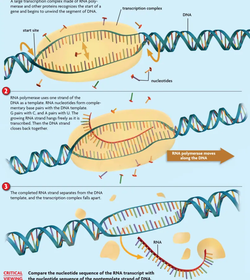
FIGURE 8.11 shows the basic steps of transcription in eukaryotic cells.
1
With the help of other proteins and DNA sequences, RNA polymerase recognizes the transcription start site of a gene. A large transcription complex consisting of RNA polymerase and other proteins assembles on the DNA strand and begins to unwind a segment of the DNA molecule, until the two strands separate from each other.2
RNA polymerase, using only one strand of DNA as a template, strings together a complementary strand of RNA nucleotides. RNA base pairing follows the same rules as DNA base pairing, except that uracil, not thymine, pairs with adenine. The growing RNA strand hangs freely as it is transcribed, and the DNA helix zips back together.3
Once the entire gene has been transcribed, the RNA strand detaches completely from the DNA. Exactly how RNA polymerase recognizes the end of a transcription unit is complicated. It varies with the type of RNA. Transcription produces three major types of RNA molecules. Not all RNA molecules code for proteins, but most play a role in the translation process. Each type of RNA molecule has a unique function.• Messenger RNA (mRNA) Messenger RNA (mRNA) is an intermediate message that is translated to form a protein.
• Ribosomal RNA (rRNA) Ribosomal RNA (rRNA) forms part of ribosomes, a cell’s protein factories.
• Transfer RNA (tRNA) Transfer RNA (tRNA) brings amino acids from the cytoplasm to a ribosome to help make the growing protein.
Remember that the RNA strand must be processed before it can exit the nucleus of a eukaryotic cell. This step occurs during or just after transcription. However, we will next examine translation and then return to processing.
RNA polymerase moves along the DNA
1
nucleotides transcription complex DNA start site2
3
A large transcription complex made of RNA poly-merase and other proteins recognizes the start of a gene and begins to unwind the segment of DNA.
RNA polymerase uses one strand of the
DNA as a template. RNA nucleotides form comple-mentary base pairs with the DNA template. G pairs with C, and A pairs with U. The growing RNA strand hangs freely as it is transcribed. Then the DNA strand closes back together.
The completed RNA strand separates from the DNA template, and the transcription complex falls apart.
RNA
FIGURE 8.11
Transcription
Transcription produces an RNA molecule from a DNA template. Like DNA replication, this process takes place in the nucleus in eukaryotic cells and involves both DNA unwinding and nucleotide base pairing.
Compare the nucleotide sequence of the RNA transcript with the nucleotide sequence of the nontemplate strand of DNA.
CRITICAL VIEWING
8.4
A SS E SS M E N T
Connecting
CO N C E P TSONLINE QUIZ
ClassZone.com
FIGURE 8.12 This TEM shows
DNA in a eukaryotic cell being transcribed into numerous mRNA strands by many RNA polymer-ases. The mRNA strands near the start of each gene are shorter than those near the end. (TEM;
magnifica-tion 13,000⫻) one gene
REVIEWING MAIN IDEAS 1. What is the central dogmacentral dogma?
2. Why can the mRNAmRNA strand made during transcriptiontranscription be thought of as a mirror image of the DNA strand from which it was made?
3. Why might a cell make lots of rRNArRNA
but only one copy of DNA?
CRITICAL THINKING
4. Apply If a DNA segment has the nucleotides AGCCTAA, what would be the nucleotide sequence of the complementary RNARNA strand?
5. Synthesize What might geneticists learn about genes by studying RNA?
6. Cell Cycle You know that a healthy cell cannot pass the G2 checkpoint until all of its
DNA has been copied. Do you think that a cell must also transcribe all of its genes into RNA to pass this checkpoint? Explain.
MAIN IDEA
The transcription process is similar to replication.
The processes of transcription and replication share many similarities. Both processes occur within the nucleus of eukaryotic cells. Both are catalyzed by large, complex enzymes. Both involve unwinding of the DNA double helix. And both involve complementary base pairing to the DNA strand. In addition, both processes are highly regulated by the cell. Just as a cell does not replicate its DNA without passing a critical checkpoint, so, too, a cell carefully regulates which genes are transcribed into RNA.
The end results of transcription and replication, how-ever, are quite different. The two processes accomplish very different tasks. Replication ensures that each new cell will have one complete set of genetic instructions. It does this by making identical sets of double-stranded chromosomes. This double-stranded structure makes DNA especially well suited for long-term storage because it helps protect DNA from being broken down and from potentially harmful interactions with other molecules. Replication occurs only once during each round of the cell cycle because each cell needs to make only one copy of its DNA.
In contrast, a cell may need hundreds or thousands of copies of certain proteins, or the rRNA and tRNA mol-ecules needed to make proteins. Transcription enables a cell to adjust to changing demands. It does so by making a single-stranded complement of only a segment of DNA and only when that particular segment is needed. In addition, many RNA molecules can be transcribed from a single gene at the same time to help produce more protein. Once RNA polymerase has transcribed one portion of a gene and has moved on, another RNA polymerase can attach itself to the beginning of the gene and start the transcription process again. This process can occur over and over again, as shown inFIGURE 8.12.
Compare How are the processes of transcription and replication similar?
DNA
A codoncodon is a sequence of three nucleo-tides that codes for an amino acid. VISUAL VOCAB HZ\bZcid[bGC6 8 J J J < 6 codon for methionine (Met) codon for leucine (Leu)
Biochemistry Recall from
Chapter 2 that amino acids are the building blocks of proteins. Although there are many types of amino acids, only the same 20 types make up the proteins of almost all organisms.
Connecting
CO N C E P TS8.5
Translation
KEY CONCEPT Translation converts an mRNA message into a polypeptide, or protein. MAIN IDEAS• Amino acids are coded by mRNA base sequences.
• Amino acids are linked to become a protein. VOCABULARY translation, translation, p. 243 codon, codon, p. 243 stop codon, stop codon, p. 244 start codon, start codon, p. 244 anticodon, anticodon, p. 245 Review peptide bond
Connect As you know, translation is a process that converts a message from one
language into another. For example, English words can be translated into Spanish words, into Chinese characters, or into the hand shapes and gestures of sign language. Translation occurs in cells too. Cells translate an RNA message into amino acids, the building blocks of proteins. But unlike people who use many different languages, all cells use the same genetic code.MAIN IDEA
Amino acids are coded by mRNA base sequences.
TranslationTranslation is the process that converts, or translates, an mRNA message into a polypeptide. One or more polypeptides make up a protein. The “language” of nucleic acids uses four nucleotides—A, G, C, and T in DNA; or A, G, C, and U in RNA. The “language” of proteins, on the other hand, uses 20 amino acids. How can four nucleotides code for 20 amino acids? Just as letters are strung together in the English language to make words, nucleotides are strung to-gether to code for amino acids.
Triplet Code
Different words have different numbers of letters. In the genetic code, however, all of the “words,” called codons, are made up of three letters. Acodoncodon is a three-nucleotide sequence that codes
for an amino acid. Why is the genetic code read in units of three nucleo-tides? Well, we can’t entirely answer that question, but consider the possi-bilities. If one nucleotide coded for one amino acid, RNA could code for only four amino acids. If two nucleo-tides coded for one amino acid, RNA could code for 16 (42) amino acids—
still not enough. But if three nucleo-tides coded for one amino acid, RNA
could code for 64 (43) amino acids, plenty to cover the 20 amino acids used to
Second base First bas e Thir d bas e UUU phenylalanine (Phe) UCU serine (Ser) UAU tyrosine (Tyr) UGU cysteine (Cys) U
UUC UCC UAC UGC C
UUA leucine
(Leu)
UCA UAA STOP UGA STOP A
UUG UCG UAG STOP UGG tryptophan (Trp) G CUU leucine (Leu) CCU proline (Pro) CAU histidine (His) CGU arginine (Arg) U CUC CCC CAC CGC C
CUA CCA CAA glutamine
(Gln) CGA A CUG CCG CAG CGG G AUU isoleucine (Ile) ACU threonine (Thr) AAU asparagine (Asn) AGU serine (Ser) U
AUC ACC AAC AGC C
AUA ACA AAA lysine
(Lys)
AGA arginine
(Arg)
A
AUG methionine (Met) ACG AAG AGG G
GUU valine (Val) GCU alanine (Ala)
GAU aspartic acid
(Asp) GGU glycine (Gly) U GUC GCC GAC GGC C
GUA GCA GAA glutamic acid
(Glu)
GGA A
GUG GCG GAG GGG G
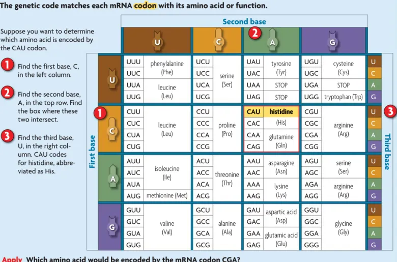
Apply Which amino acid would be encoded by the mRNA codon CGA?
J 8 6 < J 8 6 <
1
2
3
FIGURE 8.14 Codons are read as
a series of three nonoverlapping nucleotides. A change in the reading frame changes the resulting protein.
Reading frame 1
Reading frame 2
8 < 6 J 6 8 6 < J 6 < 8
Arg Tyr Ser Ser
8 < 6 J 6 8 6 < J 6 < 8
Asp Thr Val
CAU histidine
Suppose you want to determine which amino acid is encoded by the CAU codon.
FIGURE 8.13
Genetic Code: mRNA Codons
The genetic code matches each mRNA codoncodon with its amino acid or function.
As you can see inFIGURE 8.13, many amino acids are coded for by more than one codon. The amino acid leucine, for example, is represented by six different codons: CUU, CUC, CUA, CUG, UUA, and UUG. There is a pattern to the codons. In most cases, codons that represent the same amino acid share the same first two nucleotides. For example, the four codons that code for alanine each begin with the nucleotides GC. Therefore, the first two nucleotides are generally the most important in coding for an amino acid. As you will learn in Section 8.7, this feature makes DNA more tolerant of many point mutations.
In addition to codons that code for amino acids, threestop codonsstop codons signal the end of the amino acid chain. There is also onestart codon,start codon, which signals the start of translation and the amino acid methionine. This means that translation always begins with methionine. However, in many cases, this methionine is removed later in the process.
For the mRNA code to be translated correctly, codons must be read in the right order. Codons are read, without spaces, as a series of three nonoverlap-ping nucleotides. This order is called the reading frame. Changing the reading frame completely changes the resulting protein. It may even keep a protein from being made if a stop codon turns up early in the translation process. Therefore, punctuation—such as a clear start codon—plays an important role in the genetic code.FIGURE 8.14 shows how a change in reading frame changes
Find the first base, C, in the left column.
1
Find the second base, A, in the top row. Find the box where these two intersect.
2
Find the third base, U, in the right col-umn. CAU codes for histidine, abbre-viated as His.
3
FIGURE 8.15 TRANSLATION MACHINERY Ribosomes The large and small ribosomal subunits pull mRNA through the ribosome, reading it one codon at a time.
tRNA In cells, tRNA forms a characteristic L shape. One end of the L has an anticodon that recognizes an mRNA codon. The other end is attached to an amino acid.
large subunit binds to tRNA ribosome small subunit binds to mRNA binding sites amino acid anticodon tRNA
the resulting protein. When the mRNA strand is read starting from the first nucleotide, the resulting protein includes the amino acids arginine, tyrosine, and two serines. When the strand is read starting from the second nucleotide, the resulting protein includes aspartic acid, threonine, and valine.
Common Language
The genetic code is shared by almost all organisms—and even viruses. That means, for example, that the codon UUU codes for phenylalanine when that codon occurs in an armadillo, a cactus, a yeast, or a human. With a few minor exceptions, almost all organisms follow this genetic code. As a result, the code is often called universal. The common nature of the genetic code suggests that almost all organisms arose from a common ancestor. It also means that scientists can insert a gene from one organism into another organism to make a functional protein.
Calculate Suppose an mRNA molecule in the cytoplasm had 300 nucleotides. How many amino acids would be in the resulting protein?
MAIN IDEA
Amino acids are linked to become a protein.
Let’s take a step back to look at where we are in the process of making proteins. You know mRNA is a short-lived molecule that
carries instructions from DNA in the nucleus to the cyto-plasm. And you know that this mRNA message is read in sets of three nucleotides, or codons. But how does a cell actually translate a codon into an amino acid? It uses two important tools: ribosomes and tRNA molecules, as illustrated in
FIGURE 8.15.
Recall from Chapter 3 that ribosomes are the site of protein synthesis. Ribosomes are made of a combination of rRNA and proteins, and they catalyze the reaction that forms the bonds between amino acids. Ribosomes have a large and small subunit that fit together and pull the mRNA strand through. The small subunit holds onto the mRNA strand, and the large subunit holds onto the growing protein.
The tRNA acts as a sort of adaptor between mRNA and amino acids. You would need an adaptor to plug an appliance with a three-prong plug into an outlet with only two-prong openings. Similarly, cells need tRNA to carry free-floating amino acids from the cytoplasm to the ribosome. The tRNA molecules fold up in a characteristic L shape. One end of the L is attached to a specific amino acid. The other end of the L, called the anticodon, recognizes a specific codon. An
anticodon
anticodon is a set of three nucleotides that is complementary to an mRNA codon. For example, the anticodon CCC pairs with the mRNA codon GGG.
BZi BZi AZj AZj 8nh 8nh BZi BZi AZj AZj peptide bond
2
3
The ribosome pulls the mRNA strand the length of one codon. The first tRNA is shifted into the exit site, where it leaves the ribosome and returns to the cytoplasm to recharge. The first site is again empty, exposing the next mRNA codon.
BZi BZi 6g\ 6g\ AZj AZj 8nh 8nh 8nh 8nh J 6 <
The ribosome continues to translate the mRNA strand until it reaches a stop codon. Then it releases the new protein and disassembles.
1
The exposed codon in the first site attracts a complementary tRNA bearing an amino acid. The tRNA anticodon pairs with the mRNA codon, bringing it very close to the other tRNA molecule.
BZi BZi AZj AZj 8 J J < 6 6 incoming tRNA mRNA methionine start codon stop codon cytoplasm
Translation occurs in the cytoplasm of both eukaryotic (illustrated) and prokaryotic cells. It starts when a tRNA carrying a methionine attaches to a start codon.
tRNA ribosome
mRNA nucleus
amino acid
The ribosome forms a peptide bond between the two amino acids and breaks the bond between the first tRNA and its amino acid.
leucine
CRITICAL VIEWING
FIGURE 8.16
Translation
Translation
Translation converts an mRNA transcript into a polypeptide. The process consists of three repeating steps.
The figure above shows how the first two amino acids are added to a growing protein. Draw a series of sketches to show how the next two amino acids are added.
8.5
A SS E SS M E N T
Connecting
CO N C E P TSONLINE QUIZ
ClassZone.com To learn more about protein synthesis, visit scilink.org. Keycode: MLB008
CHI6 hX^a^c`h#dg\
Translation, shown inFIGURE 8.16, has many steps and takes a lot of energy from a cell. It happens in the cytoplasm of both prokaryotic and eukaryotic cells. Before translation can begin, a small ribosomal subunit must bind to an mRNA strand in the cytoplasm. Next, a tRNA with methionine attached binds to the AUG start codon. This binding signals a large ribosomal subunit— which has three binding sites for tRNA molecules—to join. The ribosome pulls the mRNA strand through itself one codon at a time. As the strand moves, the start codon and its complementary tRNA molecule shift into the second site inside the large subunit. This shift leaves the first site empty, which exposes the next mRNA codon. The illustration shows the process in one ribosome, but in a cell many ribosomes may translate the same gene at the same time.
1
The exposed codon attracts a complementary tRNA molecule bearing an amino acid. The tRNA anticodon pairs with the mRNA codon. This action brings the new tRNA molecule very close to the tRNA molecule occupying the second site.2
Next, the ribosome helps form a peptide bond between the two amino acids. The ribosome then breaks the bond between the tRNA molecule in the second site and its amino acid.3
The ribosome pulls the mRNA strand the length of one codon. The tRNA molecule in the second site is shifted into the third site, which is the exit site. The tRNA leaves the ribosome and returns to the cytoplasm to be charged with another amino acid. The tRNA molecule that was in the first site shifts into the second site. The first site is again empty, exposing the next mRNA codon.Another complementary tRNA molecule is attracted to the exposed mRNA codon, and the process continues. The ribosome moves down the mRNA strand, attaching new amino acids to the growing protein, until it reaches a stop codon. Then it lets go of the new protein and falls apart.
Summarize Explain the different roles of the large and small ribosomal subunits.
REVIEWING MAIN IDEAS 1. Explain the connection between a
codon
codon and an amino acid.
2. Briefly describe how the process of
translation
translation is started.
CRITICAL THINKING 3. Synthesize Suppose a tRNA
molecule had the anticodonanticodon
AGU. What amino acid would it carry?
4. Hypothesize The DNA of eukary-otic cells has many copies of genes that code for rRNA molecules. Suggest a hypothesis to explain why a cell needs so many copies of these genes.
5. Biochemical Reactions
Enzymes have shapes that allow them to bind to a substrate. Some types of RNA also form specific three-dimensional shapes. Why do you think RNA, but not DNA, catalyzes biochemical reactions?
VOCABULARY
The word promote comes from the Latin prefix pro-, meaning “forward,” and the Latin word movere, meaning “to move.”
8.6
Gene Expression
and Regulation
KEY CONCEPT Gene expression is carefully regulated in both prokaryotic and eukaryotic cells. MAIN IDEAS
• Prokaryotic cells turn genes on and off by controlling transcription.
• Eukaryotic cells regulate gene expression at many points.
VOCABULARY promoter, promoter, p. 248 operon, operon, p. 248 exon, exon, p. 251 intron, intron, p. 251
Connect Ours is a world of marvels. So many, in fact, that we may overlook
what seem like little ones, such as plumbing. The turn of a handle sends clean water to your sink or shower. One twist and the water trickles out; two twists and it gushes forth. Another turn of the handle and the water is off again. But think about the mess and waste that would result if you couldn’t control its flow. In a similar way, your cells have ways to control gene expression. Depending on an organism’s needs, a gene can make a lot of protein, a little protein, or none at all.MAIN IDEA
Prokaryotic cells turn genes on and off by
controlling transcription.
The regulation of gene expression allows prokaryotic cells, such as bacteria, to better respond to stimuli and to conserve energy and materials. In general, this regulation is simpler in prokaryotic cells than in eukaryotic cells, such as those that make up your body. DNA in a prokaryotic cell is in the cytoplasm. Transcription and translation can happen at the same time. As a result, gene expression in prokaryotic cells is mainly regulated at the start of transcription.
A gene includes more than just a protein-coding sequence. It may have many other nucleotide sequences that play a part in controlling its expression. The start of transcription is largely controlled by these sequences, including promoters and operators. Apromoterpromoter is a DNA segment that allows a gene to be transcribed. It helps RNA polymerase find where a gene starts. An operator is a DNA segment that turns a gene “on” or “off.” It interacts with proteins that increase the rate of transcription or block transcription from occurring.
Bacteria have much less DNA than do eukaryotes, and their genes tend to be organized into operons. Anoperonoperon is a region of DNA that includes a promoter, an operator, and one or more structural genes that code for all the proteins needed to do a specific task. Typically, operons are found only in prokaryotes and roundworms. Thelac operon was one of the earliest exam-ples of gene regulation discovered in bacteria. It will serve as our example. Thelac operon has three genes, which all code for enzymes that play a role in
9C6 L^i]djiaVXidhZhl^iX]ZYd[[ GC6edanbZgVhZWadX`ZY \ZcZh[dgZconbZhi]ViY^\ZhiaVXidhZ egdbdiZg gZegZhhdg deZgVidg L^i]aVXidhZhl^iX]ZYdc GC6edanbZgVhZigVchXg^WZh \ZcZh[dgZconbZhi]ViY^\ZhiaVXidhZ egdbdiZg gZegZhhdg aVXidhZ deZgVidg \gdl^c\GC6
operator. This means that although we’re dealing with several genes, they act together as a unit.
Thelac operon is turned on and off like a switch. When lactose is absent from the environment, thelac operon is switched off to prevent transcription of thelac genes and save the cell’s resources. When lactose is present, thelac
operon is switched on to allow transcription. How does this happen? Bacteria have a protein that can bind specifically to the operator. When lactose is absent, this protein binds to the operator, which blocks RNA poly-merase from transcribing the genes. Because the protein blocks—or re-presses—transcription, it is called a repressor protein.
When lactose is present it binds to the repressor, which makes the repressor change shape and fall off thelacoperon. RNA polymerase can then transcribe the genes in thelacoperon. The resulting transcript is translated and forms three enzymes that work together to break down the lactose.
Analyze Explain how the lac operon is turned on or off like a switch.
MAIN IDEA
Eukaryotic cells regulate gene expression at
many points.
You have already learned that every body cell in an organism has the same set of DNA. But your cells are not all the same. Cells differ from each other because different sets of genes are expressed in different types of cells. Eukary-otic cells can control the process of gene expression at many different points because of their internal compartments and chromosomal organization. As in prokaryotic cells, however, one of the most highly regulated steps is the start of transcription. In both cell types, RNA processing is a part of the transcrip-tion process. In eukaryotic cells, however, RNA processing also includes the removal of extra nucleotide segments from an mRNA transcript.
FIGURE 8.17
Starting Transcription
Transcription factors that bind to promoterspromoters and other DNA sequences help RNA polymerase recognize the start of a gene in a eukaryotic cell.
Zc]VcXZg igVchXg^ei^dc [VXidgh GC6edanbZgVhZ I6I6 Wdm egdbdiZg 9C6 \ZcZ
Predict Does an enhancer have to be close to the start site of a gene? Explain.
Connecting
Animals As you will learn in Chapter 23, most animals have homeobox genes. These genes are among the earliest that are expressed and play a key role in development. The micrograph below shows the expression of homeobox genes in a fruitfly embryo.
CO N C E P TS
Starting Transcription
The start of transcription in eukaryotic cells is controlled by many elements that work together in complex ways. These elements include regulatory DNA sequences and proteins called transcription factors, as shown inFIGURE 8.17. They occur in different combinations in different types of cells. The interplay between these elements results in specialized cells and cell responses.
Eukaryotes have many types of regulatory DNA sequences. These se-quences are recognized by transcription factors that bind to the DNA strand and help RNA polymerase know where a gene starts. Some DNA sequences, such as promoters, are close to the start of a gene. Others are far away from the genes they affect. However, DNA can loop and bend, bringing these sequences with their transcription factors into close contact with the others.
Each gene has a unique combination of regulatory sequences. Some are found in almost all eukaryotic cells. For example, most eukaryotic cells have a seven-nucleotide promoter (TATAAAA) called the TATA box. Eukaryotic cells also have other types of promoters that are more specific to an individual gene. DNA sequences called enhancers and silencers also play a role by speed-ing up or slowspeed-ing down, respectively, the rate of transcription of a gene.
Some genes control the expression of many other genes. Regulation of these genes is very important because they can have a large effect on develop-ment. One such gene codes for a protein called sonic hedgehog. This protein was first found in fruit flies, but many other organisms have very similar proteins that serve a similar function. Sonic hedgehog helps establish body pattern. When missing in fruit flies, the embryos are covered with little prickles and fail to form normal body segments.
mRNA Processing
Another important part of gene regulation in eukaryotic cells is RNA process-ing, which is shown inFIGURE 8.18. The mRNA produced by transcription is similar to a rough cut of a film that needs a bit of editing. A specialized nucleotide is added to the beginning of each mRNA molecule, which forms a cap. It helps the mRNA strand bind to a ribosome and prevents the strand from being broken down too fast. The end of the mRNA molecule gets a string of A nucleotides, called the tail, that helps the mRNA molecule exit the nucleus.