The Brain
•
The Adult Human Brain
•
Contains almost 97 percent of the body’s neural tissue
•
Average weight about 1.4 kg (3 lb)
•
Six Regions of the Brain
1.
Cerebrum
2.
Cerebellum
3.
Diencephalon
4.
Midbrain
5.
Pons
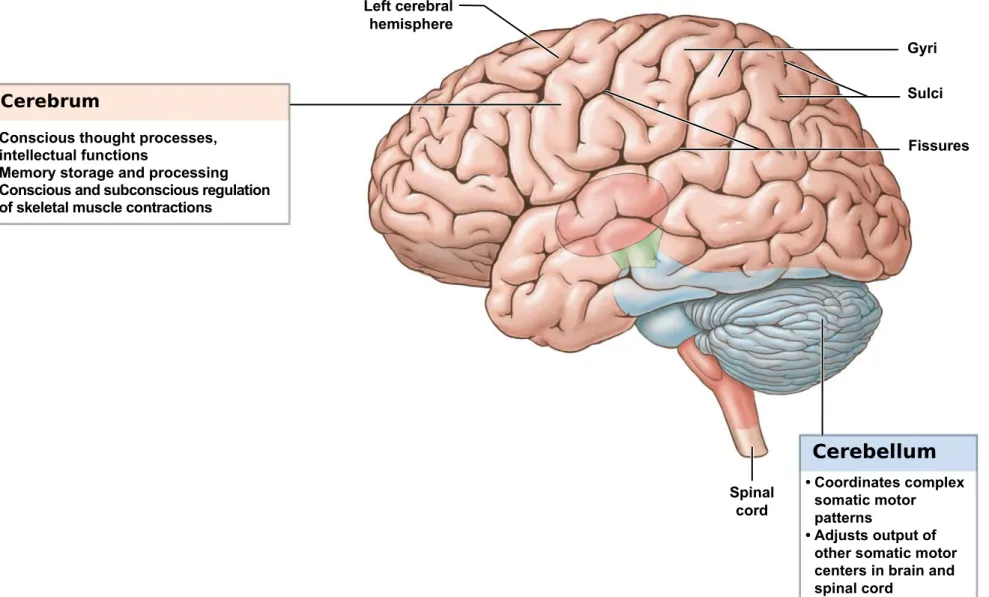
Figure 14-1 An Introduction to Brain Structures and Functions (Part 1 of 2).
Cerebrum
• Conscious thought processes, intellectual functions
• Memory storage and processing • Conscious and subconscious regulation
of skeletal muscle contractions
Left cerebral hemisphere
Gyri
Fissures
Cerebellum • Coordinates complex
somatic motor patterns
• Adjusts output of other somatic motor centers in brain and spinal cord
Sulci
Figure 14-1 An Introduction to Brain Structures and Functions (Part 2 of 2).
• Relay and processing centers for sensory information
Diencephalon
Thalamus
Hypothalamus
• Centers controlling emotions, autonomic functions, and
hormone production
Midbrain
• Processing of visual and auditory date • Generation of reflexive
somatic motor responses • Maintenance of
consciousness
Brain stem
Pons
• Relays sensory information to cerebellum and thalamus
• Subconscious
somatic and visceral motor centers
Medulla oblongata
• Relays sensory information to thalamus and to other portions of the brain stem
Ventricles of the Brain
• Each cerebral hemisphere contains one large lateral ventricle
• Separated by a thin medial partition (septum pellucidum)
• Third ventricle
• Ventricle of the diencephalon
• Lateral ventricles communicate with third ventricle
• Via interventricular foramen (foramen of Monro)
• Fourth ventricle
• Extends into medulla oblongata
• Becomes continuous with central canal of the spinal cord
• Connects with third ventricle
Figure 14-2a Ventricles of the Brain.
Cerebral hemispheres
Pons
Medulla oblongata
Spinal cord Central canal
Ventricles, lateral view
Figure 14-2b Ventricles of the Brain.
Ventricles, anterior view
Ventricles of the Brain
Lateral ventricles
Interventricular foramen
Third ventricle
Cerebral aqueduct
Fourth ventricle
Central canal
Cerebral hemispheres
Cerebellum
•
Physical Protection of the Brain
•
Bones of the cranium
•
Cranial
meninges
•
Cerebrospinal fluid
•
Biochemical Isolation
•
Blood–brain
barrier
•
The
Cranial
Meninges
•
Have three layers
1. Dura mater
2. Arachnoid mater
3. Pia mater
•
Are continuous with spinal meninges
•
The Cranial Meninges
•
Dura
mater
• Inner fibrous layer (meningeal layer)
• Outer fibrous layer (periosteal layer) fused to periosteum
• Venous sinuses between two layers – dural sinus
•
Arachnoid
mater
• Covers brain
• Contacts epithelial layer of dura mater
• Subarachnoid space between arachnoid mater and pia mater
•
Pia
mater
Figure 14-3a The Relationship among the Brain, Cranium, and Cranial Meninges.
Subarachnoid space Pia mater Cerebral cortex
Cranium Dura mater (periosteal layer)
Dural sinus
Dura mater (meningeal layer)
Subdural space Arachnoid mater Cerebrum Cerebellum Medulla oblongata Spinal cord A lateral view of the brain, showing its position in
the cranium and the organization of the meninges
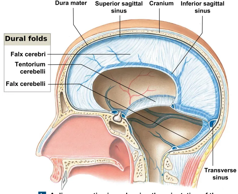
•
Dural
Folds
•
Folded inner layer of dura mater
•
Extend into cranial cavity
•
Stabilize and support brain
•
Contain collecting veins (
dural
sinuses
)
•
Three largest dural folds
1.
Falx
cerebri
- projects between the cerebral hemispheres
•
Contains
superior
sagittal
sinus
and
inferior
sagittal
sinus
2. Tentorium
cerebelli -
separates cerebellum and cerebrum
•
Contains
transverse
sinus
Figure 14-3b The Relationship among the Brain, Cranium, and Cranial Meninges.
Dura mater Superior sagittal sinus
Cranium Inferior sagittal sinus Transverse sinus Falx cerebri Tentorium cerebelli Falx cerebelli
A diagrammatic view, showing the orientation of the three largest dural folds: the falx cerebri, tentorium cerebelli, and falx cerebelli
b
•
Cerebrospinal Fluid (CSF)
•
Surrounds all exposed surfaces of CNS
•
Interchanges with interstitial fluid of brain
•
Functions of CSF
• Cushions delicate neural structures • Supports brain
• Transports nutrients, chemical messengers, and waste products
•
Choroid
plexus
• Specialized ependymal cells and capillaries • Secrete CSF into ventricles
• Remove waste products from CSF • Adjust composition of CSF
•
Cerebrospinal Fluid (CSF)
•
CSF circulates:
• From choroid plexus
• Through ventricles
• To central canal of spinal cord
• Into subarachnoid space via two lateral apertures and one median aperture around the brain, spinal cord, and cauda equine
•
CSF in subarachnoid space
• Arachnoid villi
• Extensions of subarachnoid space
• Extend through dura mater to superior sagittal sinus
• Arachnoid granulations
• Large clusters of villi
Figure 14-4 Formation and Circulation of Cerebrospinal Fluid (Part 2 of 3). Choroid plexus of fourth ventricle Choroid plexus
of third ventricle
Spinal cord Filum terminale Arachnoid mater Cauda equina Dura mater Central canal
Cerebrospinal fluid then flows through the subarachnoid space surrounding the brain, spinal cord, and cauda equina.
4 3
The CSF reaches the sub-arachnoid space through two lateral apertures and a single median aperture in the roof of the fourth ventricle.
2
•
Blood Supply to the Brain
•
Supplies nutrients and oxygen to brain
•
Delivered by
internal
carotid
arteries
and
vertebral
arteries
•
Removed
from dural sinuses by
internal jugular veins
•
Blood – brain barrier
= biochemical isolation
•
Isolates CNS neural tissue from general circulation
•
Formed by network of tight junctions between endothelial cells of CNS
capillaries
•
Lipid-soluble compounds (O
2, CO
2), steroids, and prostaglandins diffuse into
interstitial fluid of brain and spinal cord
•
Astrocytes control blood–brain barrier by releasing chemicals that control
•
Blood–CSF
Barrier
•
Formed by special ependymal cells
•
Surrounds capillaries of choroid plexus
•
Limits movement of compounds transferred
•
Allows chemical composition of blood and CSF to differ
•
Four Breaks in the BBB
1. Portions of hypothalamus
• Secrete hypothalamic hormones
2. Posterior lobe of pituitary gland
• Secretes hormones ADH and oxytocin
3. Pineal gland
• Pineal secretions
4. Choroid plexus
Gross anatomy -Cerebrum
•
CEREBRAL HEMIPHERES-
right and left separated by the longitudinal fissure
•
Surface layer of gray matter (neural cortex) -
folded surface increases surface area • Conscious thought and intelligence are produced in the neural cortex of the cerebral hemispheres• GYRI = folds ; gyrus ( singular)
•
PRECENTRAL GYRUS ( motor function)
&
POSTCENTRAL GYRUS ( sensorial
function )
•
GROOVES
•
FISSURES: deep grooves
•
CENTRAL SULCUS- separates frontal from parietal lobes
•
LATERAL SULCUS- separates frontal and parietal from temporal lobe
•
PARIETO OCCIPITAL SULCUS- separates parietal from occipital lobe
LOBES
•
FRONTAL LOBE
•
TEMPORAL LOBE
•
PARIETAL LOBE
•
OCCIPITAL LOBE
•
INSULA
• PRE CENTRAL GYRUS ( structural name)- the gyrus before the central sulcus, part of frontal lobe IT CONTEINS THE PRIMARY MOTOR AREA = Primary motor cortex ( functional name)
Function : - TO CONTROL VOLUNTARY MUSCLE MOVEMENT
• POSTCENTRAL GYRUS- the gyrus after the central sulcus, part of the parietal lobe IT CONTEINS THE PRIMARY SENSORY AREA = Primary sensory cortex
Function : - TO RECEIVE SENSORY INFO FROM GENERAL SENSORY RECEPTORS - touch, pressure, pain, vibration, taste, temperature
•
Special Sensory Cortexes
•
Visual cortex -
Information from sight receptors
•
Association Areas
•
Somatic sensory association area
• Interprets input to primary sensory cortex (e.g., recognizes and responds to touch)
•
Visual association area
• Interprets activity in visual cortex
•
Auditory association area
• Monitors auditory cortex
•
Somatic motor association area
(
premotor
cortex
)
• Coordinates motor responses (learned movements
•
Integrative areas
•
Prefrontal cortex
of frontal lobe
• Integrates information from sensory association areas
•
General Interpretive Area
• Also called Wernicke’s area
• Present in only one hemisphere, in temporal and parietal lobes
• Receives information from all sensory association areas
• Coordinates access to complex visual and auditory memories
• helps in understanding speech and using the correct words to express our thoughts.
• Wernicke's aphasia - a patient may be able to produce speech, but cannot understand the speech of others.
•
Broca's area
- motor speech area in the frontal lobe
• helps in movements required to produce speech.
•
White Matter of the Cerebrum
•
Association
fibers
•
Connections within one hemisphere
Arcuate fibers
• Are short fibers
• Connect one gyrus to another
•
Longitudinal
fasciculi
• Are longer bundles
• Connect frontal lobe to other lobes in same hemisphere
•
Commissural
fibers
• Bands of fibers connecting two hemispheres
•
Corpus callosum
•
Anterior commissure
•
Projection
fibers
• Connect cerebrum with lower areas : Diencephalon, brain stem, cerebellum, and spinal cord
•
Hemispheric Lateralization
•
Functional differences between left and right hemispheres
•
Each cerebral hemisphere performs certain functions that are not ordinarily
performed by the opposite hemisphere
•
The Left Hemisphere
•
In most people, left brain (
dominant
hemisphere
) controls:
• Reading, writing, and math
• Decision making
• Speech and language
•
The Right Hemisphere
•
Right cerebral hemisphere relates to:
• Senses (touch, smell, sight, taste, feel)
Inferior view of the cerebrum
• OLFACTORY BULBS - CARRY SENSORY INFO ABOUT SMELL
• OLFACTORY TRACS -CARRY SENSORY INFO ABOUT SMELL
• OPTIC CHIASMA- WHERE THE OPTIC NERVES CROSS (IN THE MIDDLE)
• OPTIC TRACTS- CARRY VISUAL INFORMATION
• PITUITARY GLAND (HYPOPHISYS)- PRODUCES MANY HORMONES
Medial view – sagittal section
•
CORPUS CALLOSUM- white matter connecting the hemispheres
•
SEPTUM PELLUCIDUM- membrane between lateral ventricles
•
FORNIX – tract of white matter connects hypothalamus with the
hippocampus ( limbic system )
•
DIENCEPHALON:
•
THALAMUS
•
HYPOTHALAMUS
•
EPITHALAMUS
•
BRAIN STEM:
•
MESENCEPHALON
•
PONS
•
The
Thalamus
– five groups of nuclei
•
Filters ascending sensory information for primary sensory cortex
•
Relays information between basal nuclei and cerebral cortex
•
The third ventricle - separates
left
thalamus
and
right
thalamus
•
Interthalamic
adhesion
•
Projection of gray matter
•
Extends into ventricle from each side
•
Anterior
nuclei -
part of limbic system (emotions)
•
Medial
group -
provides awareness of emotional states
•
Ventral group -
Relays sensory information
•
Posterior
group
• Pulvinar nucleus (sensory)
• Lateral geniculate nucleus (visual) • Medial geniculate nucleus (auditory)
•
Lateral
group
• Affects emotional states
•
The
Basal
Nuclei
•
Are masses of gray matter
•
Are embedded in white matter of cerebrum
•
Direct subconscious activities
•
Caudate
nucleus
• Curving, slender tail
•
Lentiform nucleus
• Globus pallidus
• Putamen
•
Functions of Basal Nuclei
•
Involved with:
• The subconscious control of skeletal muscle tone