Abstract
Recent studies show that the anti-diabetic drug metformin exhibits anticancer effects
against a broad spectrum of cancers including endometrial cancer. Due to its hydrophilic nature
and net positive charge at physiological pH, metformin requires cation-selective transporters to
enter cells. This study investigated the expression of metformin transporters in human
endometrial cancer cell lines, and the role of these transporters in metformin cellular uptake and
accumulation in two human endometrial cancer cell lines, Ishikawa and ECC-1. Total RNA
isolated from these two cell lines was subjected to real time polymerase chain reaction (RT-PCR)
to determine the expression levels of common cation-selective transporters that are known to
transport metformin. MATE 1 and 2 were the predominant transporters in Ishikawa and ECC-1
cells while OCT 1-3 expression in both cell lines was relatively poor. Time-dependent [14C]
metformin uptake into Ishikawa and ECC-1 cell lines was measured by quantifying intracellular
radioactivity. To demonstrate the role of transporters in the uptake of metformin in these two cell
lines, metformin uptake was measured in the presence and absence of cation-selective transporter
inhibitors. Metformin uptake was saturated in both cell lines, and transporter inhibitors decreased
metformin uptake in Ishikawa cells. RT-PCR results demonstrate that Ishikawa and ECC-1 cells
are relevant in vitro models for investigating metformin treatment in endometrial cancer because
expression of metformin transporters in human endometrial cancer tissues is comparable to that
observed in endometrial cancer cell lines. The finding that metformin uptake is decreased by
inhibitors only in Ishikawa cells warrants further study.
Introduction
Metformin is one of the most widely prescribed anti-diabetic drugs in the United States.
including endometrial, breast, prostate, colon, ovarian, gastric, and lung cancers1-5. Endometrial
cancer, a cancerous growth in the lining of the uterus, is the most common cancer of the female
reproductive organs. In 2013, the American Cancer Society estimated that over 48,500 women
would be diagnosed with endometrial cancer, and over 8,000 women would die from the disease.
The 1-year relative survival rate for uterine cancers is 92% while the 5-year survival rate is 95%,
67%, or 16%, depending if the cancer is diagnosed at a local, regional, or distant stage
respectively6.
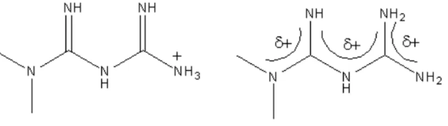
Metformin is a hydrophilic drug that is positively charged at physiological pH (Figure 1).
As a result, metformin requires transporters to pass through lipophilic cell membranes.
Cation-selective transporters known to transport metformin include organic cation transporter
(OCT) 1-3, plasma monoamine transporter (PMAT), and multidrug and toxin extrusion
transporter (MATE) 1-2. To elicit its anti-diabetic effects, metformin is transported into hepatic
cells via OCT 1 where it inhibits hepatic gluconeogenesis by first activating adenosine
monophosphate-activated protein kinase (AMPK), which suppresses regulatory gluconeogenic
genes7 (Figure 2).
Figure 1: Metformin
The anti-cancer effects of metformin are proposed to occur, in part, through activation of AMPK,
which inhibits the downstream mammalian target of rapamycin (mTOR) pathway that regulates
cell proliferation. Previous studies have demonstrated that metformin arrests cell cycle
progression and inhibits proliferation in Ishikawa and ECC-1 endometrial cancer cell lines8.
Therefore, to induce its anti-cancer effects in endometrial cancer, metformin uptake in
endometrial cancer cells and tissues must be mediated by specific cation-selective transporters.
Elucidating metformin transport mechanisms in endometrial cancer will be central to
understanding the role of transporters and transporter variability in metformin anticancer effects
and clinical outcomes in cancer patients. This study aims to investigate the expression of
metformin transporters in human endometrial cancer cell lines and tissues, and the role of these
transporters in the uptake of metformin into endometrial cancer cells. Methods
Cell Culture
ECC-1 cells were cultured in Roswell Park Memorial Institute (RPMI) 1640 medium and Figure 2: The mechanism of action of metformin in a liver cell, and the
Ishikawa cells were cultured in minimum essential medium (MEM) with 5% fetal bovine serum
(FBS), 5% L- Glutamine 200mM, and 1% antibiotic-antimycotic. Both cell lines were cultured at
37oC in 5% CO2 and 90% relative humidity.
RT-PCR
Total RNA was isolated from endometrial cancer cell lines and tissues using the Qiagen RNeasy
Plus kit, and cDNA was synthesized using a FirstStrand Synthesis kit (Invitrogen). The mRNA
levels for each transporter were determined by Taqman assay (Applied Biosystems), and
normalized to the 18s rRNA eukaryotic housekeeping gene.
Uptake Studies
Cells were seeded at 250,000 cells/cm2 and cultured for 4-7 days. Growth medium was
changed every other day and the day before experimentation. Cells were incubated with transport
buffer (HBSS with 10mM HEPES and 25mM D-glucose) for 30 minutes prior to each study. For
time-dependent studies, cells were exposed to 50µM [14C] metformin for 1-30 minutes. For
concentration-dependent studies, cells were exposed to [14C] metformin concentrations ranging
from 100µM to 20mM for 5 minutes. Chemical inhibition studies utilized 50µM [14C] metformin
alone and in combination with the transporter inhibitors quinidine (Quin, 500µM),
pyrimethamine (Pyr, 0.4µM), mitoxantrone (Mito, 25µM), corticosterone (Cort, 1µM),
desipramine (Des, 200µM), and MPP+ (MPP+, 5mM). Transporters inhibited by these chemical
inhibitors are shown in Figure 3. In each study, cells were washed 3 times with ice cold buffer
after the desired time point, lysed (0.1M NaOH with 1% SDS), and analyzed by liquid
scintillation spectrometry. Metformin uptake was normalized to protein content that was
Inhibitor Inhibited Transporters
Quinidine 500µM All transporters
Pyrimethamine 0.4µM MATE 1 & 2
Mitoxantrone 25µM OCT 1 & MATE 2
Corticosterone 1µM OCT 3
Desipramine 200µM OCT 1-3 & PMAT
MPP+ 5mM All transporters
Statistical Analysis: Data was analyzed by one-way ANOVA and Bonferroni post-hoc test. Data represents mean ± SD; n=3, *p<0.05 compared to control.
Results RT-PCR
All six transporters of interest were present in both Ishikawa and ECC-1 cell lines (Figure
4). MATE 1 and 2 showed the highest expression in both cell lines, with MATE 2 expression
being 6-fold higher than MATE 1 expression in Ishikawa cells and 5-fold higher in ECC-1 cells.
Expression of OCT 1-3 was relatively poor in both cell lines.
Uptake Studies
Uptake of [14C]metformin was linear in both Ishikawa and ECC-1 cell lines, and reached
saturation at 10 minutes (Figure 5).
Concentration-dependent uptake of metformin at 5 minutes in Ishikawa cells was linear up to
1mM and saturated at 2mM (Figure 6A), while in ECC-1 cells uptake of metformin at 5 minutes
was linear up to 5mM and saturated at 7.5mM (Figure 6B).
Metformin uptake was significantly decreased in the presence of the pan transporter inhibitor
quinidine (66% decrease) and the OCT 1 and MATE 1 inhibitor, mitoxantrone (54% decrease)
A B
Figure 5: Time-‐dependent uptake of metformin in (A) Ishikawa cells and (B) ECC-‐1 cells.
(Figure 7A). There was a trend toward a decrease in metformin uptake with the use of the OCT 3
inhibitor, corticosterone (36% decrease) and another pan transporter inhibitor, MPP+ (38%
decrease). Treatment with the MATE 1 and 2 inhibitor pyrimethamine and the OCT 1-3 and
PMAT inhibitor desipramine did not inhibit metformin uptake. Metformin uptake was not
significantly decreased in ECC-1 cells with any inhibitor used (Figure 7B).
Discussion
RT-PCR results suggest that cation-selective transporters known to transport metformin
are present in both the Ishikawa and ECC-1 cell lines. Studies by others in the Thakker
laboratory showed that MATE 1 was the primary transporter in RT-PCR analysis of 15 human
endometrial tumor tissues and 5 adjacent non-malignant tissues, with PMAT and OCT 3 also
expressed in significant amounts. The predominant expression of MATE 1 in both human
endometrial tissues types, and its high expression in the two endometrial cancer cell lines
analyzed suggest that the Ishikawa and ECC-1 cell lines are relevant in vitro models to evaluate the role of transporters in the anticancer efficacy of metformin in endometrial cancer.
The saturation of metformin uptake in the Ishikawa cell line with respect to time and
concentration suggests that uptake was transporter mediated. The significant decrease in
metformin uptake in the presence of transporter inhibitors in Ishikawa cells further establishes
the role of transporters in metformin uptake in this cell line. The significant decrease observed
with quinidine (500µM), a pan inhibitor at the concentration used, and with mitoxantrone
(25µM), which inhibits MATE 1 and OCT 3 suggests that these two transporters play an
important role in the uptake of metformin into Ishikawa cells. Metformin uptake was also
decreased in the presence of corticosterone (1µM) which also inhibits OCT 3, although this
decrease was not statistically significant. Interestingly, pyrimethamine (0.4µM), which inhibits
MATE 1 and MATE 2 did not significantly decrease metformin uptake. The wide variability
observed in metformin uptake in the presence of pyrimethamine has made it challenging to
accurately interpret this result.
The uptake of metformin in the ECC-1 cell line was also saturated with respect to time
and concentration, which suggests that uptake was mediated by transporters. However,
transporter inhibitors did not decrease metformin uptake, which was unexpected. The chemical
inhibition studies in both cell lines were performed the same day, using the same dosing
solutions, suggesting that this finding is not due to procedural error. Furthermore, the compact
standard deviations observed in this experiment, minimize the possibility that a significant
decrease in metformin uptake in the presence of chemical inhibitors was obscured by an outlying
result. Therefore, these chemical inhibition studies in ECC-1 cells require further investigation.
The current cornerstone of endometrial cancer treatment is hysterectomy, although
patients may also receive radiation, chemotherapy, and/or hormonal therapy based on the cancer
side effects that include diarrhea and nausea, which can be minimized with dose titrations. The
safety profile of metformin, compared to chemotherapy and radiation, makes it is an attractive
agent for the treatment of endometrial cancer. Understanding the relative contributions of
cation-selective transporters to the uptake of metformin in endometrial cancer cells is important for
conducting future in vivo studies which will be crucial to optimizing metformin therapy in
endometrial cancer, and understanding potential variability in response to metformin treatment
due to genetic and physiologic variability of transporters.
In conclusion, the Ishikawa and ECC-1 cell lines are reasonable endometrial cancer
models in which to study the critical role of transporters in the anticancer efficacy of metformin
in endometrial cancer.
References
1. Dowling RJ, Zakikhani M, Fantus IG, Pollak M, Sonenberg N: Metformin inhibits mammalian target of rapamycin-dependent translation initiation in breast cancer cells. Cancer Res 2007, 67:10804-10812
2. Ben Sahra I, Laurent K, Loubat A, Giorgetti-Peraldi S, Colosetti P, Auberger P, Tanti JF, Le Marchand-Brustel Y, Bost F: The antidiabetic drug metformin exerts an antitumoral effect in vitro and in vivo through a decrease of cyclin D1 level.
3. Buzzai M, Jones RG, Amaravadi RK, Lum JJ, DeBerardinis RJ, Zhao F, Viollet B, Thompson CB: Systemic treatment with the antidiabetic drug metformin selectively impairs p53-deficient tumor cell growth. Cancer Res 2007, 67:6745-6752.
4. Gotlieb WH, Saumet J, Beauchamp MC, Gu J, Lau S, Pollak MN, Bruchim I: In vitro metformin anti-neoplastic activity in epithelial ovarian cancer. Gynecol Oncol 2008, 110:246-250.
5. Kiyohito K, Gong J, Iwama H, et al. “The antidiabetic drug metformin inhibits gastric cancer cell proliferation in vitro and in vivo” Mol Cancer Ther Published OnlineFirst January 5, 2012.
6. American Cancer Society.: Cancer Facts and Figures 2013. Atlanta, Ga: American Cancer Society, 2013. 7.Zakikhani M, Dowling R, Fantus IG, Sonenberg N, Pollak M: Metformin is an AMP kinase-dependent growth inhibitor for breast cancer cells. Cancer Res. 66(21), 10269–10273, 2006.