Analysis of microRNAs expression changes in human
breast cancer cell lines following exposure to ionizing
radiation
INTRODUCTION
The most frequent carcinoma and second reason of cancer-related mortality in women, is
breast cancer (1). Today, the main clinical obstacle to the successful therapy of breast cancer is treatment resistance such as radio-resistance, which results in metastasis and
relapse. Growing evidence show that one of the most important reasons behind this is the presence of a rare population of stem-like cells
called breast cancer stem cells (BCSCs). Therefore finding a way to reduce BCSCs radio-resistance results in better clinical outcomes for patients with breast cancer (2).
MicroRNAs (miRNAs) constitute a group of evolutionarily conserved endogenous, single
stranded and non-coding RNA molecules
post-transcriptional regulators for gene expression in the cell cycle, DNA damage response, and apoptosis pathway. It should be
noted that the mRNA degradation or translational inhibition functions of these molecules are exerted through their sequence-specific binding to the 3′ un-translated
regions of target mRNAs (3). Therefore, specific miRNAs are often up- or down-regulated in response to irradiation; hence more information about miRNAs regulation and function will be critical to develop radiation therapy (4).
MicroRNA 21 (miR-21) regulates some tumour suppressor genes, such as phosphatase
and tensin homolog (PTEN), programmed cell death 4 (PDCD4), tropomyosin 1 (TPM1) and cell division cycle 25A (Cdc25A) (5, 6). So it could be involved in the cellular response to IR and
S. Dehghan Kouhestani
1, M. Forouzandeh
1*, M. Aghili
21Department of Medical Biotechnology, Faculty of Medical Sciences, Tarbiat Modares University, Tehran, Iran 2Department of Radiation Oncology, Cancer Institute, Tehran University of Medical Sciences, Tehran, Iran
ABSTRACT
Background: The expression alterations of specific miRNAs were analysed in response to gamma-irradiation in two breast cancer cell lines MCF-7 and MDA-MB-231 with low and high BCSCs content to design probable strategies for sensitizing breast cancer stem-like cells MDA-MB-231 to radiation. Materials and Methods: The expression levels of hsa-miR-34a, hsa-let-7i and hsa-miR-21 were assessed by QRT-PCR in MCF-7 and MDA-MB-231 breast cancer cells at three time points after gamma irradiation using three different doses. Results: The results showed that the over-expression of hsa-miR-34a in MCF-7 cells was much more significant than MDA-MB-231 cells. Furthermore, there was a considerable over-expression of hsa-miR-21 in MDA-MB-231 cells following exposure to ionizing radiation (IR). The hsa-let-7i expression changes were different, depending on radiation dose, time post irradiation and cell type. Conclusion: According to the results, we may be able to sensitize stem-like MDA-MB-231 breast cancer cells to radiation by increasing miR-34a and decreasing miR-21 expression levels simultaneously.
Keywords: miR-34a; miR-21; let-7i; ionizing radiation; breast cancer. *Corresponding authors:
Dr. M. Forouzandeh, Fax: +98 21 82883861
E-mail: [email protected] Revised: May 2016
Accepted: June 2017
Int. J. Radiat. Res., July 2018; 16(3): 383-388
►
Short Report
DOI: 10.18869/acadpub.ijrr.16.3.383
miR-34a is a tumour suppressor, which targets several genes involved in DNA damage response (DDR) such as Bcl-2, Notch1, high mobility group A2 (HMGA2), Cyclin D1, Cyclin E2, cell division cycle 2 (CDK1), cyclin dependent kinase 4 and 6 (CDK4 and CDK6), MET and sirtuin 1(SIRT1) (8) and may be used as a marker and therapeutic target for cell response to radiation (9). Also, miR
-34a ⁄ Notch1 is involved in regulating breast
cancer stem cell (10). Let-7 is an important miRNA family consisting of 12 members that are frequently down-regulated in a number of human cancers (11). They target RAS, CDK6,
Cyclin D2, and Cdc25A genes which their
over-expression lead to a pro-survival phenotype in response to IR (12). Let-7 effect on
several breast stem cell-like properties by inhibiting more than one target (13).
We hoped that the study of miRNA expression changes in response to IR in breast
cancer stem-like cells could be a starting point to a better radiotherapy in breast cancer disease. Thus, we decided to focus on three miRNAs,
hsa-miR-21, hsa-miR-34a, and hsa-let-7i since
they are associated with the modulation of cell damage response pathways (7, 9, 12). Furthermore, we analysed these miRNAs expression changes after gamma-irradiation in the MDA-MB-231 cells with the highest CD44+/CD24-/low stem-like population, at approximately 98% (14) and MCF-7 cells which harbour just 3.5% of CD44+/CD24-/ low phenotype (15). The aim of this study was to evaluate the role of miRNAs, related to cancer stem cells in treatment by radiation. The specific miRNAs (miR-21, miR-34a, Let-7i) expression
were investigated in response to gamma radiation in MCF7 (with low BCSCs content) and
MDA-MB1-231 (with 80% BCSCs).According to the results obtained from miRNAs expression, we designed probable strategies for sensitizing MDA-MB-231 cells to radiation.
MATERIALS AND METHODS
Cell cultureThe human breast adenocarcinoma cell lines MCF-7 (IBRC C10082, MCF-7 type) and MDA-MB
384
-231 from National Center of Genetic and Biological Reservoirs (NCGBR, Iran) and Pasteur
Institute of Iran (IPI) were maintained in
DMEM/F12 and RPMI 1640 medium respectively, supplemented with 10% fetal
bovine serum (FBS) and 1% penicillin-streptomycin. All cells were incubated
in a humidified atmosphere of 5% CO2 at 37°C. The culture medium, penicillin-streptomycin solution and FBS were purchased from GibcoTM Life Technologies (Carlsbad, CA, USA), all other supplements were from Sigma Aldrich (St Louis, MO, USA).
Ionizing radiation treatment
Adjusted number of 3×10⁵ monolayer cells in the logarithmic growth phase was irradiated at a dose rate of 0.89 Gy/min with gamma-rays from a Co60 source (Theratron 780C, Theratronix, Canada) available at Cancer Institute of Iran. The cells received a total dose of 2; 4 or 8 Gy (9, 16) for
the definite time at room temperature and corresponding controls were sham irradiated.
The exposed and sham irradiated cells were
subsequently incubated in humidified atmosphere at 37°C under 5% of CO2 and harvested after indicated time points (4, 8 and
24 hours after irradiation) to extract RNAs. The
experiment was repeated for each dose in duplicate.
Quantitative Real-time PCR amplification
After irradiation, total RNAs including miRNAs total were isolated from all samples using miRCURY™ RNA Isolation Kit (Exiqon A/S,
Vedbaek, Denmark) according to the manufacturer’s instructions. The cDNAs were
made from the isolated RNAs using the miRCURY™ LNA Universal cDNA synthesis kit II
(Exiqon A/S, Vedbaek, Denmark) according to the manufacturer’s protocol.
For the detection of specific miRNAs expression, the ExiLENT SYBR® Green master
mix kit (Exiqon A/S, Vedbaek, Denmark) was
applied and the QRT-PCR reactions were prepared as below-mentioned protocol. 4 μl diluted RT products (80x) were used as the templates in a 10 μl reaction containing 1 μl
primer sets and 5 μl PCR Master Mix. All primer sets specific for mature hsa-miR-34a, hsa-let-7i,
hsa-miR-21 (table 1) and U6 snRNA (internal
control) were purchased from Exiqon Company (Vedbaek, Denmark). The expression of mature
hsa-miR-21, hsa-miR-34a and hsa-let7i were
measured by a Rotor-Gene 3000TM Real-Time
PCR Detection System (Corbett, Sydney, Australia). The reaction conditions were as following: denaturing at 95°C for 10 min and 40 cycles of denaturing at 95°C for 10 sec and annealing at 60°C for 60 sec followed by a melting curve analysis. Endogenous control U6 snRNA was used for normalization. The results were presented as fold change of each miRNA in
each specific treated cell relative to
corresponding un-treated cells. The experiments were repeated twice, and all reactions were run in duplicate.
Data and statistical analysis
Data analysis was performed using the software supplied with the real-time PCR instrument to obtain raw threshold cycle (Ct) values. Melting curves were determined for each primer set following all RT-PCR runs using the Rotor-Gene 3000 software. Ct values for each microRNA were normalized to the respective Ct values for the reference gene U6 snRNA. Data were obtained as fold change in each miRNA gene expression using the 2-ΔΔCt method (17) and were expressed as log2 values.
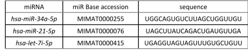
Table 1. MicroRNAs for QRT-PCR analysis in radiated cells.
miRNA miR Base accession sequence
hsa-miR-34a-5p MIMAT0000255 UGGCAGUGUCUUAGCUGGUUGU
hsa-miR-21-5p MIMAT0000076 UAGCUUAUCAGACUGAUGUUGA
hsa-let-7i-5p MIMAT0000415 UGAGGUAGUAGUUUGUGCUGUU
RESULTS
MDA-MB-231 and MCF-7 cells growing in steady state were treated with 2, 4 and 8 Gy doses of gamma rays and investigated at 4, 8, and 24 hours post irradiation for the specific microRNAs (miR-34a, miR-21, and Let-7i) expres-sion changes using Real-Time PCR.
The miRNAs expression after gamma irradiation in MCF-7 cells
The expression of miR-21 was induced in the MCF-7 cell line only 4 h after radiation, with a decline in expression at later time points in all doses. So, the miR-21 expression was not considerably changed at different irradiation doses during the experiment times compared to
miR-34a and let-7i, in this cell line. Accordingly,
the results illustrated that miR-34a was the most up-regulated miRNA post irradiation in MCF-7 cells. . Also, the expression of miR-34a increased to the maximum level at 24 h after radiation in all three doses. In fact, excluding 2Gy, 8h
permanent increase of miR-34a expression, in a dose- and time dependent fashion in the cells. Consequently, we observed considerable changes in expression of let-7i miRNA, at higher doses of 2 Gy, which peek at 8 h time point in 4 and 8 Gy transiently, with a decline in its expression at later time point (figure 1).
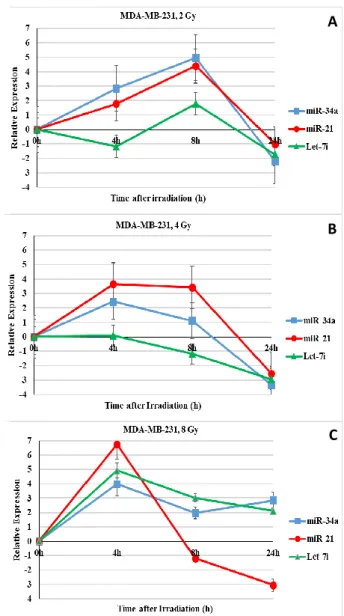
The miRNAs expression after gamma-irradiation in MDA-MB-231 cells
The results showed a similar expression pattern for miR-34a and miR-21 at 2 and 4 Gy in
MDA-MB-231 breast cancer cell line. Accordingly, these two miRNAs expression induced at 4 and 8 hours post irradiation but decreased at 24 hour. In 8 Gy, the expression of all three miRNAs especially miR-21 were strongly induced at 4 h post-irradiation. In addition, Let-7i microRNA expression was different depending on dose and time post irradiation (figure 2). Interestingly, the miR-21
increase was more significant in MDA-MB-231 cells compared to MCF-7 cells.
386
A
B
C
A
B
C
Figure 1. Relative expression of miR-34a, miR-21 and let-7i microRNAs in MCF-7 cells after exposure to different doses of
ɤ-rays compared to the un-irradiated sham corresponding control. The miRNAs expression was assessed at 0, 4, 8, and 24 h
post irradiation. The data is expressed as Log2 values. A. 2 Gy dose, B. 4 Gy dose, C. 8 Gy dose. (■) miR-34a, (●) miR-21,
(▲) Let-7i.
Figure 2. Relative expression of miR-34a, miR-21 and let-7i microRNAs in MDA-MB-231 cells after exposure to different
doses of ɤ-rays compared to the un-irradiated sham corresponding control. The miRNAs expression was assessed at 0, 4, 8, and 24 h post irradiation. The data is expressed as
Log2 values. A. 2 Gy dose, B. 4 Gy dose, C. 8 Gy dose. (■) miR-34a, (●) miR-21, (▲) Let-7i.
DISCUSSION
Today, the main reason of tumour treatment resistance is the breast cancer initiating cells (BCICs). It has been reported that 98 and 3.5 percentage of stem-like cells existed in MDA-MB -231 (14) and MCF-7 (15) cell lines, respectively. Since three specific miRNAs, miR-21, miR-34a
and let-7i are involved in DNA repair pathways,
apoptosis and cell cycle arrest (7, 9, 12), in this study, we analysed expression changes of them after gamma radiation in MDA-MB-231 and MCF -7 (IBRC C10082, MCF-7 type) cells, as breast cancer stem cells (BCSCs) and non-BCSC models, respectively.
It has been reported that IR-Induced miR-21 facilitates the repression of PTEN as its target, and the activation of PI3K/AKT to enhance the
DDR and cell survival (6, 7). In the study conducted by Wang and colleagues found that
decreased expression of Cdc25A by miR-21 led to cell cycle inhibiting to repair radiation-related cell damage (18). Therefore, miR-21 can act as a
radio-resistant miRNA when transiently over-expressed after radiation treatment (6).
Also, we detected miR-21 over-expression in
both MCF-7 and MDA-MB-231 cells post irradiation. Interestingly, up-regulation of
miR-21 was more significant in irradiated
MDA-MB-231 than MCF-7, which was only observed in primary hours.
Liu et al. (2011) reported that miR-34a could
be induced in several mice organs by radiation. They also showed that induction of miR-34a by radiation was in a p53 dependent manner, followed by down-regulation of anti-apoptotic molecule Bcl-2 that leads to apoptosis, cell-cycle arrest or senescence. On the other hands, the transcription factor encoded by the p53 tumour suppressor gene is post-transcriptionally activated by DNA damaging agents/radiation (9).
Accordingly, we found that miR-34a expression level was up-regulated by gamma irradiation in breast cancer cells (MCF-7 and MDA-MB-231). Interestingly, the over-expression of miR-34a
was much more significant in MCF-7 than MDA-MB-231, since MCF-7 cells harbour a wild
type functional p53 gene.
Chaudhry (2009) found that the expression levels of several members of the let-7 family miRNA were increased after ionizing radiation treatment in Jurkat cells but were decreased in TK6 cells (19). Taylor (2014) presented that elevated expression of let-7 in human malignancies elicits down-regulation of Ras
family members, HMGA2, and c-Myc, thereby suppressing the development, progression, and therapeutic resistance of human tumours (20). Also, we observed let-7i microRNA expression
changes following exposure to gamma irradiation were different as up- or down regulation depending on dose, time post irradiation and cell type, similar to other studies
that reported up- or down-regulation of let-7 family miRNAs expression in different gamma irradiation exposed cells (19).
we may be able sensitize somewhat stem-like MDA-MB-231 breast cancer cells to radiation by
increasing miR-34a and decreasing miR-21 expression levels simultaneously.
ACKNOWLEDGEMENTS
This study was funded by Tarbiat Modares University grant. Also, the authors wish to thank Department of Radiation Oncology of Tehran Uni-versity of Medical sciences for supporting this re-search.
Conflicts of interest: Declared none.
REFERENCES
1. Bombonati A and Sgroi DC (2011) The molecular pathology of breast cancer progression. J Pathol, 223: 308-18. 2. Gangopadhyay S, Nandy A, Hor P, Mukhopadhyay A (2013)
Breast cancer stem cells: a novel therapeutic target. Clin Breast Cancer, 13: 7-15.
3. Farazi TA, Spitzer JI, Morozov P, Tuschl T (2011) MiRNAs in human cancer. J Pathol, 223: 102-15.
4. Czochor JR and Glazer PM (2014) MicroRNAs in cancer cell response to ionizing radiation. Antioxid Redox Signal, 21: 293-312.
5. Zhu S, Wu H, Wu F, Nie D, Sheng S, Mo YY (2008) Mi-croRNA-21 targets tumor suppressor genes in invasion and metastasis. Cell Res, 18: 350-9.
6. Frankel LB, Christoffersen NR, Jacobsen A, Lindow M, Krogh A, Lund AH (2008) Programmed cell death 4 (PDCD4) is an important functional target of the microRNA
miR-21 in breast cancer cells. J Biol Chem, 283: 1026-33. 7. Anastasov N, Höfig I, Vasconcellos IG, Rappl K, Braselmann
H, Ludyga N, et al (2012) Radiation resistance due to high expression of miR-21 and G2/M checkpoint arrest in breast cancer cells. Radiat Oncol, 7: 206.
8. Hermeking H. The miR-34 family in cancer and apoptosis (2010) Cell Death Differ, 17: 193-9.
9. Liu C, Zhou C, Gao F, Cai S, Zhang C, Zhao L, et al. (2011)
MiR-34a in age and tissue related radio-sensitivity and serum miR-34a as a novel indicator of radiation injury. Int J Biol Sci, 7: 221-33.
10. Kang L, Mao J, Tao Y, Song B, Ma W, Lu Y, et al. (2015) MicroRNA-34a suppresses the breast cancer stem cell-like characteristics by downregulating Notch1 pathway. Cancer Sci, 106: 700-8.
11. Kumar MS, Erkeland SJ, Pester RE, Chen CY, Ebert MS, Sharp PA, et al. (2008) Suppression of non-small cell lung
Natl Acad Sci USA, 105: 3903-8.
12. Johnson CD, Esquela-Kerscher A, Stefani G, Byrom M, Kelnar K, Ovcharenko D, et al. (2007) The let-7 microRNA represses cell proliferation pathways in human cells. Can-cer Res, 67: 7713-22.
13. Yu F, Yao H, Zhu P, Zhang X, Pan Q, Gong C, et al (2007) Let
-7 regulates self renewal and tumorigenicity of breast cancer cells. Cell, 131: 1109-23.
14. An H, Kim JY, Oh E, Lee N, Cho Y, Seo JH (2015) Salinomy-cin promotes anoikis and decreases the CD44+/CD24- stem -like population via inhibition of STAT3 activation in MDA-MB-231 Cells. PLoS One, 10: 1-17.
15. Chen L, Xiao Z, Meng Y, Zhao Y, Han J, Su G, et al (2012) The enhancement of cancer stem cell properties of MCF-7 cells in 3D collagen scaffolds for modeling of cancer and anti-cancer drugs. Biomaterials, 33: 1437-44.
16. Gwak H, Kim HT, Jo HG, Kim Y, Kwak H, Kim HJ (2012)
Si-lencing of MicroRNA-21 Confers Radio-Sensitivity through Inhibition of the PI3K/AKT Pathway and Enhancing Au-tophagy in Malignant Glioma Cell Lines. PLoS One, 7: 1-12. 17. Livak J K and Schmittgen DT (2001) Analysis of relative
gene expression data using Real-Time quantitative PCR and the 2-△ △CT method. Methods, 25: 402–408.
18. Wang P, Zou F, Zhang X, Li H, Dulak A, Tomko RJ, et al.
(2009) MicroRNA-21 negatively regulates Cdc25A and cell cycle progression in colon cancer cells. Cancer Res, 69: 8157-65.
19. Chaudhry MA (2009) Real-time PCR analysis of micro-RNA expression in ionizing radiation-treated cells. Cancer Bi-other Radiopharm, 24: 49-56.
20. Taylor MA, Schiemann WP (2014) Therapeutic opportuni-ties for targeting microRNAs in cancer. Mol Cell Ther, 2: 30.