Effect of talocrural joint mobilizations on restricted ankle dorsiflexion and the kinematics of squatting tasks
Molly Smith
A thesis submitted to the faculty of the University of North Carolina at Chapel Hill in partial fulfillment of the requirements for the degree of Master of Arts in the Department of Exercise & Sport Science (Athletic Training) in the College of Arts & Sciences.
Chapel Hill 2013
Approved by:
© 2013 Molly Smith
Joint mobilization treatments aimed at increasing ankle dorsiflexion range of motion (DF-ROM) may affect DF-ROM and squat kinematics in healthy subjects with restricted dorsiflexion. Measures of DF-ROM and squat kinematics (knee valgus displacement, medial knee displacement, and dorsiflexion displacement) were assessed in 43 subjects. Subjects were randomly assigned to a control (calf stretching and sham mobilization) or treatment (calf stretching, mobilization with movement treatment, and anterior to posterior talocrucal joint mobilization) group. All subjects, regardless of group, demonstrated significantly improved DF-ROM at post testing. During squatting tasks, dorsiflexion displacement increased significantly from pre- to post-testing in both double and single leg squats. No significant differences were observed for knee valgus displacement or medial knee displacement. Thus, calf stretching improved passive and active dorsiflexion range of motion in subjects with dorsiflexion restrictions. Joint mobilizations did not have an additive effect on dorsiflexion gains nor affect squatting kinematics at the knee.
ABSTRACT
MOLLY SMITH: Effect of talocrural joint mobilizations on restricted ankle dorsiflexion and the kinematics of squatting tasks
TABLE OF CONTENTS
LIST OF FIGURES ... VII LIST OF TABLES ... VIII
CHAPTER I ... 1
BACKGROUND ... 1
INDEPENDENT VARIABLES ... 6
DEPENDENT VARIABLES ... 6
RESEARCH QUESTIONS AND HYPOTHESES ... 6
STATISTICAL HYPOTHESES ... 8
OPERATIONAL DEFINITIONS ... 8
ASSUMPTIONS ... 9
DELIMITATIONS ... 9
LIMITATIONS ... 9
SIGNIFICANCE OF THE STUDY ... 10
CHAPTER II ... 11
INTRODUCTION ... 11
RELEVANT ANATOMY ... 11
Knee ... 11
Ankle ... 15
Non-Contact Anterior Cruciate Ligament (ACL) Injuries ... 17
Risk Factors ... 17
ACL Reconstruction Prognosis ... 19
Knee Osteoarthritis ... 19
Ankle Dorsiflexion and Knee Injuries... 20
ACL Prevention Programs ... 21
ANKLE CONDITIONS ... 21
Acute Ankle Sprains ... 21
Chronic Ankle Instability ... 22
Ankle Dorsiflexion and Ankle Injuries ... 22
CAUSES OF RESTRICTED DORSIFLEXION ... 23
TALAR POSITION,GLIDE, AND LAXITY ... 24
Talar Position ... 25
Posterior Talar Glide ... 25
Talar Laxity ... 26
AREAS OF NEEDED RESEARCH ... 27
CONCLUSION ... 27
CHAPTER III ... 28
SUBJECTS ... 28
Inclusion criteria ... 28
Exclusion criteria ... 28
MEASUREMENT AND INSTRUMENTATION ... 29
Equipment ... 29
PROCEDURES ... 31
Screening Session ... 31
Testing Session ... 33
DATA PROCESSING AND REDUCTION ... 36
DATA ANALYSIS ... 37
CHAPTER IV... 38
OVERVIEW ... 38
INTRODUCTION ... 39
METHODS ... 41
RESULTS ... 46
DISCUSSION ... 47
FIGURES ... 52
TABLES ... 57
APPENDICES ... 64
LIST OF FIGURES
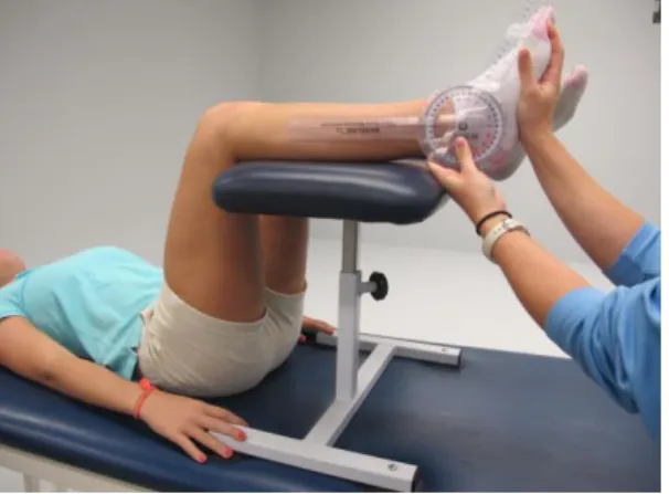
Figure 1: Dorsiflexion Range of Motion Measure with Knee Extended………..52
Figure 2: Dorsiflexion Range of Motion Measure with Knee Flexed... …………...52
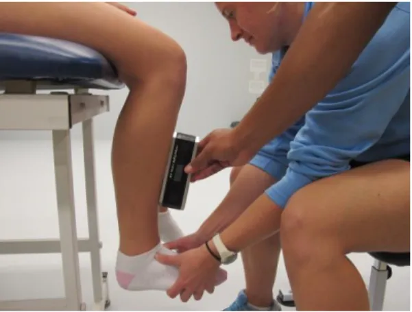
Figure 3: Weight-Bearing Lunge………..53
Figure 4: Posterior Talar Glide Test for Talar Laxity………...53
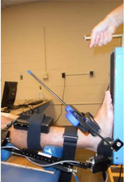
Figure 5: Ankle Arthrometer Test for Ankle Stiffness……….54
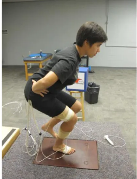
Figure 6: Double Leg Squat with Electromagnetic Motion Capture Sensors…………...54
Figure 7: Single Leg Squat with Electromagnetic Motion Capture Sensors………55
Figure 8: Mulligan with Movement Joint Mobilization Treatment………..55
Figure 9: Sham Mobilization Treatment………...56
LIST OF TABLES
Table 1: Statistical Methods………..57 Table 2: Intraclass Correlation Coefficients and Standard Error of the Measurement….58 Table 3: Group Characteristics Presented As Means ± SD For Each Group………59 Table 4: Ankle Dorsiflexion Range of Motion (Degrees) presented as
Means ± SD (95% Confidence Intervals) for each group at Pre
and Post time point………..60 Table 5: Ankle Laxity (Degrees) and Stiffness (mm) presented as Means
± SD (95% Confidence Intervals) for each group at Pre and Post
Time point………...61 Table 6: Knee and Ankle Kinematics During the Double Leg Squatting
Task presented as means ± SD (95% Confidence Intervals) for
each Group at Pre and Post time points.… ………62 Table 7: Knee and Ankle Kinematics During the Single Leg Squatting
Task presented as means ± SD (95% Confidence Intervals) for
CHAPTER I INTRODUCTION
BACKGROUND
Recreational and competitive sports are widely popular in the United States, and while an active lifestyle is healthy, sports can also cause injuries. Common injuries from sports such as running, basketball, and soccer include acute knee injuries, acute ankle sprains, and chronic ankle instability (CAI). Such injuries can be painful, expensive, and may lead to altered lower extremity biomechanics, permanent disability, and the development of early osteoarthritis.
for further reconstructions or to degenerative joint disease (Freedman, Glasgow et al. 1998; Griffin, Albohm et al. 2006). It is estimated that surgery and rehabilitation for each ACL injury costs approximately $11,000-17,000, for a total of millions of dollars spent annually because of ACL injuries (Hewett, Myer et al. 2005; Gianotti, Marshall et al. 2009). ACL injuries may also cause increased risk of knee osteoarthritis, with up to 50% of people with reconstructed ACLs showing signs of articular degeneration 15 years after surgery (Lohmander, Ostenberg et al. 2004; Meunier, Odensten et al. 2007; Roos, Englund et al. 2007; Hui, Salmon et al. 2011).
Ankle injuries are the most common lower extremity injury in the recreational and athletic settings with more than 25,000 ankle sprains occurring daily in the United States (Mickel, Bottoni et al. 2006; Wikstrom and Hubbard 2010). The greatest predisposing factor for ankle sprains is a history of at least one ankle sprain, and suffering repetitive ankle sprains can lead to chronic ankle instability (Milgrom, Shlamkovitch et al. 1991; Bahr and Bahr 1997; McKay, Goldie et al. 2001; Hertel 2002; Beynnon, Webb et al. 2004). The recurrence rate of ankle sprains is greater than 70%, and up to 75% of people who sprain their ankle develop some level of chronic functional ankle instability (Wikstrom and Hubbard 2010). Repetitive ankle sprains have also been linked to an increased risk of osteoarthritis and articular degeneration at the ankle (Harrington 1979; Hertel 2002).
Decreased or restricted dorsiflexion predisposes athletes to patellar tendinopathy and has been shown to alter biomechanics potentially contributing to injury (Malliaras, Cook et al. 2006; Backman and Danielson 2011). For example, decreased dorsiflexion range of motion has been associated with factors that increase ACL injury risk during a jump landing task. These include less knee-flexion displacement, greater knee-valgus displacement and greater ground reaction forces (Fong, Blackburn et al. 2011). Similarly, decreased dorsiflexion range of motion is associated with increased frontal plane knee excursion during a drop land task in young female soccer players (Sigward, Ota et al. 2008). It has also been found that affording individuals more ankle dorsiflexion with the use of a heel lift during a squat eliminated the presence of medial knee displacement (MKD), which is associated with tight and weak ankle musculature and can increase the risk of ACL injury and patellofemoral pain syndrome (Bell, Padua et al. 2008). However, there seem to be a number of factors that can contribute to restricted dorsiflexion range of motion.
et al., 2002; T. J. Hubbard, Kaminski, Vander Griend, & Kovaleski, 2004; Nauck, Lohrer, & Gollhofer, 2010).
Since dorsiflexion range of motion is related to a variety of lower extremity injuries, an accurate measurement of dorsiflexion range of motion is essential to identify deficits and create injury prevention and intervention strategies. Measures of lower extremity range of motion can be taken passively, actively, or functionally. While passive and active range of motion measurements are easier clinical measures that allow for identification of range of motion impairments and tracking of changes over time, functional measurements may allow for a better representation of how the individual moves during physical activity. Double and single leg squat tasks represent functional lower extremity movements and provide information on a number of variables including functional ankle dorsiflexion and medial knee displacement. Double and single leg squatting tasks have been used in a variety of research studies looking at variables such as dorsiflexion motion, muscle strength, and neuromuscular characteristics (Bell, Padua, & Clark, 2008; Padua, In Review; Macrum, In Review; (Dill, Begalle et al. In Review).
passive dorsiflexion range of motion, talar laxity, and double and single leg squat kinematics in subjects with restricted dorsiflexion.
INDEPENDENT VARIABLES Group
o Control Group: Stretching plus sham mobilization o Intervention Group: Stretching plus joint mobilization Time
o Pre-treatment o Post-treatment DEPENDENT VARIABLES
Passive dorsiflexion range of motion o Weight-bearing lunge
o Passive, knee extended o Passive, knee flexed Posterior talar laxity
Ankle stiffness
o Anterior-posterior o Medial-lateral
Double and single leg squat knee and ankle kinematics o Dorsiflexion displacement
o Medial knee displacement o Knee valgus displacement
RESEARCH QUESTIONS AND HYPOTHESES
Research Question #1: Is there a significant difference between the effect of ankle joint mobilizations plus stretching and stretching alone on measures of passive range of motion, ankle stiffness, and posterior talar laxity in individuals with restricted dorsiflexion ROM?
o Research Question #1a: Is there a significant difference between the effect of ankle joint mobilizations plus stretching and stretching alone on
measures of passive range of motion?
Research Hypothesis #1a: There will be significant increases in measures of passive range of motion for both groups, and
o Research Question #1b: Is there a significant difference between the effect of ankle joint mobilizations plus stretching and stretching alone on
measures of ankle stiffness?
Research Hypothesis #1b: There will be a significant increase between the joint mobilization group and the stretching group on measures of ankle stiffness.
o Research Question #1c: Is there a significant difference between the effect of ankle joint mobilizations plus stretching and stretching alone on
measures of posterior talar laxity?
Research Hypothesis #1c: There will be a significant increase between the joint mobilization group and the stretching group on measures of posterior talar laxity.
Research Question #2: Is there a significant difference between the effect of ankle joint mobilizations plus stretching and stretching alone on measures of
dorsiflexion displacement and medial knee displacement during double and single leg squatting tasks in individuals with restricted dorsiflexion ROM?
o Research Question #2a: Is there a significant difference between the effect of ankle joint mobilizations plus stretching and stretching alone on
measures of dorsiflexion displacement during double and single leg squatting tasks in individuals with restricted dorsiflexion ROM?
Research Hypothesis #2a: There will be significant increases in measures of dorsiflexion displacement during double and single leg squatting tasks for both groups, and significantly greater increases among the joint mobilization group than the stretching only group.
o Research Question #2b: Is there a significant difference between the effect of ankle joint mobilizations plus stretching and stretching alone on
measures of medial knee displacement during double and single leg squatting tasks in individuals with restricted dorsiflexion ROM?
Research Hypothesis #2b: There will be significant decreases in measures of medial knee displacement during double and single leg squatting tasks for both groups, and significantly greater decreases among the joint mobilization group than the stretching only group.
o Research Question #2c: Is there a significant difference between the effect of ankle joint mobilizations plus stretching and stretching alone on
measures of knee valgus displacement during double and single leg squatting tasks in individuals with restricted dorsiflexion ROM?
squatting tasks for both groups, and significantly greater decreases among the joint mobilization group than the stretching only group.
STATISTICAL HYPOTHESES Statistical Hypothesis #1
o H0: EXP=CON
o HA: EXP≠CON
o HR1a:EXP >CON
o HR1b:EXP >CON
o HR1c:EXP >CON
Statistical Hypothesis #2 o H0: EXP=CON
o HA: EXP≠CON
o HR2a:EXP >CON
o HR2b:EXP <CON
o HR2c:EXP <CON
OPERATIONAL DEFINITIONS
Healthy subject: Subjects that have no history of lower extremity surgery, no history of knee or ankle injury in the past six months (i.e. an injury that caused the subject to refrain from activity from two or more days), and are not currently doing rehabilitation on any ankle or knee injuries.
Double leg squat: Participants perform a squat maneuver, beginning with their feet shoulder-width apart, toes pointing straight ahead, and arms extended over their head. Subjects then flex their knees such as when sitting into a chair, to a depth of at least 60 degrees of knee flexion.
Single leg squat: Participants perform a single leg squat maneuver, beginning by standing on their dominant leg with their hands on their waist and their non-dominant leg flexed to 45 degrees at the hip and 90 degrees at the knee. Subjects will then squat to a depth of at least 60 degrees of knee flexion.
Restricted dorsiflexion: Equal to or less than 40 degrees of passive dorsiflexion measured with the weight-bearing lunge test.
Talocrural joint mobilization treatment: A single treatment session of 3-30 second bouts of a Mulligan’s mobilization with movement (MWM) talocrural joint mobilizations.
Sham mobilization treatment: A single treatment session of 3-30 second bouts of a sham mobilization consisting of passive knee flexion with the ankle held in a neutral orthoplast splint.
Peak knee valgus: The maximum frontal plane knee angle that occurs during the descent phase of a double or single leg squatting task.
Knee valgus displacement: The difference between initial knee valgus angle and the peak knee valgus angle that occurs during the descent phase of a double or single leg squatting task.
Medial knee displacement: The difference between initial frontal plane knee angle and the peak frontal plane knee angle that occurs during the descent phase of a double or single leg squatting task.
Peak ankle dorsiflexion: The maximum ankle dorsiflexion angle that occurs during the descent phase of a double or single leg squatting task.
Ankle displacement: The difference between initial ankle dorsiflexion angle and the peak ankle dorsiflexion angle that occurs during the descent phase of a double or single leg squatting task.
ASSUMPTIONS
The use of a standard goniometer to measure passive range of motion is representative of the joint’s range of motion.
All subjects will truthfully report current and past medical conditions which may exclude them from the study.
The testing equipment will not prevent normal body motions.
DELIMITATIONS
All subjects will be healthy, uninjured students at the University of North Carolina, Chapel Hill.
Subjects will be classified as having restricted dorsiflexion based on the criteria established by previous research.
LIMITATIONS
The motion of the posterior talar laxity test and ankle arthrometer test are similar to a joint mobilization movement.
Subjects may participate in activity or stretching between the pre-testing screening session to determine inclusion and the testing session.
The individual effort put into correctly performing the double and single leg squatting tasks cannot be assessed.
SIGNIFICANCE OF THE STUDY
CHAPTER II
REVIEW OF THE LITERATURE
INTRODUCTION
Common injuries in sport involve acute knee injuries, chronic knee pain, acute ankle sprains, and chronic ankle instability. Such injuries can be expensive and painful and may lead to disability and osteoarthritis. Thus, prevention of knee and ankle injuries is important. Both knee and ankle injuries have been linked to decreased ankle dorsiflexion range of motion. This study investigated the effect of treatments commonly used to increase dorsiflexion range of motion: stretching and joint mobilizations, in an effort to correct risky movement patterns and prevent common lower extremity injuries.
RELEVANT ANATOMY
Understanding the functional anatomy of the lower extremity is crucial for understanding the biomechanics, kinematics, and relationship between the knee and ankle during lower extremity movement. Dynamic and static stabilizers of both the knee and ankle joints are of interest for this investigation.
Knee
the medial femoral condyle is larger and more distal than the lateral condyle (Chhabra, Elliott et al. 2001). This asymmetry of the femoral condyles is an important component of the “screw home” mechanism, which occurs during the final 30 degrees of knee extension (Voight, Hoogenboom et al. 2007). The “screw home” mechanism refers to the internal rotation of the femur on the tibia as the knee extends in a closed chain, due to the larger size of the medial condyle. The “screw home” mechanism locks the knee into full extension and provides additional stability to the fully extended knee (Prentice 2004). The patella is a sesamoid bone that articulates with the femur and serves the purpose of increasing the quadriceps moment arm, providing continuity between the quadriceps tendon and patellar tendon, protecting the knee joint, and reducing pressure on the patellar tendon (Kaufer 1971; Chhabra, Elliott et al. 2001).
The femur and tibia are separated by two menisci; crescent-shaped fibrocartilaginous structures which serve to improve bony congruency between the round femoral condyles and the flat tibial plateau (Lee and Fu 2000). The menisci also assist with load bearing, shock absorption, joint stability, joint lubrication, and proprioception (Lee and Fu 2000; Englund 2008). The large, C-shaped medial meniscus is tightly attached to the tibia, joint capsule, and medial collateral ligament (Chhabra, Elliott et al. 2001). The medial meniscus is more commonly injured due to its tight attachments and inability to slide out of the way of large loads. The smaller, O-shaped lateral meniscus is an important weight-bearing structure that is attached to the tibia via the ligaments of Humphrey and Wrisberg and the popliteus tendon (Lee and Fu 2000).
the anterior cruciate ligament, posterior cruciate ligament, medial collateral ligament, and lateral collateral ligament (Hughston, Andrews et al. 1976). The anterior cruciate ligament (ACL) resists anterior translation of the tibia on the femur, prevents knee hyperextension, provides rotary and varus/valgus stability, and guides tibial and femoral motion during flexion and extension (Voight, Hoogenboom et al. 2007). The ACL extends from the posterior lateral femoral condyle to the anterior tibial spine and is comprised of two bundles which wrap around each other and have varying tautness depending on the knee flexion angle: the anteromedial bundle is tight in knee flexion and the posterolateral bundle is tight in knee extension (Girgis, Marshall et al. 1975; Hughston, Andrews et al. 1976; Arnoczky 1983). At any position of the knee, a portion of the ACL is under tension and is functional. The ACL is intraarticular, extrasynovial, and receives vascularization from the middle geniculate artery (Arnoczky 1983).
The posterior cruciate ligament (PCL) extends from the posterior medial femoral condyle to the inter-articular surface of the tibia and resists posterior translation of the tibia on the femur as well as extreme varus/valgus and rotation motion (Girgis, Marshall et al. 1975). The PCL consists of two bundles: the larger anterolateral bundle, which is tight in knee flexion, and the smaller posteromedial bundle, which is tight in knee extension. The PCL is also intraarticular and extrasynovial and receives vascular supply from the middle geniculate artery. The PCL is assisted with preventing posterior translation of the tibia by the ligaments of Humphrey and Wrisberg, which are not present in all knees (Chhabra, Elliott et al. 2001; Voos, Mauro et al. 2012).
posterior to the pes anserinus insertion and just inferior to the tibial articular surface (Chhabra, Elliott et al. 2001). Like the ACL and PCL, the MCL has two portions: the superficial medial collateral ligament and the deep medial capsular ligament (Warren and Marshall 1979).
The lateral aspect of the knee is stabilized primarily by the lateral collateral ligament (LCL), which resists varus stress and external rotation of the knee. The LCL extends from the lateral femoral condyle to the fibular head and does not have a connection to the joint capsule (Chhabra, Elliott et al. 2001; Voight, Hoogenboom et al. 2007). Other posterolateral stabilizers of the knee that work to resist posterior translation, external rotation, and varus forces include the iliotibial (IT) band, biceps femoris, patellar retinaculum, patellofemoral ligaments, popliteus tendon, popliteofibular ligament, arcuate ligament, fabellofibular ligament, and joint capsule (Chhabra, Elliott et al. 2001).
aspera of the femur. The biceps femoris has two heads and inserts on the lateral tibial condyle (short head) and the fibular head and lateral tibia (long head). The semimembranosus has many distal attachments, including the oblique popliteal ligament, posterior capsule, posterior tibia, popliteus, and medial meniscus. The semitendinosus joins with the gracilis and sartorius tendons to form the pes anserinus tendon, which attaches on the anteromedial tibia and stabilizes against valgus forces. The IT band is a continuation of the tensor fascia latae and inserts at Gerdy’s tubercle on the anterolateral aspect of the tibia. The IT band assists with knee flexion and stabilizes against anterior tibial translation and varus force (Chhabra, Elliott et al. 2001; Voight, Hoogenboom et al. 2007). Furthermore, the popliteus muscle and gastrocnemius/soleus complex provide additional stabilization against anterior and posterior tibial translation, varus forces, and antero-/posterolateral rotational instabilities (Voight, Hoogenboom et al. 2007).
Ankle
(Hertel 2002). The ankle complex is relatively stable due to bony congruency, static ligamentous support, and dynamic muscular restraints (Hertel 2002; Prentice 2004).
The talocrural joint, which is sometimes referred to as the ankle joint, is a hinge joint formed from the articulation of the talar dome, medial malleolus, tibial plafond, and lateral malleolus and allows dorsiflexion and plantarflexion movements. The talocrural joint receives ligamentous stability from the articular capsule, deltoid, anterior talofibular, posterior talofibular, and calcaneofibular ligaments (Hertel 2002; Prentice 2004; Taser, Shafiq et al. 2006). The anterior talofibular ligament (ATFL) is the most commonly injured ankle ligament (Hertel 2002). The subtalar joint consists of the articulation between the talus and the calcaneus and allows pronation and supination motions (Hertel 2002; Prentice 2004). This articulation is supported by an extensive network of ligaments that are not well understood. Ligaments can be categorized as deep, peripheral, and retinacular and work to provide stability to this joint. The distal tibiofibular joint is a syndesmosis that allows for accessory gliding between the tibia and fibula, which is essential for normal ankle mechanics. This joint is stabilized by the interosseous membrane and the anterior and posterior tibiofibular ligaments. Little motion occurs at this joint, but it can be injured during eversion ankle injuries (Hertel 2002).
slowing the plantarflexion component of supination and thus preventing lateral ligament injury (Hertel 2002).
KNEE CONDITIONS
Non-Contact Anterior Cruciate Ligament (ACL) Injuries
Each year between 80,000 and 250,000 ACL injuries occur and 70% of such injuries are due to non-contact mechanisms, which may be preventable (Hewett, Myer et al. 2005; Griffin, Albohm et al. 2006). ACL tears are most common among young athletes 15 to 25 years of age. Furthermore, female athletes are more susceptible than male athletes (Arendt, Agel et al. 1999; Griffin, Albohm et al. 2006). Each ACL injury costs approximately $17,000 for a total of between $1.3 and $4.3 billion annually for ACL surgery and rehabilitation (Hewett, Myer et al. 2005).
One mechanism of non-contact ACL tears is during tibial external rotation, when there is slack in the ACL. The ACL can impinge on the lateral femoral condyle, which causes a shearing force on the ACL and can result in a tear. This position of tibial external rotation is common in sports involving cutting tasks such as basketball and soccer (Olsen, Myklebust et al. 2004; Bahr and Krosshaug 2005).
Risk Factors
dynamic knee valgus, internal tibial rotation, and foot pronation have been linked to increased incidence of ACL injury (Woodford-Rogers, Cyphert et al. 1994; Loudon, Jenkins et al. 1996; Allen and Glasoe 2000; Ford, Myer et al. 2003; Hewett, Myer et al. 2005).
Neuromuscular risk factors also play a role in ACL injury. Research has analyzed jump landing, cutting, and pivoting tasks in an effort to identify risky movement patterns. Movements with decreased knee flexion, decreased hip flexion, increased knee valgus, increased hip internal rotation, increased tibial internal or external rotation, less hamstring stiffness, and quadriceps dominant contractions may all play a role in increased ACL injury risk (Huston and Wojtys 1996; Aune 1997; Colby, Francisco et al. 2000; Besier, Lloyd et al. 2001; Malinzak, Colby et al. 2001; Chappell, Yu et al. 2002; Lephart, Ferris et al. 2002; Decker, Torry et al. 2003; Pollard, Davis et al. 2004; McLean, Walker et al. 2005; Padua, Carcia et al. 2005). While risky movement patterns may be inherently present in certain individuals, fatigue may also cause altered movement patterns and increase ACL injury susceptibility (Chappell, Daniel et al. 2005). Muscle stiffness and its relationship to ACL injury is being investigated. Gender differences have been identified for hamstring stiffness, with females exhibiting decreased hamstring stiffness compared to males, which may be a factor in increased female ACL injury (Kibler and Livingston 2001; Granata, Padua et al. 2002; Granata, Wilson et al. 2002; Wojtys, Ashton-Miller et al. 2002; Wojtys, Huston et al. 2003; Blackburn, Norcross et al. 2011).
control of the hip and knee during a dynamic landing task and postural stability task (Paterno 2010).
ACL Reconstruction Prognosis
ACL reconstruction surgery is common after an ACL tear, and while surgeries are often successful, they do not guarantee a successful return to sport or even to normal knee function. Fifteen years after ACL reconstruction with patellar tendon autograft, nearly 60% of patients had further ACL injury either to the reconstructed knee or the contralateral knee (Hui, Salmon et al. 2011). However, positive outcomes have also been identified, with 97% of patients reported normal or near-normal knee function 10 years after ACL reconstruction (Pinczewski, Lyman et al. 2007).
Knee Osteoarthritis
Fifteen years after ACL reconstructive surgery, approximately 50% of people have been shown to have radiographic evidence of OA and 12 years after ACL rupture, 75% of female soccer players reported significant symptoms that affected their knee-related quality of life (Lohmander, Ostenberg et al. 2004; Meunier, Odensten et al. 2007; Roos, Englund et al. 2007; Hui, Salmon et al. 2011).
Ankle Dorsiflexion and Knee Injuries
ACL Prevention Programs
A variety of ACL injury prevention programs exist and include many tasks such as stretching, strengthening, aerobic conditioning, agilities, plyometrics, and risk awareness training focusing on soft landings, control on landing, dynamic balance, and agility skills (Griffin, Albohm et al. 2006). Prevention programs aim to decrease risk factors and reduce non-contact ACL injuries. Thus far, dorsiflexion range of motion and gastrocnemius/soleus flexibility have not been a focus in injury prevention programs. This study aims to determine if range of motion efforts should be included in existing ACL prevention programs.
ANKLE CONDITIONS Acute Ankle Sprains
Chronic Ankle Instability
Chronic ankle instability (CAI) refers to repetitive bouts of lateral ankle instability, leading to numerous ankle sprains (Hertel 2002). CAI may be caused by mechanical instability, functional instability, or a combination of both. Mechanical instability is caused by altered mechanics within the ankle complex and is the result of pathologic ankle ligament laxity, impaired arthrokinematics, synovial inflammation and impingement, and degenerative changes. Functional instability is the recurrence of ankle instability and the feeling of joint instability due to proprioceptive and neuromuscular control, postural control, and/or strength deficits (Hertel 2002). Of patients with previous ankle sprains, 32-74% report some type of chronic symptoms, and 32-47% report some level of functional ankle instability (i.e. sense of giving way) (Arnold, Wright et al. 2011). Repetitive sprains have also been linked to an increased risk of osteoarthritis and articular degeneration at the ankle, and a previous history of at least one ankle sprain represents the greatest predisposing factor for subsequent ankle sprains (Harrington 1979; Milgrom, Shlamkovitch et al. 1991; Bahr and Bahr 1997; McKay, Goldie et al. 2001; Beynnon, Murphy et al. 2002; Hertel 2002).
Ankle Dorsiflexion and Ankle Injuries
may have been lacking dorsiflexion in both ankles prior to injury (Tabrizi, McIntyre et al. 2000; Hertel 2002). It is thought that decreased dorsiflexion could predispose individuals to ankle sprains, because if the talocrural joint is unable to fully dorsiflex, the joint will remain in an open-packed position during movement and will be able to invert and internally rotate more easily, thus increasing risk of ankle sprains (Hertel 2002). Ankle dorsiflexion motion has also been shown to be decreased after acute inversion ankle sprains and in people with CAI (Drewes, McKeon et al. 2009; Youdas, McLean et al. 2009). Another study, however, found no link between abnormal ankle range of motion and injury in a group of dancers (Wiesler, Hunter et al. 1996).
CAUSES OF RESTRICTED DORSIFLEXION
can restore arthrokinematic motion, improve joint mobility, and allow full, pain-free range of motion at a joint (Prentice 2004). Joint mobilizations have been shown to increase range of motion and decrease pain after ankle sprains and ankle immobilization, and in people with CAI (Green, Refshauge et al. 2001; Collins, Teys et al. 2004; Reid, Birmingham et al. 2007; Landrum, Kelln et al. 2008; Hoch and McKeon 2010). There are two types of talocrural joint mobilizations generally used by practitioners; passive anterior-posterior talocrucal mobilizations and Mulligan’s mobilization with movement (MWM) talocrucal joint mobilizations. Both passive and MWM mobilizations have been shown to increase ankle range of motion, and it has been proposed that MWM treatments effectively change joint mechanics by allowing proper posterior talar gliding during ankle dorsiflexion (Green, Refshauge et al. 2001; Collins, Teys et al. 2004; Vicenzino, Branjerdporn et al. 2006; Reid, Birmingham et al. 2007; Landrum, Kelln et al. 2008; Hoch and McKeon 2010). TALAR POSITION, GLIDE, AND LAXITY
Talar Position
It has been speculated that the mechanism of an inversion ankle sprain may cause the talus to sublux anteriorly on the tibia (Mulligan 2004). The talus has no muscular attachments; therefore if the ligaments that hold it in place are slack, injured, or ruptured, they may allow movement of the talus (Denegar, Hertel et al. 2002). An anterior positional fault of the talus would lead to altered arthrokinematics and osteokinematics of the ankle, and thus cause restricted dorsiflexion (Mulligan 2004; Wikstrom and Hubbard 2010). Studies have shown talar position to be more anterior in CAI limbs than in non-CAI limbs and in individuals with CAI than in healthy individuals (Hubbard, Olmsted-Kramer et al. 2005; Wikstrom and Hubbard 2010). Anterior fibular positional faults have also been found in subjects with ankle sprains (Hubbard and Hertel 2008). Joint mobilizations may correct bony positional faults, which can explain why joint mobilizations cause an increase in ankle dorsiflexion (Green, Refshauge et al. 2001; Collins, Teys et al. 2004; Reid, Birmingham et al. 2007; Landrum, Kelln et al. 2008; Hoch and McKeon 2010). Positional faults may also be seen in healthy ankles with restricted dorsiflexion, and may be a cause of dorsiflexion restriction in uninjured individuals.
Posterior Talar Glide
dorsiflexion range of motion measures. This suggests that normal range of motion may be achieved with extensive calf musculature stretching (Denegar, Hertel et al. 2002; Hertel 2002). This may mean, however, that the ankle adopted an abnormal axis of rotation in order to achieve full motion, which could possibly lead to future injury or joint dysfunction. The impact of this joint dysfunction has not been explored thus far, but it suggests that joint mobilization is needed to restore proper ankle motion (Denegar, Hertel et al. 2002). A study looking at the effects of a Mulligan’s mobilization with movement joint mobilization determined that the mobilization restored normal posterior talar glide and increased ankle dorsiflexion (Vicenzino, Branjerdporn et al. 2006).
The relationship between posterior talar glide and anterior talar position has not been studied, but it is hypothesized that an anteriorly displaced talus would lead to excessive anterior talar glide and restricted posterior talar glide, which would cause further abnormal arthrokinematics and/or loss of motion in both healthy and injured populations (Denegar, Hertel et al. 2002).
Talar Laxity
AREAS OF NEEDED RESEARCH
While a significant amount of research exists regarding knee and ankle injuries, risk factors, and treatment techniques, there are still many areas where further research is needed. Future research should compare interventions, which address both soft tissue (stretching) and bony involvement (joint mobilizations) in dorsiflexion restriction. Additionally, future studies could examine the effect of ankle joint mobilizations on ankle and knee kinematics during functional movement patterns. The concept of a talar positional fault is accepted among some therapists, but studies are still inconclusive on the presence of this fault. Further investigation into the presence of an anterior talar positional fault is needed. Lastly, much of the research regarding dorsiflexion restriction uses injured ankles. Research is needed to determine the effects of joint mobilizations on healthy subjects with restricted dorsiflexion in order to determine if joint mobilizations can be used as an injury prevention strategy.
CONCLUSION
CHAPTER III METHODOLOGY
SUBJECTS
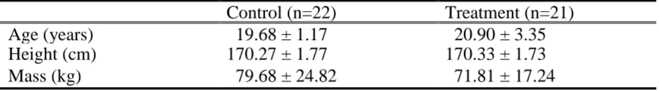
A total of forty-three individuals (23 females, 20 males) were selected for this study from a larger group of individuals who volunteered for the study. All participants were selected from a convenience sample of students from the University of North Carolina at Chapel Hill. Subjects were randomly assigned to either the control group or the treatment group. The control group contained twenty-two participants, twelve females and ten males; the treatment group contained twenty-one participants, eleven females and ten males. There were no significant differences between groups for height, weight, and age.
Inclusion criteria
All study participants were between 18 and 35 years old. All participants self-reported being in good physical condition and physically active, defined as consistent participation in at least 90 minutes of physical activity a week for the past six months. Subjects had 40 degrees or less of passive dorsiflexion during a weight-bearing lunge test. This cut-off point has been shown to be significant (Dill, Begalle et al. In Review).
Exclusion criteria
ankle or knee injuries. Subjects were also excluded if they have greater than 40 degrees of passive dorsiflexion during a weight-bearing lunge test. Additional exclusion criteria included any known vestibular, balance, or neurological disorder.
MEASUREMENT AND INSTRUMENTATION Equipment
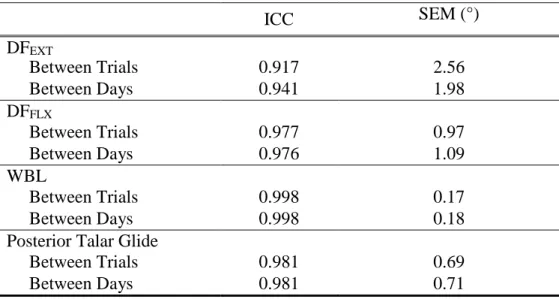
Ankle dorsiflexion range of motion was measured on the dominant limb of each subject in three positions; the weight-bearing lunge test and non-weight-bearing with the knee fully extended and the knee flexed to 90 degrees to incorporate both gastrocnemius and soleus flexibility (Piva, Fitzgerald et al. 2006). A standard 19-inch goniometer was used for measures of knee extended and knee flexed passive ankle dorsiflexion. A digital inclinometer was used during a functional weight-bearing lunge technique to measure the tibia angle relative to the vertical start position and for assessing posterior talar glide (Bennell, Techovanich et al. 1998). Inter-rater reliability between trials and between days was calculated with intraclass coefficients (ICC) and standard errors of the measurement (SEM) for each range of motion measurement (ICC3,1 range .917-.998; SEM range
.17-2.56) (Table 2).
internal-external rotational laxity characteristics of the ankle-subtalar joint complex (Kovaleski, Hollis et al. 2002; Hubbard, Kaminski et al. 2004). Reliability (Hubbard, Kaminski et al. 2004; Nauck, Lohrer et al. 2010) and validity (Kovaleski, Hollis et al. 2002; Hubbard, Kaminski et al. 2004; Nauck, Lohrer et al. 2010) have been demonstrated for the ankle arthrometer.
displacement, and ankle dorsiflexion displacement during double and single leg squat tasks.
Definition of Measures
Medial knee displacement: The difference between initial frontal plane knee
angle and the peak frontal plane knee angle that occurs during the descent phase of double and single leg squat tasks.
Ankle dorsiflexion displacement: The difference between initial ankle
dorsiflexion angle and the peak ankle dorsiflexion angle that occurs during the descent phase double and single leg squat tasks.
Knee valgus displacement: The difference between initial knee valgus angle
and the peak knee valgus angle that occurs during the descent phase double and single leg squat tasks.
PROCEDURES
Subjects reported to the Sports Medicine Research Laboratory for a screening session lasting approximately 15 minutes and, if they qualified for the study, returned within 10 days for a testing session lasting approximately one and a half hours.
Screening Session
After completing the questionnaire, anthropometric measurements of height (cm) and mass (kg) were taken.
Subjects then underwent a weight-bearing lunge dorsiflexion range of motion test to confirm inclusion/exclusion criteria (Krause, Cloud et al. ; Denegar, Hertel et al. 2002). The researcher marked 15 cm below the middle of the tibial tuberosity, which served as the point for the middle of the digital inclinometer to be held on the tibia. The subject stood on the dominant leg, held onto the wall for balance, and rested the non-dominant leg in a comfortable position on the floor. The subject then bent the dominant knee and lunged forward as far as possible while keeping the dominant foot in line with the long axis of the leg and the heel on the ground. A researcher held the heel on the ground to ensure that it did not lift off the ground. The foot was then moved posteriorly until the maximum range of dorsiflexion was reached, which was identified by the heel lifting off the ground. The digital inclinometer measurement was taken at the point of maximum dorsiflexion and the distance between the great toe and the wall was also recorded (Krause, Cloud et al. ; Bennell, Techovanich et al. 1998; Denegar, Hertel et al. 2002). Three measurements were taken. Subjects with an average of greater than 40 degrees of dorsiflexion motion were dismissed from the study at this point. Subjects with an average of equal to or less than 40 degrees of dorsiflexion motion continued with the screening procedure.
taken when a firm end-feel was felt. Three measurements were taken (Denegar, Hertel et al. 2002; Hubbard, Olmsted-Kramer et al. 2005).
Subjects then underwent an ankle arthrometer test (Hubbard, Kaminski et al. 2004). Subjects lay supine with the foot extended off the table. The dominant foot was positioned and secured onto the arthrometer with the ankle placed in neutral (0 degrees of dorsiflexion). A force load was applied by the researcher in-line with the forceplate and total anterior/posterior talar displacement and internal/external rotation was measured.
Posterior talar glide and arthrometer tests were done during the screening session because the motions of the tests were similar to that of a posterior talar joint mobilization. In order to ensure that control subjects were not receiving a movement similar to a mobilization prior to treatment and post-treatment testing, these tests were done during the screening session.
Testing Session
Subjects meeting the inclusion and exclusion criteria returned for a testing session within 10 days of the screening session. Subjects began with a five-minute upper body bike warm-up at a moderate intensity equal to 3 out of 10 on a rating of perceived exertion scale (RPE). Testing included pre-treatment measurements, treatment, and post-treatment measurements. The order of the pre- and post-treatment measurements was counterbalanced.
in the screening session (Krause, Cloud et al. ; Denegar, Hertel et al. 2002). Three measurements were taken and averaged. If a subject no longer met the dorsiflexion restriction criteria, they were excluded from the study at this point. Next, subjects lay supine on a treatment table with a foam roller under the distal shank and knee in full extension. Motion was measured with a standard goniometer while the researcher moved the foot so that the ankle was in dorsiflexion until restriction was felt. The axis of the goniometer was centered over the lateral malleolus, the stationary arm aligned with the fibular shaft and the mobile arm aligned with the 5th metatarsal. (Bell, Padua et al. 2008; Cosby 2011). Three measurements were taken and averaged. Next, passive motion was measured with the knee flexed to 90 degrees (Bell, Padua et al. 2008; Cosby 2011; Fong, Blackburn et al. 2011). Subjects lay supine with their hip and knee flexed. A goniometer was used to determine 90 degrees of knee flexion and a block will be used to maintain the position. Motion measurement was taken with a standard goniometer, in the same manner as with the knee extended measurement. Three measurements were taken and averaged.
participant squatted to a depth of at least 60 degrees; 3) the task was completed at the appropriate rate; 4) the heels maintained contact with the ground and; 5) the task was completed in a fluid motion. Next, subjects performed a series of single leg squats. Subjects were instructed to stand on their dominant leg with their hands on their waist and their non-dominant leg flexed to 45 degrees at the hip and 90 degrees at the knee. Subjects then squatted to a depth of at least 60 degrees of knee flexion. After 1-3 practice trials, subjects performed squats to the beat of a metronome (60 beats/minute), descending for 2 beats and returning to standing in 2 beats until 5 successful squats were recorded. A squat was deemed successful if 1) the participant maintained proper testing position throughout the entire motion; 2) the participant squatted to a depth of at least 60 degrees; 3) the task was completed at the appropriate rate; 4) the heels maintained contact with the ground and the legs did not touch together; 5) the participant did not touch down with the non-dominant foot and; 6) the task was completed in a fluid motion.
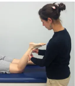
weight-bearing Mulligan’s mobilization with movement (MWM) joint mobilizations with a 20 second rest period between each set and 3x30 second bouts of passive Grade III anterior-posterior talocrural joint mobilizations with a 20 second rest period between each set. The MWM mobilization was performed with the subject standing on a treatment table. A nonelastic belt was placed around the subject’s distal leg and around the clinician’s hips. The subject stood on the dominant leg and placed the nondominant leg on the table for balance. The clinician stabilized the dominant foot and applied a posterior force to the anterior talus while applying an anterior force on the distal leg with the belt. The subject was asked to perform a slow dorsiflexion movement with the dominant leg until the first onset of pain or end of range. Once this end point is reached, the subject returned to a standing position and then immediately repeated the dorsiflexion movement. Approximately 15 movements were performed per 30 second bout of mobilizations. The Grade III mobilizations were performed at a rate of 1 oscillation per second. Oscillations were large amplitude movements from the joint’s mid-range to end-range of motion.
Post-treatment measurements were counterbalanced and followed the same procedures as the screening and pre-treatment measurements. Measurements included ankle dorsiflexion range of motion of the weight-bearing lunge; passive, knee extended; and passive, knee flexed to 90 degrees. Additional measurements consisted of double and single leg squat tasks, posterior talar glide test, and the ankle arthrometer test, as previously described.
DATA PROCESSING AND REDUCTION
(Euler sequence y, x’, z”). Knee flexion/extension was defined as the shank relative to the thigh about the y-axis, knee valgus/varus was defined as the shank relative to the thigh about the x-axis, and ankle dorsiflexion/plantarflexion was defined as the foot relative to the shank about the y-axis. Data were collected for 20 seconds for the double leg squat task, corresponding with 5 double leg squats and for 20 seconds for the single leg squat task, corresponding with 5 single leg squats. Kinematic data were sampled at a frequency of 100 Hz and filtered with a 4th order Butterworth with a 14.5 Hz low-pass filter. Data
were recorded throughout the tasks and analyzed over the descent phase of the squat from the start of the trial to the point of maximum knee flexion and were averaged across trials for each participant.
DATA ANALYSIS
CHAPTER IV MANUSCRIPT OVERVIEW
Objective: To determine the effects of mobilization with movement and anterior to posterior talocrural joint mobilizations on passive dorsiflexion range of motion and double and single leg squat kinematics in healthy subjects with restricted dorsiflexion.
Design: Randomized double-blinded controlled study. Setting: Sports medicine research laboratory.
Participants: Forty-three healthy subjects (23 females, 20 males) with restricted ankle dorsiflexion (≤40 degrees of weight-bearing lunge).
Interventions: All subjects: 2x30 seconds gastrocnemius stretching (knee extended), 2x30 seconds soleus stretching (knee flexed). Control group: 3x30 seconds sham
mobilization. Treatment group: 3x30 seconds mobilization with movement mobilization, 3x30 seconds Grade III anterior to posterior talocrural joint mobilization.
Main Outcome Measures: Dorsiflexion range of motion with the knee extended, knee flexed, and a weight-bearing lunge test and double and single leg squatting kinematics of knee valgus displacement, medial knee displacement, and dorsiflexion displacement. Results: All subjects, regardless of group, demonstrated significantly improved DF-ROM at post testing (DFEXT (F1, 41 =12.39, p=0.001), DFFLX (F1, 41 =18.83, p<0.005), WBL (F1, 41
p=0.030) and single (F1, 41 =14.862, p<0.005) leg squats. No significant differences were
observed for knee valgus displacement or medial knee displacement.
Conclusions: Calf stretching improved passive and active dorsiflexion range of motion in subjects with dorsiflexion restrictions. Joint mobilizations did not have an additive effect on dorsiflexion gains nor affect squatting kinematics at the knee.
Key Words: Dorsiflexion, talocrural joint mobilization, knee valgus
INTRODUCTION
Injury to the anterior cruciate ligament (ACL) is unfortunately commonplace in both competitive and recreational sports. Such injuries are painful, expensive, and often lead to long-term disability, decreased physical activity, and the development of early knee osteoarthritis (Lohmander, Ostenberg et al. 2004; Hewett, Myer et al. 2006; Siegel, Vandenakker-Albanese et al. 2012). Due to the high prevalence of ACL injuries, ongoing research is working towards identifying the best methods to prevent and rehabilitate ACL injuries in order to promote return to sport and limit risk of re-injury. In order to do so, identifying modifiable factors that predispose individuals to demonstrating lower extremity movement patterns known to increase the risk of ACL injury is essential.
has been associated with increased MKD during squatting tasks (Bell, Padua et al. 2008; Mauntel, Begalle et al. 2013). Additionally, affording individuals more ankle dorsiflexion with the use of a heel lift during a squat eliminated the presence of MKD (Bell, Padua et al. 2008; Padua, Bell et al. 2012).
Ankle DF-ROM is modifiable and restrictions can be caused by decreased osteokinematic or arthrokinematic motion and/or bony positional faults (Denegar, Hertel et al. 2002; Mulligan 2004; Grindstaff 2009). Restrictions in osteokinematic motion are caused by contractile tissue such as muscle, tendon, and fascia (Prentice 2004; Radford, Burns et al. 2006), whereas arthrokinematic motion restrictions are due to inert connective tissue such as ligaments and joint capsule (Prentice 2004). Bony positional faults in the ankle such as an anteriorly positioned talus may limit the amount of posterior talar glide during dorsiflexion, thus limiting DF-ROM (Mulligan 2004).
involvement in dorsiflexion restriction. This is an important area to address given the multiple factors that may ultimately limit DF-ROM.
As previously discussed, limited ankle DF-ROM is also associated with altered lower extremity biomechanics, such as greater MKD. Thus, in addition to improving ankle DF-ROM concomitant improvements in lower extremity biomechanics may also be observed following appropriately designed interventions to increase ankle DF-ROM. To our knowledge, no research has examined the acute effects of increasing ankle DF-ROM through either static stretching or joint mobilization techniques on lower extremity biomechanics. This study will identify the specific contributions of joint mobilizations in addition to stretching, and will also look at a variety of ankle and knee kinematics prior to and immediately following intervention during functional movement. Therefore, the purpose of this study is to determine the effects of Mulligan’s mobilization with movement (MWM) and anterior to posterior (AP) talocrucal joint mobilizations on passive DF-ROM and double and single leg squat kinematics in subjects with restricted dorsiflexion. This study aims to determine if efforts to increase ROM should be included in existing ACL prevention programs.
METHODS
Subjects were excluded if they had a history of lower extremity surgery, knee or ankle injury in the past 6 months, or a known vestibular, balance, or neurological disorder that would prevent them from completing the movement tasks. Subjects were randomly assigned to either the control group or treatment group, using a random number generator. All subjects read and signed an informed consent form approved by the University’s institutional review board prior to testing.
Screening of approximately 100 potential subjects was performed to identify those with less than or equal to 40 degrees on a WBL test. This cut point was based on previous research conducted in our laboratory (Dill, 2013, In Review). A digital inclinometer was used to measure the tibia angle relative to the vertical start position during the WBL (Bennell, Techovanich et al. 1998). The inclinometer was held 15cm distal to the middle of the tibial tuberosity. The subject stood on the dominant leg and rested the non-dominant leg in a comfortable position on the floor. The subject then bent the dominant knee and lunged forward as far as possible while keeping the dominant foot in line with the long axis of the leg and the heel on the ground. A researcher held the heel on the ground to ensure that it did not lift off the ground. The foot was then moved posteriorly until the maximum range of dorsiflexion was reached. The digital inclinometer measurement was taken at the point of maximum dorsiflexion (Krause, Cloud et al. ; Bennell, Techovanich et al. 1998; Denegar, Hertel et al. 2002) (Figure 10).
DF-ROM and squatting tasks, and the order of the DF-DF-ROM measurements was counterbalanced. Ankle DF-ROM was measured on the dominant limb of each subject by three methods; WBL and non-weight-bearing with the knee fully extended (DFEXT) and the
knee flexed to 90 degrees (DFFLX) (Piva, Fitzgerald et al. 2006). Subjects lay supine on a
treatment table with a foam roller under the distal shank and knee in full extension. Motion was measured with a standard goniometer while the researcher moved the foot so that the ankle was in dorsiflexion until restriction was felt. The axis of the goniometer was centered over the lateral malleolus, the stationary arm aligned with the fibular shaft and the mobile arm aligned with the 5th metatarsal (Bell, Padua et al. 2008; Cosby 2011). DFFLX motion
measurement was taken in the same manner as with the knee extended, and a block was placed under the subject’s thigh to ensure 90 degrees of knee flexion. The WBL test was performed in the same manner as during the screening. Three measurements were taken and averaged for each DF-ROM test.
digitized medial and lateral femoral condyles and medial and lateral malleoli, respectively. Joint angles were calculated with Euler angles (Euler sequence y, x’, z”) and were defined as the distal segment relative to the proximal segment. Kinematic data were sampled at a frequency of 100 Hz and filtered with a 4th order Butterworth with a 14.5 Hz low-pass filter. Data were recorded throughout the tasks and analyzed over the descent phase of the squat from the start of the trial to the point of maximum knee flexion and were averaged across trials for each participant. These data were used to measure knee valgus displacement, medial knee displacement, and ankle dorsiflexion displacement. Subjects then performed 5 successive double leg squats to the beat of a metronome (60 beats/minute). Subjects were instructed to perform a double leg squat maneuver, beginning with their feet shoulder-width apart, toes pointing straight ahead, heels on the ground, and arms extended over their head. Subjects flexed their knees as if sitting into a chair, and were asked to squat as low as possible. 1-3 practice squats were performed before data collection. Next, subjects were instructed to stand on their dominant leg with their hands on their waist and their non-dominant leg flexed to 45 degrees at the hip and 90 degrees at the knee. Subjects performed 5 successive single leg squats in a similar manner to double leg squats. Trials were repeated if subjects heels came off the ground, peak knee flexion was less than 60 degrees, or the non-dominant foot touched down during single leg squatting.
All subjects first received a single treatment session of 2 x 30 second bouts of knee extended calf stretching and 2 x 30 second bouts of knee bent calf stretching on a slant board, with a 20 second rest period between each stretch. Following stretching, the control group received a single treatment session of 3 x 30 second bouts of a sham mobilization consisting of passive knee flexion with subject prone and the ankle held in a neutral orthoplast splint, with a 20 second rest period between each set (Reid, Birmingham et al. 2007). Following stretching, the intervention group received both a 3 x 30 second bout of Mulligan’s mobilization with movement (MWM) joint mobilizations followed by 3 x 30 second bouts of passive Grade III AP talocrucral joint mobilizations. There was a 20 second rest period between each set. The MWM mobilization was performed with the subject standing on a treatment table. A nonelastic belt was placed around the subject’s distal leg and around the clinician’s hips. The subject stood on the dominant leg and placed the nondominant leg on the table for balance. The clinician stabilized the dominant foot and applied a posterior force to the anterior talus while applying an anterior force on the distal leg with the belt. The subject was asked to perform a slow dorsiflexion movement with the dominant leg until the first onset of pain or end of range. Once this end point was reached, the subject returned to a standing position and then immediately repeated the dorsiflexion movement. Approximately 15 movements were performed per 30 second bout of mobilizations. The Grade III mobilizations were performed at a rate of 1 oscillation per second. Oscillations were large amplitude movements from the joint’s mid-range to end-range of motion (Figures 8-9).
Dependent variables included DF-ROM measures as well as knee valgus displacement, medial knee displacement, and ankle dorsiflexion displacement during the descent phase of both squats. Knee valgus displacement is defined as the difference between initial knee valgus angle and the peak knee valgus angle, medial knee displacement is the difference between initial frontal plane knee angle and the peak frontal plane knee angle, and dorsiflexion displacement is the difference between initial ankle dorsiflexion angle and the peak ankle dorsiflexion angle. Significance was defined by ∝< 0.05. Data were analyzed with SPSS (Version 19.0, Chicago, IL).
RESULTS
Prior to testing there were no significant differences between groups on demographic data indicating randomization was successful (Table 3).
Inter-rater reliability was established prior to testing with intraclass correlation coefficients (ICC) and standard errors of measurement (SEM) performed for the WBL, ankle DFFLX and DFEXT measurements (ICC3,1 range 0.917-0.998; SEM range 0.17-2.56).
All subjects, regardless of group, demonstrated improved DF-ROM at post testing as a main effect for time was observed for the WBL (F1, 41 =32.65, p≤0.001), DFEXT (F1, 41
=12.39, p=0.001), and DFFLX (F1, 41 =18.83, p≤0.001). There was no significant Group x
A main effect of time was also observed for ankle dorsiflexion displacement during both squatting tasks, whereby displacement increased significantly from pre- to post-testing in both the double (F1, 41 =5.078, p=0.030) and single (F1, 41 =14.862, p≤0.001) leg
squats. Both the control group and treatment group had small to moderate effect sizes for dorsiflexion displacement during double (control d=0.08, tx d=0.16) and single (control d=0.27, tx d=0.16) leg squats (Cohen 1992). No main effects for group or interaction effects for time by group were detected for double or single leg squats (Tables 6-7). These results suggest that subjects did not display significant changes in movement patterns between pre- and post-testing.
DISCUSSION
Our most important finding was that all subjects had increased ankle DF-ROM at post-test, however the joint mobilizations did not promote greater gains in comparison to stretching alone. Improvements in ankle DF-ROM were accompanied by significant increases in ankle dorsiflexion displacement during the squat tasks. However, this did not translate into concomitant changes in frontal plane knee kinematics during the squat tasks.
did not attempt to determine if subjects had a positional fault of the talus nor if they truly needed a joint mobilization treatment. Clinically there is no objective way of identifying if a patient needs a joint mobilization, especially in a healthy population. Research has shown benefits of ankle mobilizations on ROM after ankle sprains or immobilization (Collins, Teys et al. 2004; Vicenzino, Branjerdporn et al. 2006; Landrum, Kelln et al. 2008) but to our knowledge, there has not been a study investigating the effect of mobilizations on a healthy but restricted population. It is possible that our subjects’ movement restrictions were not due to a bony restriction and thus they did not receive additional benefits from the joint mobilization treatments.
joint mobilization may have caused larger increased in DF-ROM. Future research should determine the effects of different treatment parameters and types of mobilizations.
We were surprised to see no changes in frontal plane knee kinematics given the significant improvements in both ankle DF-ROM and ankle dorsiflexion displacement during the squat tasks. Previous research demonstrated improvements in frontal plane knee motion when subjects are afforded more available ankle dorsiflexion during an overhead squat by placing the heels on a 2-inch block (Bell, Padua et al. 2008). Thus, we expected that by increasing both passive and active ankle motion, subjects would exhibit decreased knee valgus and/or medial knee displacement during squatting tasks. Contrary to our original hypotheses, subjects did not show any consistent changes in knee motion post-treatment. This may be because subjects did not achieve a clinically significant increase in DF-ROM. Despite gaining motion after treatment, all but one subject remained in the “restricted” category based on WBL measurements. The changes in motion seen in our study were on average less than 2 degrees, which is much smaller than the 10-15 degrees of DF-ROM afforded by a 2-inch block (Bell, Padua et al. 2008). Subjects in our study may not have had large enough gains in DF-ROM to facilitate alterations in knee frontal plane motion.
restrictions when compared to controls (Bell, Padua et al. 2008; Mauntel, Begalle et al. 2013), but by screening only for subjects with decreased ankle motion we may have found a population without excessive medial knee motion. Further research is needed to investigate the effect of stretching and mobilizations on subjects with excessive MKD.
Another explanation for subjects not altering their movement patterns is that while subjects did gain ankle motion, they did not receive any neuromuscular control training or movement pattern feedback. It is likely that the muscle firing patterns these subjects have been using for years would not change immediately, especially without any coaching. The treatment may have given subjects the potential to move better but it may be necessary to alter habitual movement patterns and teach proper techniques in order for people to move more effectively once they are afforded more motion. Research exploring the effect of neuromuscular control training on movement patterns is warranted.
Clinical recommendations based on this study are to use stretching and joint mobilizations for patients or athletes with decreased ankle dorsiflexion. We suggest more than one treatment session in order to see clinically significant gains in motion and to incorporate training sessions to teach proper movement patterns during lower body exercises. Through a combination of increased motion and repetitive movements, we believe faulty movement patterns can be corrected.
The following limitations may have impacted the results of our study. First, only one treatment session of the mobilization was administered. Future authors should
FIGURES
Figure 1: Dorsiflexion Range of Motion Measure with Knee Extended
Figure 3: Weight-Bearing Lunge
Figure 5: Ankle Arthrometer Test for Ankle Stiffness
Figure 7: Single Leg Squat with Electromagnetic Motion Capture Sensors
Figure 9: Sham Mobilization Treatment
TABLES
Table 1: Statistical Methods
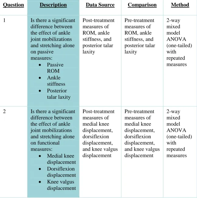
Question Description Data Source Comparison Method
1 Is there a significant difference between the effect of ankle joint mobilizations and stretching alone on passive measures: Passive ROM Ankle stiffness Posterior talar laxity Post-treatment measures of ROM, ankle stiffness, and posterior talar laxity Pre-treatment measures of ROM, ankle stiffness, and posterior talar laxity 2-way mixed model ANOVA (one-tailed) with repeated measures
2 Is there a significant difference between the effect of ankle joint mobilizations and stretching alone on functional measures:
Medial knee displacement Dorsiflexion
displacement Knee valgus
Table 2: Intraclass Correlation Coefficients and Standard Error of the Measurement
ICC SEM (°)
__________ DFEXT
Between Trials 0.917 2.56
Between Days 0.941 1.98
DFFLX
Between Trials 0.977 0.97
Between Days 0.976 1.09
WBL
Between Trials 0.998 0.17
Between Days 0.998 0.18
Posterior Talar Glide
Between Trials 0.981 0.69
Table 3: Group Characteristics Presented As Means ± SD For Each Group Control (n=22) Treatment (n=21)
Age (years) 19.68 ± 1.17 20.90 ± 3.35
Height (cm) 170.27 ± 1.77 170.33 ± 1.73
Table 4: Ankle Dorsiflexion Range of Motion (Degrees) presented as Means ± SD (95% Confidence Intervals) for each group at Pre and Post time point
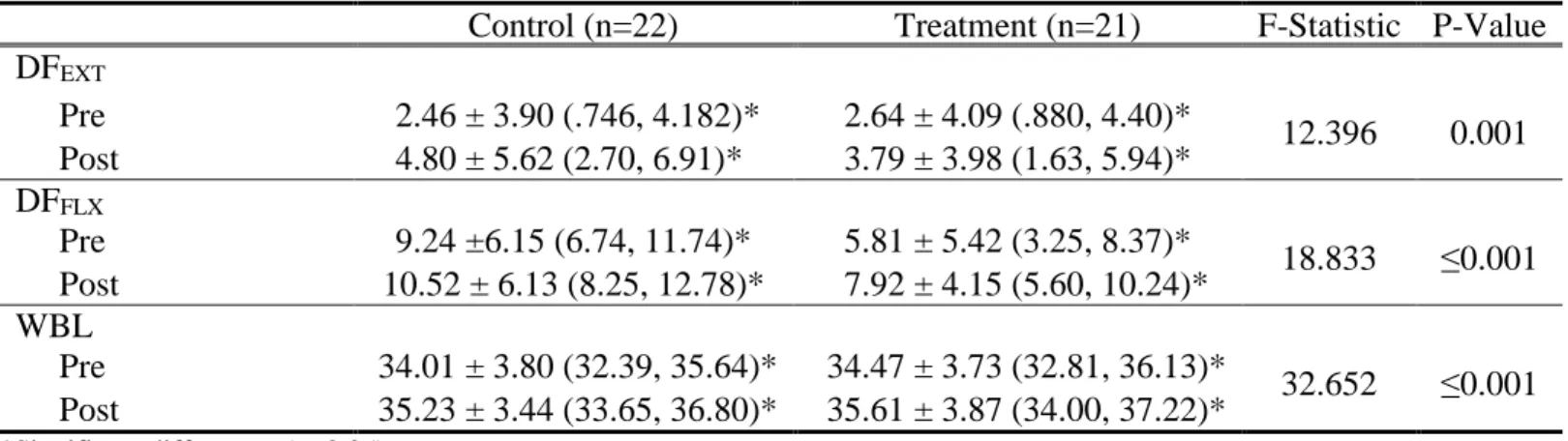
Control (n=22) Treatment (n=21) F-Statistic P-Value DFEXT
Pre 2.46 ± 3.90 (.746, 4.182)* 2.64 ± 4.09 (.880, 4.40)*
12.396 0.001 Post 4.80 ± 5.62 (2.70, 6.91)* 3.79 ± 3.98 (1.63, 5.94)*
DFFLX
Pre 9.24 ±6.15 (6.74, 11.74)* 5.81 ± 5.42 (3.25, 8.37)*
18.833 ≤0.001 Post 10.52 ± 6.13 (8.25, 12.78)* 7.92 ± 4.15 (5.60, 10.24)*
WBL
Pre 34.01 ± 3.80 (32.39, 35.64)* 34.47 ± 3.73 (32.81, 36.13)*
32.652 ≤0.001 Post 35.23 ± 3.44 (33.65, 36.80)* 35.61 ± 3.87 (34.00, 37.22)*
*Significant differences (p<0.05)