MODULATION OF SIGNAL TRANSDUCTION AND TRANSFORMATION BY KAPOSI’S SARCOMA-ASSOCIATED HERPESVIRUS
Penny M. Anders
A dissertation submitted to the faculty of the University of North Carolina at Chapel Hill in partial fulfillment of the requirements for the degree of Doctor of Philosophy in the
Department of Microbiology and Immunology in the School of Medicine
Chapel Hill 2016
Approved by: Nancy Raab-Traub Blosssom Damania Dirk Dittmer
ABSTRACT
Penny M. Anders: Modulation of Signal Transduction and Transformation by Kaposi’s Sarcoma-Associated Herpesvirus
(Under the direction of Blossom Damania)
Kaposi’s sarcoma associated-herpesvirus (KSHV) is a double-strand DNA gamma herpesvirus that establishes lifelong latent infection in the human host. KSHV infection is associated with several cancers including the endothelial cell cancer, Kaposi’s sarcoma (KS); and two B-cell lymphomas, primary effusion
lymphoma (PEL) and multicentric Castleman’s disease. This dissertation focuses on the importance of cellular signal transduction pathways in KSHV-associated cancers and focuses on two KSHV viral proteins, K1 and vPK, that modulate these signaling pathways.
of both pathways was no more effective at reducing cell viability and tumor burden in vivo than inhibition of the PI3K pathway alone.
Some viral proteins modulate host cellular proteins and thereby promote KSHV pathology. The KSHV K1 viral protein is thought to be a major contributor to KSHV-induced oncogenesis since the expression of K1 has been shown to lead to
transformation in vitro and to tumor development in vivo.
We identified AMPKγ1 as a K1-associating protein, and the K1 N-terminus is important for association with AMPKγ1. AMPK is a metabolic regulator that responds to many types of cellular stress by regulating pathways to maintain energy
homeostasis. We found that K1 expression gives cells a survival advantage when cells are stressed by serum starvation or when AMPK is inhibited, and that this survival advantage is dependent on K1’s association with AMPK. AMPK activity is also increased in K1-expressing cells following exposure to metabolic stress.
ACKNOWLEDGEMENTS
TABLE OF CONTENTS
LIST OF TABLES ... X LIST OF FIGURES ... XI LIST OF ABBREVIATIONS ... XIII
CHAPTER 1. INTRODUCTION ... 1
INTRODUCTION ... 2
KSHV-ASSOCIATED CANCERS ... 2
KAPOSI’S SARCOMA-ASSOCIATED HERPESVIRUS, HHV-8 ... 3
KSHV LATENCY PROTEINS THAT PROMOTE TUMORIGENESIS ... 5
K1 PROTEIN PROMOTES CELL SURVIVAL AND TUMORIGENESIS ... 7
VIRAL PROTEIN KINASE (ORF36) ... 8
CELLULAR SIGNAL TRANSDUCTION PATHWAYS ... 10
MAPK SIGNALING PATHWAY ... 10
PI3K SIGNALING PATHWAY ... 10
KSHV AND ACTIVATION OF THE PI3K AND MAPK PATHWAYS ... 12
AMPK, A CELLULAR KINASE ... 13
CHAPTER 2. DUAL INHIBITION OF THE PI3K/MTOR AND MAPK PATHWAYS IN NON-HODGKIN LYMPHOMA ... 15
OVERVIEW ... 15
RESULTS ... 16
DISCUSSION ... 23
CHAPTER 3. THE KSHV K1 PROTEIN MODULATES AMPK FUNCTION TO ENHANCE CELL SURVIVAL ... 25
OVERVIEW ... 25
INTRODUCTION ... 26
RESULTS ... 29
DISCUSSION ... 53
MATERIALS AND METHODS ... 56
CHAPTER 4. THE KSHV ORF36 PROTEIN KINASE AUGMENTS TUMORIGENESIS IN A MOUSE MODEL SYSTEM ... 65
OVERVIEW ... 65
INTRODUCTION ... 66
RESULTS ... 68
DISCUSSION ... 83
MATERIALS AND METHODS ... 85
CHAPTER 5. SUMMARY, CONCLUSIONS AND FUTURE DIRECTIONS ... 89
GENERAL SUMMARY ... 89
DUAL INHIBITION OF THE PI3K/MTOR AND MAPK PATHWAYS IN NON-HODGKIN LYMPHOMA ... 91
THE KSHV K1 PROTEIN MODULATES AMPK FUNCTION TO ENHANCE CELL SURVIVAL ... 92
THE KSHV ORF36 PROTEIN KINASE AUGMENTS TUMORIGENESIS IN A MOUSE MODEL SYSTEM ... 96
LIST OF TABLES
Table 4.1: Immune cell subsets in spleens from WT and vPK transgenic mice. ... 70 Table 4.2: Immune cell subsets in spleens from WT and vPK (line 2)
transgenic mice ... 71 Table 4.3: Immune cell subsets in spleens form WT and vPK (line 1)
LIST OF FIGURES
Figure 2.1. Simultaneous inhibition of the PI3K/mTOR and MEK/ERK
pathways fails to synergistically reduce NHL cell proliferation in vitro. ... 18
Figure 2.2. Combined inhibition of the MEK/ERK and PI3K/mTOR pathways is no more effective at reducing tumor burden than single blockade of the PI3K/mTOR pathway in vivo. ... 22
Figure 3.1. KSHV K1 mutant infected cells exhibit decreased survival following nutrient deprivation. ... 32
Figure 3.2. Cells infected with KSHV containing WT K1 are more resistant to AMPK inhibition than cells infected with KSHV K1 mutants. ... 35
Figure 3.3. K1 expression provides a survival advantage in cells treated with compound C. ... 37
Figure 3.4. K1 associating proteins determined by mass spectrometry. ... 39
Figure 3.5. K1 associates with AMPKγ1. ... 41
Figure 3.6. The K1 N-terminus is important for association with AMPKγ1. ... 43
Figure 3.7. K1 and AMPK associate in membranes and the perinuclear area of the cell. ... 45
Figure 3.8. K1 N-terminus is important for K1 association with AMPKγ1 in the membrane fraction ... 47
Figure 3.9. K1 and AMPK association is important for cell survival following exposure to stress. ... 49
Figure 3.10. K1-expression facilitates AMPK activity in stressed cells. ... 51
Figure 3.11. K1 maintains an active PI3K pathway despite AMPK activation following 8 hours of serum starvation ... 53
Figure 4.1. Development of a vPK transgenic mouse model system. ... 69
Figure 4.2. Immunization protocol for vPK transgenic and WT mice with NP-KLH or PBS ... 72
Figure 4.3. Body and spleen weights for NP-KLH immunized vPK transgenic and WT mice. ... 73
Figure 4.5. The percentages of B cells in spleens from vPK mice
immunized with PBS were comparable to WT immunized with NP-KLH. ... 75 Figure 4.6. Activation status of B cell subsets from the spleens of
immunized vPK transgenic and WT mice. ... 76 Figure 4.7. vPK transgenic mice have increased percentages of germinal
center B cells... 77 Figure 4.8. Splenic T cell subsets in immunized vPK and WT mice. ... 78 Figure 4.9. vPK spleens have increased percentages of CD44 positive
T helper cells compared to WT. ... 79 Figure 4.10. CD86 expression is increased on vPK T helper and
cytotoxic T cells. ... 80 Figure 4.11. Aged-vPK transgenic mice are more prone to developing
LIST OF ABBREVIATIONS
4E-BP1 eukaryotic translation initiation factor 4E-binding protein 1
AICAR 5-Aminoimidazole-4-carboxamide ribonucleotide
AIDS acquired immune deficiency syndrome
AMP adenosine monophosphate
AMPK 5' adenosine monophosphate-activated protein kinase
ANOVA analysis of variance
AP-1 activator protein-1
APC allophycocyanin
ATCC American Type Culture Collection
ATP adenosine triphosphate
BAC16 bacterial artificial chromosome 16
BCA bicinchoninic acid assay
BCR B-cell receptor
BL Burkitt lymphoma
CaMKKβ calcium/ calmodulin dependent protein kinase kinase–beta
CD cluster of differentiation
CDK6 cyclin-dependent kinase 6
CHOP cyclophosphamide, hydroxydaunorubicin, oncovin, prednisone
CO2 carbon dioxide
CSR class switch recombination
CTLA-4 cytotoxic T-lymphocyte-associated protein 4
DLBCL diffuse large B-cell lymphoma
DMEM Dulbecco's Modified Eagle Medium
DNA deoxyribonucleic acid
EBM Endothelial Basal Medium
EBV Epstein–Barr virus
EDTA ethylenediaminetetraacetic acid
EGM Endothelial Growth Medium
eIF4B eukaryotic translation initiation factor 4B
eIF4E eukaryotic translation initiation factor 4E
ER endoplasmic reticulum
ERK extracellular signal–regulated kinases
FACS fluorescence-activated cell sorting
FAK focal adhesion kinase
FITC fluorescein isothiocyanate
FL follicular lymphoma
FOXO forkhead box O
GPCR G-protein-coupled receptors
Grab 2 growth factor receptor-bound protein 2
GSK-3β glycogen synthase kinase 3 beta
GTP guanosine-5'-triphosphate
H&E hematoxylin and eosin
HCMV human cytomegalovirus
HDAC histone deacetylases
HEK human embryonic kidney
HHV-6 human herpesvirus 6
HI-FBS heat inactivated fetal bovine serum
HSV herpes simplex virus
hTERT human telomerase reverse transcriptase
HUVEC human umbilical vein endothelial cells
IgH immunoglobulin heavy locus
IL-2 interleukin-2
IL-6 interleukin -6
IP3Rs inositol 1,4,5-trisphosphate receptors
ITAM immunoreceptor tyrosine-based activation motif
JAK3 janus kinase 3
JNK c-Jun N-terminal kinase
KS Kaposi’s Sarcoma
KSHV Kaposi’s Sarcoma Associated Herpesvirus
LANA latency-associated nuclear antigen
LBK1 liver kinase B1
LPS lipopolysaccharides
MALDI Matrix Assisted Laser Desorption/Ionization/
TOF Time of Flight Mass Spectrometry
MAPK mitogen-activated protein kinase
MAPKK mitogen-activated protein kinase kinase
MAPKKK mitogen-activated protein kinase kinase kinase
MEF mouse embryonic fibroblasts
MKK4 mitogen-activated protein kinase kinase 4
MKK7 mitogen-activated protein kinase kinase 7
mTOR mammalian target of rapamycin
NaCL Sodium chloride
NFAT nuclear factor of activated T-cells
NHL Non-Hodgkin lymphoma
NP-KLH keyhole limpet hemocyanin
NP-40 nonyl phenoxypolyethoxylethanol.
PBS phosphate buffered saline
PCR polymerase chain reaction
PDK1 3-phosphoinositide dependent protein kinase-1
PE R-Phycoerythrin
PEL Primary Effusion Lymphoma
PI3K phosphoinositide 3-kinase
PIP2 phosphatidylinositol 4,5-bisphosphate
PIP3 phosphatidylinositol (3,4,5)-trisphosphate
PKCθ protein kinase C theta
PLCγ2 phospholipase C, gamma 2
Ras GAP Ras GTPase-activating protein
Rb retinoblastoma protein
REV revertant
RTA replication and transcription activator
S6KB1 S6 kinase B1
SDS-PAGE sodium dodecyl sulfate polyacrylamide gel electrophoresis
SERCA sarco/endoplasmic reticulum
SH2 Src homology 2
SYK spleen tyrosine kinase
TAK1 transforming growth factor beta-activated kinase 1
TSC2 tuberous sclerosis complex 2
v-FLIP viral fas-associated death domain-like interleukin-1β-converting
enzyme-inhibitory protein
v-IRF3 viral-interferon regulatory factor 3
Vpk viral protein kianse
VZV varicella zoster virus
WT wild-type
CHAPTER 1. INTRODUCTION
OVERVIEW
Kaposi’s sarcoma-associated herpesvirus (KSHV) or human herpesvirus-8 (HHV-8) is the causative agent of the endothelial cancer Kaposi’s sarcoma (KS) and two B cell lymphomas, primary effusion lymphoma (PEL) and multicentric
Castleman’s disease (MCD) [1-3]. These KSHV-associated diseases mainly occur in immune-suppressed individuals such as those who are HIV positive or who are organ transplant recipients although they can also occur in seemingly immune competent individuals. KS is one of the most common cancers among sub-Saharan African men, and the most prevalent cancer in several countries in sub-Saharan Africa [4, 5].
Treatment options for individuals with KS include antiretroviral therapy for HIV- positive individuals, therapies that boost the immune system, and/or excision of the KS lesion. Individuals with PEL typically are treated with CHOP (cyclophosphamide, doxorubicin, vincristine, and prednisone) chemotherapies. PEL is often resistant to chemotherapies and median survival following diagnosis is approximately 6 months [6]. Thus, effective therapies for KSHV malignancies are warranted and
INTRODUCTION
KSHV-ASSOCIATED CANCERS
Kaposi’s sarcoma (KS) is a cancer that is believed to be of endothelial cell origin. During the 1980s, cases of Kaposi’s sarcoma started appearing in immune-compromised individuals who were HIV positive. Although the KS lesion is typically viewed as a red patch that impacts the dermis on extremities, KS can also impact the mucosa and internal organs [7-9]. The KS lesion is composed of KSHV-infected spindle cells and a profusion of neovascular spaces [10]. The newly developed neovascular spaces are weak and allow leakage of fluids into the extravascular spaces, forming the characteristic KS red patch [9]. The KS lesions can progress from a patch, to a plaque, and then into a nodule. Although the KS lesion is
composed of spindle cells, it is also filled with inflammatory cells including T, B and monocytic cells [11, 12].
Inflammation has been found to play an integral part in the development and progression of KS. In the early stages of KS, there’s extensive inflammatory infiltrate including an influx of CD8+ and CD4+ -interferon-γ-producing T cells and the
production of Th1-inflammatory cytokines [13]. Inflammatory cytokines re-activate KSHV from latency and stimulate endothelial cells to produce angiogenic factors thereby promoting KS progression [14]. In vivo, KS has also been found to localize to surgical sites or areas of skin trauma, further emphasizing the importance of inflammation to KS development [15, 16].
Eastern European origin. Post-transplant KS impacts some transplant recipients and often resolves with the reduction or withdrawal of immunosuppressants although patients are then at risk for graft rejection [17]. African-KS, a more aggressive form that can impact the organs is found in children and young adults. AIDS-KS is the most aggressive form, causing lesions and impacting organs in HIV positive individuals. Anti-retroviral therapy has reduced the incidence of AIDS-KS [18].
Primary effusion lymphoma (PEL) is a monoclonal B cell lymphoma that is under the diagnostic umbrella of non-Hodgkin lymphoma [19]. It is a B cell-rich fluid localized to the body cavities. PEL occurs predominantly in HIV positive individuals. All PEL cells are latently infected with KSHV and can be cultured ex vivo [20, 21]
Multicentric Castleman’s disease (MCD) is another B lymphoproliferative disease although it differs from PEL in that it is polyclonal and found as solid tumor affecting the lymphatic organs [22, 23]. MCD can occur in HIV positive and negative individuals, but when it is associated with KSHV infection, it usually occurs in
individuals who are HIV positive [3].
KAPOSI’S SARCOMA-ASSOCIATED HERPESVIRUS, HHV-8
Chang et al. first isolated KSHV from KS lesions in 1994 [1]. KSHV is a double-strand DNA gamma herpesvirus that has a genome of approximately 165 to 170 kb long that code for over 85 open reading frames and 12 micro RNAs [24-30]. The KSHV genome contains multiple open reading frames that are conserved among other herpesviruses, and genes K1-K15 that are unique to KSHV [7]. Similar to other gammaherpesviruses, KSHV can vacillate between latent and lytic programs
KSHV can infect a variety of cell types in vitro and invivo. In vivo, KSHV has been found in monocytes, B cells, endothelial/spindle cells, and epithelial cells [10, 31-33]. Upon infection, KSHV typically establishes latency by forming an episome. The latency-associated nuclear antigen (LANA), binds the viral terminal repeats and tethers the viral genome to the host chromosome [7]. In this way, the viral genome is copied by host replication machinery and passed on to daughter cells during cell division.
During latency, a subset of viral genes is expressed including LANA, FLIP, v-Cyclin, kaposins A, B and C, v-IRF-3 as well as the twelve pre-microRNAs [34]. Although predominantly expressed during the lytic cycle, recent studies indicate that the proteins viral interleukin-6 (vIL-6) and K1 are expressed at low levels during latency as well [24, 35]. Upon reactivation, which can be induced in vitro with various compounds such as 12-O-tetradecanoylphorbol-13-acetate (TPA), histone
deacetylase (HDAC) inhibitors, and TLR 7/8 ligands, KSHV enters the viral lytic cycle resulting in the production of infectious virions [24, 36].
KSHV LATENCY PROTEINS THAT PROMOTE TUMORIGENESIS The KSHV viral proteins and microRNAs expressed during latency are
implicated in making major contributions to KSHV-induced cancers. Expression of the latency locus in B cells, which contains the genes for LANA, K13 (v-FLIP), a cellular cyclin D2 homolog (v-Cyclin), K12 (kaposin), and all viral microRNAs, in transgenic mice results in chronic mature B cell activation and B cell hyperplasia [37]. B cells are hyper-responsive to T-dependent antigen as well as
lipopolysaccharide (LPS), resulting in increased germinal center responses and an increase in the number of plasma cells. B cells expressing the latency locus are poised and responsive to unrelated pathogen, making these cells susceptible to expansion; and yet only a fraction of the latency locus transgenic mice go on to develop lymphomas [37].
Thus, the latency locus most likely contributes to, but is not the sole driver, of KSHV-induced oncogenesis. When transgenic mice expressing the latency locus are combined with mice containing a weak IgH Cα Myc allele, the latency locus/Myc mice develop lymphoma at a higher rate than the single transgenic mice expressing either the latency locus or Myc [38]. Again these data support the idea that the KSHV latency locus cooperates with host factors such as myc to promote oncogenesis.
tumorigenesis by altering cell cycle and/or survival. LANA binds to p53, and p53 expressing cells have reduced sensitivity to p53-dependent apoptosis [24]. LANA interacts with the retinoblastoma protein (Rb) and increases E2F activity, suggesting that LANA inhibits Rb, promotes E2F activity and as a consequence promotes cell cycle progression [7].
Another latency protein that appears to have a major impact on promoting cellular transformation is Fas-associated death domain-like IL-1 converting enzyme inhibitor protein (v-FLIP). In vitro, v-FLIP inhibits caspase-8 and facilitates activation of NF-κB [40]. Viral-FLIP expression has also been shown to transform rodent fibroblasts, and the transforming potential of v-FLIP is dependent on NF-κB activation [40]. The expression of v-FLIP under the major histocompatibility H2kb promoter leads to constitutive NF-κB activation in various lymphoid organs and an increased incidence of lymphoma of various subsets compared to controls [41]. Viral-FLIP expressing mice do not have impaired Fas-induced apoptosis [41].
Kaposin (K12), another latent gene, may have oncogenic potential. The kaposin gene results in the generation of kaposins A, B and C due to translation of kaposin mRNA from different sites of initiation [43]. Kaposin A expression can transform rodent fibroblasts and nude mice injected with Kaposin A expressing cell lines develop tumors [44].
In this brief summary describing the oncogenic potential of some KSHV latent proteins, we can observe that KSHV latent proteins play a role in promoting cell survival by regulating cell cycle or by inhibiting apoptosis. Based on the in vivo models, we can also see that expression of some of these proteins alone or in concert with a cellular oncogene such as myc can result in tumor formation. Recent studies suggest that the expression of some latent and lytic proteins may not be assigned exclusively to either the latent or lytic program. The KSHV K1 protein was presumed to be expressed during the lytic cycle, but more recently has been found to be expressed during latency albeit at lower levels than during the lytic cycle [24, 35, 45].
kinases [49]. K1 can interact with various SH2 (Src-Homology 2) containing signaling molecules, including Lyn, Syk, p85, PLCγ2, RasGAP, Vav, Grab 2 and SH2-containing tyrosine phosphatases 1/2 via the phosphorylated tyrosines in its ITAM [50]. Activation of K1 results in a signaling cascade leading to calcium mobilization, upregulation of the transcription factors NFAT and AP-1, and the
production of inflammatory cytokines [50]. Expression of K1 induces the activation of phosphoinositide 3-kinase (PI3K) by phosphorylation of the p85 subunit of PI3K. Activation of PI3K leads to activation of downstream kinases including Akt and mammalian target of rapamycin (mTOR), which leads to the inhibition of forkhead transcription factor family and reduced cellular apoptosis [51]. K1 has also been shown to inhibit Fas-induced apoptosis [52, 53]. K1 may promote tumorigenesis by modulating signaling pathways that are important for cell survival and proliferation.
K1 is located in the KSHV genome at a position equivalent to the saimiri transforming protein (STP) of herpesvirus saimiri (HVS), a member of the
rhadinovirus subgroup of gamma herpesvirues [54]. Replacement of oncogenic STP with K1 in HVS results in IL2-independent immortalization of T cells and the
development of lymphoma in marmosets following infection with the K1 containing-recombinant HVS [54]. K1 expression also transforms rodent fibroblasts and immortalizes primary endothelial cells [54, 55]. K1 transgenic mice develop lymphomas and sarcomas at low incidence [56].
VIRAL PROTEIN KINASE (ORF36)
ectopically in cells [57]. ORF36 shares limited homology with the protein kinases expressed by the other herpesviruses including UL13 (HSV), ORF47 (VZV), BGLF4 (EBV), UL97 (HCMV), and U69 (HHV-6) [58]. However, there are also some
differences. Viral PK appears to have diverse functions. Viral PK phosphorylates and activates the cellular targets, c-Jun N-terminal kinase (JNK), MKK4 and MKK7 as well as, the viral target K-bZIP, a transcription regulator that represses the latent/lytic switch protein, RTA (replication and transcription activator) protein [59, 60]. Kuny et al. also showed that vPK exhibits cellular cyclin dependent kinase-like activity, in that it can phosphorylate Rb thereby inactivating it and promoting cell cycle progression [57]. By binding to the activated form of interferon regulatory factor 3 (IRF-3), vPK also inhibits the anti-viral type I interferons [61].
Our lab has recently shown that vPK resembles and mimics the activity of the cellular protein S6 kinase (S6KB1) [62]. S6KB1 is downstream in the PI3K/mTOR pathway and is phosphorylated by mTOR. Activated S6KB1 phosphorylates multiple targets some of which are involved in protein synthesis such as ribosomal S6
protein, a protein that is part of the 40S ribosome; and eIF4B, a protein that is part of the translation pre-initiation complex [63]. Expression of vPK results in increased de novo protein synthesis, tubule formation and anchorage independent growth,
compared to control cells, suggesting that vPK may be possess oncogenic properties [62].
promote the production of chemokines and cytokines that support angiogenesis, and survival and growth of KSHV infected latent cells. Additionally, it is also possible that lytic proteins can be expressed in the absence of complete viral lytic replication. For example, vPK/ORF36 contains hypoxia responsive elements in its promoter, which may cause it to be expressed in isolation and in the absence of full-blown lytic replication [64, 65]. Thus, lytic proteins such as vPK, in addition to latent proteins, may be contributors to oncogenesis particularly in a changing cellular milieu.
CELLULAR SIGNAL TRANSDUCTION PATHWAYS MAPK SIGNALING PATHWAY
There are 4 major cascades of the mitogen-activated protein kinase (MAPK) pathway. Each cascade is comprised of multiple nodes that are activated
sequentially, MAPKKK-MAPKK-MAPK [66]. In response to various stimuli such as growth factors, stress or cytokines; there is engagement of the cell surface receptor and this event initiates the activation of kinases that subsequently activate one or more cascades of the MAPK pathway. We will be focusing on the RAF (MAPKKK)-MEK1/2 (MAPKK)-ERK1/2 (MAPK) cascade of the MAPK pathway, which is
stimulated by growth factors, and involved in regulating cell survival and proliferation [66].
PI3K SIGNALING PATHWAY
tyrosine kinases and G-coupled protein receptors (GPCRs) and by small GTPases. The Class IA PI3Ks are composed of a p85-like regulatory subunit (p85α, p85β, and p55γ) and a p110 catalytic subunit (p110α, p110β, p110γ and p110δ) [67].
When inactive, the p85 subunit prevents activation of the p110 subunit. Upon activation, the p85 subunit localizes to the plasma membrane thereby relieving p110 so that it can phosphorylate PtdIns 4, 5-bisphosphate (PIP2) generating PtdIns (3, 4, 5) P3 (PIP3). The serine threonine kinase, Akt, binds PIP3 via its pleckstrin homology (PH) domain resulting in translocation of Akt to the membrane. Akt is phosphorylated at Thr308 by phosphoinositide-dependent kinase-1 (PDK1); and for full activation, Akt is also phosphorylated at S473 on its C-terminus by mTOR complex 2
(mTORC2) [68]. Akt phosphorylates many substrates including the proapoptotic factors Bad, GSK-3β, and the FOXO transcription family members resulting in the inhibition of these factors, thereby promoting cell survival and proliferation [69].
Activation of Akt also leads to the direct phosphorylation of tuberous sclerosis 2 (TSC2), which releases its inhibitory effect on the serine/threonine kinase,
autophagy [74]. Mammalian TORC1 directly phosphorylates eukaryotic translation factor 4E (eIF4E)-binding protein 1 (4E-BP1) and S6 kinase 1 (S6K1), and promote protein synthesis [74]. Unlike mTORC2, mTORC1 is rapapmycin-sensitive [75].
KSHV AND ACTIVATION OF THE PI3K AND MAPK PATHWAYS The PI3K and MAPK pathways are activated early and appear important for KSHV infectivity. Binding of KSHV virions to integrins on the cell surface, facilitates phosphorylation of focal adhesion kinase (FAK) [76]. Activation of FAK stimulates the activation of Src family tyrosine kinases, which results in the activation of PI3K and Rho GTPases [76, 77]. Activation of PI3K and Rho GTPases following KSHV infection facilitates cytoskeletal rearrangements and actin polymerization [76]. Inhibition of PI3K reduces KSHV infectivity [76, 78, 79], suggesting that activation of PI3K is important for KSHV infection. The MAPK pathway is also activated upon receptor-ligand binding and viral entry [79]. Although activation of the MAPK
pathway is not necessary for viral DNA entry or nuclear delivery, it does promote the expression of various viral latent and lytic genes, including LANA and RTA
respectively, as well as host genes thereby promoting an intracellular environment that is permissive to infection [79].
KSHV-endothelial xenograft model in which the virus is latent [83]. These data suggest that the PI3K pathway is important for latent KSHV cell survival.
Activation of the MAPK pathway is also important in facilitating KSHV
reactivation from latency [82, 84]. Inhibition of ERK, JNK, and p38 MAPK pathways during TPA-mediated KSHV reactivation results in reduced expression of viral lytic genes and production of infectious virions [82]. Because both the PI3K and MAPK pathways support KSHV infection throughout its life cycle, we sought to evaluate inhibition of both pathways in non-Hodgkin lymphoma, which includes PEL.
AMPK, A CELLULAR KINASE
AMPK is a serine-threonine kinase composed of an alpha catalytic sub-unit and two regulatory units, beta and gamma [85]. AMPK is a major metabolic regulator that maintains energy homeostasis by responding to stress conditions that increase the AMP: ATP ratio such as during nutrient deprivation, oxidative stress or hypoxia [86, 87]. Activated AMPK leads to the activation of catabolic pathways that generate ATP and inhibition of anabolic pathways that consume ATP. Binding of AMP to the gamma subunit allosterically activates AMPK and promotes phosphorylation of AMPKα at Thr172 by the upstream kinase liver kinase B1 (LKB1) [88-90]. Bound AMP or ADP at higher concentrations prevents dephosphorylation of Thr172 [88, 91]. AMPK can also be phosphorylated by Ca2+/calmodulin-dependent protein
The role of AMPK as a tumor promoter is actively being explored [102, 103]. Some studies suggest that AMPK promotes tumor cell survival in vitro and in vivo. Inhibition of AMPK results in reduced pancreatic cell survival and apoptosis under normal and stressed conditions [104, 105]. AMPK also promotes survival in multiple myeloma, colorectal and glioma cancer cell lines [106-109]. In vivo, AMPK signaling is elevated in developing tumors in a glioblastoma rat model [110]. Moreover, mouse embryonic fibroblasts (MEFs) that are null for LBK1, a kinase that
CHAPTER 2. DUAL INHIBITION OF THE PI3K/MTOR AND MAPK PATHWAYS IN NON-HODGKIN LYMPHOMA1
OVERVIEW
NHL subtypes are classified based on clinical and histologic characteristics, as well as lymphocyte developmental stage. NHL sub-types include diffuse large B-cell lymphoma (DLBCL), follicular lymphoma (FL), mantle cell lymphoma (MCL), Burkitt lymphoma (BL), primary effusion lymphoma (PEL) to name just a few. Many
lymphomas such as DLBCL, MCL, and AIDS-associated lymphomas are very
aggressive and rapidly fatal if unresponsive to therapy. Indolent lymphomas, such as FL are also many times not curable with conventional chemotherapy. Clearly, novel treatment paradigms are needed to improve clinical outcomes of patients with NHL.
INTRODUCTION
We had previously shown that the phosphatidylinositol 3-kinase (PI3K) and mammalian target of rapamycin (mTOR) pathway was important for NHL survival and that single and dual inhibitors of this pathway effectively inhibited NHL
1 Anders P, Bhende PM, Foote M, Dittmer DP, Park SI, Damania B., Accepted manuscript of
proliferation in vitro and in mouse xenograft models of NHL including FL and PEL [80, 113].
Another pathway that is linked to tumorigenesis is the mitogen activated protein kinase (MAPK), and many sub-types of NHL display an activated MAPK pathway [114-116].
Although conventional chemotherapeutic drugs are useful in some cases, a significant number of NHL patients will eventually relapse and develop resistance to chemotherapy, which necessitates determining additional therapeutics. Since both PI3K and MAPK pathways are activated in NHL, we decided to investigate
combination therapy involving inhibition of both pathways. It has been demonstrated that such therapy is effective against certain solid tumors [117, 118].
RESULTS
In this study we used a combination of BEZ234 and AZD6244. NVP-BEZ235 is an imidazo[4,5-c]quinoline derivative compound, shown to inhibit PI3K and mTOR kinase activity both in vitro and in vivo including PEL and FL [119, 120]. AZD6244 is an inhibitor of MEK1 and MEK2 that does not compete with ATP
binding. It is a potent and selective MEK1/2 inhibitor that demonstrates high activity in solid tumor models in vitro and in vivo [121].
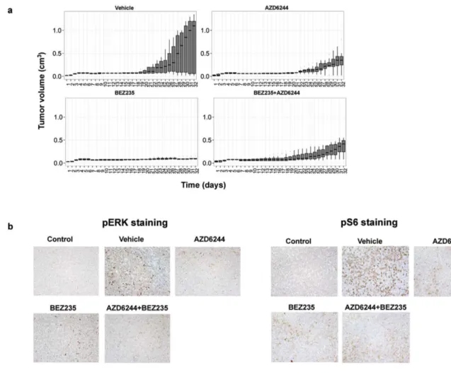
Figure 2.1. Simultaneous inhibition of the PI3K/mTOR and MEK/ERK pathways fails to
synergistically reduce NHL cell proliferation in vitro. (a) Proliferation of NHL cell lines treated
with drug(s) or vehicle were evaluated by MTS Assay using the Cell Titer 96 Aqueous One
Solution Cell Proliferation Assay (Promega) according to the manufacturer’s instructions.
Cells (1X10⁵/mL) were incubated with drug(s) or vehicle for 72 hours in a 12-well plate.
Twenty microliters of MTS reagent was added to 100µL of cell suspension in a 96-well plate
on the y-axis. The percent of viable cells for untreated cells was set as 100% for each
sample. Elevated absorbance values are indicative of metabolically active cells. The
COLO-205 cells (5X10³ cells/100µL) were added to each well of a 96-well plate. Increasing
concentrations of drug(s) or vehicle were added the following day. The MTS reagent was
added after 72 hours following the addition of the drug(s) or vehicle, and incubated with the
cells for 4 hours at 37°C. Absorbance was measured at 490nm using a FLUOstar OPTIMA
spectrophotometer. Percent viable cells after 72 hours treatment is shown on the y-axis. The
percent of viable cells for vehicle treated was set as 100% for each sample. Elevated
absorbance values are indicative of metabolically active cells. (b) Phosphorylation of ERK
1/2 and S6 kinase were determined by Western blot of NHL cell lines treated with AZD6244
(1µM) and/or NVP-BEZ235 (30nM) for 24 hours. Equal volume of DMSO (0.1%) was added
for vehicle control. (c) Proliferation of cells with 25 µM CAL-101 and increasing
concentrations of AZD6244 was determined by MTS assay after 48 hours following the
addition of the drug(s) or vehicle as described in panel a. (d) Phosphorylation of Akt, ERK
1/2 and S6 kinase were determined by Western blot of NHL cell lines treated with AZD6244
(1µM) and/or CAL-101 (25µM) for 24 hours.
presence of increasing concentrations of the chemically unrelated MEK inhibitor, PD184352 [123]. We found that PD184352 was ineffective at reducing cell viability in various NHL cell lines except at the highest dose of 10 µM (data not shown). This data supports our observations made with the MEK inhibitor, AZD6244, and suggests that NHL cells are not addicted to the MEK/ERK pathway for survival.
To verify that each of the drugs was inhibiting their respective targets, we performed Western blot analysis on the NHL cells that were treated with the above therapies for 24 hours (Figure 2.1b). We used phosphorylated S6K as a downstream marker for active PI3K/mTOR as we previously described [80, 113], and we used phosphorylated ERK1/2 as a downstream marker for MAPK activation [121]. We found that NVP-BEZ235 inhibited PI3K signaling as measured by reduced
phosphorylated S6K levels, and that AZD6244 inhibited MAPK signaling as measured by reduced ERK1/2 phosphorylation (Figure 2.1b).
(Figure 2.1c), although each drug appeared to be effective in inhibiting downstream effectors of the pathway. Treatment with CAL-101 inhibited phosphorylation of Akt and S6K, and AZD6244 inhibited phosphorylation of ERK1/2 (Figure 2.1d). We found similar results for FL-18 and Raji cells (data not shown). Thus, neither a dual inhibitor of the PI3K/mTOR pathway (NVP-BEZ235) nor a single inhibitor of PI3K (CAL-101) appears to synergize with AZD6244 in our system.
We next investigated the combination of NVP-BEZ235 and AZD6244 in a mouse xenograft model [113]. Four-to-five-week-old athymic nude-Foxn1^nu female mice were injected intraperitoneally with anti-asialo GM1 antibody (Wako). The following day, 1x10⁶ FL-18 lymphoma cells were subcutaneously injected with growth-factor reduced matrigel as we previously described [113]. Upon
Figure 2.2. Combined inhibition of the MEK/ERK and PI3K/mTOR pathways is no more
effective at reducing tumor burden than single blockade of the PI3K/mTOR pathway in vivo.
(a) Athymic nude-Foxn1^nu 4-5-week-old female mice were injected subcutaneously into
the right flank with 200 µL (1X10⁶) of FL-18 cells. Five mice per group were treated with
equal volume of vehicle, 45 mg/kg NVP-BEZ235, 45 mg/kg AZD6244, or 25 mg/kg each of
NVP-BEZ235 and AZD6244 (combination therapy). Treatment involved oral gavage 6 times
per week. Tumor volume was determined daily (L x W x D) and plotted over time. Shown for
each time point is a box and whisker plot of the results per group. The horizontal bar
indicates the median, the box indicates 25% and 75% of the data and the whiskers
represent 1.5 x the interquartile range, which encompasses 50% of the data. (b)
Phosphorylation of ERK 1/2 or S6 protein from each treatment group was evaluated on
with hematoxylin, and visualized at magnifications of 200X using a Leica DMLS microscope.
Images were acquired using the Leica DFC480 camera and associated Leica Firecam
software. Stored TIFF images were evaluated using Adobe Photoshop CS5.1.
The mice were sacrificed when maximal tumor size was reached. Tumors were excised, sectioned, and subjected to immunohistochemical analysis as we
previously described [80]. S6 was consistently phosphorylated in almost all cells in the vehicle control group, however we found reduced phosphorylated levels of S6, a downstream effector of the PI3K pathway in the NVP-BEZ235 treated grafts, and reduced phosphorylated ERK1/2, an effector of the MAPK pathway in the AZD6244 treated grafts (Figure 2.2b). Both markers were reduced in animals treated with both drugs. This suggests that both drugs were delivered to the tumors and inhibited their respective targets in the tumor cells in vivo. Overall, our data demonstrate that the combined inhibition of the PI3K and MAPK pathways was not synergistic or additive in preventing tumor growth of NHL cells in vitro or in vivo.
DISCUSSION
growth in similar pre-clinical models [117, 118]. Davies et al. found that tumors with activating Ras mutations are more sensitive to AZD6244 than tumors with WT Ras [122]. Since activating mutations in Ras are infrequent in NHL [126], inhibition of the MEK/ERK pathway may not be as effective for NHL as for solid tumors despite the fact that ERK was highly phosphorylated in the cell lines tested.
Even though MEK/ERK and PI3K/mTOR pathways are active in NHL [80, 115, 116], our data suggests that NHL are more reliant on the PI3K/mTOR pathway than the MEK/ERK pathway for cell survival. Normal B-lymphocyte survival is also
dependent on the PI3K pathway [127] and studies have shown that B-lymphocytes lacking BCR signaling do not survive while these cells can be rescued from
apoptosis by being engineered to have constitutively active PI3K [127].
Although the MEK/ERK pathway may be important in NHL pathology, we found that targeting this pathway alone is not sufficient to reduce NHL cell survival in vitro or in vivo. There are multiple arms of the MAPK pathway [66] and to see additional benefit in NHL, we may need to modulate a different and/or multiple arms of this pathway along with PI3K/mTOR inhibition to see an enhancement in the reduction of NHL cell viability. Based on these results, we propose that further preclinical studies should be performed before a combination of MEK 1/2 and PI3K inhibitors is
CHAPTER 3. THE KSHV K1 PROTEIN MODULATES AMPK FUNCTION TO ENHANCE CELL SURVIVAL2
OVERVIEW
Kaposi’s sarcoma-associated herpesvirus (KSHV) is the etiologic agent of Kaposi’s sarcoma (KS) as well as two lymphoproliferative diseases, primary effusion lymphoma and multicentric Castleman’s disease. KSHV encodes viral proteins, such as K1, that alter signaling pathways involved in cell survival. Expression of K1 has been reported to transform rodent fibroblasts, and K1 transgenic mice develop multiple tumors, suggesting that K1 has an important role in KSHV pathogenesis. We found that cells infected with a KSHV virus containing a WT K1 gene had a survival advantage under conditions of nutrient deprivation compared to cells infected with KSHV K1 mutant viruses. 5’ adenosine monophosphate-activated protein kinase (AMPK) responds to nutrient deprivation by maintaining energy homeostasis, and AMPK signaling has been shown to promote cell survival in various types of cancers. Under conditions of AMPK inhibition, we also observed that cells infected with KSHV containing a WT K1 gene had a survival advantage compared to KSHV K1 mutant virus infected cells. To explore the underpinnings of this phenotype, we identified K1-associated cellular proteins by tandem affinity
purification and mass spectrometry. We found that the KSHV K1 protein associates with the gamma subunit of AMPK (AMPKγ1). We corroborated this finding by independently confirming that K1 co-immunoprecipitates with AMPKγ1.
Co-immunoprecipitations of wild-type K1 (K1WT) or K1 domain mutants and AMPKγ1, revealed that the K1 N-terminus is important for the association between K1 and AMPKγ1. We propose that the KSHV K1 protein promotes cell survival via its association with AMPKγ1 following exposure to stress.
INTRODUCTION
Kaposi’s sarcoma-associated herpesvirus (KSHV) is the causative agent of the endothelial cancer, Kaposi’s sarcoma (KS), and two B-cell lymphomas including primary effusion lymphoma (PEL) and multicentric Castleman’s disease (MCD) [1-3]. KSHV-related malignancies primarily arise in immune-suppressed individuals
including HIV-positive individuals and organ transplant recipients, although these cancers can also occur in the absence of immunosuppression. KS is a common cancer in some sub-Saharan African countries [4, 128].
inhibitors, and TLR 7/8 ligands, KSHV enters the viral lytic cycle resulting in the production of infectious virions [7, 36].
Both latent and lytic phases appear to be important for KSHV pathology. Expression of latent genes generally promotes the survival of the infected cell and persistence of infection during cell division. Lytic gene expression results in the production of inflammatory cytokines, pro-angiogenic factors and viral proteins that subvert the host immune system and promote virion production. KSHV K1 is
primarily expressed during the lytic phase although recent studies indicate that K1 is also expressed at low levels during latency [24, 35, 45].
K1 is a 46-kDa transmembrane glycoprotein that contains a C-terminal
immunoreceptor tyrosine-based activation motif (ITAM) analogous to the signaling molecules in the B-cell receptor (BCR) signaling complex [47]. The K1 ITAM has been found to interact with various SH2 containing signaling molecules, including among others, the p85 regulatory unit of phosphoinositide-3-kinase (PI3K) [50]. K1 has been shown to initiate a signaling cascade leading to intracellular calcium mobilization, upregulation of NFAT and AP-1 transcription factors, and production of inflammatory cytokines [47, 50]. It is thought that K1 is maintained in an activated state by oligomerization of the K1 ectodomain and subsequent phosphorylation of the ITAM tyrosines by Src family kinases [49].
development [54-56]. These cancerous phenotypes may be due to K1’s modulation of cellular proteins in signaling pathways that are important for cell survival. We and others have previously shown that K1 activates the PI3K/Akt/mTOR pathway and protects against Fas-mediated apoptosis [51-53].
In our current studies, we observed that cells infected with KSHV viruses containing a wild-type K1 gene (KSHV-K1WT and KSHV-K1REV) displayed a survival advantage under conditions of nutrient deprivation compared to viruses containing mutant K1 genes (KSHV-K15XSTOP and KSHVΔK1). To understand the underpinnings of this phenotype, we performed tandem affinity purification and mass spectrometry to identify K1 binding proteins. We found that KSHV K1 associates with the gamma subunit of 5’adenosine monophosphate-activated protein kinase (AMPKγ1).
AMPK is a heterotrimeric serine/threonine kinase composed of an alpha
catalytic sub-unit and two regulatory subunits, beta and gamma [85]. Each subunit is part of a larger isoform family including the following subunit isoforms: α1, α2, β1, β2, γ1, γ2, and γ3 [130-133]. The isoforms of each subunit are found in different
activation of AMPK, and the regulatory subunits stabilize expression of the catalytic α subunit [137].
AMPK responds to stresses that reduce ATP levels by inhibiting anabolic and activating catabolic pathways to maintain energy homeostasis [86]. Binding of adenosine monophosphate (AMP) to the gamma subunit allosterically activates AMPK and promotes phosphorylation of AMPKα at Thr172 by upstream kinases [88-90]. AMPK also responds to environmental stress factors that reduce cellular ATP levels such as hypoxia [111, 138-140].
The role of AMPK as a tumor promoter is actively being explored [102, 103]. Some studies suggest that AMPK promotes tumor cell survival in vitro and in vivo. Inhibition of AMPK reduces prostate cell survival and increases apoptosis under normal and stressed conditions [104, 105]. AMPK promotes survival in multiple myeloma, colorectal and glioma cancer cell lines [107-109]. In vivo, AMPK signaling is elevated in developing tumors in a glioblastoma rat model [110]. Moreover, xenografts that have been prepared from AMPKα1/α2-null MEFs exhibit reduced tumor growth in comparison to wild-type (WT) MEFs [111]. Thus, there is
accumulating evidence suggesting that AMPK may promote cancer cell survival and tumor development.
Here we report that K1 binds AMPKγ1 and that this interaction is important for K1’s ability to enhance cell survival.
RESULTS
made as previously described [141]. Immortalized human umbilical vein endothelial cells (HUVEC) [55] or iSLK cells were infected with BAC16 recombinant viruses containing WT K1 (KSHV-K1WT and KSHV-K1REV) or mutant K1 (KSHV-K15XSTOP and KSHVΔK1) genes [141]. These recombinant BAC16 viruses contain a GFP marker to monitor cell infectivity [141]. Both HUVEC and iSLK cells were stably selected until 100% of cells were green indicating that all cells were infected with these viruses.
Each HUVEC cell line was then subjected to stress by withdrawing serum and growth factors. KSHV-K1WT and KSHV-K1REV (revertant) harbor a wild type K1 gene while the KSHV-K15XSTOP and KSHVΔK1 lack K1 (Fig. 3.1A). We evaluated cell viability at various time-points by MTS
([3-(4,5-dimethylthiazol-2-yl)-5-(3-carboxymethoxyphenyl)-2-(4-sulfophenyl)-2H-tetrazolium, inner salt), which is a measurement of metabolic activity, and trypan blue exclusion assay. At 24, 48, and 72 hours following nutrient deprivation, HUVEC infected with KSHV-K1WT and KSHV-K1REV had more viable cells compared to the KSHV-K15XSTOP and KSHVΔK1 HUVEC as observed using the MTS assay (Fig. 3.1B). The differences were
greatest at 72 hours post-nutrient deprivation.
compared to KSHV-K1WT and KSHV-K1REV infected cells at 24, 48, and 72 hours post-starvation (Fig. 3.1C).
Furthermore, iSLK cell lines stably infected with these same viruses (KSHV-K1WT, KSHV-K1REV, KSHV-K15XSTOP and KSHVΔK1) were also evaluated following serum withdrawal. Every two days for a total of 12 days, we evaluated cell viability by trypan blue exclusion. At all time-points following serum withdrawal, we observed that KSHV-KTWT and KSHV-K1REV infected iSLK cells had increased viability
Figure 3.1. KSHV K1 mutant infected cells exhibit decreased survival following
nutrient deprivation. (A) KSHV-K1WT and KSHV-K1REV (revertant) harbor a wild type K1
gene. The KSHV-K15XSTOP has a K1 mutant gene that contains 5 stop codons. Three of the
two downstream ATG codons at positions 481 and 763 in the K1 gene. The KSHVΔK1
mutant has had the K1 gene replaced by a RpsL-Neo cassette. Hence, the KSHV-K15XSTOP
and KSHVΔK1 are not able to express the K1 protein. The grey box represents FLAG and
horizontal lines represent the stop codons in KSHV-K15XSTOP. HUVEC infected with
KSHV-K1WT, KSHV-K15XSTOP, KSHVΔK1 or KSHV-K1REV were starved of serum and growth factors
for 24, 48 and 72 hours. (B) The proportion of viable cells within each group was determined
using an MTS assay. (C) The number of viable cells/well for each group was determined by
trypan blue exclusion assay. Error bars are the standard deviation of biological triplicates.
(D) KSHV-K1WT, KSHV-K15XSTOP, KSHVΔK1, and KSHV-K1REV infected iSLK cells were
cultured without serum. Cell viability was determined by trypan blue exclusion. Error bars
are the standard deviation of biological triplicates. GraphPad Prism was used to determine
one-way ANOVA and Tukey’s post-test. *P<0.05 ,**P<0.005
absence of AMP, compound C has a Ki of 109 ± 16 nM [143]. Compound C significantly prevents AMPK activation in vitro at 20 µM and 40 µM in cells treated with the AMPK activators metformin or AICAR [143]. According to Zhou et al.,
compound C has minimal impact on structurally related kinases such as ZAPK, SYK, PKCθ, PKA and JAK3 [143]. Inhibition of AMPK by compound C has been shown to induce cell death in various cell lines [105, 144].
We observed increased cell viability in KSHV-K1WT and KSHV-K1REV infected cells compared to KSHV-K15XSTOP infected cells by MTS assay (Fig. 3.2A).
Corroborating these results, we also observed increased cell viability in KSHV-K1WT and KSHV-K1REV compared to KSHVΔK1 infected cells, suggesting that cells
Figure 3.2. Cells infected with KSHV containing WT K1 are more resistant to AMPK
inhibition than cells infected with KSHV K1 mutants. HUVEC infected with viruses
containing a WT or mutant K1 gene were treated with 20 µM of compound C or vehicle
control (DMSO = 0.2%) for 48 hours. The percent of metabolically active cells was
determined using an MTS assay. Percentages were derived from normalization to vehicle
treated samples. (A) The percent of metabolically active HUVEC stably infected with the
recombinant viruses KSHV-K1WT, KSHV-K15XSTOP, and KSHV-K1REV. (B) The percent of
metabolically active of HUVEC stably infected with KSHV- K1WT, KSHVΔK1, and
KSHV-K1REV. Error bars represent the standard deviation of biological triplicates. GraphPad Prism
was used to determine one-way ANOVA and Tukey’s post-test. *P<0.02, **P<0.01,
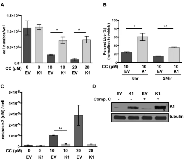
To evaluate the impact of K1 expression by itself on cell survival following treatment with the AMPK inhibitor, compound C, we created FLAG epitope-tagged K1 (K1) or empty vector (EV) HEK-293 stable cell lines. The EV or K1 HEK-293 cells were treated with increasing concentrations of compound C, and cell viability was evaluated by trypan blue exclusion assay. We observed an increased number of viable cells in the K1 expressing cells compared to EV expressing cells using two different concentrations (10 µM and 20 µM) of compound C (Fig. 3.3A). We
observed this difference at both 8 and 24 hours following incubation of the cells with 10 µM compound C (Fig. 3.3B). In order to determine whether K1 protected from apoptosis induced by AMPK inhibition, we also performed an assay to detect active caspase-3, which is an indicator of apoptosis. Active caspase-3 levels were
Figure 3.3. K1 expression provides a survival advantage in cells treated with
compound C. (A) HEK-293 cells stably expressing empty vector (EV) or FLAG-K1 (K1)
were treated in duplicate with 0 µM/ DMSO (0.2%), 20 µM compound C (CC) or 10 µM
compound C for 6-8 hours. (B) HEK-293 cells stably expressing EV or K1 were treated in
duplicate with 0 µM/DMSO (0.1%) or 10 µM compound C (CC) for 8 and 24 hours. Cell
viability was determined by trypan blue exclusion assay. Percentages were derived from
normalization to vehicle treated samples. (C) The level of caspase-3 activity was determined
and normalized to cell number. (D) EV or K1 HEK-293 cells treated with either 0 µM/DMSO
The KSHV K1 protein associates with the gamma subunit of AMPK
(AMPKγ1). To investigate how K1 may be promoting cell survival following exposure to metabolic stress, we wanted to determine cellular proteins associated with K1. We identified K1-associated cellular proteins by performing tandem affinity purification of K1 from HEK-293 cells and subjecting cellular proteins bound to K1 to mass
spectrometry.
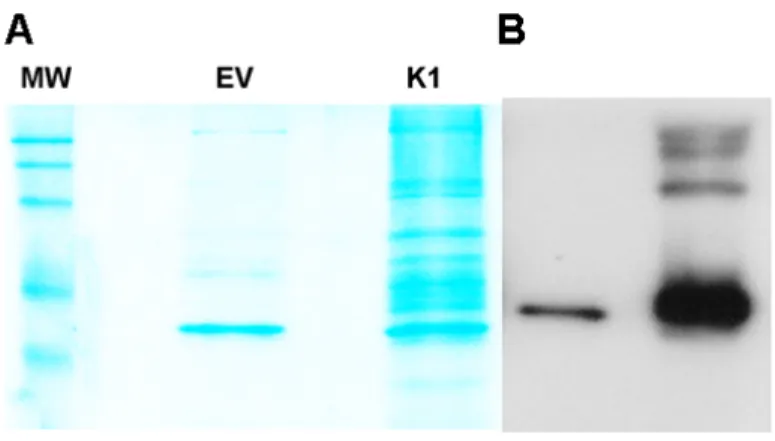
Stable cell lines expressing a FLAG and HA double epitope-tagged version of K1 and EV HEK-293 cells were generated as previously described [145]. For tandem affinity purification, FLAG-HA-K1 or EV HEK-293 expressing cells were lysed with NP40 buffer. The clarified lysates were incubated with anti-FLAG M2 affinity gel, washed with NP40 buffer, and then eluted with 3X FLAG peptide. The eluates were subsequently incubated with an anti-HA resin, washed with NP40 buffer and eluted. The eluates were resolved by sulfate polyacrylamide gel
electrophoresis (SDS-PAGE) and Coomassie stained. Only bands that were present in the K1 lane and absent in the EV lane were isolated and submitted for
Figure 3.4. K1 associating proteins determined by mass spectrometry. A)Stable cell
lines expressing a FLAG and HA double epitope-tagged version of K1 and EV HEK-293
cells were lysed and the lysates were incubated with anti-FLAG M2 affinity gel, washed and
eluted with 3X FLAG peptide. The eluates were subsequently incubated with an anti-HA
resin, washed with NP40 buffer and eluted. The eluates were resolved by sulfate
polyacrylamide gel electrophoresis (SDS-PAGE) and Coomassie stained. (B)The eluates
from (A) were also subjected to SDS-PAGE and Western blot analysis with an anti-FLAG
antibody to detect the presence of the monomer and oligomeric forms of K1.(C and D)
Mass spectrometry analysis identified HSP90 (C) and AMPKγ1 also known as PRKAG1 (D)
as K1 associating proteins. The peptide sequences of Hsp90 and AMPKγ1 identified by
mass spectrometry are shown. Asterisk indicates modified amino acids.
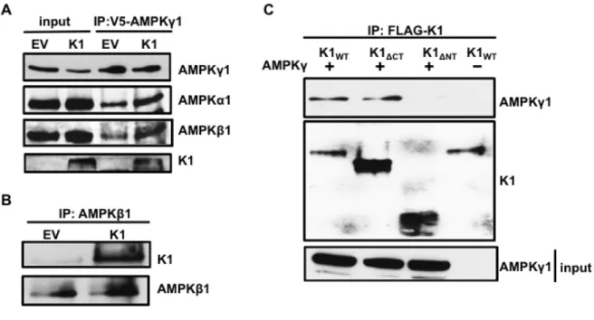
We also confirmed successful pull-down of FLAG-HA-K1 by detecting FLAG by SDS-PAGE and Western blot (Fig. 3.4B). Using mass spectrometry, we found AMPKγ1 associated with K1 (Figs. 3.4D and 3.5A). We also observed an
Figure 3.5. K1 associates with AMPKγ1. (A) Number of PRKAG1 (AMPKγ1) and HSP90
peptides identified in association with K1 by mass spectrometry. (B) HEK-293 cells stably
expressing empty vector (EV) or FLAG-K1 (K1) were transfected with V5-AMPKγ1
(AMPKγ1). AMPKγ1 was immunoprecipitated, and immunoblotted for K1 or AMPKγ1. (C)
HEK-293 cells stably expressing EV or K1 were transfected with AMPKγ1 or EV (pcDNA3).
K1 was immunoprecipitated and immunoblotted for AMPKγ1 or K1.
spectrometry, we transiently expressed AMPKγ1 in HEK-293 cells stably expressing EV or FLAG epitope-tagged K1 (K1). We performed a co-immunoprecipitation by incubating EV- or K1-expressing HEK 293 clarified lysates containing equal amounts of protein with anti-V5 antibody to pull down the V5 epitope-tagged AMPKγ1. We detected the multimer and monomer forms of K1 co-immunoprecipitating with
AMPKγ1 in K1-expressing cells, but not from EV control cells (Fig. 3.5B). To further substantiate the association between K1 and AMPKγ1, we performed the reverse immunoprecipitation and immunoprecipitated K1. We observed that AMPKγ1 co-immunoprecipitated with K1 from K1-expressing cells but not from EV control cells (Fig. 3.5C).
In the cell, AMPKγ1 complexes with AMPKα1 and AMPKβ1. In addition to AMPKγ1, we next wanted to determine whether the other AMPK subunits were part of the protein complex associated with K1. We transfected V5-AMPKγ1 in HEK-293 cells stably expressing empty vector (EV) or FLAG-K1 (K1), immunoprecipitated V5-AMPKγ1, and probed for endogenous AMPKα1 and AMPKβ1. In addition to
detecting K1 as we previously observed, we also observed the expression of
further confirming an association between K1 and the endogenous AMPK complex (Fig. 3.6B).
Figure 3.6. The K1 N-terminus is important for association with AMPKγ1. (A) HEK-293
cells stably expressing empty vector (EV) or FLAG-K1 (K1) were transfected with
V5-AMPKγ1 (AMPKγ1). AMPKγ1 was immunoprecipitated, and blots were probed for AMPKγ1,
endogenous AMPKα1, endogenous AMPKβ1 and K1. (B) Endogenous AMPKβ1 was
immunoprecipitated from EV or K1 expressing HEK-293 cells. The blots were probed for K1
or AMPKβ1. (C) HEK-293 cells were transiently transfected with AMPKγ1 and with one of
the following: K1WT, K1ΔCT or K1ΔNT. An equivalent amount of EV (pcDNA3) was also
transfected along with K1WT. We immunoprecipitated the various K1 domain mutants, and
immunoblotted for AMPKγ1 or K1.
domain deletion mutants and tagged AMPKγ1. We transiently expressed V5-AMPKγ1 (V5-AMPKγ1) along with one of the following in HEK-293 cells: K1WT, K1 lacking the C-terminus (K1ΔCT), or lacking the N-terminus (K1ΔNT). We also transfected an equivalent amount of EV (pcDNA3) and K1WT as a control. The construction of the FLAG-tagged K1 mutants has previously been described [145]. We immunoprecipitated K1WT, K1ΔCT, or K1ΔNT and probed for V5-tagged AMPKγ1 (Fig. 3.6C). As previously observed, we detected co-immunoprecipitation of AMPKγ1 and K1WT (Fig. 3.6C, lane 1). We also detected co-immunoprecipitation of AMPKγ1 and K1ΔCT indicating that the K1 C-terminus is not important for K1 and AMPKγ1 association (Fig. 3.6C, lane 2). We did not observe co-immunoprecipitation of AMPKγ1 and K1ΔNT suggesting that AMPKγ1 associates with K1 via the K1 N-terminus (Fig. 3.6C, lane 3).
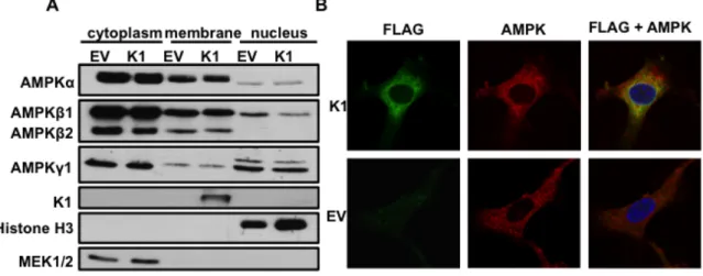
K1 and AMPK subunits are localized to cellular membranes. About 10-20% of K1 is localized to the plasma membrane with the major fraction of K1 being found in the endoplasmic reticulum [46]. K1 can also be internalized and internalization is associated with K1 signaling [146]. Additionally, all three subunits of AMPK have been shown to localize to the cellular membrane fraction [135]. We wanted to evaluate the localization of the endogenous AMPK subunits in EV- and expressing stable HEK-293 cells. We lysed equal numbers of EV- and
K1-expressing HEK-293 cells and separated the cellular fractions. The fractions were then resolved by SDS-PAGE and Western blot. The blots were probed using
in the cellular membrane fraction (Fig. 3.7A). Along with the AMPK subunits, K1 was also found in the membrane fraction (Fig. 3.7A). We evaluated the purity of the cytoplasmic, membrane, and nuclear fractions by probing for MEK1/2, K1, and histone H3 respectively (Fig. 3.7A). These proteins are restricted to each of these fractions. Based on these findings, we conclude that K1 and multiple AMPK subunits and isoforms (without over expression) are localized to the cellular membrane
fraction, suggesting that K1 associates with AMPK in the cellular membrane.
Figure 3.7. K1 and AMPK are visualized in membranes and the perinuclear area of the
cell. (A) HEK-293 cells stably expressing empty vector (EV) or FLAG-K1 (K1) were lysed
and fractionated into cytoplasmic, membrane and nuclear fractions. Immunoblots were
probed for endogenous AMPK subunit and isoforms. (B) HUVEC stably expressing empty
(EV) or FLAG-K1 (K1) were fixed and permeabilized. Cells were then stained with anti-FLAG
directly conjugated to FITC antibody, and an AMPKβ1/2 antibody, followed by an anti-rabbit
conjugated to Alexa Fluor 647. Nuclei were stained with DAPI. Stained HUVEC were
We subsequently evaluated localization of FLAG-K1 (K1) and endogenous AMPK by immunofluorescence staining. Because our AMPKγ1 antibody is not appropriate for immunofluorescence staining, and we had determined that AMPKβ1 co-localized with K1 (Fig. 3.6B), we stained for K1 and endogenous AMPKβ1/2 in EV- or K1- stably expressing HUVEC. We fixed the cells with formaldehyde, washed and then permeabilized the cells with Triton-X-100. We stained cells with FLAG-K1-FITC and AMPKβ1/2 antibodies followed by an anti-rabbit Alexa Fluor 647
secondary antibody. By confocal microscopy, we acquired z-stacks on fully stained EV and FLAG-tagged K1 HUVEC (Fig. 3.7B). We also completed z-stacks on controls containing only the secondary anti-rabbit AF647 in order to demonstrate that the staining for AMPKβ1/2 is specific and not due to non-specific secondary staining. We observed co-localization of K1 and endogenous AMPKβ1/2 in the perinuclear area (Fig. 3.7B) as determined by a Mander’s overlap coefficient of 0.83, which was determined using ImageJ software. The EV transfected cells stained positive for AMPKβ1/2 but not K1, as expected.
In Fig. 3.6C we had observed that K1ΔNT does not associate with AMPKγ1 by co-immunoprecipitation. K1ΔNT, lacks the signal peptide sequence, therefore we wanted to confirm that its lack of association with AMPKγ1 is not due to being mislocalized in the cell. Thus, we transiently expressed K1ΔNT, and fractionated the cell lysates into cytoplasmic, membrane and nuclear components. We then
the fractionation efficiency by immunoblotting for MEK1/2, AIF, and Histone H3, indicators for the cytoplasm, membrane and nucleus, respectively. To corroborate our previous findings, we confirmed that K1WT and AMPKγ1 associate in the membrane fraction and that there is loss of K1ΔNT and AMPKγ1 association in the membrane fraction by co-immunoprecipitation. Thus, we immunoprecipitated EV (pcDNA3), K1WT, K1ΔCT, or K1ΔNT and probed for V5-tagged AMPKγ1. We detected K1WT and AMPKγ1 association, and we did not detect K1ΔNT and AMPKγ1
association in the membrane fraction (Fig. 3.8B).
Figure 3.8. K1 N-terminus is important for K1 association with AMPKγ1 in the
membrane fraction. (A) HEK-293 cells were transfected with EV (pcDNA3), K1WT, K1ΔCT,
and K1ΔNT, and then fractionated into cytoplasmic (cyto.), membrane (memb.) and nuclear
fractionation efficiency was evaluated by detecting AIF (membrane), MEK1/2 (cytoplasm),
and Histone H3 (nucleus). (B) HEK 293 cells were transfected as described in (A) with the
addition of V5-AMPKγ1. The cells were fractionated, and FLAG was immunprecipiated using
the membrane fraction. V5-AMPKγ1 was detected by immunoblot.
K1 and AMPK association is important for cell survival following exposure to stress. Thus far, we have observed that KSHV K1 promotes survival in stressed cells, and K1 associates with AMPK via the K1 N-terminus. We next wanted to determine whether the association of K1 and AMPK is important for the survival advantage observed in stressed cells. We generated lentivirally transduced HEK-293 cells stably expressing FLAG epitope-tagged K1WT, K1ΔCT, K1ΔNT and empty vector (EV). We treated these cells with the AMPK inhibitor, compound C, and evaluated cell viability using the MTS assay. We observed an increased percentage of viable K1WT expressing cells when AMPK was inhibited, compared to cells
expressing K1ΔNT. This result suggests that the association between K1 and AMPK is important for survival in stressed cells (Fig. 3.9A). Surprisingly, we also observed that K1ΔCT expressing cells appear sensitive to AMPK inhibition, indicating that the K1-C terminus is also important for survival in stressed cells (Fig. 3.9A). We
confirmed expression of the various K1 constructs by completing a Western blot using an anti-FLAG antibody or an anti-K1 antibody (Fig. 3.9B). When we
expression but no K1ΔCT expression since the K1 antibody is directed towards an epitope on the K1 C-terminus (Fig. 3.9C).
Figure 3.9. K1 and AMPK association is important for cell survival following exposure
to stress. (A) HEK-293 cells stably expressing FLAG-tagged K1WT, K1ΔCT, K1ΔNT or empty
vector (EV = pLenti CMV) were treated with 10 or 7.5 µM compound C for 48 hours. Cell
cells was determined by normalization to the DMSO (0.1%) control. Error bars represent the
standard deviation of biological triplicates. (B) Western blot showing expression of EV, K1WT,
K1ΔCT, and K1ΔNT using an anti-FLAG antibody. (C) Western blot from (Fig. 7B) was stripped
and probed for K1 using an anti-K1 antibody. GraphPad Prism was used to determine
one-way ANOVA and Tukey’s post-test **P<0.005
K1 facilitates AMPK activity in stressed cells. We found that K1 associates with AMPK and this association is important for the survival advantage in stressed cells. We next wanted to determine the status of AMPK activity in stressed EV and K1 expressing cells. We exposed HUVEC stably expressing empty vector (EV) or FLAG-tagged K1 (K1) to media without serum and growth factors containing either compound C or DMSO control for 24 hours. We then performed an AMPK-specific kinase activity assay. We incubated lysate from EV or K1 expressing HUVEC with or without an AMPK substrate, a synthetic peptide called SAMS peptide
Figure 3.10. K1-expression facilitates AMPK activity in stressed cells. (A) HUVEC
stably expressing empty vector (EV) or FLAG-K1 (K1) were deprived of serum and growth
factors (starve) for 24 hours. At the time of starvation, 5 µM compound C or DMSO (0.05%)
was added. Each condition for EV and K1 was completed in triplicate. AMPK-specific activity
was determined by subtracting counts per minute (cpm) derived for each sample incubated
without SAMS peptide from cpm derived from each sample incubated with SAMS peptide.
These values were then normalized to total protein for each sample. The error bars
represent the standard deviation of triplicates. (B) Western blot of K1 and tubulin using
lysate from the AMPK activity assay. *P<0.05 Student’s t test
Figure 3.11. K1 maintains an active PI3K pathway despite AMPK activation following 8
hours of serum starvation. HUVEC stably expressing EV or K1 were treated with vehicle
(DMSO), serum starved for 8 hours, or treated with 2 µM oligomyicn, an ATP synthase
inhibitor one hour prior to cell harvest. The levels of phosphorylated and total proteins were
determined by Western blot.
DISCUSSION
Cell survival during KSHV infection is paramount to the establishment of life-long infection. Upon infection, KSHV primarily enters a latent state. During latency, KSHV expresses a limited number of proteins and microRNAs that enable it to successfully persist in the cell by avoiding the immune response and by promoting cell survival [150]. When KSHV reactivates and enters the lytic stage, the cell must remain viable throughout viral replication and virion assembly so that infectious virions are generated. Cell death prior to completion of the lytic program would result in defective viral replication.
We observed that KSHV infected cells containing a WT K1 gene had a survival advantage compared to cells infected with KSHV K1 mutants following exposure to stress. To explore the underpinnings of this phenotype, we completed tandem affinity purification and mass spectrometry to identify K1-associating proteins. We identified AMPKγ1 as a K1-associating protein. We corroborated this finding independently and found that K1 co-immunoprecipitated with AMPKγ1. By
between K1 and AMPK is important for the survival advantage in stressed cells because we observed reduced cell viability in cells expressing K1ΔNT compared to K1WT. Our studies indicate that KSHV K1 promotes survival via its association with AMPK, and KSHV K1 facilitates AMPK activity in stressed cells.
Under normal culture conditions, our lab and others have shown that K1 activates the PI3K/Akt/mTOR pathway [50, 51]. It has been reported that cells having overly active Akt and consequently a high glycolytic rate are more sensitive to cell death following starvation compared to control cells [151]. Moreover, when starved-cells are treated with an activator of AMPK, cells with active Akt are protected from cell death [151]. KSHV-infected cells also have an active
PI3K/Akt/mTOR pathway and a high glycolytic rate [80, 152, 153]. Furthermore, simultaneous activation of AMPK, Akt and mTOR, has been observed in liver cancer cells following nutrient starvation [154]. Thus, low levels of AMPK activation may promote metabolic adaptation and consequently, increase KSHV-infected cell
survival. We propose that K1 promotes AMPK activity during metabolic stress and in this way enhances KSHV-infected cell survival.
SERCA and/or IP3Rs can be modulated by pro-tumorigenic factors such as BCL-2 and myc, thus, promoting cell survival in a changing tumor microenvironment [155].
AMPKα2 expression promotes SERCA activity and reduces ER stress in HUVEC cells [157]. Although interaction between K1, SERCA and AMPK has not been confirmed by co-immunoprecipitations, we speculate that K1 modulates SERCA activity by altering AMPK activity and consequently, alleviates ER stress. Increased ER stress can be detrimental to the cell and result in apoptosis [158].
The role of AMPK during herpesvirus infection is complicated, and whether it promotes viral replication or inhibits it may depend on a variety of factors. During human cytomegalovirus (HCMV) infection, AMPK has been found to promote a metabolic environment that is conducive to viral replication [159, 160]. AMPK
inhibition blocks increased glycolysis that is induced by HCMV infection and inhibits viral DNA synthesis [160]. Interestingly, treatment of HCMV-infected fibroblasts with the AMPK activator, AICAR [159, 161] or the inhibitor, compound C, results in reduced HCMV replication [159]. During human herpes simplex virus-1 (HSV-1) infection, AMPK activity facilitates neuron survival and reduces viral production [162]. Thus, AMPK appears to impact viral production differently during HCMV and HSV-1 infection.