Abstract
The cellular interior is crowded, with macromolecules occupying from 10% to 40%
of the volume.1 Under these conditions, proteins experience hard-core repulsions and
chemical interactions with cytoplasmic components.2,3 Hard-core repulsions stabilize
globular proteins, whereas chemical interactions can be either repulsive and stabilizing,
or attractive and destabilizing.2,3 Several studies have considered crowding effects on
globular protein stability4–7, but there are few such studies on protein-protein interactions. We used 19F NMR to quantify the effects (298 K, pH 7.5) of macromolecular cosolutes on
a variant of the B1 domain of protein G (GB1) that forms a domain-swapped homodimer.8
At a concentration of 200 g/L, the monomer of the synthetic polymer polyethylene glycol
(PEG) destabilizes the dimer by 0.30 kcal/mol, while at the same concentration, 3.3
kDa-, 8 kDa- and 20 kDa- PEG stabilize the dimer by 0.08kDa-, 0.39 and 0.4 kcal/molkDa-, respectively.
These data indicate a stabilizing, but saturable, macromolecular effect. We also showed
that the physiologically-relevant cosolutes bovine serum albumin (BSA) and lysozyme
have opposite effects; the former (at 100 g/L) stabilizes the dimer by 0.51 kcal/mol, and
the latter (at 50 g/L) destabilizes the dimer by 0.12 kcal/mol. These results can be
explained by the differences in charge. BSA has the same charge as GB1, resulting in
stabilizing repulsions. Lysozyme and GB1 have complementary charges, resulting in
destabilizing attractions. The differing effects of PEG and the protein cosolutes indicate
that synthetic polymers are poor mimics of the cellular interior because they do not
Background: Proteins are mostly studied in dilute buffer, where the concentration of macromolecules is <10 g/L. However, the cytoplasm is highly crowded milieu where the
concentration of macromolecules can exceed 300 g/L.9 At these concentrations, the
effects of cosolutes on protein stability can no longer be ignored.2 Although such
interactions can be described as weak and transient, they play a major role in protein
function and stability.4,7
These transient cosolute-protein interactions can be fall into one of two categories:
hard-core repulsions and chemical interactions.2 Hard-core repulsions are steric
interactions that arise from a decrease in the available volume.2 Chemical interactions
can be briefly defined as nonspecific interactions between the protein and molecules in
solution (hydrogen bonding, polar/nonpolar interactions, etc.).2
Large polymers, such as polyethylene glycol (PEG) and Ficoll, have often been used
to simulate these high concentration conditions. These solutes cause for the weak
transient interactions to manifest themselves in the form of hard-core repulsions and
chemical interactions.2 Hard-core repulsions are steric interactions that arise from a
decrease in the available volume.3 A decrease in volume pushes a single globular
protein to a more compact state.2
Then, chemical interactions may be either repulsive or attractive. These arise from
transient interactions between the protein of interest and the cosolutes in solution.2 If
they are favorable (hydrogen bonding, polar/nonpolar interactions, etc.), then they lead
to the globular protein favoring a more open state (opening yields more points of contact
globular protein becomes more compact (closing yields less points of contact for
unfavorable interactions).2
However, all of these observations are made for single globular proteins. Although it
is easier to think of cells as a mass of globular proteins acting in tandem, it is incorrect
to say that they act alone. There are a myriad of protein-protein interactions (PPIs) that
are involved in everything from metabolism, to cellular structure, to even disease.10–12 A variant (L5V;F30V;Y33F;A34F) of the monomeric B1 domain of protein G (GB1)
will serve as the test protein for
understanding the effects of
crowding on PPIs (figure 1).
The mutations destabilize the
monomeric GB1 protein.8
However, the variant undergoes
intermolecular domain swapping
through exchanging second -hairpins each, forming a thermodynamically favorable
structure compared to the destabilized monomer.8
GB1 contains one tryptophan,
which can be fluorine-labeled using
the metabolic precursor
5-fluoroindole, allowing 19F nuclear
magnetic resonance (NMR) to be
used to observe both the dimer
and monomer states. 19F is NMR–active, 100% abundant, rarely found in biological Figure 2: The addition of the 5-fluoroindole (left) to the media allows us to monitor the monomer and dimer states by 19F NMR (right).
systems, has 83% of the NMR sensitivity of proton, and a chemical shift highly sensitive
to environment.13,14
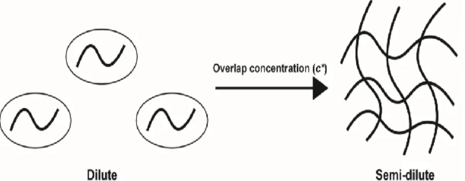
The physical behavior of polymers vary based on concentration, and these shifts in
behavior have been shown have an effect on proteins.4 Polymers exist in several
different states based on concentration, two of which are dilute and semi-dilute.15 In the
dilute state, the individual polymer molecules can be thought of as individual molecules
that are not interacting. The semi-dilute state is much different, where the polymer
molecules stop acting like individual spheres and begin to interact, forming a mesh.15
The concentration at which this transition from the dilute to the semi-dilute regime
occurs is called the overlap concentration (c*).15 This behavior is summarized in Figure
3. These polymer effects are governed by many factors, one of which is their chain
length. To better
understand the effects of
macromolecular
polymerization on
dimerization, we will study
the effects of long-chain
polymers on dimer stability.
Results
Hard core repulsions, monomers of polymers. The polymers 8 kDa PEG and 70 kDa
Ficoll were used to probe hard core-repulsions. Solutions were made to a final
concentration of 200 g/L and 300 g/L of 8 kDa PEG and 70 kDa Ficoll, respectively.
Ethylene glycol (the monomer of PEG) and sucrose (the monomer of Ficoll) were used
at the same concentration as their polymer counterparts. Buffer containing 300 g/L 70
kDa Ficoll yielded a KD→ M of 27 ± 2 μM, and buffer containing 200 g/L 8 kDa PEG
yielding a KD→ M of 46 ± 3 μM. Buffer containing 200 g/L ethylene glycol resulted in a
KD→M of 153 ± 12 μM, while buffer
containing 300 g/L sucrose
yielding a KD→M of 58 ± 4 μM.
These data were used to calculate
the ΔΔ𝐺𝐷→𝑀′𝑜 , and uncertainties
were calculated as standard
deviation of the mean from
triplicate analysis. These results
are summarized in figure 4.
Chemical interactions. We first tested dimer stability in two controls: 100 g/L urea and
38 g/L trimethylamine-N-oxide (TMAO). Urea was used since it is a protein denaturant
from its high degree of backbone interactions.16 As such, we expected it to favor the
open, unstable monomer state, making it a good control for attractive chemical
interactions. TMAO has the opposite effect of urea, in that it excludes backbone instead
of interacting with it.17 We expected it to favor the state with the most excluded
backbone, in that we expect it to favor the compact, structured dimer state, making it a
good control for repulsive interactions. This was shown to be true, in that the addition of
100 g/L urea to the buffer led to a KD→M of 840 ± 40 μM and the addition of 38 g/L
TMAO to the buffer led to a KD→M of 68 ± 4 μM.
Next, we were interested in picking an experimental cosolute to probe attractive
interactions, and another that could probe repulsive interactions. This choice was made
easier when we considered protein charge. At pH 7.5, the GB1 variant has a charge of
-8.3. As such, we wanted to choose
one protein which is positively charged
at pH 7.5 for probing attractive
interactions and another which is
negatively charged at pH 7.5 for
probing repulsive interactions. We
chose lysozyme and BSA, which have
charges of +7.1 and -37.9 at pH 7.5,
respectively. The addition of 100 g/L
BSA to buffer resulted in a KD→M of 38
± 3 μM and the addition of 50 g/L
lysozyme to buffer resulted in a KD→M of 110 ± 5 μM. A lower amount of lysozyme was
added than that of BSA because the peaks were too broad to be analyzed at 100 g/L
lysozyme. These data were used to calculate the ΔΔ𝐺𝐷→𝑀′𝑜 , and uncertainties were
calculated as standard deviation of the mean from triplicate analysis. These results are
summarized in figure 5.
The effects of polymer chain length. To test the effects of the molecular weight of the
cosolute, we tested the stability of the domain swapped dimer in several different types
of PEG. We used 20 kDa PEG and 3.35 kDa PEG to test the effects of decreasing and
increasing the polymer molecular weight on dimer stability.
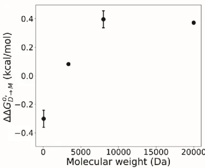
The concentration in
buffer of the 3.35 kDa and 20
kDa PEG was 200 g/L. The
3.35 kDa PEG adjusted the
stability by 0.08 kcal/mol, while
the 20 kDa PEG adjusted the
dimer stability by 0.37 kcal/mol.
These results were compared
with the 8 kDa PEG and
ethylene glycol data in figure 6
and.
Discussion
Domain swapped proteins vary greatly in function, with functions varying greatly
from toxins to circadian clock regulators.18,19 As such, it is of great importance to
understand the effects of the cellular interior on dimers formed in this manner. We
characterized the effects of many different cosolutes on the GB1 domain-swapped
homodimer, and all facets of the data have yielded interesting results.
Figure 6: Plot of molecular weight versus ΔΔ𝐺𝐷→𝑀𝑜 ′
The synthetic polymer cosolutes used to generate hard-core repulsions led to
stabilizing of the GB1 domain swapped homodimer. The addition of 200 g/L 8 kDa PEG
led to a stabilization of the dimer by 0.39 ± 0.06 kcal/mol, and the addition of 300 g/L 70
kDa Ficoll led to a stabilization by 0.71 ± 0.06 kcal/mol. The monomers had a different
or diminished effects than the polymers they constituted. Although 70 kDa Ficoll
stabilized the dimer by 0.71 ± 0.06 kcal/mol, the sucrose monomer only stabilized the
dimer by 0.26 ± 0.06 kcal/mol. Surprisingly, while 8 kDa PEG stabilized the dimer by
0.39 ± 0.06 kcal/mol, the ethylene glycol monomer destabilized the dimer by 0.30 ± 0.06
kcal/mol. The differences in the cosolute effect of the monomers and polymers suggest
that upon formation of the polymer, changes
occur in how the molecules interact with the
protein. This is called the macromolecular effect,
and has been previously observed with PPIs.20,21
This best manifests itself in ethylene glycol and 8
kDa PEG. Ethylene glycol contains two hydroxyl
groups (figure 7). These hydroxyl groups most likely have attractive chemical
interactions with the protein surface. These attractive interactions are maximized in the
monomer state since it has the most exposed surface area, pushing the equilibrium
towards the monomer. Upon formation of the PEG polymer, many of these hydroxyl
groups will become buried, and excluded volume will play a larger role as these groups
become buried. Now, the more compact dimer state is favored.
The macromolecular effect observed with the hard core repulsions experiments
prompted us to carry out experiments to verify this observation. To do so, we carried out
the experiments with the different sizes of PEG. The data show more evidence for a
macromolecular effect. The stability data from 3.35 kDa PEG fill the gap between
ethylene glycol and 8 kDa PEG. Since the 3.35 kDa point lies between the 8 kDa PEG,
this is more conclusive evidence that a macromolecular effect is present upon
polymerization of the chain. With the effects that are observed, I hypothesize that the
effects of PEG will approach ethylene glycol as the chain length is decreased.
These data are also interesting due to the 20 kDa point, in that there was not an
increase in stability as the PEG lengthens from 8 kDa to 20 kDa. This inform us that the
macromolecular effect present is saturable. Then, it also highlights some of the
importance of chemical interactions. If the crowder were to be a hard, impenetrable
sphere, we might expect that the protein would be more stable with a larger polymer.
However, this is not what a true polymer acts as, since it will have chemical interactions
with the test protein. Furthermore, we must also consider the polymer overlapping
shown in Figure 3. Although I have not measured the overlap concentrations, others
have measure the overlap concentrations of different PEGs. It was found that 6 kDa and
20 kDa PEG had a c*of 119.2 and 50.9 g/L, respectively.22 Since c*is directly
proportional to molecular weight22, this tells us that at 200 g/L, we must be over the c*
concentration for 8 kDa and 20 kDa PEG. As such, we would want to carry out
experiments below the c* of the PEG crowders to better understand the effects of
polymer overlap on protein stability.
Urea and TMAO had similar effects on PPIs as they do on protein folding.5 Urea
modulated dimer stability by -1.31 ± 0.05 kcal/mol, and TMAO modulated dimer stability
of the protein cosolutes. BSA has a charge of -18 at a pH of 7.5, while lysozyme has a
charge of +8 at a pH of 7.5. Lysozyme modulated dimer stability by -0.12 ± 0.05
kcal/mol. This destabilization arises due to the positive changes on the surface of
lysozyme interacting with the negative charges on the surface of GB1 (figure 8). Since
the monomeric species have more surface area available for interacting with the
lysozyme, the monomer is favored. BSA has the opposite effect, where it modulates
dimer stability by 0.51 ± 0.07 kcal/mol.
Here, the negatively charged BSA has
repulsive interactions with the
negatively charged patches of GB1. To
minimize these interactions, the dimer
is favored since it has less exposed
surface area than the individual
monomers.
Conclusions
Our study emphasizes the importance of macromolecular crowding on a
domain-swapped homodimer. We used a variety of cosolutes to test the effects of hard-core
repulsions and chemical interactions. We found that hard-core repulsions favored
dimerization. The use of polymers and their monomers led to us observing a
macromolecular effect. This result was supported by the experiments with differing
molecular weights of PEG, where we found that there was evidence of a saturable
macromolecular effect. Chemical interactions varied for each of the selected cosolutes, 180⁰
+
-
but we found that repulsive interactions stabilized the dimer while attractive interactions
destabilized the dimer. Due to the stark differences between the protein and polymer
cosolutes, this work shows that polymer cosolutes are not good models of the cellular
interior. The results of the macromolecular effect experiments are interesting in that they
agree with past work showing similar effects for PPIs.20,21 Those results also show a
sharp dichotomy between protein folding and PPIs. With protein folding, it was found
that monomers were more effective at stabilizing proteins than the polymers they make
up5, whereas it was found that polymers were more effective at stabilizing the dimer
(figure 4). The future directions of this work could include sampling concentrations
above and below the c* of the polymers to better understand the effects of polymer
overlap on dimer stability and also carrying out temperature experiments to calculate
the enthalpic and entropic components of the modulation of dimer stability.5
Materials and Methods
Vector. A pET11a plasmid containing the GB1 A34F variant was used as the wild-type vector. Agilent’s QuickChange mutagenesis kit was then used to induce the other
mutations (L5V;F30V;Y33F) to make the domain-swapped homodimer variant.
Protein expression and purification. The plasmid encoding the GB1 mutant was transformed into competent BL21 (DE3) Gold Escherichia coli cells and spread onto
LB-agar plates containing 100 µg/mL ampicillin. Following overnight incubation at 37 ºC, a
single colony from the plates was used to inoculate a 25-mL overnight culture in LB
225 rpm at 37 ºC (New Brunswick Scientific, model I26). This overnight culture was
used to inoculate a 975 mL culture in M9 minimal medium (50 mM Na2HPO4, 20 mM
KH2PO4, 9 mM NaCl, 1 g/L NH4Cl, 4 g/L glucose, 2 mM MgSO4, 10 mg/mL thiamine
HCl, 10 mg/mL biotin, 100 µM CaCl2, and 100 µg/mL ampicillin). The culture was grown
with shaking at 37 ºC, and its optical density at 600 nm (OD600) was monitored by
UV-vis spectroscopy (Biorad Spectra Plus). Once an OD600 of 0.400 was reached, 500 mg
of glyphosphate were added to inhibit aromatic amino acid synthesis, along with 60 mg
of L-phenylalanine, 60 mg of L-tyrosine, and 70 mg of 5-fluoroindole. Once the culture
reached an OD600 of 0.600, protein expression was induced by the addition of 1 mM
isopropyl-β-D-thiogalactopyranoside. After 2 h, cells were harvested for 30 min at 4000
rpm(RC-3B Refrigerated Centrifuge; Sorvall Instruments).
Harvested cells were resuspended in 25 mL buffer A (20 mM Tris-HCl, pH 7.65),
and 300 µL of protease inhibitor (Roche) were added prior to cell lysis. Cell lysis was
carried out by sonication (Fischer Scientific Sonic Dismembrator model 500) using the
following parameters: 15% amplitude, 0.50 s on, 0.50 s off, 20 min. The lysate was then
spun down (RC-5B Refrigerated Superspeed Centrifuge; Sorvall Instruments) at 10,000
rpm for 1 h.
The supernatant was filtered through a 0.22-µm syringe-driven unit (Millex), and
loaded onto an anion exchange column with Q Sepharose exchange resin (16 mm x
200 mm Q Sepharose; GE Healthcare) at 4 ºC on an AKTA Pure FPLC (GE
Healthcare). Buffer A was used to equilibrate, load lysate, and elute impurities. Buffer B
(20 mM Tris-HCl, 2 M NaCl at pH 7.65) was used to produce a linear gradient of 0-500
SDS-PAGE (4-20% Criterion TGX gels; Biorad) stained with Coomasie Brilliant Blue R-250.
Fractions containing the GB1 mutant were concentrated using a 3000 Da MWCO
centrifugal concentrator (Millipore). The concentrated sample was then filtered through
a 0.22-µm syringe-driven unit (Millex), and loaded onto a size exclusion
chromatography column (16 mm x 600 mm Superdex 75; GE Healthcare) at 4 ºC. The
column was equilibrated with two column volumes of 5 mM Na2HPO4, 2 mM KH2PO4,
0.9 mM NaCl buffer at pH 7.5. The eluate was assessed again using SDS PAGE, and
fractions containing the GB1 mutant were concentrated using a 3000 Da MWCO
centrifugal concentrator (Millipore). The concentrated protein was then exchanged thrice
into 18.00 MΩ deionized water. The protein concentration was measured using UV-vis
spectrophotometry (NanoDrop One). An extinction coefficient at 280 nm of 8400 L M-1
cm-1 was used. The protein was split into 500 µM aliquots and lyophilized for 12-16 h
(Labonco Freezone).
Crowder preparation. All crowders were dissolved in 20 mM sodium phosphate buffer, and the pH was adjusted to 7.5. For the preparation of protein cosolutes, lyophilized
lysozyme and bovine serum albumin were purchased from Sigma-Aldrich.
Concentration of the BSA and lysozyme solutions were monitored using using extinction
coefficients at 280 nm of 6700 L mg-1 cm-1 and 26400 L mg-1 cm-1, respectively. Fluorine-19 NMR. Fluorinated protein was resuspended in buffer (with or without cosolutes) to a final concentration of 500 µM. Experiments were consucted using a
bruker Avance III HD spectrometer operating at a 19F Larmor frequence of 470 MHz
equipped with a cryogenic QCI probe with an H/F channel. Over at least 128 scans,
100 pm, and a sweep width of 100 ppm. Samples were composed of 10% D2O for
locking.
Data analysis. The NMR spectra were analyzed using Topspin3.5pI6. An exponential
line broadening fucniton of 10 Hz was applied to each free induction decay prior to FT
analysis. The monomer and dimer peaks were integrated, and the fraction dimer (Fd)
was calculated as the ratio of the area of the dimer peak divided by the sum of the area
under the dimer and monomer peaks. These data were fit to equation [1] using
MATLAB (R2017A), where Pt is the total protein concentration and KD->M is the
equilibrium constant for dissociation.20
𝐹𝑑 =4𝑃𝑡+𝐾𝑑−√𝐾𝑑
2+∗8𝑃 𝑡𝐾𝑑
4𝑃𝑡 [1]
Acknowledgments
I would like to thank the Pielak lab for help on this project and past projects. I would
like to thank Annelis Gorensek-benitez for training me in the basics of protein
biochemistry and biophysics, and Alex Guseman for helping me along the way with this
specific project. Dr. Gary Pielak, my faculty advisor, provided useful scientific discussion
on this project and was a great mentor. I would also like to thank Dr. Gregory Young for
NMR spectrometer maintenance and help in carrying out the NMR experiments. Lastly, I
References
1. Rivas, G., Ferrone, F. & Herzfeld, J. Life in a crowded world. EMBO Rep. 5, 23–
27 (2004).
2. Sarkar, M., Li, C. & Pielak, G. J. Soft interactions and crowding. Biophys. Rev. 5,
187–194 (2013).
3. Minton, A. P. Excluded volume as a determinant of protein structure and stability.
Biopolymers20, 2093–2120 (1981).
4. Acosta, L. C., Goncalves, G. M. P., Pielak, G. J. & Annelise, H. Large cosolutes ,
small cosolutes and dihydrofolate reductase activity. Protein Sci. 26, 2417–2425
(2017).
5. Gorensek-benitez, A. H., Smith, A. E., Stadmiller, S. S., Goncalves, G. M. P. &
Pielak, G. J. Cosolutes, Crowding, and Protein Folding Kinetics. J. Phys. Chem. B
121, 6527–6537 (2017).
6. Monteith, W. B., Cohen, R. D., Smith, A. E., Guzman-Cisneros, E. & Pielak, G. J.
Quinary structure modulates protein stability in cells. Proc. Natl. Acad. Sci. U. S.
A.112, 1739–1742 (2015).
7. Smith, A. E., Zhou, L. Z., Gorensek, A. H., Senske, M. & Pielak, G. J. In-cell
thermodynamics and a new role for protein surfaces. Proc. Natl. Acad. Sci. U. S.
A.113, 1725–1730 (2016).
8. Byeon, I. J. L., Louis, J. M. & Gronenborn, A. M. A protein contortionist: Core
333, 141–152 (2003).
9. Zimmerman, Steven B.; Trach, S. O. Estimation Of Macromolecule Concentration
And Excluded Volume Effect For The Cytoplasm Of E.coli. J. Mol. Biol.222, 599–
620 (1991).
10. Pérez-Bercoff, Å., McLysaght, A. & Conant, G. C. Patterns of indirect protein
interactions suggest a spatial organization to metabolism. Mol. Biosyst.7, 3056 (2011).
11. Mondal, S. et al. Regulation of the actin cytoskeleton by an interaction of IQGAP
related protein GAPA with Filamin and Cortexillin I. PLoS One 5, (2010). 12. Gonzalez, M. W. & Kann, M. G. Chapter 4: Protein Interactions and Disease.
PLoS Comput. Biol.8, (2012).
13. Chen, H., Viel, S., Ziarelli, F. & Peng, L. 19F NMR: a valuable tool for studying
biological events. Chem Soc Rev42, 7971–7982 (2013).
14. Harper, D. B. & O’Hagan, D. The fluorinated natural products. Nat. Prod. Rep.11,
123–133 (1994).
15. M, R. & RH, C. Polymer Physics. (Oxford University Press, 2003).
16. Stumpe, M. C. & Grubmüller, H. Interaction of urea with amino acids: Implications
for urea-induced protein denaturation. J. Am. Chem. Soc. 129, 16126–16131
(2007).
17. Hu, C. Y., Lynch, G. C., Kokubo, H. & Montgomery Pettitt, B. Trimethylamine
Struct. Funct. Bioinforma.78, 695–704 (2010).
18. Bell, C. E. & Eisenberg, D. Crystal structure of diphtheria toxin bound to
nicotinamide adenine dinucleotide. Biochemistry 35, 1137–1149 (1996).
19. Ye, S., Vakonakis, I., Ioerger, T. R., LiWang, A. C. & Sacchettini, J. C. Crystal
Structure of Circadian Clock Protein KaiA from Synechococcus elongatus. J. Biol.
Chem.279, 20511–20518 (2004).
20. Guseman, A. J. & Pielak, G. J. Cosolute and Crowding Effects on a Side-By-Side
Protein Dimer. Biochemistry56, 971–976 (2017).
21. Munari, F. et al. Identification of primary and secondary UBA footprints on the
surface of ubiquitin in cell-mimicking crowded solution. FEBS Lett.591, 979–990
(2017).
22. Ziębacz, N., Wieczorek, S. A., Kalwarczyk, T., Fiałkowski, M. & Hołyst, R.
Crossover regime for the diffusion of nanoparticles in polyethylene glycol