Application of Intermittent Vibration to Fluctuating Pressure Pain Nathan Ahlgrim
University of North Carolina at Chapel Hill Spring 2014
A thesis presented to the faculty of The University of North Carolina at Chapel Hill in partial fulfillment of the requirements for the Bachelor of Science degree with Honors in Psychology.
Advisor _________________________
Committee Member _________________________
Abstract
The effects of chronic pain are debilitating and long-lasting, which make traditional tools like pharmaceuticals impractical and ineffective. One alternative treatment is vibratory stimulation to promote pain gating: decreased pain in the presence of innocuous tactile stimuli. However, evidence concerning the parameters to maximize pain modulation is contradictory. Earlier work has shown that vibration can even increase pain in those who sensitize rapidly to repetitive heat stimuli, suggesting that the application of vibration to increasing pain can exacerbate the change (Hollins, Harper & Maixner, 2011). The present study changed the force of a pressure stimulus to elicit large changes in pain and selectively applied vibration while subjects reported their pain as increasing or decreasing. Although the vibration condition did not cause a change in overall pain, the presence of vibration attenuated the pain fluctuations. A similar phenomenon was found when runs were grouped by order: earlier runs created smaller increases and decreases of pain. These findings suggest that offset analgesia is related to vibratory pain gating and
Acknowledgements
First and foremost, I would like to express my gratitude to my advisor, Dr. Mark Hollins, for his guidance and advice throughout this project. I was always challenged to expand my understanding of the topic, and his mentorship throughout the year was a driving factor in the success of this project. His approach to mentorship not only helped me through this project, but supported my professional growth as well.
I would also like to thank the members of my honors committee, Drs. Mark Hollins, Joseph Hopfinger, and Glenn Matsushima. Their willingness to donate their time and expertise is a testament to their commitment to continuing education and mentorship.
The help and support I received in the Somatosensory Lab, especially from Dr. Mark Hollins, Mr. Daniel Harper, and Ms. Kelly Barry, was invaluable in my approach and analysis of the project. The expertise of Mr. Nathan Roach and Mr. Daniel Horschler in LabView
programming was instrumental in the construction of this project.
Finally, I would like to thank the Psychology Department Honors Program, including the instructors Dr. Beth Kurtz-Costes and Dr. Mark Hollins, for facilitating this experience. I am thankful for the experience granted me over the past year, which facilitated my post-graduate opportunities.
Intermittent Application of Vibration to Fluctuating Pressure Pain
For the majority of the population, pain is an unavoidable experience. Although pain is by its nature an undesirable feeling, it often serves a useful and beneficial purpose. Experiencing pain alerts individuals to danger, whether from a hot stove or a damaging movement, and the desire to rest while in pain promotes the healing process. Even so, individuals suffering from acute or chronic pain often seek relief through painkilling medication to optimally perform throughout the day. Many methods of analgesia exist, but the most promising treatments for chronic pain would not require drugs or any foreign substance. In an effort to create a minimally intrusive treatment to relieve pain, we hope to exploit the organization of the somatosensory system to beneficially manipulate the perception of pain.
The Importance and Mechanics of Pain
Perhaps the most colorful illustration of the utility of pain comes from those who live without that experience. People with congenital insensitivity to pain never feel the burning of a hot stove, the throbbing of a bruised shin, or any other painful experience. This condition often leads to severe and chronic injury patterns because this population never feels the warning signs to protect themselves from painful stimuli (Nagasako, Oaklander & Dworkin, 2003).
musculoskeletal injury. Since their pain is not derived from a typical source like inflammation, traditional analgesics like anti-inflammatory drugs often have a limited or absent effect
(Leventhal, 1999), so other avenues of pain relief are greatly needed.
To effectively modulate the pain experience, the mechanisms of pain must first be understood. Importantly, the perception of pain is influenced not only by the physical stimulus, but also many subjective factors. Cognitive processes can significantly influence how
individuals experience and deal with pain. The intensity of pain subjects report can increase or decrease based on attention directed towards or away from the painful stimulus, respectively (Bantick et al., 2002; Wiech, Ploner & Tracey, 2008). Such a method does not exploit a known physiological mechanism to counteract or block a pain pathway, and yet directed attention practices like meditation have still been used effectively to reduce pain (Perlman, Salomons, Davidson & Lutz, 2010). A slightly different cognitive process of expectation has also consistently been shown to be an effective analgesic in the form of the placebo effect. With nothing more than belief in a treatment, many populations can have pain reduced by the
application of a completely inert substance (Wager et al., 2004). Neurophysiological recordings of subjects expecting an effective treatment have shown significant changes in cortical
somatosensory-evoked potentials, suggesting the modulation of pain intensity to be of cortical origin (Fiorio et al., 2012). Successful analgesia in the absence of physiological manipulation highlights the subjective nature of pain.
Bingham, Bathon & Haythornthwaite, 2006). In experimental settings, those who regularly catastrophize their pain report the same stimulus intensity as more painful when compared to healthy controls. This negative outlook is often coupled with hypervigilance, a heightened sensitivity to and awareness of painful, unpleasant, or worrisome sensations (McDermid, Rollman & McCain, 1996). The processes that increase pain can interact with and effectively negate those that alleviate pain. In a study conducted by Campbell and colleagues (2010), distraction away from a painful stimulus reduced pain ratings in all subjects, yet this reduction was delayed in the subjects who catastrophized about their pain. Their results suggest an
interplay between the two mechanisms. Thus, even if chronic pain patients are taught to distract themselves from their pain, negative cognitive processes may block the therapeutic benefits of distraction. These cognitive processes combine with the physical sensation of chronic pain, and make such diseases even more debilitating.
conditions for temporal summation as well as conditions with a longer interstimulus interval that does not allow for temporal summation (Hollins, Harper & Maixner, 2011).
Data from chronic pain patients were key in determining at what level the magnification of pain occurred during sensitization. The rate subjects sensitized to the stimulus depended on the stimulus intensity, not the subjective pain rating, and subjects exhibiting hypervigilance or catastrophizing did not show a more profound sensitization. Such a pattern suggests that the phenomenon occurs at a precortical level (Hollins et al., 2011). Many phenomena like central sensitization of second pain that modulate pain perception have been found to act before
associated cognitive processes. The interactions of multiple types of afferent fibers in the spinal cord have been found to play crucial roles in precortical pain modification. The spinal cord is the first place where tactile and noxious signals meet and interact, so tactile stimuli can also be manipulated to change the pain experience (Le Bars, Dickenson & Besson, 1979).
Touch, Vibration and Other Somesthetic Sensations
The direct interaction of non-noxious stimulation and the perception of pain is demonstrated in the phenomenon of allodynia, the sensation of pain in reaction to normally innocuous stimuli. When subjects are experiencing delayed-onset muscle soreness (DOMS), mechanical stimulation like gentle pressure or vibration causes pain. In a study using large-fiber nerve blocks to impede mechanoreceptor signals, allodynia derived from DOMS was found to be a product of distortion of signals from mechanoreceptors. That is, pain is still felt even when nociceptors do not fire (Weerakkody et al., 2003). The fact that stimulation of mechanoreceptors can create a painful sensation suggests that the two modalities are physiologically linked.
Connecting the two sensations opens possibilities of modulating pain through tactile stimuli Vibration has long been a useful tactile stimulus in experimental settings because its parameters can be strictly defined and controlled. This type of stimulation preferentially activates two distinct mechanoreceptors in the skin: rapidly adapting Meissner corpuscles and Pacinian corpuscles. In contrast to Meissner corpuscles, Pacinian corpuscles show spatial and temporal summation, and have greater sensitivity at higher frequencies (around 100 Hz and greater). Such distinctions allow the two pathways to be individually stimulated to adapt one receptor class at a time, demonstrating that they are indeed separate channels (Gescheider, Wright & Verillo, 2009). These and other tactile afferent fibers project to the dorsal horn of the spinal cord, which is the first site of integration of multiple stimuli. Wide-dynamic range
knowledge, it appears that enhancing the inhibitory inputs to WDR neurons can lessen the perceived intensity of noxious stimuli.
Pain Modulation
One of the first theories to explain the interaction between painful and innocuous
sensations was Melzack and Wall’s Gate Control Theory (1965). The premise behind the theory was that if a touch signal and a pain signal were coming from the same area on the body, they would both project to the same dorsal horn neuron. The nociceptor would be excitatory and stimulate the circuit to the brain, whereas the mechanoreceptor would be inhibitory and close the gate, terminating communication from the painful stimulation. The theory attempts to explain phenomena like how rubbing a bruised area on the body can lessen the pain of the bruise. Although the mechanisms of the Gate Control Theory have not been wholly supported by more recent data, the research base has continued to build and elucidate how the interactions between modalities occur.
loop that first projects to the brainstem before descending to the area of primary pain, which allows for inhibition across dermatomes (Le Bars, Villanueva, Willer & Bouhassira, 1991). However, this phenomenon is restricted to distal stimulation of delta or C fibers. Since A-delta and C fibers are activated exclusively in response to noxious stimuli, innocuous mechanical stimulation away from the site of pain has not proven to be effective. Alleviating one source of pain by inducing another is not ideal in a clinical situation.
The desire for an innocuous method of pain modulation has prompted extensive research on the efficacy of vibrotactile stimulation. When compared to other methods of tactile analgesia, vibration decreased experimental pain more than application of cold, heat or pressure (Bini et al., 1984). However, the mechanisms behind vibratory pain modulation have rendered seemingly contradictory findings. For instance, 33 Hz vibration has been found to be more effective at reducing pain than 100 Hz when applied on the hand (Hollins, Harper & Maixner, 2011) and 100 Hz vibration has been found to be more effective than 20 Hz when applied to the face (Roy, Hollins & Maixner, 2003). It is the Pacinian corpuscles that respond more strongly to high frequency, and yet the face does not contain Pacinian corpuscles. However, spatial summation has been shown to affect the extent of pain modulation when tested in other areas, which is a unique feature of the Pacinian system (Roy et al., 2003). This discrepancy may simply indicate that vibration modulates pain through multiple pathways, but an exact map of those pathways is unknown.
cutaneous fascicles, therefore bypassing the nerve endings themselves (Bini et al., 1984). The influence of late cortical processes was also assessed by comparing the effects of vibration in healthy controls and patients with chronic pain conditions like fibromyalgia and
temporomandibular disorder. These disorders are characterized in part by central enhancement of pain as a result of cortical processes like catastrophizing and hypervigilance. Therefore, a difference between these clinical populations and healthy controls would suggest that cortical processes were involved in vibratory pain modulation. No such differences have been found across multiple studies, so vibrotactile stimulation likely modulates pain primarily at a
precortical and/or early cortical level (Staudl, Robinson, Goldman & Price, 2011; Hollins et al., 2011; Kosek & Hansson, 1997).
The importance of early cortical mechanisms in somatosensory processing has been highlighted by the recent data showing substantial crosstalk between areas 3a and 3b/1 of the primary somatosensory cortex. The inhibitory interneurons of these two areas are more responsive than the excitatory interneurons, so a multimodal stimulus appears to have a net inhibitory effect on perceived intensity compared to a one-modality stimulus (Vierck, Whitsel, Favorov, Brown & Tommerdahl, 2013). Again, this network does not rule out the role of segmental processes in vibratory pain modulation. However, one phenomenon characteristic of spinal mechanisms is a excitatory-center/inhibitory-surround receptive field (Hollins,
The subjective nature of pain has raised questions of whether modulation through vibration is truly due to the physical stimulation, or by another cognitive factor. This concern is pertinent because the majority of testing on vibratory analgesia is done on experimentally induced pain, such as heat pulses or pressure, which can be repetitive and predictable to the subject. As such, a sudden vibratory stimulus could serve as a distraction away from the pain, which could be the main cause of a drop in pain ratings. Longe and colleagues (2001) studied this effect by applying a noxious and vibratory stimulus, and asking subjects to attend to the pain, vibration, or a neutral stimulus. Analgesia was only found when attending to the vibration or neutral stimulus, but attending to the pain rendered the vibration ineffective. This does not mean, however, that vibration modulates pain solely at a cortical level. Vibration capturing the attention of subjects cannot be a full explanation, because it still has a modulatory effect when subjects are distracted from the vibratory stimulus (Staudl et al., 2011). The totality of the evidence rules out vibratory pain modulation acting solely through cognitive processes, but like many other sensations, cognition may play a role in addition to earlier integration.
central mechanism that acts only at the area of noxious stimulus. If that is the case, the effects of offset analgesia may influence the central mechanism of pain gating.
Pain relief from offset analgesia was shown by Grill and Coghill (2002) to last for 15 seconds at most with a changing thermal stimulus. More importantly, fluctuating continuous thermal stimuli showed that both decreasing and increasing intensities had a lasting effect on subsequent ratings (Vierck, Riley III, Wong, King & Mauderli, 2010). Increases produced a sensitization-like effect, while decreases produced the analgesic effect described earlier.
Bidirectional temporal mechanisms of pain modulation may play a role in chronic pain patients, with whom the mechanisms of temporal sensitization could be inappropriately activated. Like other central pain modulation systems, offset analgesia is reduced or absent in many chronic pain conditions (Niesters, Hoitsma, Sarton, Aarts & Dahan, 2011). Traditional analgesic medication are also less effective, leaving many patient populations without a reliable physiological
treatment.
The present study will be using a pressure stimulus (instead of thermal stimuli like the studies described above), so the exact temporal constraints may differ, but such effects must be considered whenever fluctuating stimuli are used.
Purpose of the Present Study
had their pain worsened by vibration (Hollins et al., 2011). This correlational evidence is the basis for the current study: the application of vibration during pain will be manipulated to determine if its effect on pain depends on the context of pain at the time it is applied. If so, the direction of pain modulation could depend not on the parameters of the vibratory stimulus, but the timing of its application in relation to how pain intensity is changing. This problem will be tested by applying a vibratory stimulus only when pain is increasing or decreasing to measure its effects during different pain contexts. One alternative explanation of the previous results is that subjects who sensitize more rapidly to repetitive stimuli process pain differently in some central mechanism (e.g. at the spinal cord or early cortical levels) which alters the effect of vibration.
In order to accurately test this hypothesis, the vibratory stimulus will be programmed to respond to a digital Visual Analog Scale (VAS), a linear scale used to continuously record the intensity of a sensation. This connection will allow the application of vibration to be precisely tuned to the moment-to-moment fluctuations in pain that a subject may experience. Pain is known to spontaneously fluctuate in chronic pain patients (Apkarian, Baliki & Geha, 2009), but these fluctuations will be made salient to the subject and predictable to the experimentor by changing the pressure stimulus throughout an experimental run. Such a level of synchrony would not be possible with a traditional analog VAS.
repeated use of a VAS to measure pain in chronic pain patients has demonstrated high internal consistency (Price, McGrath, Rafii & Buckingham, 1983), showing that even if VAS ratings are subject to bias, the ratings are a valid representation of a given subject’s pain experience. The conditions of the current study have been designed to eliminate the biases listed above. Obtaining pain ratings through a VAS should not confound the results.
Pain ratings from the VAS will be collected to assess the hypothesis. Based on the previous data showing vibration to exacerbate pain in subjects with a steep sensitization slope (Hollins et al., 2011), it is hypothesized that the direction of vibratory pain modulation will depend on which direction a subject’s pain is changing at the time of vibratory stimulation. Specifically, it is hypothesized that vibration applied while a subject’s pain is increasing will increase pain, and applying vibration while a subject’s pain is decreasing will decrease pain over the pain reported when no vibration is applied. Extending this reasoning, vibration may
generally affect increases and decreases in pain differently. If such a pattern is true, our results could provide clarification of the mechanisms of vibratory pain modulation at the segmental level. Ideally, this information could be used in a clinical setting to establish more effective vibratory analgesic treatments for chronic pain patients by selectively applying vibration during periods of spontaneous decreases in pain.
Methods Participants
All subjects were enrolled in PSYC 101 in Fall 2013 or Spring 2014 at the University of North Carolina at Chapel Hill. 50 subjects were enrolled for the study. The age of subjects ranged from 17-22 (M = 18.8, SD = .89). 45 subjects were right-handed.
Participants were excluded if any of the safety precautions were triggered (described below). Participation was immediately terminated in this case. Reporting insubstantial pain, defined as a lack of reported pain of at least 10 in a given run or reporting ‘No Pain’ during a run, was also grounds for exclusion. Data was excluded after the experiment was completed if the participant failed to report substantial pain.
Thirty-six subjects completed the experiment and were included in the final data set, with fourteen being excluded. Five were excluded because they reported a 90 on the VAS, which caused an automatic termination of the experiment for safety precautions. Eight subjects did not report substantial pain. One subject was excluded because a computer error prevented the
completion of the experiment.
Of the archival subjects, there were 24 females and 12 males, with ages ranging from 17-22 (M = 18.7, SD = .86). Thirty-two were right-handed.
Participants who were excluded due to intense pain or insubstantial pain still provided useful data for exploratory analyses. All subjects (with the exception of the one excluded from computer error) were included in the analysis of finger size and pain ratings. Excluded subjects were only used for this analysis, which could possibly serve to screen participants (i.e. excluding a participant with small fingers because he/she would experience intense pain) in future studies. The archival data set was used for all other analyses.
Survey Measures
Subjects were given three pencil-and-paper questionnaires.
The intensity and unpleasantness of current and recent pain were reported in a separate questionnaire, as well as the location of any current pain.
Subjects completed the Cognitive Failures Questionnaire (Broadbent, Cooper, FitzGerald & Parkes, 1982). This data was recorded specifically to assess the distractibility subscore of the questionnaire, which includes questions such as “Do you read something and find you haven’t been thinking about it and must read it again?” (Wallace, Kass, & Stanny, 2002).
Visual Analog Scale and Pain Ratings
Subjects reported their pain on a digital VAS. All programming was completed using LabView software (National Instruments). The VAS appeared on a computer monitor with the left end labeled as “No Pain” and the right labeled as “The Most Intense Pain Imaginable”. No other demarcations or labels were given. The VAS was presented on the screen as 23.5 cm in length and 4.25 cm in height. It was given on a black background, and the VAS was grey when empty. Pain ratings were reported during a run by moving a computer mouse to fill the VAS with a red bar.
Pain Stimulus
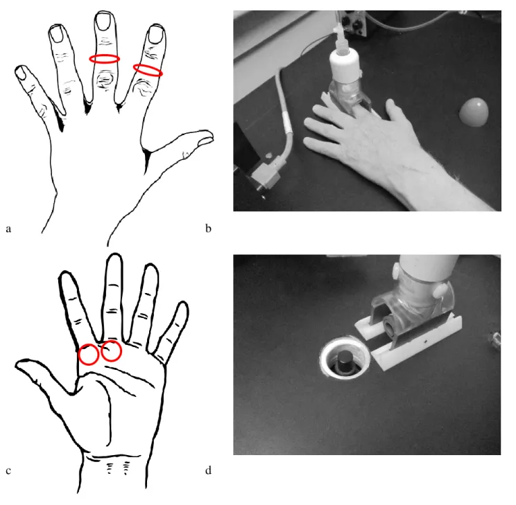
The pain stimulus was a hard plastic ridge with a radius of curvature of 2.25 mm, which was applied to the dorsal side of the middle phalanx of a finger of the subject’s hand. The stimulus was applied to the index and middle fingers of the left hand over the course of the experiment. Participants placed his/her finger in an open cylinder with a diameter of 31 mm and length of 57 mm which housed the plastic ridge. The apparatus could move towards or away from the vibratory stimulus (described in the following section) along two plastic tracks secured to the table. The subject’s hand rested on a table, palm down, while the stimulus was being applied (Figure 1).
In a given run, the pressure was manipulated to elicit fluctuations in stimulus intensity and pain ratings. The relationship between the pressure in the system and the force on the plastic ridge was non-linear with the apparatus used, so forces were calibrated for each condition
sequentially using a digital balance. The pressures were calibrated to produce a 1.2 kg force for 75 seconds, followed by 0.48 kg for 17 seconds, 1.1 kg for 17 seconds, 0.48 kg for 17 seconds, 1.0 kg for 17 seconds, and 0.48 kg for a final 17 seconds. Each experimental run lasted 160 seconds. Changes in pressure were computer-controlled.
A parameter of the pressure stimulus was modified during data collection in response to several subjects reporting intense pain. Changes between high and low pressure were virtually instantaneous for the first 8 archival subjects, but the pressure changed more slowly
(approximately 250 g/second) for the remaining 28 subjects. Safety Precautions
experimenter to end the run, or reported a pain rating of 90 on the VAS. All subjects were informed of these three methods of ending their participation during the consent process. Vibratory Stimulus
Vibration was applied using a circular contactor with a diameter of 15 mm, raised 1.0 mm above the table. The contactor protruded through a circular gap in the table with a diameter of 45 mm. Subjects placed the base of the middle or index finger of the left hand (to correspond to the finger used for the pain stimulus) palmar side down on the vibratory contactor (Figure 1). 60 Hz vibration was applied, and the system was calibrated for each subject to apply an
amplitude of 0.80 mm.
Application of vibration was controlled through the same LabView program that subjects used to report the intensity of pain on the digital VAS. In all conditions, vibration could only be applied in the final 100 seconds of the run; starting 60 seconds after the initiation of the run. The final 100 seconds of each run was therefore termed the ‘vibration period’. The delay was
incorporated to allow pain intensities to stabilize before the experimental manipulation. Experimental Design
The experiment consisted of four conditions, differing only with regard to when vibration was applied. In the random condition (RAN), vibration was randomly applied for 25 seconds during the final 100 seconds. The randomization was calculated in LabView at the start of the run. No vibration (NO) was applied in a separate condition. In the increasing (INC) or
compared the VAS ratings of two consecutive 0.1 second increments to determine if the necessary condition was met, and if it was, would then apply vibration for 0.2 seconds. The minimum possible vibration duration was 0.2 seconds; continuous vibration could be applied in response to a continuous change of VAS rating.
Procedure
Subjects were given a brief description of the study upon entering the laboratory. Specifically, all subjects were told that the purpose of the study was to examine moment-to-moment fluctuations in pain and factors that can influence those fluctuations. However, subjects were not told of the parameters of the experimental runs or the hypothesis that the effect of vibration on pain is dependent on the context of its application. The surveys were completed after the consent process. Subjects were then trained to use the VAS by completing an auditory intensity rating task. Using a VAS labeled with “No Loudness” to “The Loudest Music
Imaginable”, subjects rated the loudness of a 108.5 second excerpt of Peter Ilyich Tchaikovsky’s “Dance of the Reed Flutes”. The subjects listened to the music through Sennheiser 265 HD Linear headphones, with the computer volume set to 80% of maximum. The presentation of the VAS was identical to that in the experimental runs with the exception of the aforementioned labels.
random order. Position of the plastic ridge was assigned in pseudo-random order, with the constraint being that the ridge had to alternate between index and middle fingers.
During each run, participants listened to moderate-loudness pink noise through headphones to mask the sound of the vibratory stimulus and pneumatic pressure system. The pink noise was generated using Rane AC22 and RA27 components. Low/Mid frequency was set to 500 Hz.
All runs began with the plastic ridge already lowered on the subject’s finger. Subjects continually rated their pain during the run using their right hand to control the mouse. Pain ratings were recorded at 0.1 second intervals throughout the entire run. At the end of the run, the subject removed his/her hand from the stimulator and was given a short break of approximately five minutes. The plastic ridge was repositioned to stimulate the correct location on the finger for the new run, and then the second run was administered using the same protocol. A 10 minute break was given between the second and third runs, and another short break was given between the 3rd and 4th runs.
All procedures were approved by the Institutional Review Board at UNC-CH. Analysis
Data from the surveys and pain ratings were analyzed using IBM SPSS 22 and Microsoft Excel.
The effects of condition and run number were analyzed using repeated-measures ANOVA tests. Where appropriate, post-hoc tests were run using pairwise comparisons of estimated marginal means at a 95% confidence interval with Bonferroni corrections.
Subjects reported an average distractibility of 16.4 (range: 7-30, SD = 5.1) from the Cognitive Failures Questionnaire. 13 subjects reported pain at the time of the experiment (M = 11.1, SD = 12.6), but none reported pain in the left hand. The average diameter of subjects’ index and middle fingers of the left hand was 17.0 mm (SD = 1.5).
Manipulation Check
Changes in the pressure stimulus were performed to create large fluctuations in pain ratings. If this was successful, more vibration would occur during the high pressure phases in the INC run, and more vibration would occur during the low pressure phases in the DEC run. The manipulation was tested by grouping the three high pressure phases (1.2 kg, 1.1 kg, 1.0 kg) together and three low pressure phases (.48 kg, .48 kg, .48 kg) together (of the vibration period) for separate analysis.
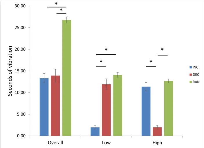
Overall, vibration duration (in seconds) for INC (13.34 ± 1.09) and DEC (13.93 ± 1.48) were similar, and RAN displayed approximately twice as much vibration (26.78 ± .70).
Vibration duration was significantly different across conditions in high, F(1.49,52.27) = 100.9, p = .001, ηp2 = .742 and low pressure phases, F(1.78,62.29) = 149.3, p = .001, ηp2 = .810. INC
(2.0 ± .373)was significantly less than DEC (11.9 ± 1.24) and RAN (14.1 ± .519) in low pressure phases, and DEC (2.00 ± .445) was significantly less than INC (11.4 ± .973) and RAN (12.7 ± .454) in high pressure phases (Figure 2).
The relationship between vibration duration and condition indicates that the pressure manipulation produced the desired changes in pain, granting validity to the following analyses. Pain Ratings
The subjects who experienced the instantaneous changes in pressure experienced
significantly greater pain throughout the entire run than those who experienced gradual changes t(34) = 2.86, p = .007. On average, instantaneous changes produced a pain of 38.8 (SD = 12.6), and gradual changes produced a pain of 25.0 (SD = 11.9). Similarly, there was a significant between-subjects effect when comparing pain of the vibration condition by the two pressure paradigms with a 4 x 2 mixed-measures ANOVA, F(1,34) = 6.130, p = .018, ηp2 = .153.
However, there was no significant interaction effect, F(1.91,65.04) = .202, p= .808. When the runs were ordered temporally and not by vibration condition, the same pattern existed. There was a significant effect of pressure paradigm on pain ratings by run number, F(1,34) = 8.19, p = .007, ηp2 = .194, and no significant interaction between pressure paradigm and run number,
F(3,102) = .319, p = .811.
Lacking a significant interaction effect, the subjects from both pressure paradigms were grouped together for the main data analysis. All further analyses comparing the four vibration conditions were completed using a one-way, repeated-measures ANOVA.
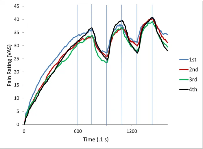
Analyzing the effect of order on pain ratings did not require a normalization of ratings. The independent variable (the order number) affected the run from start to finish, so there was no need to normalize any variance at the beginning of the run. Order number also did not affect pain ratings. There was no significant within-subjects effect of order on pain ratings, F(3,105) = .865, p = .462 (Figure 4).
Fluctuations in pain ratings
Pain ratings were further analyzed by breaking down the run into high and low pressure phases, in the same way that vibration duration was analyzed. Then, the changes in pain over the course of a given phase was computed by subtracting pain at the beginning of the phase from pain at the end of the phase. The difference from the three high pressure or three low pressure phases were summed to give total pain increase and total pain decrease, respectively.
Increases during the high pressure phases were significantly different across conditions, F(3,105) = 6.15, p = .001, ηp2 = .149. RAN produced the smallest increase in pain ratings (17.80
± 2.56). NO (30.47 ± 3.51) and DEC (29.77 ± 3.74) produced significantly larger increases in pain. INC (26.58 ± 3.69) produced qualitatively larger increases in pain that approached significance (Figure 5).
Vibration condition also had a significant effect on decreases in pain rating during low pressure phases, F(3,105) = 2.931, p = .037, ηp2 = .077. The overall effect was significant, but
the data did not hold enough power to determine which conditions were statistically significant. Qualitatively, RAN (-19.28 ± 4.40) produced the smallest decrease when compared to NO (-31.77 ± 5.71), INC (-27.76 ± 5.11) and DEC (-31.62 ± 5.54) (Figure 5).
The fourth run (32.92 ± 3.67) produced significantly larger increases than the first (21.28 ± 3.25) and second (23.18 ± 3.19) runs. The third run (27.24 ± 3.60) was not significantly different from the fourth (Figure 6).
Order also had a significant effect on decreases in pain during low pressure phases, F(3,105) = 4.70, p = .004, ηp2 = .118. The fourth run (-37.42 ± 6.13) produced significantly
larger increases than the first (-21.06 ± 4.43) and second (-23.40 ± 4.37) runs. The third run (-28.58 ± 5.56) was not significantly different from the fourth (Figure 6).
Pain Levels and Response to Vibration
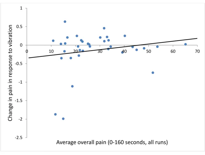
Bini and colleagues (1984) reported that pain closer to tolerance level was less reduced by vibration, so it was hypothesized that subjects who reported greater overall pain (0-160 seconds) would not be affected by vibration as much as those who reported less pain. The analysis was done by subtracting the average pain rating of NO from average pain rating of the three other conditions to assess the general response to vibration. Averaging the three conditions did not raise concerns of variance because there was no significant effect of condition on pain rating. Analysis of this response to vibration with overall pain showed no significant correlation between the two variables, r(34) = .189, p = .271 (Figure 7).
Distractibility and Pain Perception
The distractibility subscale of the Cognitive Failures Questionnaire allowed comparison of a participant’s distractibility and the response to vibration. Vibration has been hypothesized to act at least in part through distracting the participant away from the painful stimulus, so greater distractibility may have influenced response to vibration. No significant correlation was observed between distractibility and response to vibration, r(34) = -.069, p = .691.
Finger Width and Overall Pain Ratings
Since the participants’ contact with the plastic ridge could change based on the width of the subject’s finger, we hypothesized that finger width would be correlated with overall pain. There was no significant correlation between finger width and overall pain when the archival subjects alone were analyzed, r(34) = .006, p = .974.
However, many subjects were excluded based on pain ratings that were either too high or too low. A separate analysis was completed using all participant data. Participants who reached 90 on the VAS were assigned a score of 90, and those who did not reach 10 in a run were
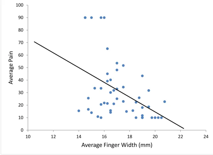
assigned a score of 10. In this analysis, finger width and overall pain was moderately correlated in a negative direction, r(47) = -.439, p = .002. Larger finger width significantly predicted a lower overall level of pain, R2 = .193, F(1,48) = 11.239, p = .002 (Figure 8).
Discussion
Applying intermittent vibrations to fluctuating pain did not reduce or exacerbate experimental pressure pain. Lacking an effect one way or the other, the question posed by Hollins and colleagues (2011), of whether vibratory hyperalgesia was caused by increasing pain or a central process in subjects who rapidly sensitize, could not be directly addressed. This study was unique in applying intermittent vibrations during changing pain stimuli, so there is not a large literature base to compare the results against. However, the prospect of minimizing fluctuations in pain could be useful in a clinical setting. Attenuated fluctuations in pain would create a more constant pain experience, which could allow for a more accurate management of pain and an easier psychological control over it.
patient populations experience (Apkarian et al., 2009) than the commonly-used static thermal stimulus. Pressure pain is not as easy a modality to work with when compared to heat pain, and working with it brings in a new set of variables like finger size and previous injury. However, pressure pain more accurately represents the musculoskeletal pain so often seen in chronic pain conditions like fibromyalgia. For that reason, the study into analgesic mechanisms specific to pressure pain should be stressed in the context of chronic pain research.
Factors Related to Overall Pain
Many participants were excluded from the archival data set due to intense or insubstantial pain. The significant correlation between finger width and overall pain (Figure 8) could allow for a further screening measure to prevent exclusions in future studies of a similar setup. Such an effect likely demonstrates a relationship between finger size and the area of contact between the ridge and finger. A smaller area of contact would increase the pressure on the subject even under the same force.
Intermittent Vibrations Do Not Change Overall Pain
No application paradigm of vibration produced a significant change in pain during the vibration period. A lack of effect is in contradiction with many previous studies, which have shown vibration to decrease pain (Hollins et al., 2014; Bini et al., 1984) or increase pain in certain participants (Hollins et al., 2011). A lack of effect may be a result of the short overall duration of vibration. Depending on the condition, subjects experienced approximately 14 or 27 seconds of vibration during the 100 second manipulation. This is in contrast to 80 seconds of vibration applied to a pressure pain that produced a median of 10% pain reduction (Hollins et al., 2014). If vibration duration has a linear relationship with the magnitude of analgesia, only a 3% decrease would be expected, which would likely not be detected with the number of subjects run.
An alternative hypothesis is that fluctuating pain decreases the efficacy of vibratory analgesia. Large changes in pain could prompt participants to attend to their pain, which has been shown to erase any analgesic effects of brief vibration (Longe et al., 2001). If that was the case, analgesic effects may be strengthened by instructing participants to either attend to the vibratory stimulus or a neutral stimulus.
Intermittent Vibration and Order Affected Pain Fluctuations
Vibration did decrease both positive and negative fluctuations in pain during RAN. The other vibration conditions did not produce a significant difference in fluctuations, but
qualitatively the changes were smaller than during NO. The disparity between conditions may be due to the longer vibration application during RAN, or a distraction phenomenon arising from the unpredictable stimulus.
effect of a therapy. Again, the result may be due to the unpredictable stimulus distracting participants away from the changes in pressure, or the vibration could be interacting with the endogenous mechanism of offset analgesia. The relationship between pressure and pain intensity during offset analgesia is likely different than that of heat and pain. The pressure stimulus
pushes down on the finger to change its conformation, exposing different tissue and stimulating different populations of afferents as the ridge “settles”. Conformational changes in the finger would explain the continuous increase in pain during the first 60 seconds (Figures 3,4). Even so, pressure offset did produce noticeable decreases in pain, which were attenuated by vibration. Further studies would need to find a way to produce an additive (instead of a cancellation) effect of offset and vibratory analgesia to crease a useful treatment.
The other issue to consider with offset analgesia is that, during rapid changes of thermal stimuli, the effects were seen to last up to 15 seconds (Grill & Coghill, 2002). Pressure changed every 17 seconds during the vibration period, so the effect of one change could blend into the next pressure phase. Also, if stimulus fluctuations can bidirectionally modulate the pain response (Vierck et al., 2010), it is possible that the sensitization from the high pressure and analgesia from the low pressure cancelled each other out over the course of the run. Such considerations could be further explored by extending the time of each pressure phase to better isolate the effects of one change. This would come at the cost of pain leveling out at the end of each phase, so high or low pressure could not be accurately described as increasing and
decreasing pain periods.
have a randomly assigned vibration condition, displayed the greatest increases and decreases in pain, even though the overall pain throughout the run was no different (Figure 4).
Qualitatively, each consecutive run produced larger pain fluctuations than the previous one. A pattern like this would suggest a sensitization-like effect, but one would assume that sensitization would translate to overall pain levels as well. Like sensitization phenomena in other studies (Price et al., 1977), the effect seems to be an artifact of a central mechanism, because runs alternated fingers. A peripheral mechanism would likely show the 1st and 2nd, and
the 3rd and 4th to be the same.
Since the analyses of run order were a product of the data and not manipulated in isolation, a concrete explanation is not possible. One possibility is that participants grew more accustomed to the procedure with each consecutive run, so they attended more to the pressure fluctuations instead of their pain. Another explanation is that the mechanisms behind central sensitization interact with offset analgesia, and increased sensitization increases offset analgesia. Such an interaction would support Grill and Coghill’s initial hypothesis (2002) that offset
analgesia could act to reinforce escape behavior. Sensitization occurs when a noxious stimulus is presented repeatedly, and recurrent damage would be a strong cue for escape from that environment.
Conclusions
significant pain modulation is taking place. What remains to be seen is whether shrinking fluctuations would reduce unpleasantness of pain in a clinical setting.
Since the manipulation of pain fluctuations was effective in the current study, follow-up studies with alterations in vibratory application could clarify the relationship between vibration duration and pain fluctuations. It is possible that the primary question, that vibration during increasing or decreasing pain modulates pain differently, could be clarified if vibration was applied constantly during high or low pressure phases. Also, the phenomenon of RAN producing a larger effect merits further study. It is possible that the unpredictable nature of random vibration caused greater distraction to account for the larger effect, or simply the longer application. RAN produced twice as much vibration as INC or DEC so that high (for RAN and INC) and low (for RAN and DEC) pressure phases would have the same amount of vibration (Figure 2), but equalizing all three conditions could clarify this point.
References
Apkarian, A. V., Baliki, M. N., & Geha, P. Y. (2009). Towards a theory of chronic pain. Progress in Neurobiology, 87, 81-97. Doi:10.1016/j.pneurobio.2008.09.018 Bantick, S. J., Wise, R. G., Ploghaus, A., Clare, S., Smith, S. M., & Tracey, I. (2002). Imaging
how attention modulates pain in humans using functional MRI. Brain, 125, 310-319. Bini, G., Cruccu, G., Hagbarth, K.-E., Schady, W., & Torebjörk, E. (1984). Analgesic effect of
vibration and cooling on pain induced by intraneural electrical stimulation. Pain, 18, 239-248.
Broadbent, D.E., Cooper, P.F., FitzGerald, P., & Parkes, K.R. (1982). The Cognitive Failures Questionnaire (CFQ) and its correlates. British Journal of Clinical Psychology, 21, 1-16. Campbell, C. M., Witmer, K., Simango, M., Carteret, A., Loggia, M. L., Campbell, J. N., . . .
Edwards, R. R. (2010). Catastrophizing delays the analgesic effect of distraction. Pain, 149, 202-207. doi:10.1016/j.pain.2009.11.012
Cohen, M., Quintner, J., & Buchanan, D. (2013). Is chronic pain a disease? Pain Medicine, 14, 1284-1288. doi:10.1111/pme.12025
Edwards, R. R., Bingham, C. O., Bathon, J., & Haythornthwaite, J. A. (2006). Catastrophizing and pain in arthritis, fibromyalgia, and other rheumatic diseases. Arthritis Care & Research, 55, 325-332. doi:10.1002/art.21865
Fiorio, M., Recchia, S., Corrà, F., Simonetto, S., Garcia-Larrea, L., & Tinazzi, M. (2012). Enhancing non-noxious perception: Behavioural and neurophysiological correlates of a placebo-like manipulation. Neuroscience, 217, 96-104.
Forgione, A. G., & Barber, T. X. (1971). A strain gauge pain stimulator. Psychophysiology, 8(1), 102-106.
Gescheider, G. A., Wright, J. H., & Verrillo, R. T. (2009). Information-processing channels in the tactile sensory system. New York: Psychology Press.
Grill, J.D. & Coghill, R.C. (2002). Transient analgesia evoked by noxious stimulus offset. J. of Neurophysiology, 87, 2205-2208. doi: 10.1152/jn.00730.2001
Hollins, M., Harper, D., & Maixner, W. (2011). Changes in pain from a repetitive thermal stimulus: The roles of adaptation and sensitization. Pain, 152, 1583-1590.
doi:10.1016/j.pain.2011.02.049
Hollins, M., McDermott, K. & Harper, D. (2014). How does vibration reduce pain? Perception, 43, 70-84. doi:10.1068/p7637
Kemp, J., Despres, O., Dufour, A. (2012). Unreliability of the Visual Analog Scale in Experimental Pain Assessment: A Sensitivity and Evoked Potentials Study. Pain Physician, 15, E693-E699.
Kosek, E., & Hansson, P. (1997). Modulatory influence on somatosensory perception from vibration and heterotopic noxious conditioning stimulation (HNCS) in fibromyalgia patients and healthy subjects. Pain, 70, 41-51.
Le Bars, D., Dickenson, A. H., & Besson, J. M. (1979). Diffuse noxious inhibitory controls (DNIC). I. Effects on dorsal horn convergent neurones in the rat. Pain, 6(3), 283-304. Le Bars, D., Villanueva, L., Willer, J. C., & Bouhassira, D. (1991). Diffuse noxious inhibitory
controls (DNIC) in animals and in man. Acupuncture in Medicine, 9, 47-56.
Longe, S. E., Wise, R., Bantick, S., Lloyd, D., Johansen-Berg, H., McGlone, F., & Tracey, I. (2001). Counter-stimulatory effects on pain perception and processing are significantly altered by attention: an fMRI study. Neuroreport,12, 2021-2025.
McDermid, A. J., Rollman, G. B., & McCain, G. A. (1996). Generalized hypervigilance in fibromyalgia: Evidence of perceptual amplification. Pain, 66(2–3), 133-144. doi: 10.1016/0304-3959(96)03059-X
Melzack, R., & Wall, P. D. (1965). Pain mechanisms: A new theory. Science, 150, 971-979. Nagasako, E. M., Oaklander, A. L., & Dworkin, R. H. (2003). Congenital insensitivity to pain:
An update. Pain, 101, 213-219. doi:10.1016/S0304-3959(02)00482-7
Niesters, M., Hoitsma, E., Sarton, E., Aarts, L., & Dahan, A. (2011). Offset analgesia in neuropathic pain patients and effect of treatment with morphine and ketamine. Anesthesiology, 115, 1063-1071. doi: 10.1097/ALN.0b013e31822fd03a
Perlman, D. M., Salomons, T. V., Davidson, R. J., & Lutz, A. (2010). Differential effects on pain intensity and unpleasantness of two meditation practices. Emotion, 10, 65-71.
doi:10.1037/a0018440
Price, D. D., & Dubner, R. (1977). Neurons that subserve the sensory-discriminative aspects of pain. Pain, 3, 307-338. doi:10.1016/0304-3959(77)90063-X
Price, D. D., Hu, J. W., Dubner, R., & Gracely, R. H. (1977). Peripheral suppression of first pain and central summation of second pain evoked by noxious heat pulses. Pain, 3, 57-68. doi:10.1016/0304-3959(77)90035-5
Roy, E. A., Hollins, M., & Maixner, W. (2003). Reduction of TMD pain by high-frequency vibration: A spatial and temporal analysis. Pain, 101, 267-274. doi:10.1016/S0304-3959(02)00332-9
Salter, M. W., & Henry, J. L. (1990). Differential responses of nociceptive vs. non-nociceptive spinal dorsal horn neurones to cutaneously applied vibration in the cat. Pain, 40, 311-322. doi:10.1016/0304-3959(90)91128-6
Staudl, R., Robinson, M. E., Goldman, C. T., & Price, D. D. (2011). Attenuation of experimental pain by vibro-tactile stimulation in patients with chronic local or widespread
musculoskeletal pain. European Journal of Pain, 15, 836-842. doi:10.1016/j.ejpain.2011.01.011
Vierck, C. J., Riley III, J. L., Wong, F., King, C. D., & Mauderli, A. P. (2010). Psychophysical demonstration of bidirectional pain modulation (sensitization and desensitization) by ascending or descending progressions of thermal stimulus intensity. Brain
Research, 1347, 58-64. doi:http://dx.doi.org/10.1016/j.brainres.2010.06.006
Vierck, C. J., Whitsel, B. L., Favorov, O. V., Brown, A. W., & Tommerdahl, M. (2013). Role of primary somatosensory cortex in the coding of pain. Pain, 154, 334-344.
Wager, T. D., Rilling, J. K., Smith, E. E., Sokolik, A., Casey, K. L., Davidson, R. J., ... & Cohen, J. D. (2004). Placebo-induced changes in FMRI in the anticipation and experience of pain. Science, 303, 1162-1167.
Yelle, M. D., Rogers, J. M., & Coghill, R. C. (2008). Offset analgesia: A temporal contrast mechanism for nociceptive information. Pain, 134, 174-186.
Wallace, J. C., Kass, S. J., & Stanny, C. J. (2002). The Cognitive Failures Questionnaire revisited: Dimensions and correlates. Journal of General Psychology, 129(3), 238-256. Weerakkody, N. S., Percival, P., Hickey, M. W., Morgan, D. L., Gregory, J. E., Canny, B. J., &
Proske, U. (2003). Effects of local pressure and vibration on muscle pain from eccentric exercise and hypertonic saline. Pain, 105, 425-435. doi:10.1016/S0304-3959(03)00257-4 Wiech, K., Ploner, M., & Tracey, I. (2008). Neurocognitive aspects of pain perception. Trends in
a b
c d
Figure 2. Vibration duration across condition. NO is not displayed. Low and High designate the periods of the run in which low or high pressure was applied. Error bars denote SEM.
* denotes p < .001 0.00
5.00 10.00 15.00 20.00 25.00 30.00
Overall Low High
Sec
ond
s of
vibr
ation
INC
DEC
RAN
*
*
*
*
*
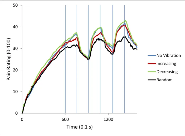
Figure 3. Pain ratings of each vibration condition averaged across all subjects. Vertical lines designate changes in pressure. Vibration period began at 60 seconds (first vertical line). Vibration condition did not significantly affect pain ratings.
0 10 20 30 40 50
0 600 1200
Pain
R
atin
g
(0
-100)
Time (0.1 s)
Figure 4. Pain ratings of runs averaged across all subjects. Vertical lines designate changes in pressure. Run order did not significantly affect pain ratings.
0 5 10 15 20 25 30 35 40 45
0 600 1200
Pain
R
atin
g
(V
AS
)
Time (.1 s)
1st
Figure 5. Fluctuation in pain during high or low pressure conditions by vibration condition. Pressure changes are displayed as absolute values, but all low pressure changes represent decreases in pain. Error bars denote SEM.
* denotes p < .05
0 5 10 15 20 25 30 35 40
High Pressure
Low Pressure
Figure 6. Fluctuation in pain during high or low pressure conditions by run order. Pressure changes are displayed as absolute values, but all low pressure changes represent decreases in pain. Error bars denote SEM.
* denotes p < .05 ** denotes p < .01
0 5 10 15 20 25 30 35 40 45 50
High Pressure
Low Pressure
Figure 7. Average pain over all four runs was not significantly correlated with the change in pain from vibration. Change in pain in response to vibration was calculated by subtracting the average pain of NO from the average pain of the three vibration conditions.
-2.5 -2 -1.5 -1 -0.5 0 0.5 1
0 10 20 30 40 50 60 70
C
h
an
ge i
n
p
ain
in r
esp
onse
to
vibr
ation
Figure 8. Correlation between average finger width and overall pain. Excluded subjects were assigned a pain of 90 if exclusion was due to reaching 90, and 10 if the subject failed to reach 10 during a run. Regression analysis shows finger width to significantly predict overall pain. * p = .002
0 10 20 30 40 50 60 70 80 90 100
10 12 14 16 18 20 22 24