Azathioprine and 6-Mercaptopurine Induced Liver Injury: Clinical
Features and Outcomes
Einar S. Björnsson, MD, PhD1,2, Jiezhun Gu, Phd3, David E. Kleiner, MD4, Naga Chalasani, MD5, Paul H. Hayashi, MD6, and Jay H. Hoofnagle, MD1 for the DILIN Investigators
1Liver Disease Research Branch, Division of Digestive Diseases and Nutrition, National Institutes
of Health, Bethesda, MD 2The Faculty of Medicine, University of Iceland, National University
Hospital of Iceland, Reykjavik, Iceland 3Duke Clinical Research Institute, Durham, NC 4Laboratory
of Pathology, National Cancer Institute, National Institutes of Health 5Indiana University School of
Medicine, Indianapolis, IN 6University of North Carolina, Chapel Hill, NC
Abstract
Goals—To define the clinical, biochemical and histologic features of liver injury from thiopurines.
Background—Azathioprine (Aza) and 6-mercaptopurine (6-MP) can cause liver injury but no large series exist.
Methods—Clinical and laboratory data and 6-months outcomes were analyzed from patients with thiopurine hepatotoxicity from the Drug-Induced Liver Injury Network Prospective Study.
Results—22 patients were identified, 12 due to Aza and 10 6-MP, with a median age of 55 years and the majority females (68%). Inflammatory bowel disease was the indication in 55%, and median thiopurine dose 150 (range 25–300) mg daily. The median latency to onset was 75 (range 3 to 2584) days. Injury first arose after a dose escalation in 59% of patients; the median latency after dose increase being 44 (range 3 to 254) days. At onset, the median alanine aminotransferase was 210 U/L, alkaline phosphatase 151 U/L and bilirubin 7.4 mg/dL (peak 13.4 mg/dL). There were no major differences between Aza and 6-MP cases, but anicteric cases typically had non-specific symptoms and a hepatocellular pattern of enzyme elevations, whereas icteric cases experienced a cholestatic hepatitis with modest enzyme elevations in a mixed pattern. One patient with pre-existing cirrhosis required liver transplantation, all others resolved clinically. One patient still had moderate alkaline phosphatase elevations 2 years after onset.
Conclusions—Nearly three-quarters of patients with thiopurine-induced liver injury present with self-limited, cholestatic hepatitis, typically within 3 months of starting or a dose increase. The prognosis is favorable except in patients with pre-existing cirrhosis.
Correspondence: Jay H. Hoofnagle, Liver Disease Research Branch, Room 655, Democracy II, 6707, Democracy Blvd, Bethesda, MD
HHS Public Access
Author manuscript
J Clin Gastroenterol
. Author manuscript; available in PMC 2017 July 11.Published in final edited form as:
J Clin Gastroenterol. 2017 January ; 51(1): 63–69. doi:10.1097/MCG.0000000000000568.
A
uthor Man
uscr
ipt
A
uthor Man
uscr
ipt
A
uthor Man
uscr
ipt
A
uthor Man
uscr
Keywords
hepatotoxicity; drug-induced liver injury; azathioprine; 6-Mercaptopurine
Azathioprine (Aza) and 6-mercaptopurine (6-MP) are purine nucleoside analogues (thiopurines) that have potent antiproliferative and immunosuppressive activities and have been in common use for decades. 6-MP was introduced in the 1950s as an antineoplastic agent for the therapy of leukemias and lymphomas. Azathioprine, a prodrug of 6-MP, was introduced in the 1960s, but largely as an immunosuppressive agent, initially for prevention of transplant rejection and later for its corticosteroid sparing features as therapy of
autoimmune diseases such as inflammatory bowel disease (IBD) and autoimmune hepatitis. Reports on liver injury, sometimes with a fatal outcome, associated with the use of these drugs appeared shortly after their introduction (1–7). Since then more than 100 cases of liver injury attributed to Aza and 6-MP have been reported (8). In many cases, the association was confirmed by a prompt recurrence of injury upon readminstration of the agent (9–14). In large, prospective (15–16) and retrospective studies on drug-induced liver injury, Aza and 6-MP are frequently listed as common causes (17). Indeed, in a recent population-based study from Iceland, Aza was associated with the highest risk of hepatotoxicity, occurring in 1 of 133 persons in whom it was prescribed (18).
Despite the common problem of hepatotoxicity with thiopurines there is a lack of studies with a significant number of well characterized patients with this type of liver injury. Most studies have been limited to the IBD patient population and focused largely on
asymptomatic elevations of serum aminotransferase levels as the only evidence of liver injury (19–22). Clearly, both Aza and 6-MP are associated with transient elevations in serum enzyme levels, but these are usually mild, asymptomatic and self-limited and may resolve even without dose adjustment or temporary discontinuation (19). More importantly, both Aza and 6-MP have been implicated in instances of clinically apparent hepatotoxicity which can be severe leading to prolonged intrahepatic cholestasis (3, 7) or even to acute liver failure (2, 4, 5). Furthermore, both veno-occlusive disease (23) and nodular regenerative hyperplasia (24–25) have been well documented with higher doses and long-term treatment with these agents. Despite the number of case reports and case series, the severity and the overall clinical outcomes in patients with liver injury due to Aza and 6-MP have not been well defined. Furthermore, the cross-sensitivity to hepatotoxicity between Aza and 6-MP and possibly to the third thiopurine – 6-thioguanine (6-TG), has not been fully characterized.
The U.S. Drug-induced Liver Injury Network (DILIN) is a multicenter prospective cohort study which was started in 2004 with the primary aim to enroll patients with liver injury from drugs and dietary supplements to allow for better characterization of the clinical features, pathogenesis and outcomes of this poorly understood form of liver disease. The current analysis has focused on more fully characterizing the clinical, serologic and histological characteristics of the patients with liver injury attributed to the thiopurines who were enrolled in the DILIN study.
A
uthor Man
uscr
ipt
A
uthor Man
uscr
ipt
A
uthor Man
uscr
ipt
A
uthor Man
uscr
METHODS
The study design of the prospective DILIN study has been described in detail in previous publications (16, 26). In brief, patients meeting pretermined eligibility criteria were enrolled for structured assessment of potential competing etiologies and thorough analysis of clinical features and laboratory testing results. Patients were followed for at least 6 months and, if clinical or laboratory features were still abnormal, were seen again at 12 and 24 months. The causality asessment and the overall diagnosis of drug-induced liver injury were based on adjudication by the DILIN Causality Committee using expert consensus (27). The strength of the relationship between the liver injury and the implicated agent was scored as definite (greater than 95% likelihood), very likely (75–94% likelihood), probable (50–74% likelihood), possible (25–49% likelihood) or unlikely (<25% likelihood) (20). In addition, the enrolling physician also applied the Rousell-Uclaf Causality Assessment Method (RUCAM) to the clinical information which generates a score from −9 to 14 and is classified as highly probable (9–14), probable (6–8), possible (3–5), unlikely (1–2) or excluded (≤ 0) (28). Finally, a severity score was assigned as (1) mild, with serum enzyme elevations without jaundice (bilirubin <2.5 mg/dL) or coagulopathy (INR <1.5), (2) moderate, with bilirubin ≥2.5 mg/dL or INR ≥ 1.5, (3) moderate, with bilirubin or INR elevations as above and hospitalized for liver injury, (4) severe, with jaundice and signs of liver failure (such as INR ≥ 1.5, ascites or hepatic encephalopathy) and (5) fatal, with liver-related death and/or liver transplantation within 6 months of onset of liver injury.
For the current analysis, all patients enrolled between September 2004 and September 2014 with liver injury suspected to be due to thioprines were analyzed (Figure 1). Only those cases in which the causality was adjudicated as probable, very likely or definite were included. The clinical features, laboratory results and outcomes were obtained from the Duke Clinical Research Institute, the data coordinating center of the DILIN study. All patients were analysed by two of the authors (ESB, JHH) for clinical and laboratory features and outcomes such as progression to chronicity and liver-related death and need for liver transplantation. The type of liver injury was categorized on the basis of the R ratio calculated using the initial values of serum alanine aminotransferase (ALT) divided by alkaline phosphatase (Alk P), both expressed as ratios to upper limits of normal (ULN): R = ALT/ULN ÷ Alk P/ULN. By convention, R ratios less than 2 were defined as cholestatic, 2 to 5 as mixed, and greater than 5 as hepatocellular (28). The type of liver injury was also assessed using the initial ALT and Alk P values graphed against each other using criteria established by Zimmerman (29).
Statistical analysis
Descriptive statistics of demographic and clinical data of Aza cases in comparison to 6-MP cases were generated and analyzed. Description of the data was given as medians (range), frequencies and percentages. Statistical significance among groups was determined by Wilcoxon rank-sum test for continuous variables, Fisher’s exact test for binary variables, Chi-square for categorical variables, and log-rank tests for time-to-event variables. P values of <0.05 were considered statistically significant. SAS v.9.4 [SAS Institute, Cary, NC] was used for all statistical analyses.
A
uthor Man
uscr
ipt
A
uthor Man
uscr
ipt
A
uthor Man
uscr
ipt
A
uthor Man
uscr
Results
Clinical characteristics
Over the initial ten-year period of the prospective DILIN cohort study, 1434 cases of drug-induced liver injury were enrolled and underwent causality assessment, of which 27 were attributed, at least in part, to a thioprine, including 15 to Aza, 12 to 6-MP but none to 6-TG (Figure 1). For this analysis, 5 cases were excluded in which liver injury was considered only possibly related to the thiopurine. The remaining 22 cases (12 attributed to Aza and 10 to 6-MP) fulfilled the predefined criteria for inclusion and were used for the current analysis.
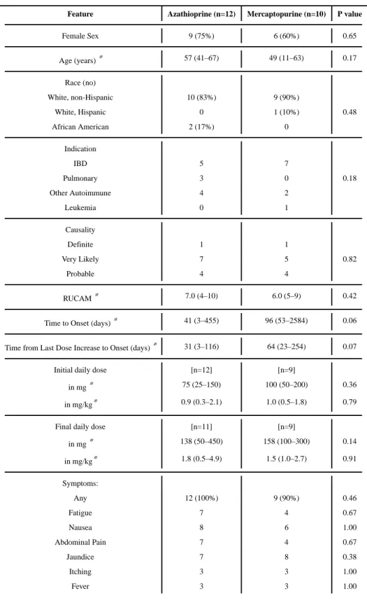
The demographics and initial clinical features of the 12 Aza and 10 6-MP cases are compared in Table 1. In most regards, the clinical features of cases associated with either drug were similar. The 22 cases included 15 women and 7 men, ages 11 to 66 years (mean 51 years) and all except one (a 11 year boy receiving 6-MP for acute lymphocytic leukemia) were adults with an autoimmune condition. Most patient were white (n=20 including one Hispanic white) and two were African Americans. The most common indication for therapy was inflammatory bowel disease (IBD) (55%), followed by pulmonary disease of
immunological nature (14%) (sarcoidosis, interstitial lung disease and idiopathic pulmonary fibrosis), autoimmune hepatitis (9%), systemic lupus erythematosus (9%) and
dermatomyositis (9%).
The expert DILIN causality assessment was definite for 2 (9%), very likely for 12 (54%), and probable for 8 (36%) cases, and the distribution was similar for Aza and 6-MP. RUCAM scores were also similar with the two agents, the median being 6.5 overall with 4 cases rated highly probable (9–10), 14 probable (6–8) and 4 possible (4–5). Viral serologies were negative for hepatitis A, B, C and E in all patients except for one patient with known but stable pre-existing chronic hepatitis C. All patients underwent imaging of the liver and biliary tree (ultrasound in 73%, computerized tomography in 59% and magnetic resonance imaging in 27%) and none had evidence of biliary obstruction. Antinuclear antibodies were present in 5 patients (22%) but were thought to be due to the underlying condition (lupus, autoimmune hepatitis in 2, IBD in 2) in each case. Overall 10 patients (46%) were receiving corticosteroids when they developed liver injury. Only one patient was started on treatment with corticosteroids for the liver injury, but did not require long-term therapy.
All except one patient had symptoms attributable to the liver injury, the most common being jaundice (73%), nausea (64%), fatigue (50%) and abdominal pain (50%). Itching was reported in 6 patients (27%) but was generally transient and mild-to-moderate in severity. Rash (23%) and fever (27%) were reported in a minority of patients and were not prominent or severe. No patient was diagnosed as having DRESS syndrome or a severe cutaneous reaction.
The latency to onset, measured as time from starting the medication to identification of laboratory abnormalities, ranged widely from 3 days to almost 10 years, with median of 75 days. Strikingly, 13 patients (59%) had an escalation of dose before the onset of liver injury and the median latency from the last dose increase (or initial dose if there were no increase) to onset was 44 days (range 3 to 254 days). The latency as measured from starting therapy
A
uthor Man
uscr
ipt
A
uthor Man
uscr
ipt
A
uthor Man
uscr
ipt
A
uthor Man
uscr
and from the time of the last dose increase is plotted in Figure 2 separately for each case of Aza and 6-MP induced liver injury. Overall 59% of cases developed laboratory evidence of DILI within 3 months of starting therapy and 86% within 3 months of the last dose increase. Using either measure, latencies were shorter for Aza than 6-MP cases, but the differences were of marginal statistical significance. The initial thiopurine dose ranged from 25 to 200 mg (median = 75 mg) daily; the final dose from 50 to 450 mg (median = 150 mg) daily. The median initial and final daily doses were similar in those taking Aza or 6-MP expressed either as total daily dose or as mg/kg/day (Table 1).
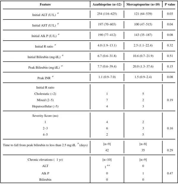
Laboratory results are given in Table 2, comparing Aza and 6-MP related cases. Serum aminotransferase levels were elevated in all 22 patients, Alk P in 18 and bilirubin (≥ 2.5 mg/dL) in 16. The initial median (and range) values for ALT were 211 (64–625) U/L, AST 153 (47–603) U/L, Alk P 151 (35–412) U/L and bilirubin 7.4 (0.6–31.8) mg/dL. The median peak values were 13.8 mg/dL for bilirubin and 1.3 for INR. The R ratio at onset of injury ranged from cholestatic to hepatocellular and the most frequent pattern was in the “mixed hepatitis” range (2–5). These R ratios, however, failed to reflect the somewhat unusual relative ratios of ALT to Alk P. In most cases with jaundice, Alk P levels were only modestly elevated and some were withi the normal range, at least at the onset of illness. Concurrent ALT levels were generally in the moderate range and never greater than 10 fold elevated. When ALT and Alk P were plotted by absolute value in quadrants using the criteria established by Zimmerman (29), the values for jaundiced cases were in the range of
“canalicular cholestasis” rather than hepatocellular, mixed or cholestatic hepatitis (Figure 3). This pattern is defined by ALT elevations less than 8 times ULN and Alk P levels less than 3 times ULN and is most typical of the bland cholestasis that occurs with estrogen or anabolic steroid induced jaundice. Interestingly, among the 6 anicteric cases, the plotted values of ALT and Alk P were usually in the hepatocellular range and the median R ratio was 8.6 (range 3–22), which was distinctly different from the more cholestatic pattern of the patients who developed jaundice (median R ratio = 2.5, range 1.1–14.2).
The severity of the acute liver injury was scored as mild in 6, moderate in 9 and severe in 7 patients (Table 2). 6-MP associated cases tended to be more severe than the Aza cases both in terms of severity scores (5 of the 7 severe cases were due to 6-MP) as well as initial and peak bilirubin and INR values, but there was considerable overlap. No patient died, but one underwent liver transplantation: a 63 year old man with autoimmune hepatitis and cirrhosis who developed persistent jaundice 2 months after having 6-MP (150 mg daily) added to his corticosteroid therapy, with worsening coagulopathy and hepatic encephalopathy, leading to liver transplantation 41 days later. The histology of the explant revealed post necrotic cirrhosis, severe cholestasis and massive necrosis.
All other patients recovered from the acute liver injury clinically and all except 2 had normal liver tests at a final follow-up assessment. The first of the 2 had pre-existing chronic
hepatitis C, and aminotransferase levels had been elevated before Aza was started. A second patient had persistent elevations in Alk P that had followed a severe cholestatic hepatitis and a liver biopsy that had shown duct injury and nodular regeneration attributed to 6-MP. At last follow up, no patient was jaundiced or had symptoms attributable to liver disease. No patient was rechallenged with Aza or 6-MP, but two gave a history of a previous episode of liver
A
uthor Man
uscr
ipt
A
uthor Man
uscr
ipt
A
uthor Man
uscr
ipt
A
uthor Man
uscr
injury due to Aza; one with a recurrence after re-exposure to Aza and one with recurrence upon treatment with 6-MP.
Liver biopsies were done during the acute episode on 8 patients, and 7 were available for central review, all among the jaundiced cases. The most common finding was cholestatic hepatitis with lobular and portal inflammation, zone 3 cholestasis and duct injury without duct loss (Figure 4a and 4b). No patient had findings typical of bland cholestasis. One patient had what appeared to be an underlying steatohepatitis and another had a severe autoimmune hepatitis with a superimposed cholestasis. Interestingly, reticulum staining showed that 3 patients had areas of nodular regeneration (Figure 4c). These 3 all had severe injury (peak bilirubin >20 mg/dL in all three) and had been treated for an extended period (73 to 460 days). One had persistent Alk P elevations even after 2 years of follow up and the other 2 resolved the injury both clinically and biochemically.
Thiopurine methyl transferase (TPMT) genotype analysis was available in only 4 patients and was normal in all. Thiopurine (6-MMP) metabolites were measured at the time of liver injury in two Aza-treated patients and were elevated in both, with values of 16,980 and 21,856 pmol per 108 red blood cells. Stored serum samples were available from many patients, but not from the time of initial onset.
Concise clinical histories of 6 representative patients are provided in the Supplementary material: 2 had self-limited cholestatic hepatitis, 2 cholestatic hepatitis superimposed upon chronic liver disease and 2 anicteric hepatitis with hepatocellular enzyme elevations. The clinical courses of other cases are available on the LiverTox website (8).
Discussion
Aza and 6-MP are thiopurines with similar chemical structures and mechanisms of action. Aza acts as a prodrug of 6-MP, differing only by addition of an imido-azo side chain on the 6′ sulfate. The side chain is removed non-enzymatically in the liver, and Aza tends to be better absorbed and perhaps better tolerated than 6-MP. Nevertheless, the two thiopurines have similar activities and side effects.
Both 6-MP and Aza are well known causes of drug-induced liver injury. At least 4 patterns of injury have been associated with their use. First, and most common, is transient and usually asymptomatic elevations in serum enzymes, mostly ALT and AST, that occurs in 5– 15% of treated subjects. In this series, 6 patients had this pattern of serum aminotransferase elevations accompanied by minor symptoms but without jaundice. More clinically
significant is a second form of hepatotoxicity, an idiosyncratic, cholestatic hepatitis that typically occurs after 1 to 3 months of treatment. Indeed, the majority of cases in the current report, had this pattern; 16 patients presenting with jaundice and most with a cholestatic pattern. Both Aza and 6-MP are also capable of causing sinusoidal obstruction syndrome, a third form of hepatotoxicity that occurs when the agents are given in high doses as with cancer chemotherapy. Finally, both agents have also been linked to cases of nodular regenerative hyperplasia when given long-term, particularly in the setting of organ
transplantation or acute leukemia. In this series, 3 of 7 patients who underwent liver biopsy
A
uthor Man
uscr
ipt
A
uthor Man
uscr
ipt
A
uthor Man
uscr
ipt
A
uthor Man
uscr
had histologic features of nodular regeneration, however, none subsequently progressed to developed signs or symptoms of portal hypertension. These four patterns of injury have also been described with thioguanine (23–25,30).
The current analysis showed that Aza and 6-MP associated liver injury accounted for 1–2% of cases of drug-induced liver injury in the DILIN network. Individually, Aza and 6-MP ranked in the top 20 causes of liver injury in the DILIN prospective study and if grouped together would have ranked as the 6th most common cause (16). In jaundiced cases, the disease was marked by appearance of fatigue, nausea, vomiting and abdominal discomfort followed by dark urine and jaundice. Itching was not common and immunoallergic features were rare, and if present generally mild. Strikingly, the latency to onset varied widely and some patients had been receiving the thiopurine for years when jaundice arose. In these patients, however, there was usually a history of recent increase in dose. Thus, the latency to onset after starting therapy or after a dose increase was generally a few weeks to 3 months, and rarely exceeded 6 months. While the course of illness could be severe, it was generally short lived and rapidly improved upon stopping treatment. Only one of the 22 patients had evidence of chronic liver injury in long-term follow up, marked by asymptomatic but persistent elevations in Alk P after a particularly severe bout of cholestatic hepatitis. No patient died with acute liver failure, although one patient with a pre-existing autoimmune hepatitis and cirrhosis developed an “acute-on-chronic” liver failure and required liver transplantation. Thus, in the absence of pre-existing liver disease, the hepatotoxicity of Aza and 6-MP tended to be benign.
In the current study an intriguing finding was the frequent occurrence of appearance of liver injury after dose escalation, a pattern that occurred in 59% of patients including 11 of 16 (69%) with jaundice and 2 of 6 (33%) anicteric cases. This finding suggests that the liver injury is partially dose-related and that these thiopurines have a degree of intrinsic hepatotoxicity at higher doses. The occurrence of sinusoidal obstruction syndrome and nodular regeneration with the thiopurines further supports their intrinsic hepatotoxicity, at least at higher doses or with longer courses of therapy. Previous experience has shown that elevated liver enzymes during thiopurine therapy can often be managed by reducing the dose with or without a transient period of withdrawal (19, 31–33). On the other hand, some degree of dose-relatedness has been shown for drugs with well defined idiosyncratic liver injury, including diclofenac, amoxicillin-clavulanic acid, flucloxacillin and bosentan (34– 36). Indeed, the majority of drugs that are believed to be hepatotoxic are given in doses of more than 50 mg daily, suggesting a threshold dose for risk of hepatic injury (36–38). Importantly, the finding of a relationship between dose escalation of Aza and MP and the risk of liver injury points to the need of informing patients about risk of liver injury and monitoring liver tests after increasing the dose. Thus, Aza and 6-MP should be added to a growing list of agents that may have both idiosyncratic and intrinsic hepatotoxic potential.
Another striking feature of the cholestatic hepatitis was the somewhat distinctive pattern of serum enzyme elevations. Thus, despite the disease being cholestatic (verified by liver biopsy), serum Alk P levels were generally low and were normal in many patients at the onset of jaundice. A similar pattern of liver enzyme elevations is typical of anabolic steroid jaundice (29). However, the clinical course and liver histology of Aza and 6-MP induced
A
uthor Man
uscr
ipt
A
uthor Man
uscr
ipt
A
uthor Man
uscr
ipt
A
uthor Man
uscr
liver injury was distinctly different from that of anabolic steroid injury which typically causes a prolonged jaundice with severe pruritus and canalicular, bland cholestasis on liver biopsy. The reasons for the minimal Alk P elevations accompanying the cholestatic hepatitis of Aza and 6-MP injury are not clear, but may reflect the activity of these agents in treating liver injury. Both agents are used for immune-mediated forms of hepatitis, and therefore might mitigate immune-mediated injury due to the compounds themselves. Another possibility, is that the relatively low Alk P levels reflect the effects of concurrent use of corticosteroids (common in the population who receive Aza and 6-MP) or to the underlying diseases themselves. Regardless, this pattern may be helpful in identifying Aza or 6-MP as a cause of liver injury in patients on multiple, potentially hepatotoxic agents or patients with other potential causes of liver injury.
The strength of the current study is the prospective collection of data, well characterized patients and good follow-up. The limitations include the lack of patients with nodular regenerative hyperplasia (NRH) related to these drugs due to the entry criteria which are based on liver enzyme elevations. Another limitation as in most other studies on drug-induced liver injury was that the incidence of liver injury among patients taking these drugs could not be estimated. Furthermore, TPMT genotyping was not a part of the study design. Some of these such as NRH related to thiopurines and the TPMT genotyping in this context have been covered in recent review (39).
Finally, there is a likelihood of cross-sensitivity to cholestatic liver injury due to these two purine analogues. Apart from a recurrence of liver injury in a patient with a prevous bout of azathioprine induced liver injury, another patient developed prominent hepatotoxicity from 6-MP after an episode of jaundice while taking Aza. Similar cases have been documented in the literature, largely in instances of clinically apparent, icteric liver injury due to these agents.
In conclusion, liver injury from Aza and 6-MP most often occurs within 3 months of exposure or dose escalation, often in women and usually presenting with non-specific symptoms followed by jaundice. Generally two patterns of injury can be seen; (1) a transient, mildly symptomatic elevation in serum aminotransferase levels and (2) a cholestatic hepatitis with modest elevations in both ALT and Alk P and moderate-to-severe jaundice. While the injury can be severe, the prognosis is generally favorable except in patients with pre-existing liver disease. Close monitoring of liver tests may be appropriate after dose escalation of Aza or 6-MP and the need for treatment in patients with pre-existing cirrhosis should be considered carefully before initating therapy.
Supplementary Material
Refer to Web version on PubMed Central for supplementary material.
Acknowledgments
Financial Support: This study was supported by the National Institute of Diabetes and Digestive and Kidney Diseases (NIDDK) under the following cooperative agreements: 1UO1DK065201, 1UO1DK065193,
1UO1DKO65184, 1UO1DK065211, 1UO1DK065238, and 1UO1DK06F5176. This research was supported in part by the Intramural Research Programs of the National Cancer Institute and NIDDK, NIH.
A
uthor Man
uscr
ipt
A
uthor Man
uscr
ipt
A
uthor Man
uscr
ipt
A
uthor Man
uscr
References
1. McIlvanie SK, MacCarthy JD. Hepatitis in association with prolonged 6-mercaptopurine therapy. Blood. 1959; 14:80–90. [PubMed: 13607581]
2. Clark PA, Hsia YE, Huntsman RG. Toxic complications of treatment with 6-mercaptopurine: two cases with hepatic necrosis and intestinal ulceration. Br Med J. 1960; 1:393–5. [PubMed: 13810473]
3. Einhorn M, Davidsohn I. Hepatotoxicity of mercaptopurine. JAMA. 1964; 188:802–6. [PubMed: 14132534]
4. Krawitt EL, Stein JH, Kirkendall WM, et al. Mercaptopurine hepatotoxicity in a patient with chronic active hepatitis. Arch Intern Med. 1967; 120:729–34. [PubMed: 4168362]
5. Shorey J, Schenker S, Suki WN, et al. Hepatotoxicity of mercaptopurine. Arch Intern Med. 1968; 122:54–8. [PubMed: 5659378]
6. Lascari AD, Givier RL, Soper RT, et al. Portal hypertension in a case of acute leukemia treated with antimetabolites for 10 years. N Engl J Med. 1968; 279:303–6. [PubMed: 5301748]
7. Sparberg M, Simon N, Del Greco F. Intrahepatic cholestasis due to azathioprine. Gastroenterology. 1969; 57:439–41. [PubMed: 4951148]
8. http://livertox.nlm.nih.gov/Azathioprine.htm
9. Lemarchand P, Desrumeaux B, Bercoff E, et al. Cholestasis and sinusoidal dilatation following treatment with azathioprine. Gastroenterol Clin Biol. 1986; 10:853–4.
10. Small P, Lichter M. Probable azathioprine hepatotoxicity: a case report. Ann Allergy. 1989; 62:518–20. [PubMed: 2735558]
12. Ramalho HJ, Terra EG, Cartapatti E, et al. Hepatotoxicity of azathioprine in renal transplant recipients. Transplant Proc. 1989; 21(1 Pt 2):1716–7. [PubMed: 2652562]
13. Sterneck M, Wiesner R, Ascher N, et al. Azathioprine hepatotoxicity after liver transplantation. Hepatology. 1991; 14:806–10. [PubMed: 1937385]
14. Hunt CM. Mitochondrial and immunoallergic injury increase risk of positive drug rechallenge after drug-induced liver injury: a systematic review. Hepatology. 2010; 52:2216–22. [PubMed: 21105110]
15. Andrade RJ, Lucena MI, Fernández MC, et al. Drug-induced liver injury: an analysis of 461 incidences submitted to the Spanish registry over a 10-year period. Spanish Group for the Study of Drug-Induced Liver Disease. Gastroenterology. 2005; 129:512–21. [PubMed: 16083708]
16. Chalasani N, Fontana RJ, Bonkovsky HL, et al. Causes, clinical features, and outcomes from a prospective study of drug-induced liver injury in the United States. Drug Induced Liver Injury Network (DILIN). Gastroenterology. 2008; 135:1924–34. {Update to 2015 paper}. [PubMed: 18955056]
17. Björnsson E, Olsson R. Outcome and prognostic markers in severe drug-induced liver disease. Hepatology. 2005; 42:481–489. [PubMed: 16025496]
18. Björnsson ES, Bergmann OM, Björnsson HK, et al. Incidence, Presentation and Outcomes in Patients with Drug-Induced Liver Injury in the General Population of Iceland. Gastroenterology. 2013; 144:1419–25. [PubMed: 23419359]
19. Gearry RB, Barclay ML, Burt MJ, et al. Thiopurine drug adverse effects in a population of New Zealand patients with inflammatory bowel disease. Pharmacoepidemiol Drug Saf. 2004; 13(8): 563–7. [PubMed: 15317038]
20. Bastida G1, Nos P, Aguas M, et al. Incidence, risk factors and clinical course of thiopurine-induced liver injury in patients with inflammatory bowel disease. Aliment Pharmacol Ther. 2005; 22(9): 775–82. [PubMed: 16225485]
21. Gisbert JP, Luna M, González-Lama Y, et al. Liver injury in inflammatory bowel disease: long-term follow-up study of 786 patients. Inflamm Bowel Dis. 2007; 13(9):1106–14. [PubMed: 17455203]
22. Shaye OA, Yadegari M, Abreu MT, et al. Hepatotoxicity of 6-mercaptopurine (6-MP) and
Azathioprine (AZA) in adult IBD patients. Am J Gastroenterol. 2007; 102(11):2488–94. [PubMed: 17764490]
A
uthor Man
uscr
ipt
A
uthor Man
uscr
ipt
A
uthor Man
uscr
ipt
A
uthor Man
uscr
23. Marubbio AT, Danielson B. Hepatic veno-occlusive disease in a renal transplant patient receiving azathioprine. Gastroenterology. 1975; 69:739–43. [PubMed: 1098955]
24. Fonseca V, Havard CW. Portal hypertension secondary to azathioprine in myasthenia gravis. Postgrad Med J. 1988; 64:950–2. [PubMed: 3256814]
25. Buffet C, Cantarovitch M, Pelletier G, et al. Three cases of nodular regenerative hyperplasia of the liver following renal transplantation. Nephrol Dial Transplant. 1988; 3:327–30. [PubMed: 3140108]
26. Fontana RJ, Watkins PB, Bonkovsky Hl, et al. Drug-induced Liver Injury Network (DILIN) prospective study: rationale, design, and conduct. Drug Saf. 2009; 32:55–68. [PubMed: 19132805] 27. Rockey DC, Seeff LB, Rochon J, et al. Causality assessment in drug-induced liver injury using a
structured expert opinion process: comparions to the Roussel-Uclaf causality asessment method. Hepatology. 2010; 51:2117–2126. [PubMed: 20512999]
28. Danan G, Benichou C. Causality assessment of adverse reactions to drugs-I. A novel method based on the conclusions of international consensus meetings: application to drug-induced liver injuries. J Clin Epidemiol. 1993; 46:1323–30. [PubMed: 8229110]
29. Zimmerman, HJ. Hepatotoxicity: The Adverse Effects of Drugs and Other Chemicals on the Liver. 2. Lippincott Williams & Wilkins; Philadelphia: 1999. p. 433
30. Seksik P, Mary JY, Beaugerie L, et al. Incidence of nodular regenerative hyperplasia in
inflammatory bowel disease patients treated with azathioprine. Inflamm Bowel Dis. 2011; 17:565– 72. [PubMed: 20848502]
31. George J1, Present DH, Pou R, Bodian C, Rubin PH. The long-term outcome of ulcerative colitis treated with 6-mercaptopurine. Am J Gastroenterol. 1996; 91:1711–4. [PubMed: 8792685] 32. Markowitz J, Grancher K, Kohn N, Daum F, et al. A multicenter trial of 6-mercaptopurine and
prednisone in children with newly diagnosed Crohn’s disease. Gastroenterology. 2000; 119:895– 902. [PubMed: 11040176]
33. Nygaard U, Toft N, Schmiegelow K. Methylated metabolites of 6-mercaptopurine are associated with hepatotoxicity. Clin Pharmacol Ther. 2004; 75:274–81. [PubMed: 15060506]
34. de Abajo FJ, Montero D, Madurga M, et al. Acute and clinically relevant drug-induced liver injury: a population based case-control study. Brit J Clin Pharmacol. 2004; 58:71–80. [PubMed:
15206996]
35. Kenyon KW, Nappi JM. Bosentan for the treatment of pulmonary arterial hypertension. Ann Pharmacother. 2003; 37:1055–62. [PubMed: 12841819]
36. Lammert C, Einarsson S, Saha C, et al. Relationship between daily dose of oral medications and idiosyncratic drug-induced liver injury: search for signals. Hepatology. 2008; 47:2003–9. [PubMed: 18454504]
37. Russo MW, Galanko JA, Shrestha R, et al. Liver transplantation for acute liver failure from drug induced liver injury in the United States. Liver Transpl. 2004; 10:1018–23. [PubMed: 15390328] 38. Lucena MI, Andrade RJ, Kaplowitz N, et al. Phenotypic characterization of idiosyncratic
drug-induced liver injury: the influence of age and sex. Hepatology. 2009; 49:2001–9. [PubMed: 19475693]
39. Gisbert JP, Gonzalez-Lama Y, Mate J. Thiopurine-induced liver injury in patients with inflammatory bowel disease: a systematic review. Am J Gastroenterol. 2007; 102:1518–27. [PubMed: 17391318]
A
uthor Man
uscr
ipt
A
uthor Man
uscr
ipt
A
uthor Man
uscr
ipt
A
uthor Man
uscr
Figure 1.
Selection and bases for exclusion of cases from the DILIN cohort.
A
uthor Man
uscr
ipt
A
uthor Man
uscr
ipt
A
uthor Man
uscr
ipt
A
uthor Man
uscr
Figure 2.
Latency to onset of laboratory abnormalities in months from start of therapy (left) and from the time of the last dose escalation (right) for 12 cases of azathioprine (blue circles) and 10 cases of 6-mercaptopurine hepatotoxicity (red circles). Calculation of latency from first starting the thiopurines yielded a wide range of latencies whereas calculation from the time of the last dose increase yielded latencies that were largely within 3 months.
A
uthor Man
uscr
ipt
A
uthor Man
uscr
ipt
A
uthor Man
uscr
ipt
A
uthor Man
uscr
Figure 3.
Use of the initial ALT and alkaline phosphatase levels and four quadrants to characterize the pattern of liver injury as suggested by Zimmerman (29). The plotting of 12 cases of
azathioprine (blue circles) and 10 cases of 6-mercaptopurine liver injury (red circles) for both icteric (closed circles) and anicteric cases (open circles) shows a similar pattern for the two drugs but a different pattern for anicteric cases which were typically hepatocellular compared to the icteric cases which were typically cholestatic but with modest alkaline phosphatase elevations which is typical of canalicular as opposed to hepatocanalicular cholestasis.
A
uthor Man
uscr
ipt
A
uthor Man
uscr
ipt
A
uthor Man
uscr
ipt
A
uthor Man
uscr
Figure 4.
Liver biopsy histology from [Case #20 described in Supplementary material] shows cholestatic hepatitis with nodular regenerative hyperplasia due to 6-mercaptopurine liver injury. a. Portal inflammation consists mainly of foamy macrophages with scattered
neutrophils (H&E, 400x). b: Bile stasis (arrowheads) is evident in zone 3 (H&E, 600x). c: A reticulin stain shows small nodular areas of hepatocyte plate expansion bounded by
narrowed, atrophic hepatocyte plates (200x).
A
uthor Man
uscr
ipt
A
uthor Man
uscr
ipt
A
uthor Man
uscr
ipt
A
uthor Man
uscr
A
uthor Man
uscr
ipt
A
uthor Man
uscr
ipt
A
uthor Man
uscr
ipt
A
uthor Man
uscr
ipt
Table 1
Clinical Features
Feature Azathioprine (n=12) Mercaptopurine (n=10) P value
Female Sex 9 (75%) 6 (60%) 0.65
Age (years) * 57 (41–67) 49 (11–63) 0.17
Race (no)
White, non-Hispanic 10 (83%) 9 (90%)
White, Hispanic 0 1 (10%) 0.48
African American 2 (17%) 0
Indication
IBD 5 7
Pulmonary 3 0 0.18
Other Autoimmune 4 2
Leukemia 0 1
Causality
Definite 1 1
Very Likely 7 5 0.82
Probable 4 4
RUCAM * 7.0 (4–10) 6.0 (5–9) 0.42
Time to Onset (days) * 41 (3–455) 96 (53–2584) 0.06
Time from Last Dose Increase to Onset (days) * 31 (3–116) 64 (23–254) 0.07
Initial daily dose [n=12] [n=9]
in mg * 75 (25–150) 100 (50–200) 0.36
in mg/kg* 0.9 (0.3–2.1) 1.0 (0.5–1.8) 0.79
Final daily dose [n=11] [n=9]
in mg * 138 (50–450) 158 (100–300) 0.14
in mg/kg* 1.8 (0.5–4.9) 1.5 (1.0–2.7) 0.91
Symptoms:
Any 12 (100%) 9 (90%) 0.46
Fatigue 7 4 0.67
Nausea 8 6 1.00
Abdominal Pain 7 4 0.67
Jaundice 7 8 0.38
Itching 3 3 1.00
A
uthor Man
uscr
ipt
A
uthor Man
uscr
ipt
A
uthor Man
uscr
ipt
A
uthor Man
uscr
ipt
Feature Azathioprine (n=12) Mercaptopurine (n=10) P value
Rash 3 2 1.00
*
A
uthor Man
uscr
ipt
A
uthor Man
uscr
ipt
A
uthor Man
uscr
ipt
A
uthor Man
uscr
ipt
Table 2
Laboratory Features
Feature Azathioprine (n=12) Mercaptopurine (n=10) P value
Initial ALT (U/L) * 254 (116–625) 121 (64–539) 0.03
Initial AST (U/L) * 197 (70–603) 100 (47–515) 0.04
Initial Alk P (U/L) * 190 (77–412) 143 (35–187) 0.08
Initial R ratio * 4.0 (1.9–13.1) 2.5 (1.1–22.4) 0.32
Initial Bilirubin (mg/dL) * 6.7 (0.6–31.8) 10.6 (0.7–21.9) 0.51
Peak Bilirubin (mg/dL) * 7.7 (0.6–39.4) 20.0 (1.3–37.6) 0.15
Peak INR * 1.1 (0.9–7.0) 1.5 (0.9–2.4) 0.08
Initial R ratio
Cholestatic (<2) 1 5
Mixed (2–5) 7 2 0.19
Hepatocellular (>5) 4 3
Severity Score (no)
1 4 2
2–3 6 3 0.16
4–5 2 5
Time to fall from peak bilirubin to less than 2.5 mg/dL * (days) [n=9] [n=8]
42 35 0.29
Chronic elevations (≥1 yr) [n=10] [n=9]
ALT 1** 0
Alk P 0 1 0.47
Bilirubin 0 0
*
Median (and range).
**