C2E2: AN ORALLY ADMINISTERED RADIONUCLIDE DECORPORATION ADENT
James E. Huckle
A dissertation submitted to the faculty of the University of North Carolina at Chapel Hill in partial fulfillment of the requirements for the degree of Doctor of Philosophy in Pharmaceutical Sciences in the Eshelman School of Pharmacy (Molecular Pharmaceutics).
Chapel Hill 2014
Approved by Michael Jay
ABSTRACT
James E. Huckle: C2E2: An Orally Administered Radionuclide Decorporation Agent Under the direction of: Michael Jay
The increasing threats of nuclear terrorism have made the development of medical countermeasures a priority for international security. Injectable formulations of
diethylenetriaminepentaacetic acid (DTPA) have been approved by the FDA, however an oral formulation is more amenable in a situation involving mass casualties. The overall objective of this thesis is to synthesize and characterize the di-ethyl ester of DTPA, named C2E2, and assess the orally bioavailability and radionuclide decorporation efficacy.
The first specific aim investigates the physiochemical properties of C2E2. The solubility, lipophilicity and pKa’s were determined. C2E2 was shown to have high aqueous solubility and lipophilicity was moderately increased over DTPA. The
The third aim evaluated both the toxicity and oral efficacy of C2E2 in rats and dogs. Oral dose-range finding toxicology studies were performed to identify no-observed-adverse-effect-levels (NOAEL). Single and multiple dose decorporation efficiency of C2E2 was determined when administered 24 hours post-contamination in rats with an intramuscular wound model with 241Am–nitrate. A single dose decorporation efficiency study was performed in dogs with contamination of 241Am–nitrate via
inhalation.
Overall C2E2 was synthesized and shown to have improved oral absorption over DTPA and retained the ability to chelate americium from the plasma. Oral administration of C2E2 exhibited low toxicity, a simple metabolic pathway and enhanced 241Am
decorporation over control. Based on this research this DTPA analog appears to be an effective orally-administered medical countermeasure for treating individuals
ACKNOWLEDGEMENTS
The completion of this work has been made possible by the contributions and support of many people. Firstly I would like to acknowledge the unreserved support of my family and Ericka. The help and encouragement from both my parents and grandparents have been the greatest driving force behind my accomplishments.
Next, I would like to thank my advisor, Dr. Michael Jay. I am very grateful for the opportunities and mentorship that he has given me and through his guidance I have developed as a scientist. He taught me how to design experiments and encouraged my
I would like to thank the past and present Jay lab members who have also contributed to this work. Drs. Pacyniak and Leed who helped guide me when I first joined the lab and provided many good times both in and out of lab. I would like to especially acknowledge Dr. Sadgrove who greatly simplified the running of the in vivo studies and has always been available for discussions regarding the interpretation of decorporation data.
The decorporation efficacy data was conducted at Lovelace I gained a large amount of knowledge through discussions with Drs. Guilmette, Weber and Doyle-Eisele who conducted the studies. The rat and beagle toxicity studies were conducted at Covance. I would also like to thank Drs. Susick and Agha for their input during the early phases of efficacy study design.
Finally I would like to thank the division of molecular pharmaceutics and the
TABLE OF CONTENTS
LIST OF TABLES………..….vii
LIST OF FIGURES………...x
LIST OF ABBREVIATIONS ………...xiii
CHAPTER I: INTRODUCTION……….……..1
CHAPTER II: C2E2: A DI-ETHYL ESTER PRODRUG OF DTPA AS AN ORALLY BIOAVAILABLE RADIONUCLIDE DECORPORATION AGENT..……..37
CHAPTER III: SCALE-UP AND CHARACTERIZATION OF POLYMOPRHIC C2E2……..……..……..……..……..……..……..………..………62
CHAPTER IV: SPECIES-DEPENDENT CHELATION OF 241AM BY DTPA DI-ETHYL ESTER..……..……..……..……..………..……75
CHAPTER V: C2E2 EFFICACY, TOXICITY & PHARMACOKINETICS IN RAT ………...…..………...96
CHAPTER VI: ORALLY ADMINISTERED DTPA DI-ETHYL ESTER FOR DECORPORATION OF 241AM: EFFICACY IN A DOG INHALATION-CONTAMINATION MODEL AND SAFETY ASSESSMENT IN DOGS...…….……132
LIST OF TABLES
Table 1.1 Plasma volume, protein and iron concentrations across species………..……13
Table 1.2 Concentrations of relevant low molecular weight ligands in plasma and their stability constants for both americium and plutonium.…………...13
Table 1.3. Comparison of the initial distribution of injected americium in various tissues across species………...13
Table 1.4 Overview of Decorporation Treatments………..………20
Table 1.5 Chelators currently approved by the FDA………..….20
Table 1.6 DTPA Binding Constants………..…..27
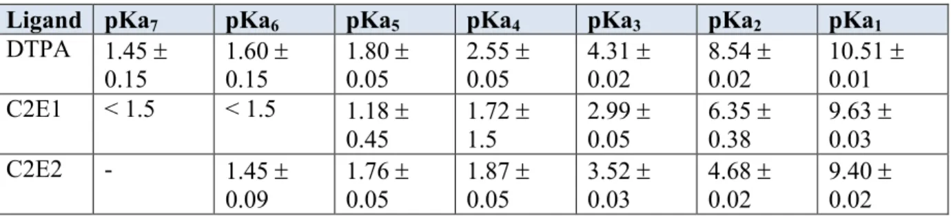
Table 4.1 The precursor product ion transitions and retention times for C2E2, C2E1 and DTPA used during LC/MS/MS quantification…...……….87
Table 4.2 Acid dissociation constants determined for C2E2 and C2E1 compared to those known for DTPA……….……..87
Table 4.3 Logistic equation parameters and calculated EC50 and EC90 values for C2E2, C2E1 and DTPA binding with 241Am……….87
Table 5.1 Toxicokinetic Bleeding Schedule………..………106
Table 5.2. Total 241Am Decorporation (Mean SD), represented as percent of the administered 241Am dose, after treatment with increasing doses of orally administered C2E2 or intravenously administered DTPA. Daily dosing began 24 h after contamination and continued for a total of 7 days……….………...…….…...…………108
Table 5.4. Total 241Am liver burden 12 days after contamination…………..…………112
Table 5.5. Total 241Am skeletal burden 12 days after contamination………..………...112
Table 5.6 Total 241Am decorporation 12 days after contamination following five days of a 600 mg/kg C2E2 dose administered either once daily
or divided between two equal doses………..114
Table 5.7 C2E2 toxicokinetic parameters in Sprague-Dawley rats after oral
gavage doses of C2E2 for 10 days………..…...118
Table 5.8 C2E1 toxicokinetic parameters in Sprague-Dawley rats after
oral gavage doses of C2E2 for 10 days………..……...119
Table 5.9 Mean ( SD) 241Am Decorporation 7 Days after IM Contamination in Male SD Rats: Treatment with a Single Oral Dose of C2E2 24 h
after Contamination……….…..130
Table 5.10 Mean ( SD) 241Am Decorporation 7 Days after IM Contamination in Female SD Rats: Treatment with a Single Oral Dose of C2E2 24 h
after Contamination………...130
Table 5.11 Percentage increase in 241Am decorporation over control, after
treatment with single doses of orally-administered C2E2 or C2E5…..…….131
Table 5.12 Total 241Am decorporation, represented as percent of the administered 241Am dose, after treatment with increasing doses of orally-administered C2E2 or C2E5………...………...131
Table 6.1 Systemic C2E2 exposure following oral administration of single
escalating doses in dogs……….146
Table A.1 Summary of Pharmacokinetic and Efficacy Data for Male
Sprague-Dawley Rats………..…...168
Table A.2. Summary of Pharmacokinetic and Efficacy Data for Female
Sprague-Dawley Rats………..…..….168
Table A.3 Summary of Pharmacokinetic and Efficacy Data for Beagle Dog…...168
Table A.4 C2E2 plasma concentrations following administration of ten oral
doses of 200 mg/kg in both male and female rats………..170
Table A.5 C2E2 plasma concentrations following administration of ten oral
doses of 400 mg/kg in both male and female rats……….………...171
Table A.6 C2E2 plasma concentrations following administration of ten oral
doses of 800 mg/kg in both male and female rats……….….172
Table A.7 C2E2 plasma concentrations following administration of a single
oral dose of 2000 mg/kg in both male and female rats………..………173
Table A.8 C2E2 plasma concentrations calculated for a 600 mg/kg dose of
C2E2 in rats………173
Table A.9 C2E2 plasma concentrations following administration of a single
oral dose of 100 mg/kg in both male and female dogs………..…174
Table A.10 C2E2 plasma concentrations following administration of a single
oral dose of 300 mg/kg in both male and female dogs………..174
Table A.11 C2E2 plasma concentrations calculated for a 500 mg/kg dose of
LIST OF FIGURES
Figure 1.1 Clearance of iv‐injected 241Amfrom the plasma volume of animals………...14
Figure 1.2 Retention of 241Am in rats after various treatment delays between
contamination and administration of either Ca- or Zn-DTPA…………...27
Figure 2.1 Synthesis of DTPA di-ethyl ester (C2E2)………....47
Figure 2.2 Differential scanning calorimetry (DSC) and thermogravimetic
analysis (TGA) chromatograms of C2E2………..………..52
Figure 2.3 X-Ray powder diffraction (XRPD) chromatograph depicting
crystalline nature of C2E2………...52
Figure 2.4 Log Dprofile for C2E2………..…..53
Figure 2.5 Stability of C2E2 in simulated gastric and intestinal fluids……….54
Figure 2.6 Dissolution profiles of HPMC C2E2 capsules after storage under
accelerated stability conditions for six months………....54
Figure 2.7 C2E2 dose response curve showing total 241Am decorporation in
male and female Sprague Dawley rats seven days after contamination……..55
Figure 2.8 C2E2 dose response curve showing 241Am liver (left) and skeletal (right) burdens in male and female Sprague Dawley rats seven days
after contamination………...………...55
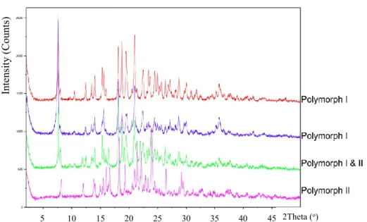
Figure 3.1 Differential scanning calorimetry chromatograph of polymorph I and
II...69
Figure 3.3 X-Ray powder diffraction (XRPD) chromatographs for polymorphs I
and II depicting the differences in crystal structure………...…...…..70
Figure 3.4 Scanning electron microscopy images of polymorph I and II,
displaying the different crystal habits of each form……….………70
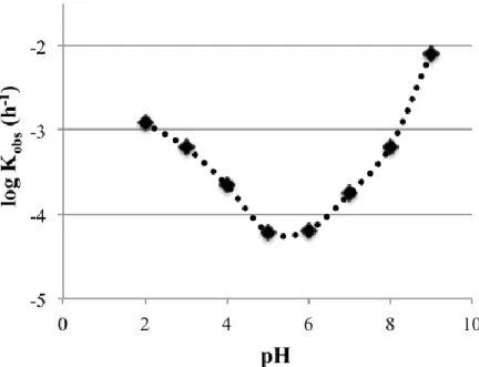
Figure 3.5 pH-rate stability profile of C2E2 at buffer concentrations of 200 mM
with constant ionic strength (I = 0.6 M)………..71
Figure 3.6 Schematic of the proposed acid and base catalyzed hydrolysis
pathways for C2E2………...………71
Figure 3.7 Stability of C2E2 in rat, beagle and human plasma………...72
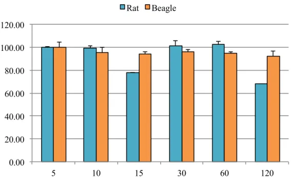
Figure 3.8 Metabolism of C2E2 by rat and beagle hepatic S9 fractions.
Graph depicts mean of triplicate samples (+ SD)………… ………..….73
Figure 3.9 Metabolism of C2E2 by rat and beagle intestinal S9 fractions.
Graph depicts mean of triplicate samples (+ SD)……….…..….73
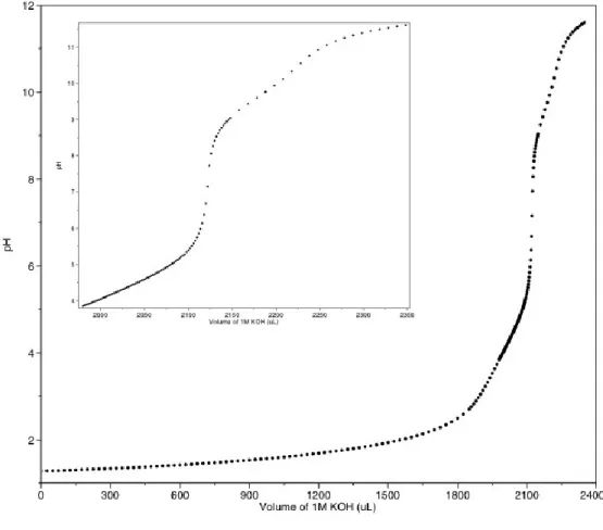
Figure 4.1 Titration of DTPA di-ethyl ester with 1 N potassium hydroxide…………....88
Figure 4.2 Distribution of 241Am as a function of total ligand concentration at I = 0.1 M (NaCl), T = 25 oC, pH = 1.3, and CHDEHP = 0.1 M in
dodecane………..89
Figure 4.3 The competitive binding of 3nM 241Am by C2E2, C2E1 and plasma proteins after 0.5 h incubation at 37 oC in rat beagle and human
plasma………..…90
Figure 4.4 The plasma concentrations of C2E2, C2E1 and DTPA after administration of a 100 mg kg-1 oral solution. The effective duration based on EC50 and EC90 values are indicated by dotted
Figure 5.1 Total 241Am Decorporation (Mean SD), represented as percent of the administered 241Am dose, after treatment with increasing doses of orally administered C2E2 or intravenously administered DTPA. Daily dosing began 24 h after contamination and continued for a
total of 7 days……….108
Figure 5.2 Daily Decorporation of 241Am from Female (A) and Male (B)
Sprague-Dawley Rats as a Percentage of the Injected Dose………111
Figure 5.3 Total 241Am Decorporation (Mean SD), represented as percent of the administered 241Am dose, after treatment with either once or twice daily orally administered C2E2 or intravenously administered DTPA to male SD rats………...…….114
Figure 5.4 Total 241Am Decorporation (Mean SD), represented as percent of the administered 241Am dose, after treatment with either once or twice daily orally administered C2E2 or intravenously administered DTPA to female SD rats………..………..115
Figure 5.5 Percentage of initial americium injection eliminated in the urine per
hour in male rats………...115
Figure 6.1 Daily urinary excretion after a single oral administration of C2E2
to dogs 24 hours after inhalation contamination with 241Am nitrate……….148
Figure 6.2 Percent of respirable 241Am dose remaining in lung, liver, total bone and kidney in dogs administered different doses of C2E2 at 24 h after
inhalation contamination with 241Am nitrate………...……..148
Figure A.1 Prediction of increase in decorporation over control from cumulative
AUC in rats………...………….169
Figure A.2 Prediction of increase in decorporation over control from cumulative
LIST OF ABBREVIATIONS
AAALAC Association for Assessment and Accreditation of Laboratory Animal Care
ALI Allowable Level of Intake
ALT Alanine Aminotransferase
Am Americium
AMAD Activity Median Aerodynamic Diameter ANOVA Analysis of Variance
AST Aspartate Aminotransferase
AUC Area Under Curve
BCS Biopharmaceutics Classification System BID Twice Daily / bis in die
C2E1 DTPA Mono-ethyl Ester
C2E2 DTPA Di-ethyl Ester
C2E3 DTPA Tri-ethyl Ester C2E5 DTPA Penta-ethyl Ester CAD Charged Aerosol Detector
CEA The Atomic Energy and Alternative Energies Commission
CHO Chinese Hamster Ovary
Ci Curies
Cm Curium
ED Effective Dose
EDTA Ethylenediaminetetraacetic Acid
F Bioavailability
FDA US Food and Drug Administration
FIH First in Humans
GFR Glomerular Filtration Rate SEM Scanning Electron Microscope
Gy Gray
HDEHP Di-(2-ethylhexyl)phosphoric Acid
HED Human Equivalent Dose
HOPO Hydroxypyridinone
HPLC High Performance Liquid Chromatography IAEA International Atomic Energy Agency ICH International Conference on Harmonisation ICRP International Commission on Radiation Protection
IM Intramuscular
IND Investigational New Drug
ITDB Incident and Trafficking Database
IV Intravenous
LD Lethal Dose
MCT Medium-chain Triglyceride
NADPH Nicotinamide Adenine Dinucleotide Phosphate NCE Normochromatic Erythrocyte
NCRP National Council on Radiation Protection & Measurements NIAID National Institute of Allergy and Infectious Diseases NIH National Institutes of Health
NMR Nuclear Magnetic Resonance NOAEL No Observed Adverse Effect Level ORAU Oak Ridge Associated Universities PCE Polychromatic Erythrocyte
PET Positron Emission Tomography PKPD Pharmacokinetic-Pharmacodynamic
PK Pharmacokinetic
PO Orally / per os
Pu Plutonium
QD Once Daily / quaque die RDD Radiological Dispersal Device
RH Relative Humidity
SGF Simulated Gastric Fluid SIF Simulated Intestinal Fluid SNS Strategic National Stockpile
SPECT Single-Photon Emission Computed Tomography
SV Sieverts
TTHA Triethylenetetraaminehexaacetic Acid
CHAPTER I
INTRODUCTION
1.1The Hazards of Radioactivity
The threat posed by radioactive materials is not new; naturally occurring isotopes, such as radium, continually expose us to low levels of radiation. In the United States the average background absorbed radiation dose is 360 mrem/y, which comes equally from both natural and manufactured sources1. Fortunately the consequences of background radiation are minimal as our cellular repair mechanisms are usually capable of responding to the effects of ionizing radiation. Therefore, chronic low-dose radiation is stochastic in nature, whereby some people may experience an effect from the exposure but the risk is low and cannot be attributed to radiation. Exposure to ionizing radiation at high-doses however leads to predictable effects that can be irreversible and often fatal.
This widespread availability gives rise to potential terrorist threats, which since September 11, 2001 has become a national security priority14. The use of unconventional terrorist tactics such as the delivery of anthrax spores in mailboxes in New York, Washington and Florida during 200115 illustrates the risk of attacks and there is an increased risk of further attacks that utilize biological, radiological or even nuclear weapons. The use of either radiological or nuclear weapons could result in significant numbers of individuals being exposed to ionizing radiation requiring urgent medical intervention. President Obama highlighted the importance of this threat in his first speech to the U.N. Security Council in New York, where he called nuclear terrorism one of the greatest threats to international security and stated that the consequences of such an attack would be enormous16.
couple of minutes could be fatal.) The loss of radioactive sources does not only occur overseas, for example nineteen tubes of 137Cs were stolen from a hospital in Greensboro, North Carolina and were never found18.
The release of radioactivity can result in localized or whole-body exposure and both internal and external contamination with radioactive materials. Exposure to radioactivity occurs when radiation penetrates the body from an external source, whereas contamination occurs when material deposits on the surface of an object. In order to minimize exposure it is important to increase distance from the source, wear shielding and minimize the time of exposure.
radiotherapy, however, it is only effective if given prophylactically and has numerous serious side effects20. Treatment of people exposed to radioactivity is thus mainly symptomatic.
When a radioactive nucleus decays it emits ionizing radiation in the form of an -particle, -particle, -rays, or a combination of these. Alpha particles consist of two protons and two neutrons. They can travel 2-3 cm in air, however they can only travel microns in tissue. They cannot penetrate paper, so are not an external hazard on skin, the outer layers of which are already dead. However, if ingested or inhaled alpha particles can cause serious damage to the adjacent tissues. Beta particles are high-energy electrons and can travel a meter in air and millimeters within tissue. They are an external hazard as they can penetrate the skin and can cause severe burns, if internalized they will also cause significant damage. Finally gamma rays are high-energy rays with short wavelengths that are very penetrating, having the ability to travel many centimeters through tissue21.
ions can form unstable hydrogen peroxide resulting in the formation of a peroxide hydroxyl radical that can become stable by combining with organic compounds in the cell. The organic hydrogen peroxide can then interfere with essential enzymes leading to cell death.
1.2Internalized Radionuclide Contamination
There are four main routes by which internal contamination can occur (a) inhalation, (b) uptake via cuts, wounds, burns or contaminated shrapnel, (c) ingestion followed by oral absorption and (d) transdermal absorption. The risk of exposure to radioactive heavy metals via the last two routes is low due to poor oral and transdermal absorption. Accidental contaminations such as syringe pricks or puncture wounds can occur in industrial, hospital or research laboratory environments. After detonation of a nuclear device, inhalation of the radioactive plume and wound contamination from radioactive shrapnel are the most likely sources of internalized contamination.
There have been a number of radiological accidents leading to contamination. The Goiânia incident in Brazil during 1987 highlighted the dangers of unsecure nuclear materials25. A radiotherapy source was stolen from an abandoned hospital and sold to a local scrapyard. The source container was opened and the blue cesium-137 was passed among the owner’s family and friends. As members of the family began to fall ill, the contents were tested and found to be radioactive; ultimately 249 people had significant contamination on their body of which 129 also had internal contamination. There were four fatalities associated with the incident, all of whom worked at the scrapyard where the radioactive source became exposed.
plutonium into the atmosphere, though in this case the levels were deemed low enough to not require treatment27.
The main focus of this doctoral research is the decontamination of americium and plutonium, which are -particle emitting radionuclides of the actinide series, with americium-241 being the radioisotope of primary interest. Each of these radionuclides has a unique biokinetic profile resulting in different patterns of tissue deposition. As such the disposition is also dependent on the route of contamination, the physio-chemical properties such as size and solubility of the ingested form, and the health status of the contaminated individual. Once internalized the tissues adjacent to the radionuclide are exposed to ionizing radiation until the contamination is removed either by excretion or treatment. The International Commission on Radiological Protection (ICRP) has summarized the distribution of americium and plutonium28.
Plutonium-239 has a half-life of 24,000 years29 and is the primary fissile isotope of plutonium used in the production of nuclear weapons and is also one of the main isotopes used as fuel in nuclear reactors. Due to its widespread use, the biodistribution of plutonium has been extensively investigated. Once it leaves the blood 50 % of the material is deposited on bone surfaces, 30 % retained in the liver and only 20 % in other tissues. Removal of plutonium from the bone and liver occurs with half-lives of 8000 and 9 years, respectively30.
different with 50 % of the radionuclide depositing in the liver and 30 % on the bone. Americium clears from the liver faster than plutonium with a half-life of 2-3 years, however loss from the skeleton is no different31.Once the radionuclide is deposited in a tissue it may re-speciate by equilibrating with other ligands and metal ions present, redistributing via the blood stream to a secondary deposition site32. It is for this reason that the half-life of americium elimination from the liver begins to increase over time, as americium redistributes from its primary disposition sites to the liver. It is easy to see from the long biological half-lives that internalized americium and plutonium can be expected to continue to irradiate the surrounding tissues for the remaining life of a contaminated individual in the absence of treatment to remove it.
The distribution and elimination kinetics of americium influence many aspects of treatment. Although the rate of tissue distribution is dependent on the site of contamination and the solubility of the internalized form, once solubilized and absorbed into the plasma the distribution and elimination kinetics are predictable and independent on the route of contamination. Therefore, though unlikely to occur, IV 241Am administration has proved useful for investigating the fate of 241Am after uptake from contamination sites. Radionuclide contamination is most likely to occur through either inhalation or a contaminated wound site. Thus during efficacy studies both inhalation and IM contamination models are more suitable as they mimic the continuous uptake of radionuclide into the plasma.
difference between species is the plasma iron concentration and subsequently the transferrin saturation. In human plasma americium is predominantly associated with transferrin, with the remainder associated with other proteins such as albumin, - and -globulins or low molecular weight ligands34. The stability constants for low molecular weight ligand complexes of americium have been determined35 (Table 1.2). Based on the stability constants and concentrations in plasma, citrate and carbonate are likely to be the predominant low molecular weight species. Any americium present as a citrate complex is ultrafiltratable by the kidneys and will be excreted in the urine.
The rate of radionuclide plasma clearance is largely dependent on its ability to form complexes with transferrin. Actinides that form more stable complexes, such as plutonium, are cleared more slowly than those with lower affinities such as americium. Animal physiology is therefore important with plasma 241Am retention being directly related to body size and inversely related with metabolic rate and renal filtration rate so that:
Human > Dog > Baboon > Monkey > Rat > Mouse
Plasma clearance of 241Am is fast with 90 % complete within 60 minutes and 99% complete in < 600 minutes (Figure 1.1). Only a small fraction is present in the urine at 24 hours and therefore the majority is deposited within the tissues.
receptor-mediated endocytosis involving the Tf-Fe complex and a cell surface Tf-receptor. Although both americium and plutonium bind with Tf, this seems to have a negative effect on hepatic uptake37. Therefore, hepatic uptake is related to the successful competition of hepatic cell binding sites for 241Am from plasma circulating complexes. Similarly skeletal binding sites, depending on their location, compete with 241Am-complexes present in the plasma or extracellular fluid. Generally there are three types of bone surface, those actively growing, resting, or resorbing and 241Am deposition is higher on resorbing and resting bone surfaces than those actively growing38. Ultimately there is continual competition between americium ions, plasma complexes and the tissues resulting in a dynamic equilibrium of species with a continual shift to the most stable complex. However actinides deposited within the bone are only released during limited structural remodeling. Therefore, once deposited within the bone the strong association prevents release back into the circulation even in the continued presence of chelating agents.
likelihood of cancer or DNA damage leading to genetic mutations. As there is no threshold, an exposure at any level carries a risk of stochastic effects.
Although any degree of contamination can lead to an increase of cancer, the increase in risk may be insignificant compared to other daily health risks. Hence the National Commission for Radiation Protection (NCRP) has suggested radiation levels below which treatment is often unwarranted. These levels are based on a conservative point, whereby treatment should start at the upper limit of radionuclide contamination permitted for radiation workers each year, known as an allowable level of intake (ALI). This ALI corresponds to an effective radiation dose of 5 rem (equivalent to 0.05 Sv), fifty times that of what is allowed for members of the general public, at which there is no measurable risk. For americium and plutonium treatment is indicated when one ALI is internalized which correlates to 0.8 Ci of ingested or 6 nCi of inhaled material40. For reference the average smoke detector contains 1 Ci of americium.
Table 1.1 Plasma volume, protein and iron concentrations across species (Taken from 33).
Mouse Rat Beagle Monkey Human
Body weight (kg) 0.035 0.25 10 6.2 70
Plasma (mL kg-1) 50 36 49 36 43
Serum Albumin (mM L-1) 0.56 0.60 0.52 0.64 0.65 Serum Globulin (mM L-1) 0.14 0.22 0.19 0.25 0.21 Transferrin (mM L-1) 0.036 0.033 0.032 0.040 0.027 Serum Iron (mM L-1) 0.053 0.039 0.025 0.032 0.018
Transferrin Saturation (%) 74 59 39 40 33
Table 1.2 Concentrations of relevant low molecular weight ligands in plasma and their stability constants for both americium and plutonium (Adapted from 35a and 35b).
Biological Ligand Blood Serum Concentration (M)
Stability constant (Log K) Americium Am3+ Plutonium Pu4+
Carbonate (CO32-) 2.5 x 10⁻² 8.0 -
Citrate 1.6 x 10⁻⁴ 8.6 15.3
Chloride 9.0 x 10⁻² 1.1 1.8
Glycine 2.6 x 10-4* 4.1 -
*Concentration from 41
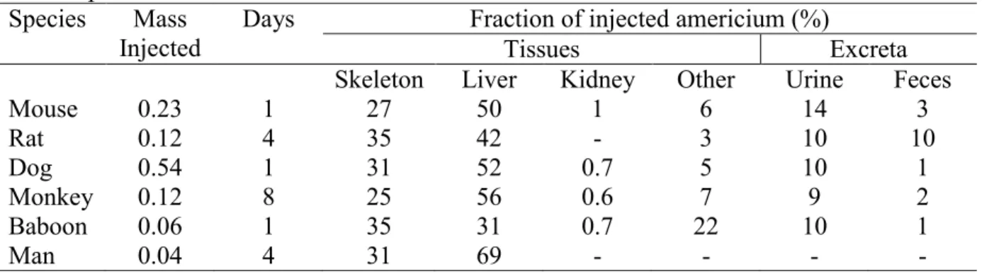
Table 1.3. Comparison of the initial distribution of injected americium in various tissues across species.
Species Mass Injected
Days Fraction of injected americium (%)
Tissues Excreta
Skeleton Liver Kidney Other Urine Feces
Mouse 0.23 1 27 50 1 6 14 3
Rat 0.12 4 35 42 - 3 10 10
Dog 0.54 1 31 52 0.7 5 10 1
Monkey 0.12 8 25 56 0.6 7 9 2
Baboon 0.06 1 35 31 0.7 22 10 1
Figure 1.1 Clearance of IV‐injected 241Am3þ from the plasma volume of animals: rat
(Turner and Taylor, 1968a); mouse (P. W. Durbin and B. Kullgren, unpublished data); beagle
(Stevens and Bruenger, 1972); monkey (Durbin, 1973); baboon (Guilmette et al., 1980).
1.3Decontamination of Internalized Radinuclides
Fortunately the dangers of plutonium and other actinide elements were perceived during early investigations, so decorporation methods were sought out by the health group of the Manhattan Project as early as 194742. For those who become internally contaminated, there are five decontamination treatment options, the use of which depends on the internalized radionuclide. The underlying hypothesis for all five treatments is that accelerating the natural rate of elimination of the radionuclide will reduce the amount of activity remaining in the body and thus reduce the dose received by sensitive tissues and the risk of radiation effects31. The currently available oral and IV decorporation treatments are listed in Table 1.4.
The first method is to surgically remove the contaminated area. This method may be employed in situations where wounds are contaminated with a long-lived isotope, such as plutonium, in order to prevent systemic uptake. A second simple measure is to administer large volumes of oral fluids, which help to dilute the radionuclides and increase urinary excretion. For example, treatment with 5-10 L/day of fluid for one week is indicated for tritium contamination43. The use of diuretics will also help to further enhance urinary elimination.
Blue), which is insoluble and binds cesium-137 and isotopes of thallium with high affinity, preventing their absorption. In addition to preventing absorption of ingested cesium-137, Prussian blue can play a role in decorporation of patients systemically contaminated with cesium-137 by interfering with its entero-hepatic circulation44. In 2003 it received US Food and Drug Administration (FDA) approval for treatment of internal contamination45. Other examples of binding agents include barium sulfate, a radiographic contrast agent, and aluminum and magnesium salts, all of which can bind to strontium and radium to decrease absorption46.
Once a radionuclide is absorbed into the systemic circulation, the first three decontamination methods have limited use. The radionuclide must now be removed by enhancing its elimination either through blocking uptake into the target organ or through the use of a chelating agent. Uptake into target organs can be blocked by supersaturating radionuclide binding sites thereby preventing accumulation and enhancing elimination. Potassium iodide has been approved by the FDA since 2002 for prevention of radioactive iodine sequestration by the thyroid47. Calcium gluconate and calcium chloride act in a similar fashion, limiting the incorporation of radioactive strontium or calcium into the bone. Mobilizing agents can be used to help remove deposited radionuclides. Sodium bicarbonate can be used to remove uranium from the kidney and ammonium chloride can help to increase elimination of internalized radiostrontium. Both of these agents work by altering the pH at their target site.
range of cations, some are bound with a higher affinity leading to preferential chelation of the most tightly bound cation. Once bound the cation is part of a stable complex and no longer acts as a free ion, allowing for excretion by the kidney. This basic principle was first reported in 1942 by Kety48 and has been applied to decorporation therapy since the 1950s. Chelator selectivity for a particular ion is determined by a number of properties including the number of vacant coordination sites, the distance between these sites and the size of the ion to be bound. If the affinity of the chelator for essential metals such as Mg2+ is higher than that of the plasma proteins toxicity can occur due to depletion of essential metals.
Chelation therapy is most effective when administered immediately after contamination, while the radionuclide is in the plasma and before deposition within the tissues has occurred. Although the highest decorporation effect is seen with the initial dose, as a large proportion of the radionuclide is in the plasma, chelators are still effective at eliminating radionuclides albeit at lower levels with sustained treatment. The continual benefit of chelation therapy may be due to metabolic recycling of the radionuclide into the extracellular fluid or in some cases be due to intracellular chelation. After exposure via the lung or a wound, the radionuclide will continue to slowly diffuse into the plasma and redistribute from the contamination site to other tissues such as the bone. In these instances chronic chelation therapy may be indicated to continue fostering elimination, reducing the total dose and residual disposition in the bone and liver. Ultimately, the duration of treatment will be guided by the biokinetics of the radionuclide and the degree of contamination.
the use of common carboxylic acids and complexing agents and showed limited efficacy. A good chelating agent should possess the following properties:
Chelating moieties should be deprotonated at physiological pH High number of chelate rings to increase stability of the complex
Actinide-ligand complex behaves differently from actinide-biological complex High affinity for actinides and low selectivity for essential metals
Low toxicity
Avoid degradation to metabolites that no longer bind the metal High oral bioavailability is desirable35b
Table 1.4 Overview of Decorporation Treatments (Modified from43)
Decorporation Agent Route Dosage Radionuclide
Ammonium Chloride Oral 1-2 g q.i.d for 6 days Strontium Ca-DTPA, Zn-DTPA i.v. 1 g in 250 ml saline over an
hour
Americium, Plutonium, Curium
Calcium Oral Generous doses Radium,
Strontium, Barium Calcium gluconate i.v. 2.5 g administered in 0.5 L
over 4h. Daily for 6 days
Strontium D-Penicillamine Oral 250 mg daily between meals
and bedtime up to 4-5g daily Cobalt, Indium, Palladium
Potassium Iodide Oral 130 mg daily Iodine
Potassium Phosphate Oral 250-500 mg q.d.s. Phosphorous
Propylthiouracil Oral 100 mg t.d.s. for 8 days Iodine Prussian blue Oral 1 g t.d.s for up to three weeks Cesium, Thallium,
Rubidium Sodium alginate Oral 10 g add water and drink Strontium
Sodium bicarbonate i.v. - Uranium
Table 1.5 Chelators currently approved by the FDA
Chelator Target Radionuclide(s)
Deferasirox Deferiprone
Deferoxamine mesylate Dimercaprol (BAL)
Edetate calcium disodium (Calcium EDTA) Penicillamine
Pentetate calcium trisodium (Ca-DTPA) Pentetate zinc trisodium (Zn-DTPA) Prussian blue Succimer (DMSA) Trientine hydrochloride Iron Iron Iron
Arsenic, Gold, Mercury Lead
Copper
Americium, Plutonium, Curium Americium, Plutonium, Curium Caesium, Thallium
1.4DTPA
Diethylenetriaminepentaacetic acid (DTPA) is a polyaminopolycarboxylic acid chelating agent that was originally synthesized in 1954 as an alternative EDTA ligand with the aim of increasing the affinity towards multivalent cations59. The additional nitrogen and carboxylic acid on DTPA provide eight potential metal coordination sites that allow binding to a wide range of metals with high affinity60 (Table 1.6). The additional binding sites improve the stability of metal chelates compared to EDTA, while the coordination of Ca ions remains similar therefore the / ratio is enhanced resulting in increased efficacy. Due to having a lower affinity for calcium compared to zinc, the Ca-DTPA salt is used preferentially within the first 24 hours after contamination where it is ten times more effective at actinide elimination than the Zn-DTPA salt61. After the initial 24-hour period both salts are equally effective, however Zn-DTPA is preferred due to zinc’s stronger affinity for DTPA, which minimizes toxicity by limiting the loss of essential traces metals.
Due to its hydrophilic nature DTPA is poorly absorbed from the GI tract (3-5%)62, however when administered intraperitoneally or intramuscularly it is completely and rapidly absorbed63. Following IV injection DTPA rapidly distributes in the plasma and extracellular fluid, though its hydrophilicity prevents it acting intracellularly. The plasma concentration in humans can be described by three exponential components with half-lives of 1.4, 14.5 and 94.4 minutes. DTPA is rapidly cleared by glomerular filtration into the urine (t½ 19 min); 99 % of the administered dose is excreted in the urine within 24 hours, with the remaining DTPA being slowly excreted over the following days64. This remaining fraction of the DTPA dose (< 1%) may remain in excess of radionuclide due to the minute quantities present and be responsible for enhanced urinary elimination of the radionuclide after 24 hours.
KMLM K CaL
Historically DTPA been used for the treatment of lead poisoning and in the paper industry for chelation of metals in pulp. In addition, Gd-DTPA is used medically as an MRI contrast agent. Its efficacy for radionuclide decorporation was established through animal studies and accidental human contaminations65.
Ca-DTPA dose administered 30 minutes after contamination resulted in significant increases in elimination. After seven days, urinary elimination was increased from 10 to 70 % of the total injected dose and fecal elimination from 1.3 to 9.8 %. In addition the liver disposition was significantly reduced from 47 to 9.7 %70. The relationship between efficacy and initial dose was established in beagles to justify a 30 µmol/kg human initial dose. Body retention of 241Am was inversely proportional to the dose, with a linear relationship seen over doses up to 300 µmol/kg. The 30 µmol/kg dose human dose was selected as doubling the dose resulted in < 25 % change in total body burden after seven days71.
In addition to animal studies there have been many cases of accidental exposure to actinides in the workplace, however there are very limited numbers of incidents in which detailed in vivo measurements have been recorded72. The 1976 Hanford incident resulted in an exposure that was around 1000 times more severe than any previous incident, the patient was contaminated solely with 241Am and the case provides the best human evidence for the extended use of DTPA therapy73.
Am(NO3)3 more than 20 and 40 Ci had already deposited in the bone and liver respectively75.
As the 241Am was slowly absorbed into the blood stream this case was suited for chelation therapy. Over the course of the next 4.5 years the patient received a total of 584 g of DTPA. For the first 25 days up to three 1 g doses were given per day, this was followed by daily injections until day 332 where injections were administered 2-3 times per week.When treatment was suspended for a year, 241Am excretion in the urine dropped and biweekly DTPA therapy was resumed as liver retention was seen to be increasing69. In total approximately 900 Ciwas eliminated via the urine, 59 % of which was removed in the first 6 days. If left untreated only 10 % of the activity would be expected to be eliminated naturally with the remainder being deposited in the liver and bone76. The administration of DTPA ensured that after 5.3 years only 1% of the 241Am that reached the plasma was retained in the bone and liver, demonstrating that in this case DTPA blocked 99 % of the 241Am predicted to deposit in the tissues69.
Due to the slow absorption of 241Am from the skin to the bloodstream the majority was prevented from depositing in the tissues. The only effects of radiation seen were depressed leukocyte, neutrophil and lymphocyte counts in the blood, which had no observable health effects. Due to the contamination at age 64, the threat of tumor development was low due to the latency period exceeding the patient’s life expectancy. In addition after intensive and extended therapy, 241Am deposited within the bone was not effectively removed. Therefore this corroborates the fact that therapy should be initiated as early as possible after exposure.
calcium resulting in increased depletion of natural metals such as zinc and magnesium. When examined in vivo, the differences in toxicities were less pronounced in non-dividing cells. In proliferating Chinese hamster cells the Zn-DTPA was non-toxic whereas Ca-DTPA resulted in significant toxicity, suggesting that it interferes with protein or DNA synthesis by altering the composition of the extracellular metal ions of the culture media. In mice, the zinc salt was 2.5 times less toxic than the calcium salt77. The LD50 values for mice administered a single IP Zn-DTPA dose a is 12.5 mmol/kg (6484 mg/kg)63, which is 1.4 times higher than that of EDTA. In general DTPA can lead to histopathological lesions in the kidneys and similarly to EDTA can be considered nephrotoxic78.
The doses at which toxicity was noted during animal studies are significantly higher than the 30 µmol/kg used in humans, and therefore it is unsurprising that in all human cases of prolonged DTPA administration, no severe side effects have been observed. The Atomic Energy and Alternative Energies Commission (CEA) in France has administered 1158 injections to 469 people from 1970 to 2003, with over 70 of these cases involving multiple injections. From all these exposures the one-single adverse event was an allergic skin reaction that resolved with no long-term effect79. In addition the Oak Ridge Associated Universities (ORAU) maintain a DTPA registry and between the period of 1958 – 1987 reported that 3077 doses of DTPA were administered to 485 patients, with the temporary loss of some essential elements being the only reported effects80.
evidence of enhanced elimination and lack of toxicity obtained during unlicensed use in accidental contamination cases was used to support regulatory approval.
Table 1.6 DTPA Binding Constants
Element Log KM
Americium 22.9
Berkelium 22.8
Californium 22.6
Curium 23.0
Neptunium 23.6
Plutonium 23.4
1.5Pro-drugs
A drug’s bioavailability (F) is the fraction of unchanged drug that reaches the systemic circulation. In the case of an IV this is 100% of the administered dose and therefore its bioavailability is 1. In all other cases of drug delivery, a drug must first overcome a cellular barrier during absorption and only a fraction of the unchanged drug reaches the systemic circulation. The composition of this barrier and the difficulty in achieving permeation varies depending on the delivery route. In order to be absorbed, the drug molecule must first be in solution. Consequently the physiochemical properties of the molecule that affect both solubility and lipophilicity play a critical role in determining its bioavailability.
Although drug absorption is predominately considered to be affected by both the solubility and permeability of the drug molecule, there are additional factors that play an important role in defining the bioavailability. The presence of efflux pumps such as P-gp on the basolateral membrane81 and first-pass metabolism also contribute to a reduction in the fraction of unchanged drug reaching the circulation.
Although effective at binding radionuclides in the plasma, the majority of chelators are highly hydrophilic, restricting entry into cells and limiting efficacy. Lipophilic prodrug chelators have previously been investigated in hope of removing radionuclides deposited within the tissues such as the liver and bone. Markley88 was the first to produce a DTPA pro-drug with the aim of promoting intracellular penetration. Esterification was chosen to allow intracellular hydrolysis, restoration of the carboxylic acids and therefore chelation sites89. Markley chose the penta-ethyl ester of DTPA so that toxicity from the liberated alcohol would be minimized. The results indicated that although the penta-ethyl ester was no better than DTPA at removing plutonium from the liver alone, when given in combination with DTPA there was an additive effect suggesting that there were two mechanisms of removal and that the penta-ethyl ester was acting intracellularly.
Although able to act intracellularly the penta-ethyl ester was far more toxic than DTPA, possibly due to chelation of natural metals within the cells88. Therefore partially esterified DTPA analogues were investigated for their potential to remove intracellular plutonium90. The effect on plutonium deposition in mice was investigated using di-methyl, -ethyl, -butyl and –octyl esters given five days after contamination. None of the esters showed benefit in terms of liver burden, however the methyl and ethyl esters did show a reduction in skeletal burden compared with untreated controls. The study also confirmed that in general the toxicity of esters increased as chain length increased, the exception being that the ethyl ester was less toxic than the methyl ester. All were significantly less toxic than the penta-ethyl ester, which suggests that toxicity is affected primarily by lipophilicity.
DTPA molecule containing an additional nitrogen and carboxylic acid increasing its coordination number to 10. TTHA molecules have been esterified with chain lengths varying from C8-C22 in order to both improve their oral absorption and traverse cellular barriers. These chelators were designed to increase removal americium or plutonium retained within the tissues and efficacy was assessed in rats contaminated with 239Pu and 241Am by IV injection. The chelators were administered fourteen days after administration of activity to allow deposition in tissues. For 30 days, rats received 80-85 mg of the C22-TTHA ligand orally, DTPA control rats were administered an equimolar concentration of DTPA by S.C. injection twice a week. At completion of the study the activity of 239Pu and 241Am in all tissues and bones was no better than DTPA therapy91. Interestingly the prodrug is predominantly excreted via the fecal route, suggesting a biliary excretion pathway, less lipophilic prodrugs however result in a larger fraction of urinary excretion92. Preliminary toxicity studies indicated that there was no apparent toxicity following 10 daily 200 mg/kg (200 µmol/kg) doses of the C22-TTHA ligand93.
circulating forms95. The increased lipophilicity resulted in efficient membrane transport, which may be a contributing factor to the high toxicity. Although successful the oral penta-ethyl ester research ceased due to a complex metabolic profile involving ten potential metabolites and due to the high toxicity. The penta-ethyl ester prodrug has also been examined topically with the aim of establishing sustained delivery of chelator to eliminate the continual release of radionuclide from the contamination site96. Topical administration and subsequent metabolism by skin esterases was also hypothesized to limit the fraction of lipophilic metabolites reaching the plasma resulting in reduced toxicity and improved efficacy. In rats contaminated with Am-nitrate (IM) and treated with a 1120 µmol/kg topical gel applied over a 6 cm2 area 24 hours after contamination, statistically significant increases in elimination of 241Am were observed97. Liver and skeletal burdens were also reduced compared to untreated control animals, demonstrating that the transdermal route may be a feasible option for chelation therapies.
Though there has been much research into alternatives for DTPA over the past fifty years there are still no new products in medical use. This may have been due to the lack of funding for a product with a limited patient population and the lack of animal rule pathway for regulatory approval.
1.6Approval of Medical Countermeasures
In late 2001 the National Institutes of Health (NIH) and National Institute of Allergy and Infectious Disease (NIAID) began to formulate a comprehensive biodefense research program, which culminated with the publication of a strategic plan for medical countermeasures against radiological and nuclear threats in 2005. One of the aims outlined in this plan was to promote research of new decorporation products that can be easily administered, safe for repeated dosing, have a long shelf life and be easy to manufacture98. In addition to the creation of a biodefense research program, the FDA introduced a new pathway for drug approval to accelerate the development of medical countermeasures (MCM) known as the “Animal Rule”99. Prior to the introduction of this rule there was no pathway for approval for these MCMs where the patient population is too small to conduct well-controlled clinical trials and it is unethical to contaminate healthy individuals in order to determine the efficacy.
In order to gain regulatory approval via the animal rule the product must meet the following criteria100:
1. A detailed understanding exists regarding the pathophysiological mechanism for toxicity of the agent in humans and also regarding the mechanism by which the proposed product reduces or prevents adverse effects in humans.
2. The prophylactic or therapeutic effect of the product is demonstrated in more than one animal species, unless the effect is demonstrated in a single animal species that represents a sufficiently well characterized animal model for predicting human response.
4. Pharmacokinetic and pharmacodynamics data for the proposed product are available and can be used to select an effective dose in humans.
For radionuclides the mechanism of toxicity is established and extensively detailed in published data. The kinetics of americium and plutonium distribution have predominantly been investigated in rats and dogs. Not only is the choice of animal important but the route of contamination must also be considered, as the animal model must mimic the effects of contamination in humans. In addition to selecting the most appropriate contamination models, the timing of treatment administration should mimic that which will be used in humans. A single model is unlikely to simulate the human response and two species can help to increase the confidence in the animal models response101. The development of a pharmacokinetic-pharmacodynamic model will help to identify the dose range likely to be effective in humans.
1.7Perspective
In response to the NIH/NIAID call for improvements of DTPA formulation, three research groups received funding, including SRI International (Menlo Park, CA), Nanotherapeutics Incorporated (Alachua, FL) and the lab of Dr. Jay at the University of Kentucky51. SRI has developed orally bioavailable formulations of DTPA by incorporating permeation enhancers into an oral capsule. Various permeation enhancers have been evaluated in vitro and the oral absorption of DTPA has been shown to increase from 5 to 12 % in rats104. The toxicity and efficacy of this novel oral formulation has also been investigated. The Nanotherapeutics approach involves the development of an orally administered nanoparticle formulation. Preclinical studies of this oral ‘NanoDTPA™ capsule formulation have demonstrated good pharmacokinetic and safety profiles in rodent and dog models105 and the product received additional funding from Biomedical Advanced Research and Development Authority (BARDA)106. For a more detailed discussion on the results of these studies in relation to the current work the reader is referred to chapter VII.
1.8Specific Aims
Specific Aim 1: To synthesize and characterize the di-ethyl ester pro-drug of DTPA, C2E2, and its major metabolite the mono-ethyl ester C2E1. In order to study C2E2 as a potential DTPA pro-drug and to determine its pharmacokinetics, the compounds of interest must be synthesized. Both C2E2 and its primary metabolite C2E1 have previously been described in the literature, however their use as an oral pro-drug of DTPA is novel. Once synthesized the compounds will be characterized by NMR, MS, HPLC, DSC, XRPD, elemental analysis and water content to determine their purity. The majority of active pharmaceutical ingredients (APIs) are administered as a solid dosage form. Regulatory agencies place a heavy emphasis on safety and efficacy; therefore the chemical purity of solids is often the primary focus. However the physical aspects of the dosage form such as stability are equally important. The measurement of pKa, solubility and lipophilicity are an integral part of lead compound profiling. Properties to be investigated include solubility, pKa, stability constants, Log P and metal binding kinetics.
Specific Aim 2: To investigate the metabolism of C2E2 in vitro. The stability of C2E2 will be determined in simulated intestinal and gastric fluids to ensure the esters are not hydrolyzed prior to absorption. Plasma binding, and stability will be determined using human, beagle dog and rat plasma. The metabolism of C2E2 by human, beagle dog and Sprague-Dawley rat S9 fractions will be evaluated to determine the conversion of C2E2 to it’s metabolites C2E1 and DTPA.
Sprague-Dawley rat and Beagle dog models. The parameters obtained such as the CMax and AUC can be incorporated into a pharmacokinetic model. The bioavailability and decorporation efficiency of C2E2 will also be determined. Tissue accumulation will also be performed.
CHAPTER II
C2E2: A DI-ETHYL ESTER PRODRUG OF DTPA AS AN ORALLY BIOAVAILABLE RADIONUCLIDE DECORPORATION AGENT
2.1. Introduction
Diethylenetriaminepentaacetic acid (DTPA) is a chelating agent with eight coordination sites making it effective at binding a wide range of metals with high affinity. It is available as both calcium and zinc tri-sodium salts (Ca-/Zn-DTPA) for intravenous injection both of which have been approved by the FDA for treatment of individuals internally contaminated with isotopes of the transuranic elements of americium (Am), curium (Cm) and plutonium (Pu)58. Once administered DTPA is rapidly distributed to the extracellular fluids where the calcium or zinc is exchanged for the higher affinity radioactive metals enhancing their elimination via urinary excretion.
exposure while the metal is in the circulation and soft tissue43. Any delay in treatment has a profound effect on the retention of americium (241Am). In beagles contaminated with IV 241Am citrate and treated with a single dose of IV Ca-DTPA, the percentage of 241Am retained increased from 3% after a 1 minute delay to 29, 58 and 73% after 30 minutes, 8 hours and 1 day delays, respectively108. Similar effects have been shown with both plutonium and curium. The radionuclide deposition kinetics are dependent on the method of contamination, species, and chemical form of the heavy metal33. Contamination is most likely to occur via inhalation or wound sites and therefore the percentage decorporation achieved after a single dose is likely to be lower than that described above due to retention of the metal in the lung or wound site. For this reason it is not uncommon for DTPA to be given over a period of days or months and in extreme cases for years73. The efficacy of DTPA in humans has been investigated after unintentional contaminations such as during the Hanford Americium Accident109. Due to the increased threat of nuclear terrorism both forms of DTPA have been added to the United States Strategic National Stockpile as radiological countermeasures.
where there may be mass contamination and casualties. During an emergency scenario delivery of oral dosage forms of DTPA would enable rapid distribution and cost-effective treatment saving resources for patients who need more urgent care. The ease of administration should also help to decrease the delay in receiving the first dose thereby improving decorporation and reducing the length of treatment required. In addition those who are severely contaminated may need months of treatment. Daily IV injections will reduce the patient’s quality of life, which would not be the case with an oral product.
2.2. Experimental Methods
2.2.1 Synthesis & Identification
scientific, Sunnyvale, CA). A reverse phase gradient separation was performed using an Alltima C18 column 250 x 2.1 mm2 internal diameter with 5 µm particle size (Grace) at 40 oC and a flow rate of 0.25 mL/min. The mobile phases were composed of water with 0.1% trifluoroacetic acid (A), acetonitrile/isopropanol 2:1 (B). The mobile phase followed a linear gradient from 94:6 to 75:25 over 14 min, the gradient then increased for 0.5 min to achieve a flow of 0:100 for 3.5 minutes followed by re-equilibration of the system at 94:6 for 6 min. The CAD analysis was performed at 25 oC with nitrogen flow at 35.1 psi.
2.2.2 Characterization of Solid C2E2
zero-background silicon plates and scanned from 2-50 2 with a step size of 0.016 and a time of 10 s per step.
2.2.3 Solubility
The apparent solubility of C2E2 was determined in deionized water. C2E2 was added to the water in excess and left to stir, at various time-points a sample of supernatant was taken and filtered to remove undissolved solid. The concentration of the supernatant was then determined by HPLC-CAD. The solubility was measured until two concurrent readings were achieved and the experiment repeated in triplicate.
2.2.4 Lipophilicity
2.2.5 Stability
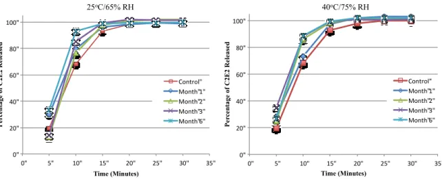
The stability of C2E2 was investigated in simulated gastric fluid (SGF) and simulated intestinal fluid (SIF). The simulated fluids were prepared as outlined in the United States Pharmacopeia using pepsin and pancreatin (Acros organics). C2E2 was dissolved in pre-heated simulated fluids, and the reaction mixture retained at 37.5oC for the duration of the experiment. At various time-points and aliquot was taken and the concentration of C2E2 was determined by HPLC-CAD. In addition C2E2 capsules were evaluated under accelerated stability conditions at both 25oC/65%RH and 40oC/75%RH. Size 0 HPMC capsules were filled with 200 mg C2E2 and placed in HDPE bottles. At 0, 1, 2, 3 and 6 months the dissolution performance of the capsules was evaluated. The C2E2 content of the capsules was measured at each of the aforementioned time-points and at 12 months. Dissolution was performed in a using dissolution bath (Hansen) equipped with USP II paddle apparatus. Capsules were placed in 900 mL of 0.1N HCl solution with constant stirring at 50 rpm with all samples were analyzed in triplicate.
2.2.6 Single Dose 241Am Decorporation
performed under both fed and fasted conditions to determine if there were any food effects. Approximately 16 hours before administration of C2E2, rats assigned to the fasted groups had access to food restricted with replacement occurring 1 hour after oral gavage treatment. At all other times all animals had ad libitum access to food. A treatment control group received a single intravenous dose of Ca-DTPA solution (13.3 mg/kg, 0.67 mL/kg) via a jugular vein catheter 24 hours after contamination under fed conditions. All animals were observed at least once daily for morbidity, mortality, and general appearance.
Excreted urine and feces were collected daily. Feces were allowed to dry overnight before transfer to 20 mL scintillation vials and weighing. Following necropsy selected tissues were removed and weighed. The 241Am gamma activity was determined by quantifying the 59.7 keV photons emitted by 241Am using a gamma counter (Wizard2 2480, Perkin Elmer). The total amount of 241Am administered to the animals was determined by counting duplicate aliquots (100 µL) of the injection solution. A counting window from 40 – 80 keV and a 60-second counting time were used for acquisition and each reading was corrected for background at acquisition. All experimental tissues and samples were counted using the same gamma counter and protocol. For all samples, 241Am content was expressed as a percentage of the initial dose. The femur from the leg opposite to the injection site was scaled by a factor of 20 to estimate total skeletal 241Am burden.
and Least squares means calculated for all the groups in this model. Comparison of means was made using the Tukey-Kramer adjustment for multiple comparisons, with p < 0.05 considered significant.
2.3. Results
2.3.1 Synthesis & Identification
The synthesis of DTPA-BA (1) resulted in a cream colored powder. Yield = 50 %; 1HNMR (400MHz, d6 DMSO) δ 3.65 (8H, s), 3.44 (2H, d), 2.74 (4H, t), 2.59 (4H, t). Addition of ethanol in the presence of pyridine yielded the DTPA diethyl ester (2), named C2E2. Yield =95.6 %; 1HNMR (400MHz, d6 DMSO) δ 4.07 (4H, q), 3.53 (4H, s), 3.45 (2H, s), 3.43 (4H, s), 2.89 (4H, d), 2.84 (4H, d), 1.19 (6H, t). MS, m/z calculated 450.2; found 450.13. Elemental analysis: Predicted for C18H31N3O10 – C, 48.10; H, 6.95; N, 9.35; O, 35.60; Actual – C, 47.59; H, 7.22; N, 8.67; O, 36.29.
2.3.2 Solid Characterization
DSC identified a single-phase transition for C2E2 at 115.8oC (Figure 2.2). The use of TGA confirmed that the transition observed during DSC studies was the melting point. There was a small loss in weight observed during TGA analysis, prior to decomposition (Figure 2.2). The loss was gradual, not attributed to a specific temperature, and non-stoichiometric suggesting that surface/incorporated water was the cause. The XRPD chromatogram was obtained and confirmed that the synthesized C2E2 was crystalline (Figure 2.3).
2.3.3 Solubility
solubility profile a range of buffers were prepared however, due to the high solubility of C2E2 the buffer capacities were exceeded even at high buffer concentrations and so the pH dependent solubility profile could not be determined.
2.3.4 Lipophilicity
The lipophilicity of C2E2 was determined by its partitioning between aqueous buffers of pH 2-9 and 1-octanol. The log D profile in Figure 2.4 shows that lipophilicity increases with decreasing pH. The Log D values ranged from -2.1 to -3.8. This is expected for C2E2, the three acids and bases have an isoelectric point at pH 2.74, therefore the highest lipophilicity would be expected at this point.
2.3.5 Stability
In order to ensure that the ethyl-ester promoeities were not hydrolyzed prior to absorption their stability was determined in both simulated gastric (SGF) and intestinal fluids (SIF). Over a 24-hour period there was a 34 % reduction in C2E2 concentration in SGF and only a 5 % loss in SIF (Figure 2.5). Therefore, with gastric emptying occurring within 2 hours in the average adult115, only a small fraction of the dose (~5 %) is expected to be lost to hydrolysis prior to absorption.
condition, with > 90 % release achieved in 15 minutes for all capsules tested (Figure 2.6), meeting FDA immediate release acceptance criteria under all the conditions tested115.
2.3.6 Single Dose 241Am Decorporation
Sixty-four rats completed the study, of these 61 rats (32 male and 29 female) were included in data analysis. Three fasted female rats (1x 600 mg/kg; 2x 1000 mg/kg) were excluded from the dataset as they did not receive a complete dose. C2E2 enhanced the elimination of americium in a dose dependent manner in male and female rats (Figure 2.7). Analysis of the total decorporation achieved in the seven days after contamination for all the rats treated with C2E2 showed significant effects for gender (F(1,44)= 17.54, p < 0.001) and C2E2 dose (F(2,44)= 40.71, p < 0.001) but not for the fed state at treatment (F(1,44)=1.51, p = 0.23). Interactions between food and other main effects were not considered significant and therefore, for subsequent analysis ad libitum and fasted groups were combined.
In male rats the total decorporation was significantly increased compared to untreated controls at doses of 600 mg/kg and above (p < 0.001) and increasing the dose from 600 mg/kg to 1000 mg/kg also significantly increased decorporation (p < 0.05). A similar trend was observed in female rats with doses ≥ 600 mg/kg inducing significantly enhanced decorporation (p < 0.001), the 1000 mg/kg dose appears to be more effective than the 600 mg/kg dose although this did not reach statistical significance.
0.056) it is consistent with the trend of C2E2 treatment reducing liver burden. A dose-dependent reduction in 241Am liver burden was observed in female rats although, only the reduction following the 1000 mg/kg dose was statistically significant compared to untreated controls (p < 0.01).
Figure 2.2 Differential scanning calorimetry (DSC, Red) and thermogravimetic analysis (TGA, Blue) chromatograms of C2E2.
Figure 2.5 Stability of C2E2 in simulated gastric and intestinal fluids.
Figure 2.6 Dissolution profiles of HPMC C2E2 capsules after storage under accelerated stability conditions for six months.
0" 20" 40" 60" 80" 100"
0" 5" 10" 15" 20" 25" 30" 35"
P er ce n ta ge o f C 2E 2 R el ea se d Time (Minutes) Control" Month"1" Month"2" Month"3" Month"6" 0" 20" 40" 60" 80" 100"
0" 5" 10" 15" 20" 25" 30" 35"
Figure 2.7 C2E2 dose response curve showing total 241Am decorporation in male and female Sprague-Dawley rats seven days after contamination.