INHIBITING THE FORMATION OF ALS-RELEVANT SOD1 OLIGOMERS
Elizabeth A. Proctor
A dissertation submitted to the faculty of the University of North Carolina at Chapel Hill in partial fulfillment of the requirements for the degree of Doctor of Philosophy in the Curriculum in Bioinformatics and Computational Biology.
Chapel Hill 2013
Approved by:
ABSTRACT
Elizabeth A. Proctor: Inhibiting the formation of ALS-relevant SOD1 oligomers (Under the direction of Nikolay V. Dokholyan)
Amyotrophic Lateral Sclerosis (ALS) is an invariably fatal neurodegenerative disease selectively affecting motor neurons. The aggregation of Cu, Zn superoxide dismutase (SOD1) has been linked to the disease, but the toxic species and mechanism of disease progression remain unknown. In this work, we explore two strategies for interfering in SOD1 aggregation: preventing the dissociation of the native homodimer, and preventing the association of misfolded SOD1 monomers into potentially toxic oligomers. In the first strategy, we elucidate the
mechanism by which a commonly found post-translational modification, glutathionylation, increases the dissociation of native SOD1. We find that the addition of glutathione moieties at their modification site near the dimer interface creates steric clashing that causes the two
monomers to twist in relation to one another, changing the shape and size of the dimer interface. These changes to the dimer interface cause a decoupling of dimerization and monomer folding processes, which in turn stabilizes a folding intermediate on the path to dimer dissociation, increasing the population of SOD1 monomer as observed in experiments. To pursue our second strategy, we develop a method to model the structure of meta-stable protein aggregation
recognized by conformational antibodies shown to bind disease-relevant misfolded SOD1 species. We use our structure to design mutations that will stabilize or destabilize the formation of SOD1 trimer in vitro, and demonstrate using time-resolved analytical size exclusion
This work is dedicated to my uncle, Dr. John H. Johnson, who succumbed to ALS in the summer of 2009. Your encouragement and quiet support helped give me the confidence and inspiration to
ACKNOWLEDGEMENTS
No journey such as this one is ever undertaken alone. I have grown so much in the past five years, but only a small fraction of that growth can be attributed to my own efforts. So here, in perhaps the most important part of my dissertation, I want to thank all of those who have contributed to my success. My sincere gratitude to:
The members of the Department of Biochemistry and Biophysics, the Program in Molecular and Cellular Biophysics, and the Curriculum in Bioinformatics and Computational Biology, for listening to various presentations of my work, making comments, asking questions, and overall making my science better. My committee, for their patience with me through these years, and their advice and support in helping me to see a problem from all angles.
My labmates, who always have the answers to all of my questions, and the perfect words that I can never formulate for myself. Thank you for giving me confidence and a sense of
belonging that I have never had until I came to this laboratory. My advisor, Nikolay Dokholyan, for his patience, encouragement, and guidance. I grew into a scientist in your hands, and I am forever grateful to you for opening my mind and teaching me to truly think.
PREFACE
Parts of the work described in this dissertation has been previously published as:
Elizabeth A. Proctor, Feng Ding, and Nikolay V. Dokholyan. “Structural and thermodynamic effects of post-translational modification in mutant and wild type Cu, Zn superoxide dismutase,” Journal of Molecular Biology 408: 555-567 (2011).
TABLE OF CONTENTS
LIST OF TABLES...xi
LIST OF FIGURES...xii
LIST OF ABBREVIATIONS AND SYMBOLS...xiv
CHAPTER 1: INTRODUCTION...1
CHAPTER 2: MECHANISM OF DIMERIC SOD1 DESTABILIZATION BY GLUTATHIONYLATION...5
2.1 Modeling and simulation of glutathionylated SOD1...6
2.2 Steric clashing of glutathione in the interface causes relative rotation of SOD1 monomers...8
2.3 Relative rotation of SOD1 monomers causes changes in dimer interface composition...10
2.4 Changes in SOD1 dimer interface lead to destabilization and decoupling of dimer dissociation and monomer unfolding...14
2.5 Glutathionylation stabilizes an SOD1 intermediate folding state...18
2.6 Possible implications of post-translational modifications of SOD1 in ALS...23
CHAPTER 3: CONTROLLING THE FORMATION OF SOD1 OLIGOMERS...26
3.1 Method for structural determination of meta-stable protein aggregation intermediates...28
3.2 SOD1 trimer constraints applied to monomer demonstrate rearrangement of monomer structure inside trimer...39
3.3 SOD1 trimer is a degenerate species with defined interfaces...41
3.4 Mutation to proposed trimer interfaces affects formation of SOD1 trimer...48
LIST OF TABLES
Table 1 - Torsional angle between monomers within SOD1 dimer...12
Table 2 - SOD1 dimer transition temperatures...17
Table 3 - Structural characterization of glutathionylated and unmodified SOD1 energetic populations...22
Table 4 - Energetic populations of glutathionylated and unmodified SOD1...22
Table 5 - Residues composing the SOD1 trimer interfaces...49
Table 6 - Effect of ALS disease mutations on SOD1 trimer stability...50
LIST OF FIGURES
Figure 1 - Proposed SOD1 aggregation pathway...3 Figure 2 - Structural effects of glutathionylation on the angle between
monomers in SOD1 dimer...11 Figure 3 - Effects of glutathionylation on SOD1 dimer interface
interactions...12 Figure 4 - SOD1 residues interacting with glutathione modification...14 Figure 5 - Thermodynamics of SOD1 unfolding in glutathionylated
and unmodified species...16 Figure 6 - Deciphering SOD1 energetic states in glutathionylated and
unmodified species...19 Figure 7 - Structural characterization of glutathionylated and
unmodified SOD1 energetic populations...21 Figure 8 - Wild type SOD1 forms a stable trimer under
destabilizing conditions...28 Figure 9 - SOD1 trimer is selectively bound by antibody raised against
disease-causative SOD1 species...29 Figure 10 - Schematic diagram for structural modeling of meta-stable
protein oligomers...31 Figure 11 - Folding of SOD1 monomer with λ=0...34 Figure 12 - Folding of SOD1 monomer with λ=0.66...35 Figure 13 - Final structures of perturbed SOD1 monomer at λ=0.66
and λ=0.99...36 Figure 14 - SOD1 trimer structures group by average number of
contacts (Nc) and radius of gyration (Rg)...37 Figure 15 - Limited proteolysis cleavage sites mapped to SOD1 native
dimer structure...40 Figure 16 - Location of limited proteolysis cleavage sites mapped to
Figure 17 - Structural model of SOD1 trimer...43 Figure 18 - Evaluation of structural quality of the SOD1 trimer model...44 Figure 19 - Replicate SOD1 trimer models feature significant variation
in tertiary and quaternary structure...45 Figure 20 - Agreement of SOD1 trimer interface residue identity
between models...47 Figure 21 - Atomic force microscopy visualization of SOD1 trimer...52 Figure 22 - Mutations destabilizing to SOD1 trimer result in increased
dimer and monomer populations...54 Figure 23 - Mutations destabilizing to SOD1 trimer result in formation
of large aggregates...55 Figure 24 - Predicted trimer-destabilizing mutant forms potentially
super-stable SOD1 trimer...57 Figure 25 - Mutation stabilizing to SOD1 trimer promotes dominant
and stable trimer...58 Figure 26 - Trimer-stabilizing mutations stabilize additional
LIST OF ABBREVIATIONS AND SYMBOLS
AFM Atomic force microscopy ALS Amyotrophic lateral sclerosis DMD Discrete molecular dynamics
ER Endoplasmic reticulum
FPLC Fast protein liquid chromatography HS-AFM High speed atomic force microscopy ITC Isothermal titration calorimetry KD Equilibrium binding constant KRMSD Kabsch root mean square deviation PDB Protein Data Bank
PDB ID Protein Data Bank identifier REX Replica exchange
RMSD Root mean square deviation SEC Size exclusion chromatography SOD1 Cu, Zn superoxide dismutase SPR Surface plasmon resonance
CHAPTER 1
INTRODUCTION
Amyotrophic lateral sclerosis (ALS) is a progressive neurodegenerative disease that selectively affects motor neurons, resulting in the eventual complete paralysis of patients. ALS is invariably fatal, with death occurring in most cases between two to five years following diagnosis. In the vast majority of cases, ALS does not affect cognitive, sensory, or autonomic processes, and so patients are fully cognizant of their decline. The most common cause of death is asphyxiation when paralysis reaches the diaphragm. Approximately 1 in 800 people will contract ALS in their lifetime, a number that grows larger as life expectancies increase1–4.
No cure or even effective treatment currently exists for ALS. The one FDA-approved medication available, riluzole, prolongs life by only a few months, mainly in patients who experience difficulty swallowing (National Institute of Neurological Disorders and Stroke, http://www.ninds.nih.gov/disorders/amyotrophiclateralsclerosis/ALS.htm). Patients often elect to stop treatment because they feel that the side effects of riluzole (nausea and fatigue) decrease quality of life to an extent that exceeds any therapeutic benefits. Other treatments are palliative only: for example, drugs to reduce fatigue, pain, and depression.
The main deterrent to the development of effective therapeutic strategies for ALS is that the underlying cause and molecular mechanism of ALS is unknown5. ALS is generally
~20% are associated with mutations to the homodimeric metalloprotein Cu, Zn superoxide dismutase (SOD1)4. The discovery in 19937,8 that mutations to the gene sod1 are linked to ALS was the first breakthrough in the search for a molecular mechanism of ALS etiology. Although SOD1 mutations account for only a small percentage of total ALS cases, symptoms and
presentation of SOD1-linked, sporadic, or non-SOD1-linked genetic cases are clinically
indistinguishable, and require a genetic test to differentiate1,9,10. These similarities motivate the general acceptance in the field that similar mechanisms underlie disease etiology in all cases of ALS, and that the study of ALS-linked SOD1 mutants will lead to understanding relevant to the treatment of all ALS sub-types1,4,9,10.
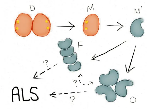
Since the initial linking of SOD1 mutations to ALS, over 140 mutations to SOD1 have been discovered to confer the disease (ALSOD database, http://alsod.iop.kcl.ac.uk). The vast majority of these mutations are destabilizing to either the individual monomers or to the monomer-monomer interface, with many of the remaining mutations destabilizing the apo-monomer11–15. Inclusion bodies containing SOD1 have been found in the motor neurons of both familial and sporadic ALS patients16–19. For these reasons, ALS is believed to be a protein misfolding disease1. The first step of SOD1 aggregation in ALS is the dissociation of the dimeric structure into two metal-bound monomers20,21 (Figure 1). The loss of the dimer interface allows the SOD1 monomers to more easily lose their bound zinc and copper ions, resulting in unstable apo-monomers that quickly form aggregates. SOD1 aggregates to form both small, soluble oligomers and insoluble amyloid fibrils. Whether these various aggregate forms lie along the same or competing aggregation pathways remains an outstanding question in the field.
Figure 1. Proposed SOD1 aggregation pathway. “D” native, metallated dimer; “M” native, metallated monomer; “M’” misfolded apo-monomer; “O” small, soluble oligomers; “F” large, insoluble aggregates, protofibrils, and fibrils.
this loss of function is not the underlying cause of ALS4. SOD1 knockout mice do not develop the disease22, nor does supplementation of wild type SOD1 to mice expressing ALS-relevant mutant SOD1 prevent or impede disease onset or progression, nor prolong life expectancy1. Therefore, it is thought that SOD1 undergoes a gain-of-function upon mutation, likely related to the resulting misfolding and aggregation. However, despite decades of research, the toxic species of SOD1 is still unknown, although several candidates have been suggested: insoluble fibrils, soluble oligomers, and misfolded monomers16–18,23–34. No toxic candidate species has been definitively ruled out to date, but several studies in ALS and other neurodegenerative diseases have supported the hypothesis that amyloid fibrils are inert and even possibly
motor neurons, having a cytotoxic effect as cells age and can no longer counter their aberrant interactions4,25,39,40. For example, ALS-relevant mutant SOD1 has been shown to interfere with conductance across the mitochondrial membrane41 and cause structural damage to the
mitochondria42, as well as induce ER stress and activate cell death pathways43, among other deleterious effects4. However, despite these advances in identifying toxic interactions of misfolded SOD1 in the cell, the identity of the SOD1 species involved could not be verified, although in each case the toxic SOD1 species was known to be small and soluble.
Aggregation and the formation of non-native species of SOD1 are intimately implicated in both genetic and sporadic forms of ALS, but little is known about the structure or toxicity of the various non-native toxic candidates. In this work, we incorporate experimental data to guide computational modeling of non-native SOD1 species. We use our models to deduce
mechanisms of misfolding and aggregation and to develop strategies to inhibit the formation of non-native SOD1 oligomers. The scientific advances we describe contribute toward knowledge of the molecular etiology of ALS and provide possible avenues for the development of
CHAPTER 2
MECHANISM OF DIMERIC SOD1 DESTABILIZATION BY GLUTATHIONYLATION
SOD1 is ordinarily a highly stable protein, with a folding free energy difference (ΔG) of 25 kcal/mol20,44–46. For comparison, a typical globular protein features a ΔG of 5 to 15
kcal/mol47. Although disease-linked mutations are usually destabilizing (as described above), these mutations only subtract, on average, 5 kcal/mol from the stability. This destabilization brings the free energy of folding to 20 kcal/mol, still significantly more stable than the typical globular protein, which leaves us with the question: how can this super-stable protein misfold in ALS?
occurrence of sporadic ALS in groups of individuals exposed to oxidizing chemicals: Italian soccer players exposed to fertilizers on the field51,52, veterans of the Gulf War exposed to chemical warfare agents53,54, and natives of Guam55,56 and Japan57 exposed to local toxins, among others4. How glutathione, itself an antioxidant, modifies an antioxidant protein to result in protein dissociation and enhanced aggregation remains unknown. To address this question, we computationally model glutathionylated wild type and mutant SOD1 and perform discrete molecular dynamics (DMD) simulations in order to determine the thermodynamic and structural effects of glutathionylation.
2.1 Modeling and simulation of glutathionylated SOD1
Modeling. We obtain structures of post-translationally modified mutant and wild type SOD1 using the known X-ray crystallographic structure of wild type SOD1 (PDBID 1SPD) as a reference structure. We constrain glutathione molecules to be covalently (permanently) bound to cysteine-111, and the zinc and copper ions to be permanently bound to their ligand residues. We obtain parameters for bond length, angle, and dihedral constraints for glutathione, metals, and disulfide bonds from the CHARMM19 force field58. All dimers are homo-modified. We then mutate the wild type structure to generate each disease-relevant mutant (A4V, G37R, G93A, H46R, and I112T) using the Eris suite59,60. We do not make structural adjustments to those residues participating in metal-binding, glutathionylation, or disulfide bond interactions unless they are necessary to the system of interest, as in the case of the H46R mutation, which coordinates the catalytic copper ion.
DMD. We perform three simulation iterations, each with a progressively lower heat exchange rate (0.2, 0.02, and 0.002 fs–1, respectively). Each iteration has a duration of 50 ps at a reduced unit temperature of 0.5 kcal/(mol!kB) (∼ 251 K).
Simulation. The discrete molecular dynamics (DMD) simulation engine is a variation on traditional molecular dynamics engines, where pairwise interactions between particles are modeled using step functions in place of continuous potentials. The DMD algorithm is
discussed in detail elsewhere61,62. We utilize the all-atom protein model62,63, in which all heavy atoms as well as polar hydrogens are explicitly represented. Bonded interactions are described by infinite square well constraints on bond lengths, angles, and dihedrals, making bonded interactions effectively permanent. Non-bonded interactions are discretized from the Medusa force field64, which includes van der Waals interactions (Lennard-Jones potential), solvation (Lazaridis-Karplus model65), and explicit hydrogen bonding (reaction algorithm66).
We utilize the replica exchange (REX) method to perform simulations of multiple copies of the same system in parallel at various temperatures67,68. The REX method is explained in detail elsewhere67,68, and is useful for exploring dynamics and thermodynamics of folding and unfolding. Briefly, at given time intervals, replicas of neighboring temperatures exchange temperature values according to a Metropolis-based stochastic algorithm. This exchange allows the system to overcome energetic barriers at higher temperatures, while retaining a realistic free energy profile at lower temperatures. We set the temperature exchange interval at 1000 DMD time steps (approximately 50 ps). We choose the temperature range of the replicas to cover the entire transition profile of the system, such that the system is disordered at the highest
simulations and construct a Temperature vs. Energy plot, from which the transition profile of the system is apparent. We then choose temperatures for replicas that cover this range, and fine-tune such that the successful exchange rate between adjoining replicas is between 0.2 and 0.7. In the monomer species, we perform 12 replica simulations with length of 100 ns at
temperatures of 0.50 (∼252 K), 0.52 (∼262 K), 0.54 (∼272 K), 0.56 (∼282 K), 0.58 (∼ 292 K),
0.60 (∼ 302 K), 0.62 (∼ 312 K), 0.64 (∼ 322 K), 0.66 (∼ 332 K), 0.68 (∼ 343 K), 0.70 (∼ 353 K)
and 0.72 (∼ 363 K) kcal/(mol!kB). In the dimer species, we perform 16 replica simulations with
length of 50 ns at temperatures of 0.48 (∼ 242 K), 0.495 (∼ 249 K), 0.51 (∼ 257 K), 0.525 (∼
264 K), 0.54 (∼ 272 K), 0.555 (∼ 280 K), 0.57 (∼ 287 K), 0.585 (∼ 295 K), 0.60 (∼ 302 K),
0.615 (∼ 310 K), 0.63 (∼ 317 K), 0.645 (∼ 325 K), 0.65 (∼ 327 K), 0.67 (∼ 337 K), 0.69 (∼ 347
K) and 0.71 (∼357 K) kcal/(mol!kB). We note that temperatures used in MD simulations,
including DMD simulations, often do not equate directly with physical temperatures. For example, the melting temperature of wild type SOD1 dimer that we calculate here differs from the experimentally-measured value of 93°C (366 K)69. However, simulations accurately reflect the relative changes in melting temperature, which are relevant to our studies.
To test for sufficient sampling, we split our simulation trajectories in half and compare the distribution of energies for the two halves. We obtain similar results for both halves, and therefore conclude that sampling is sufficient to reach equilibrium.
2.2 Steric clashing of glutathione in the interface causes relative rotation of SOD1 monomers
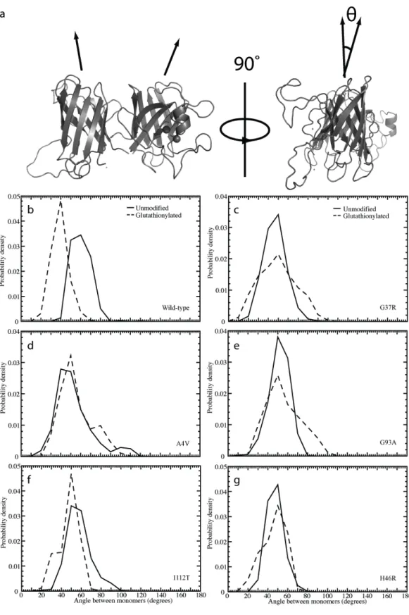
interface. We note in single-temperature simulations that glutathionylated SOD1 features a distinct change in the orientation of the two monomers with respect to one another. To quantify this change, we represent the two monomers of SOD1 as cylinders that, instead of being
oriented in a parallel fashion, are positioned at a torsional angle to one another (Figure 2). We then measure the angle between the axes of these cylinders.
We define the radial axis vector for each monomer by creating vectors along each beta-strand making up the approximately cylindrical beta-barrel, and averaging over the components. We define all beta-strand vectors to have the same directionality sign, and we length-normalize the vectors before averaging. The representative vector therefore features the three-dimensional average direction of all beta-strand vectors in each monomer. In order for these calculations to be meaningful, structures must be both folded and associated. In order to eliminate structures with monomer unfolding, we calculate the aligned RMSD (Kabsch RMSD70, KRMSD) between the beta-barrel alpha carbons of each monomer of the initial wild type-like structure and those of the corresponding monomer of each simulation snapshot (snapshots recorded every 5 ps). We impose a KRMSD cutoff of 4 Å, which we choose on the basis of the distribution of KRMSD values from all snapshots of all simulated species. To eliminate dissociated structures, we measure the distance between the centers of mass of the two monomers, which we calculate on the basis of the alpha carbons in the beta-barrels. We impose a cutoff of 35 Å, which we choose in the same manner as the cutoff for KRMSD. We retain those structures of each species that meet the criteria for both folded and associated, and measure the angle between the vectors characterizing the two monomers. We calculate the angle between the two vectors v1 and v2 as:
This torsional angle is affected by glutathionylation, likely because the bulkiness of the θ=arccos v1⋅v2
a b " #
$$ %
glutathione moieties in the dimer interface causes steric clashing that forces the two monomers to rotate in opposite directions to accommodate the modifications. However, the effect of this steric clash differs in different SOD1 mutants (Figure 2, Table 1). These differences are
potentially due to variation between the mutants in their dimer interface configuration and how they are affected by the addition of glutathione (Figure 3).
2.3 Relative rotation of SOD1 monomers causes changes in dimer interface composition The change in the relative orientation of the SOD1 monomers within the dimer
necessarily changes which residues are in contact, as well as the dimer interface area. In order to determine the extent of the change in dimer interface contacts, we construct contact maps for the dimer interface residues, and compare contact maps from unmodified structures with those of the glutathionylated structures.
We define two residues as being in contact in the dimer interface if the two alpha carbons are within 10 Å of each other. At each simulation snapshot, we evaluate contacts between the two monomeric chains. We normalize the count between every pair of residues for the total number of simulation snapshots.
Figure 2. Structural effects of glutathionylation on the angle between monomers in SOD1 dimer. (a) Depiction of the angle, θ, measured between monomers in the SOD1 dimer. Distribution of angle between
Table 1. Torsional angle between monomers within SOD1 dimer. Peak values of the distributions of the angle between monomers observed in Figure 2.
Angle between monomers (°)
Wild-type Unmodified 60
Glutathionylated 40
A4V Unmodified 45
Glutathionylated 50
G37R Unmodified 46
Glutathionylated 50
G93A Unmodified 53
Glutathionylated 51
H46R Unmodified 47
Glutathionylated 50
I112T Unmodified 55
Glutathionylated 50
introduction of glutathione induces the formation of non-native interactions in the interface (type 2) (Figure 3). In contrast, in I112T-SOD1, losses of interface contacts are accompanied by a gain in the residues directly adjacent (type 3) (Figure 3), which implies a shift in the location of the dimer interface and, therefore, a shift in its makeup. We find a similar shifting of contacts in the C-termini of G37R-SOD1, implying structural movement in that area (Figure 3).
Taken together, these results imply a shift in the residue composition and position of the dimer interface upon glutathionylation, likely due to the change in orientation of the two
monomers forced by the presence of bulky glutathione moieties in the dimer interface. A change in the identity, and in some cases the number, of dimer interface contacts can result in the loss of monomer-monomer binding affinity and increased dimer dissociation. In a parallel
experimental study50, we find a shift toward the monomer population in wild type and mutant A4V SOD1 (group 1), but an increase in the rate of monomer formation in mutant I112T-SOD1 (group 3), which suggests that the contact loss observed in glutathionylated wild type and A4V-SOD1 (Figure 3) results in a loss of monomer-monomer binding affinity, whereas the overall shift in contacts seen in I112T-GSH (Figure 3) instead affects kinetics, resulting in an increased koff for the dissociation reaction50.
compromise may introduce strain into the structure that contributes to the increased dissociation observed experimentally49,50.
We also note that the majority of SOD1 residues that interact with the glutathione moieties are located in the small loop segments connecting beta-strands (Figure 4). Interactions with glutathione thus may interrupt the side-chain alignment necessary for formation of the beta-barrels, which would result in the loss of crucial contacts and therefore dissociation.
Figure 4. SOD1 residues interacting with glutathione modification. Glutathione, copper, and zinc are shown in sphere representation. Residues with the highest frequency of interaction with glutathione are colored teal.
2.4 Changes in SOD1 dimer interface upon glutathionylation lead to destabilization and
decoupling of dimer dissociation and monomer unfolding
trajectories. Given the density of states ρ(E), the folding specific heat at constant volume, CV, is computed at various temperatures according to the partition function:
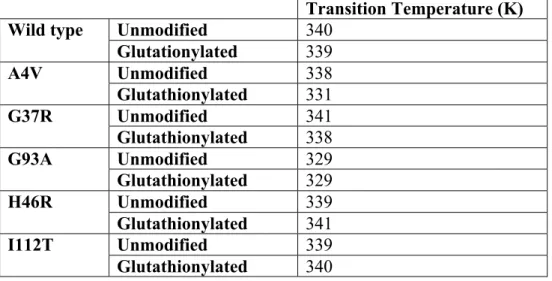
where E is the potential energy, T is the temperature, and kBis the Boltzmann constant. Because we do not observe multiple unfolding and refolding transitions in our simulations, we do not expect to obtain fully quantitative free energy landscapes for the various species of SOD1. However, we expect to gain insight into the stability of both the monomer and dimer species by using the unfolding transition temperature, which corresponds to the major peak in specific heat (Figure 5 )72. Specific heat is a measure of the amount of energy necessary to increase the temperature (kinetic energy) of the protein. Hence, a peak in specific heat corresponds to a transition between energetic states, where energy is devoted to raising the potential as opposed to the kinetic energy of the complex. Monomer species exhibit one significant thermodynamic transition, corresponding to monomer unfolding (Figure 5). Dimer species also exhibit one major transition, with the transition temperature shifted significantly to higher temperature as compared to the corresponding monomer species. Because the SOD1 dimer species feature one major peak rather than two peaks, corresponding to dimer dissociation and monomer unfolding, we conclude that dimer dissociation and monomer unfolding are highly coupled processes. The strong interactions in the dimer interface stabilize the individual monomers by shifting the unfolding transition to a higher temperature, keeping the dimer associated and folded at temperatures higher than the monomer unfolding temperature.
We observe that, in wild type and all mutant variants except A4V and I112T,
glutathionylation causes a decrease in the coupled nature of the dissociation/unfolding transition. All unmodified species exhibit a single peak in specific heat, representing a single transition,
but glutathionylated species feature a shoulder, representing a partial decoupling of the two
processes (Figure 5).
We find that glutathionylation destabilizes the dimer by shifting the transition to a lower
temperature. The exception to this trend is G93A-SOD1, where the glutathionylated species is
stabilized with respect to the unmodified form (Figure 5, Table 2).
Table 2. SOD1 dimer transition temperatures. Major transition temperature of the various SOD1 dimer species (Figure 5).
Transition Temperature (K)
Wild type Unmodified 340
Glutationylated 339
A4V Unmodified 338
Glutathionylated 331
G37R Unmodified 341
Glutathionylated 338
G93A Unmodified 329
Glutathionylated 329
H46R Unmodified 339
Glutathionylated 341
I112T Unmodified 339
Glutathionylated 340
We observe in dimer species that the maximum value of the major peak in specific heat
(corresponding to the energy input needed to raise the temperature of the complex by 1 °C
during the dimer dissociation/unfolding transition) is highest in the unmodified species, as
opposed to in the glutathionylated species (Figure 5), with the exceptions of G93A and I112T.
The absolute value of the specific heat indicates the cooperativity of the phase transition.
Therefore, this observation suggests that the dissociation/unfolding transitions are more
cooperative in the unmodified species, and that glutathionylated species exhibit a more gradual
melting transition from associated to dissociated states.
significantly affect monomer stability (Figure 5). All of these effects taken together imply that the destabilization of SOD1 dimer found experimentally is due to disruption of the coupling of dimerization and monomer folding. Interactions across the dimer interface strengthen the stability of the SOD1 monomers, but these interactions are disrupted by the twisting of the monomers in relation to each other, induced by the steric effects of bulky glutathione molecules near the dimer interface. The uncoupling decreases the stability of dimerization, increasing dimer dissociation.
2.5 Glutathionylation stabilizes an intermediate folding state in SOD1
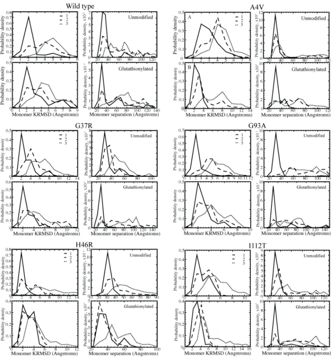
In order to study the more subtle transitions between native-like states that may further explain an increased dissociation in glutathionylated species, we examine the potential energy distributions of structures in the mid-range temperature region, 275–325 K, just reaching the lower bound of the dissociation/unfolding transition (Figure 6). These structures should include populations of native-like, dissociated, and any intermediate states. All energies discussed are potential energies of the system; contributions from internal potential energy of the modification molecules are minimal in comparison to the potential energy of the system, and therefore no adjustment is needed for comparisons of potential energy between modified and unmodified systems.
In each system, we observe the existence of at least three low-energy states with
Figure 6. Deciphering SOD1 energetic states in glutathionylated and unmodified species. Distributions of total potential energy throughout simulations, sampled at T=0.55-0.65 kcal/(mol!kB), with representative structures for
each of the three low-energy states.
significantly, and deconvolution with respect to individual structures is not possible, we identify
representative structures by clustering structures whose energies fall within 1 kcal/mol around
the mean of the respective Gaussian by RMSD using the OC suite73. Structures with energies in
this range have a higher probability of belonging to the energetic state of interest than of being a
global maximum in a histogram of pairwise RMSDs of all structures clustered. We choose the
centroid of the largest cluster as the representative structure for each state. The largest cluster is
in all cases at least four times the weight of the next-largest cluster.
The three delineated low-energy populations correspond to the native state and the early
excitation states in the dissociation/unfolding process. The first state is a low-energy state with a
native-like structure; the second state has undergone a conformational change that can be
characterized as a “loosening” of the beta-barrels and/or a slight movement outward of the two
centers of mass (Figure 6, Figure 7, Table 3); and the third state is the dissociated state, with
partial unfolding due to the coupled nature of the dissociation and unfolding processes.
The amount of differentiation between the states in partial unfolding or separation of the
monomers varies between mutants. Although in some glutathionylated species, namely A4V,
G37R, and I112T, the representative structure of the intermediate energetic state is dissociated
(Figure 6), we observe from the distributions of monomer separation and monomer unfolding
that, statistically, the intermediate state as a whole is still distinct from the dissociated state
(Figure 7, Table 3). In G93A, the unmodified species features an intermediate state that
maintains very few dimer interface contacts and, therefore, has a greater tendency to be
dissociated than the intermediate states of other mutant variants. However, glutathionylation
restores the intermediate state to a form with similar characteristics to the other mutants.
In general, we find that glutathionylation does not destabilize the native state; in most
mutant variants, the native-like state, which occurs at approximately –550 kcal/mol, does not
undergo a significant energetic change upon glutathionylation (Figure 6, Table 4). However, the
intermediate state population is shifted to a lower energy in glutathionylated species, decreasing
probability of the protein to misfold and dissociate. In many of the glutathionylated species, the
third (dissociated) state is also stabilized as compared to in the unmodified species, and the
Figure 7. Structural characterization of glutathionylated and unmodified SOD1 energetic populations.
Distributions of values for the monomer aligned KRMSD from the starting structure, used as a measure of β-barrel integrity and unfolding, and the distance between monomer centers of mass, used as a measure of dimer
Table 3. Structural characterization of glutathionylated and unmodified SOD1 energetic populations. Distribution averages for the monomer aligned RMSD from original structure, a measure of β-barrel integrity, and distance between monomer centers of mass, a measure of dissociation. Values shown in each cell are for the first, second, and third energy populations described in Figure 6. The full distributions of the properties below are shown in Figure 6.
Monomer RMSD (Å) COM-COM distance (Å)
1 2 3 1 2 3
Wild type
Unmodified 1.75 3.40 4.64 33.89 56.46 66.11
Glutathionylated 2.67 3.60 4.26 45.57 65.43 71.84
A4V Unmodified 2.47 3.32 4.12 27.55 43.94 57.86
Glutathionylated 1.98 3.75 6.71 29.31 47.37 59.16 G37R Unmodified 2.77 3.92 6.05 32.56 34.88 49.444
Glutathionylated 2.19 4.20 4.24 26.67 66.09 72.34 G93A Unmodified 1.85 4.28 5.68 31.23 72.26 76.76 Glutathionylated 2.27 3.06 4.16 42.12 58.86 66.71 H46R Unmodified 1.61 3.47 7.38 26.29 41.01 51.75 Glutathionylated 2.39 2.73 4.27 34.66 32.35 54.96 I112T Unmodified 2.35 2.99 5.09 29.23 45.35 67.88 Glutathionylated 2.44 3.28 5.99 34.57 58.86 76.44
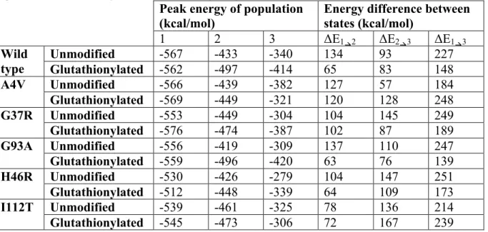
Table 4. Energetic populations of glutathionylated and unmodified SOD1. Peak values of the energetic populations observed in Figure 6, and the resulting energy differences between the states.
Peak energy of population
(kcal/mol) Energy difference between states (kcal/mol) 1 2 3 ΔE1"2 ΔE2"3 ΔE1"3
Wild type
Unmodified -567 -433 -340 134 93 227
Glutathionylated -562 -497 -414 65 83 148
A4V Unmodified -566 -439 -382 127 57 184
Glutathionylated -569 -449 -321 120 128 248
G37R Unmodified -553 -449 -304 104 145 249
Glutathionylated -576 -474 -387 102 87 189
G93A Unmodified -556 -419 -309 137 110 247
Glutathionylated -559 -496 -420 63 76 139
H46R Unmodified -530 -426 -279 104 147 251
Glutathionylated -512 -448 -339 64 109 173
I112T Unmodified -539 -461 -325 78 136 214
Glutathionylated -545 -473 -306 72 167 239
overall ΔE between the native-like and the dissociated populations is decreased (Figure 6, Table
2.6 Possible implications of post-translational modifications of SOD1 in ALS
A strong coupling of the dimer dissociation and monomer unfolding processes has
important implications for SOD1 overall protein stability and aggregation. Dimerization
significantly stabilizes the folded monomer, which implies that contacts in the dimer interface
contribute to the integrity of the monomer beta-barrels. As these contacts are broken, the
intra-monomer interactions maintaining the beta-barrel formation of each intra-monomer are also broken,
causing simultaneous dissociation and partial unfolding. The unfolding process is then more
favorable to complete upon full dissociation.
Supporting this hypothesis, both wild type and mutant unmodified species in general
dissociate and unfold more sharply than the glutathionylated forms with an increase in
temperature (Figure 5). This finding implies that the shifting or gain of non-native contacts
(Figure 3) that occurs in glutathionylated species causes a loss of cooperativity in the interface
and intra-monomer interactions. This loss of cooperativity allows some contacts to be lost much
more frequently than others, whether due to a structural change or the steric interference of the
modification molecules. A loss in interaction cooperativity may be manifest in the stabilization
of the intermediate state, causing a decreased potential energy gap between the native and
dissociated states (Figure 6, Table 4). Interactions between the glutathione moieties and their
associated monomer may also be responsible for this more drawn-out dissociation interaction
and loss of cooperativity; the modification sites are located near the dimer interface (Figure 4),
and interactions with the monomers could disrupt or weaken native dimer interface interactions.
This effect could possibly be remedied by the introduction of additional interactions in the form
of a drug that would bind, bridging the dimer interface and holding it together, as was found by
SOD1 in the first place, whether by occupation or blocking of the modification site or inhibition
of the modification binding interaction.
Here we study homo-modified species of wild type and mutant SOD1, but in vivo SOD1
may exhibit significant hetero-modified populations in addition to homo-modified species. In
the current experimental characterization of post-translational modifications from erythrocytes
(mass spectrometry)49,50, dimers are necessarily dissociated before measurement, and so it is
impossible to determine whether dimers are hetero- or homo-modified in vivo using this method. The molecular mechanism of glutathionylation is still unknown, so it is unclear whether
modification of the individual monomers is a cooperative or an independent process. However,
we find that the presence of only one glutathione moiety near the dimer interface disrupts or
changes monomer-monomer contacts and induces many of the same effects that we observe in
homo-modified species.
The results above indicate that the post-translational modification of glutathionylation
affects the energetic and structural properties of wild type and mutant SOD1. The effect varies
between mutants, suggesting that glutathionylation may have varied effects on the stability of
the dimer of the various genetic mutations, and for different reasons. For example, the A4V
dimer appears to be largely stabilized by glutathionylation, while the wild type dimer is
destabilized by glutathionylation. However, with the exceptions of A4V and I112T,
glutathionylation of SOD1 has a dramatic effect on decreasing the potential energy gap between
the native-like state and the dissociated state. We infer from this finding that the presence of
glutathione, a marker of oxidative stress in the cell75, would be detrimental in most types of
familial ALS. This finding corroborates with reports that exercise76 and electrical stimulation77
these would produce increased oxidative stress in cells and hence increased levels of
glutathionylated protein78. An environmental factor such as oxidative stress could help to explain the differences in disease progression between the various ALS-causative mutants.
Indeed, recent research shows a strong link between oxidative stress and ALS4, as oxidative stress contributes to misfolding and aberrant interactions of SOD1, inflammation, and
mitochondrial malfunction observed in the disease. Our results indicate that the oxidative
modification of SOD1 by glutathione could be a contributing factor to the linkage between
oxidative stress and ALS.
On a further note, because glutathionylation decreases the potential energy gap between
the native-like and dissociated states in wild type SOD1, and glutathionylation of SOD1 is
present even in healthy individuals49, glutathionylation caused by oxidative stress to motor neurons could be a factor in sporadic ALS. The late onset of ALS suggests that an
environmental trigger could exist for both familial and sporadic cases, which fits with the
increased occurrence of sporadic ALS in athletes52 and soldiers54 as compared to the general population; both of these groups experience more extreme and frequent oxidative stress than
does the average individual. Glutathionylation of SOD1 due to environmental factors could also
explain incomplete penetrance of some disease-linked SOD1 mutations, and the relatively small
destabilization (~5 kcal/mol) caused by disease mutations as compared to the high stability of
SOD1 (25 kcal/mol)4. Such environmental factors could possibly be counteracted with a drug or lifestyle decisions. Further investigation into the mechanism of post-translational modification
in SOD1 could illuminate preventative measures against the observed increased dissociation and
CHAPTER 3
CONTROLLING THE FORMATION OF SOD1 OLIGOMERS
The aggregation of SOD1 has been linked to ALS4, but the toxic species that causes
motor neuron death is still unknown. SOD1 is known to form several diverse species in its path
to aggregation: misfolded monomers; non-native dimers and other small, soluble oligomers;
large, insoluble aggregates and proto-fibrils; and stable, SDS-resistant fibrils16–18,23–34. However,
the road map to SOD1 aggregation remains a mystery: we do not know which species are
precursors or products of which others, which lie on the same versus competing pathways, and
which may form completely independently of others. Khare and colleagues have elucidated a
part of this puzzle by showing that SOD1 aggregation begins with the dissociation of native,
metallated dimer79. The resulting monomers then lose their copper and zinc ions, destabilizing
the monomeric structure and allowing the monomer to misfold (Figure 1)79. Khare and
colleagues found that the formation of large SOD1 aggregates increases in proportion with the
available population of monomer, demonstrating that SOD1 monomer is the seed for SOD1
aggregation.
Two types of SOD1 aggregates have been isolated in diseased cells: large, insoluble
aggregates, including SDS-stable fibrils; and small, soluble oligomers16–18,23–34. Researchers at
first thought that the large, stable amyloid-like fibrils were the cause of cell death in ALS, but
SOD1 is known to cause endoplasmic reticulum stress43, defective axonal transport80,81,
mitochondrial malfunction41,42, withdrawal of axon guidance signals82, and both oxidative and
nitrative stress83,84.
Because small, soluble oligomers have been shown to activate cell death pathways4,43,
they are prime candidates for the cytotoxic species responsible for ALS. Recent work has
demonstrated that promoting the formation of large, insoluble aggregates and fibrils decreases
the population of small, soluble aggregates and therefore may be a protective pathway in the
cell4,35–38, but an abundance of large aggregates in the cell poses its own risks and deleterious
effects. We propose to instead inhibit the formation of the potentially toxic oligomers by
interfering in the pathway to their formation, directly inhibiting the association of the misfolded
monomers into toxic oligomers. In order to implement this strategy, we need structural
knowledge of the toxic oligomers, specifically about the non-native monomer-monomer
interfaces. Recently, Redler and colleagues have identified a meta-stable, small, soluble
oligomer of SOD1: an SOD1 trimer (Redler et al., submitted). Using size exclusion
chromatography, they find that under destabilizing conditions, both wild type and mutant SOD1
will dissociate to form monomer and reassociate to form trimer over a 24-hour period (Figure 8).
This trimeric species can be isolated and is then stable at low pH (3.5), a destabilizing
environment that Khare and colleagues have shown produces accelerated but physiologically
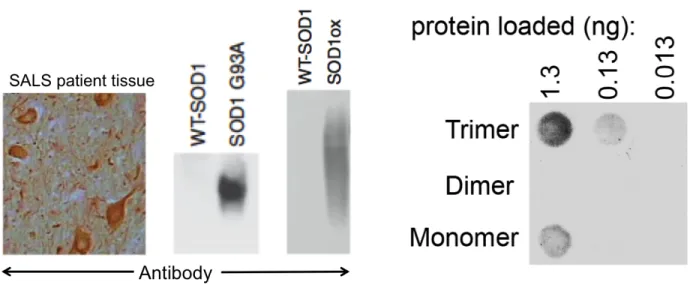
relevant misfolding and aggregation20. Antibodies developed to recognize disease-causative
mutant and misfolded SOD185,86 also specifically bind the trimer, but do not bind native dimeric
or monomeric SOD1, implicating SOD1 trimer as a potential toxic species in ALS (Figure 9)
Figure 8. Wild type SOD1 forms a stable trimer under destabilizing conditions. Aggregation of metallated wild type SOD1 from human erythrocytes under destabilizing conditions (pH 3.5) as measured over 24 hours by size exclusion chromatography. The native dimer dissociates into monomer, and then forms a trimer. Plot courtesy of Kyle Wilcox and Rachel Redler.
(approximately 48 kDa). To address this issue, we design a method that combines
low-resolution experimental data with computational modeling tools in order to obtain structures of
meta-stable protein aggregation intermediates.
3.1 Method for structural determination of meta-stable protein aggregation intermediates
We cannot determine the structure of meta-stable protein aggregates at high resolution
using traditional experimental methods because of their instability and size. Likewise, with
three protein chains totaling 459 residues, SOD1 trimer is too large to obtain a structure
computationally in an ab initio manner. Therefore, we design a method to incorporate any
Figure 9. SOD1 trimer is selectively bound by antibody raised against disease-causative SOD1 species. (Left) The conformational antibody C4F685 is shown to bind the ALS-causative mutant G93A-SOD1, as well as oxidized SOD1, which has been linked to cytotoxicity, but not wild type SOD1. Importantly, C4F6 does bind to misfolded wild type SOD1 present in SOD1 inclusions in tissue from a patient afflicted with sporadic ALS. Image adapted from Bosco et al.85 (Right) C4F6 antibody selectively binds to SOD1 trimer at various concentrations, but does not bind to native SOD1 dimer, and exhibits minimal binding to SOD1 monomer. Image courtesy of Lanette Fee.
the native SOD1 structure into a SOD1 trimer model that we verify experimentally.
Our method takes advantage of structural information obtained from limited proteolysis
experiments. Incubation of SOD1 with various proteases provides a diverse range of cleavage
site data points. Each single-enzyme experiment repetition lasts for only enough time to obtain
one to two cleavage sites (1-5 minutes, depending on enzyme and pH). Limited proteolysis
experiments are performed at 25 °C using chymotrypsin (pH 7.8), pepsin (pH 3.5), proteinase K
(pH 4), and V8 (pH 4) in 100 mM buffer appropriate to pH (pH 3.5: sodium acetate, pH 4:
phosphate, or pH 7.8: Tris). We then determine the identity of the cleavage sites using mass
spectrometry, matching the resulting fragment sizes against possible SOD1 peptide fragments
using the MASCOT database87.
The structural information gained from limited proteolysis experiments comes from the
knowledge that protease cleavage sites must by necessity be located in regions of the protein
the 11-12 residues directly surrounding the cleavage site (5-6 residues on each side) must be
flexible and unstructured in order for the region to conform to fit inside the protease active
site88,89. Using the knowledge that any cleavage site obtained from experiments will have to be
both solvent-exposed and unstructured, we can create a quantitative bias for use in discrete
molecular dynamics (DMD) simulations that will perturb the structure of native SOD1
monomers to obtain the structure of SOD1 trimer.
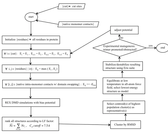
Incorporation of experimental constraints. To incorporate information from limited
proteolysis experiments into DMD simulations, we create an algorithm for converting
knowledge of the sequence positions of proteolytic cleavage sites into pairwise simulation
constraints (Figure 10). The formation of trimeric SOD1 is made possible by a perturbation to
the native state, which occurs as local unfolding in a background of native interactions. To
account for this phenomenon, we first represent the native background as a Gō potential90,91,
assigning a pairwise attraction of 1 kcal/mol between pairs of residues with Cβ-Cβ distance less
than 7.5 Å. Next, we define a bias potential based on the location of proteolytic cleavage sites:
because each cleavage site must necessarily be (i) solvent-exposed and (ii) unstructured, both
criteria which result in few interactions with other residues, we assign a repulsive interaction of
energy E0 between the cleavage site j and every other residue in the system. Because residues
near a solvent-exposed site are also likely to be solvent-exposed, and because an unstructured
region of approximately 12 residues surrounding the cleavage site is necessary for the
proteolytic enzyme to gain access to the site88,89, we apply a step-wise, decreasingly repulsive
potential to the two residues on either side of each cleavage site, such that the repulsive
interaction of the residue with each residue in the system has energy:
Figure 10. Schematic diagram for structural modeling of meta-stable protein oligomers.
For each identified cleavage site, we thus generate 5n pairwise constraints, where n is the total number of residues in the system. All Gō and experimental constraint potentials are additive.
Thus, the combined potential function is:
E=−
∑
i<jΔij•δ
ijE0+λ
∑
i<jδ
ij((Er(j)) / 2),Where Δij is the native contact matrix of the conformation, δij is the contact matrix of the current
conformation, and λ is a scaling parameter discussed in the next section. Our algorithm
produces competition between the native SOD1 structure and the information inferred from
limited proteolysis experiments about the SOD1 trimeric structure. In addition, the energy
start
{cut} cut sites
€ ≡
{native monomer contacts}
Initialize {residues} all residues in protein
€
≡
i ε {cut}: Ei = E1 , Ei±1 = E2 , Ei±2 = E3 , Ei±3 = E4
€ ∀
€ ∀
i, j ε {residues} | i≠j : Eij = max ( Ei , Ej )
[i, j] ε {native intra-monomer contacts w/ domain swapping} : Eij += -EGō
€ ∀
REX DMD simulations with bias potential
rank all structures according to LF factor:
Nc= Nci, Cβcutoff=7.5A
i∈cuts
∑ Cluster by RMSD
Select centroid(s) of highest-population cluster(s) as representative(s) adjust potential end yes no Stabilize/destabilize resulting structure using Eris suite Experimental mutagenesis: trimer promoted/obstructed?
Equilibrate at low
function rewards native interactions with some energy, E0, which will apply to both inter- and
intra-chain interactions, including the possibility of domain-swapping interactions, since regions
that tend to interact in the native structure will also favorably interact between misfolded
monomers. Thus, native interface interactions and domain-swapping interactions comprise the
attractive force between monomers that will, after including the perturbation of repulsion
obtained from experimental data, result in the trimeric interfaces.
Parameterization of bias potential. In order to scale the two terms of our bias potential,
we test the effect of various λ from 0.0 to 30 at intervals of 0.3 (from 0.0 to 3.0) or 3 (from 3 to
30) in coarse-grained62,66 replica exchange DMD simulations of apo-SOD1 monomer, which
features a well-defined folding pathway. We utilize a four-bead protein model, which represents
each residue using four beads: one for the carbon, alpha-carbon, nitrogen, and the side-chain
beta-carbon62,66. Glycine, which has no side chain, is represented using only three beads.
Because they contain fewer particles for the same size of system, and hence fewer necessary
calculations are needed to perform simulations, coarse-grained protein models are useful tools
for increasing the speed of simulation production. The four-bead protein model is an
intermediate coarse-grained model that features the speed advantage of coarse-graining while
still allowing for accurate modeling of backbone dihedrals and hydrogen bond formation. The
four-bead protein model also allows for simplicity in applying constraints, as attraction and
repulsion interactions can be applied directly to the beta-carbon bead (or alpha-carbon bead for
glycine).We utilize the DMD simulation engine to evolve our coarse-grained system over time
according to our bias potential. The DMD engine is a variation on traditional molecular
dynamics engines, where pairwise interactions between particles are modeled using step
elsewhere61,62. In the replica exchange (REX) method, we perform simulations of multiple,
identical copies of the system at a range of temperatures in parallel. The REX method is
explained in detail elsewhere67,68. Briefly, at given time intervals, replicas of neighboring
temperatures exchange temperature values according to a Metropolis-based stochastic algorithm.
This exchange allows the system to overcome energetic barriers at higher temperatures, while
retaining a realistic free energy profile at lower temperatures. We set the temperature exchange
interval at 1000 DMD time steps. The temperature range of the replicas covers the entire
transition profile of the given system, such that the system is disordered at the highest
temperatures and stable and ordered at the lowest temperatures. In order to find the ideal range
and separation between replicas, first we perform a wide range of single-temperature
simulations and construct a Temperature vs. Energy plot, from which the transition profile of
the system is apparent. We then choose temperatures for replicas that cover this range, and
fine-tune such that the successful exchange rate between adjoining replicas is between 0.2 and 0.7.
We utilize 19 replicas in our coarse-grained simulations of the SOD1 apo-monomer system:
0.450, 0.475, 0.498, 0.516, 0.534, 0.554, 0.574, 0.595, 0.616, 0.637, 0.658, 0.678, 0.699, 0.720,
0.740, 0.759, 0.779, 0.805, 0.855 kcal/(mol!kB). It is important to note that, due to the manner
in which energy is calculated, temperatures between coarse-grained and all-atom systems are
not comparable. We perform simulations for 106 time steps (approximately 50 ns) for each
replica.
We examine the resulting energetic profiles and folding trajectories and calculate the
specific heat and radius of gyration as a function of temperature for each value of λ. We select
values of λ that produce distinct folding intermediates with clear energetic transitions. The
terms, demonstrating adequate scaling between the two terms in the energy function, while
distinct folding transitions are indicative of a realistic protein folding pathway. At λ=0 (Gō
potential only), we find a single, sharp folding transition with no intermediates (Figure 11), as
expected from experimental findings79. At λ ≥ 1.3, we cannot resolve individual thermodynamic
Figure 11. Folding of SOD1 monomer with λ=0. SOD1 monomer folding features two distinct states with no folding intermediates. Transitions in energy and radius of gyration are sharp and well-defined. Trajectories from T=0.80 and T=0.85 are located at the transition, and exhibit flipping between folded and unfolded structures.
transitions, a behavior that indicates non-cooperative folding to an extent that is unrealistic for
proteins. We find that that λ values 0.66 ≤ λ ≤ 0.99 fit our criteria for selection (Figure 12), and
that values in this range produce nearly identical final structures in our test apo-monomer
system (Figure 13). In the trimeric system, the increase in the number of Gō constraints over the
monomeric system is greater than the increase in the number of experimental constraints, so we
Figure 12. Folding of SOD1 monomer with λ=0.66. SOD1 monomer folding features distinct intermediate states with mid-range λ. Transitions in energy and radius of gyration remain well-defined. Trajectories from T=0.70, T=0.75, and T=0.80 are located at the transition, and exhibit flipping between states.
SOD1.
Coarse-grained simulation. In order to obtain initial trimeric SOD1 structures that agree
with experimental data, we utilize discrete molecular dynamics61,62 replica exchange67,68
simulations (DMD REX). We apply our bias potential to a four-bead model (discussed above)
of three natively-folded SOD1 apo-monomers placed in proximity to each other but not initially
bound. For scaling parameter λ = 0.99 (discussed above), we utilize 27 replicas, with
temperatures of 0.435, 0.445, 0.455, 0.462, 0.470, 0.480, 0.490, 0.503, 0.513, 0.523, 0.538,
0.552, 0.566, 0.581, 0.595, 0.610, 0.625, 0.639, 0.654, 0.674, 0.694, 0.713, 0.733, 0.753, 0.768,
0.788, and 0.815 kcal/(mol!kB). We perform simulations for 106 time steps for each replica
In order to demonstrate agreement with limited proteolysis results, each proteolytic cut
site residue should make as few contacts with other residues as possible, since its ability to be
cleaved by proteolytic enzymes denotes its solvent accessibility and lack of participation in
secondary structure interactions. We therefore evaluate the experimental agreement of each
structural snapshot from simulation by calculating the average number of contacts made by each
proteolytic cut site residue (Nc). However, while the proteolytic cleavage sites should
demonstrate a minimum of structural interaction, size exclusion chromatography results (Redler
et al., submitted) indicate that the structure should be a compact, associated trimer. We therefore
utilize a combination of proteolytic cut site contacts and trimer radius of gyration (Rg), a
measure of the size of the complex, to select a pool of candidate structures from all
coarse-grained REX simulation trajectories. When we examine these two criteria together, we find
structures with Nc < 1.5 and Rg < 30 Å (approximately three times the Rg of native monomer)
(Figure 14). We cluster this pool of candidate structures by pairwise root mean square deviation
(RMSD), and select the centroid of the largest cluster for further simulation and structural
refinement (Figure 10).
Figure 14. SOD1 trimer structures group by average number of contacts (Nc) and radius of gyration (Rg).
All-atom simulation. We reconstruct the four-bead centroid structure that we obtain from
the DMD REX simulations described above to an all-atom model according to the Medusa force
field62,63. In the all-atom protein model62,63, all heavy atoms as well as polar hydrogens are
explicitly represented. In all-atom DMD simulations, bonded interactions are described by
infinite square well constraints on bond lengths, angles, and dihedrals, making bonded
interactions effectively permanent. Non-bonded interactions are discretized from the Medusa
(Lazaridis-Karplus model65), and explicit hydrogen bonding (reaction algorithm66). We perform
structural minimization using Chiron92 in order to remove clashes introduced by reconstruction.
Chiron uses short, high-temperature, high heat exchange simulations to resolve clashes while
minimally affecting the backbone of the protein structure. We then conduct low-temperature
simulations to accommodate the obtained trimer structure to the all-atom regime. We perform
low-temperature (below the melting transition) all-atom REX simulations using 26 replicas with
temperatures of 0.350, 0.360, 0.370, 0.380, 0.390, 0.400, 0.410, 0.420, 0.430, 0.440, 0.450,
0.460, 0.470, 0.480, 0.490, 0.500, 0.510, 0.520, 0.530, 0.540, 0.550 0.560, 0.570, 0.580, 0.590,
and 0.600 kcal/(mol!kB). We select the ideal number of replicas and spread of replica
temperatures such that exchange of replicas occurs with an acceptance rate between 0.2 and 0.7,
with exchange attempted every 1000 time steps. We perform simulations for 106 time steps for
each replica. We isolate the lowest-energy structure from all simulation replicas and continue
simulation of that structure at a temperature of 0.350 kcal/(mol!kB) for an additional 106 time
steps. Finally, we select the lowest-energy structure from this single-temperature trajectory as
our final model, and verify the quality of the model using Gaia93 (Figure 10), which compares
various structural parameters with those of high-resolution crystal structures from the Protein
Data Bank.
With the use of a high-performance computing cluster (Dell C6100 servers with 12-core,
2.93 GHz Intel processors, 12M L3 cache, and 48 GB memory), the computational protocol
from input of experimental constraints to output of a structural model can be completed for
SOD1 in two weeks of real time, with computational time totaling approximately 12,000
computational hours. Our SOD1 trimer model, discussed below, is to our knowledge the
3.2 SOD1 trimer constraints applied to monomer demonstrate rearrangement of monomer
structure inside trimer
When we map the results of the limited proteolysis experiments to the native, dimeric
SOD1 structure (Figure 15), we find that if SOD1 were to keep its native tertiary structure and
interface many of these sites would be in secondary structure elements or even in the dimeric
interface. Since enzyme cleavage is impossible under those conditions, we conclude that the
SOD1 monomer inside the trimeric structure features significant rearrangement from the native
structure.
In order to determine the extent of this rearrangement, as well as to parameterize our
experimentally-derived force field and determine the proper temperature range for the monomer
folding/unfolding transition in that force field (Section 3.1), we apply our method described in
the previous section to the single chain of the SOD1 monomer. The structure that we obtain
features high agreement with experimental constraints, in that those residues known to be
cleavage sites undergo a minimum of interactions with other residues in the protein, and are
solvent-exposed and not members of secondary structural elements (Figure 16). We further note
that, while some elements of the structure seem to be similarly arranged as in the native
structure (mainly that area directly opposite where the native dimer interface would be, which
natively contains the metal ions), the beta-strands of the N-terminal region have flipped out
from the beta-barrel and unfolded to accommodate the cluster of cleavage sites concentrated in
this area (Figure 16). Interestingly, the N-terminal region that we see here as non-natively
unfolded is a similar region as that identified by Chan and colleagues in their assessment of the
Figure 15. Limited proteolysis cleavage sites mapped to SOD1 native dimer structure. (Top) Limited proteolysis cleavage sites along the linear SOD1 sequence. Enzyme, cleavage site residue, and pH of the proteolytic reaction are indicated. Diagram courtesy of Lanette Fee. (Bottom) Cleavage sites highlighted in hot pink. We note that most cleavage sites are located in regions natively engaged in secondary structure interactions or in the dimer interface, leading us to conclude that SOD1 undergoes significant tertiary and quaternary
possibility that the SOD1 trimer is a meta-stable intermediate on the pathway to formation of
these fibrils. Alternatively, the two species could belong to different pathways competing for the
same aggregation interface surfaces on SOD1 misfolded monomers.
Figure 16. Location of limited proteolysis cleavage sites mapped to perturbed SOD1 monomer structure. Cleavage sites are highlighted in hot pink, with a sphere for each alpha-carbon for clarity.
3.3 SOD1 trimer is a degenerate species with defined interfaces
Having calibrated our hybrid structure determination method using the well-defined
SOD1 monomer system and obtained potentially interesting preliminary results, we apply our
method to obtain a structure of the SOD1 trimer. To account for the association of the three
monomers, we expand the contribution of native interactions to include the possibility of
native interaction with residue 83, then the interaction 20-83 would be rewarded, as well as all
pairwise interactions between the two sets [20, 173, 326] and [83, 236, 389] (e.g., 20-236,
326-83, 173-389). Dimer interface interactions are also incorporated to treat all monomers as
possible partners; for example, dimer interface interactions between monomers 1 and 2, 1 and 3,
and 2 and 3 would all be rewarded. We note that these interactions cannot possibly all be
fulfilled in the same structure; we design these interactions specifically to induce competition
for favorable interactions, as would occur in the real-world system.
After applying our biased force field in coarse-grained replica exchange simulations, we
obtain a representative SOD1 trimer structure by clustering all structures that fit the criteria of
being compact, associated trimers featuring the best agreement with experimental results (low
Rg and low Nc, the average number of contacts of cleavage site residues, Section 3.1). We then
equilibrate the representative structure in a physical force field for two rounds of simulation:
low-temperature replica exchange followed by low-temperature single-temperature equilibration
(Section 3.1). We select the lowest energy structure from the replica exchange simulations to
continue the temperature equilibration, and the lowest energy structure from the
single-temperature equilibration as our final structural model of SOD1 trimer (Figure 17). We note that
two of the three monomers retain some similarity to the natively-folded SOD1 monomer
(RMSD from native structure: 11.9 Å and 12.1 Å), with the native beta-barrel opening and
rearranging while retaining some native beta-strand pairings. The third monomer, with an
RMSD of 19.3 Å from the native monomeric structure, has completely unfolded and rearranged,
and is mostly unstructured (Figure 17). The SOD1 trimer model features an overall potential
energy of -1717 kcal/mol, and a free energy (ΔG) of -538 kcal/mol, as measured by the Medusa