INFLUENCE OF LOWER EXTREMITY SENSORY FUNCTION ON THRESHOLD OF INTER-LIMB GAIT ASYMMETRY PERCEPTION IN PEOPLE POST STROKE
Clinton Wutzke
A dissertation submitted to the faculty of the University of North Carolina at Chapel Hill in partial fulfillment of the requirements for the degree of Doctor of Philosophy in the School of
Medicine (Curriculum in Human Movement Science)
Chapel Hill 2014
iii ABSTRACT
Clinton Wutzke: Influence of Lower Extremity Sensation on Threshold of Inter-limb Gait Asymmetry Perception in People Post Stroke
(Under the direction of Michael D. Lewek)
People with chronic hemiparesis often walk with spatiotemporal asymmetry despite
rehabilitation programs targeted to improve walking function. The persistence of walking
asymmetry may be due, in part, to the perception of established walking patterns post stroke as ‘normal.’ As a result of perceiving this movement pattern as normal, people with chronic
hemiparesis may therefore be unable to identify walking patterns that are more symmetrical.
Also potentially contributing to this inability to perceive walking asymmetry are lower extremity
sensory impairments. Although accurate afferent feedback is delivered to the central nervous
system, inappropriate motor responses are produced leading to spatiotemporal asymmetry. The
purpose of this study was to identify sensory impairments in the lower extremity of people with
chronic hemiparesis and to determine associations between lower extremity sensation and
perception of walking asymmetry in people with chronic hemiparesis.
Thirty people post stroke completed an assessment of lower extremity sensation
including cutaneous, vibratory, and proprioceptive sensation of the paretic and non paretic limbs
as well as walking on a split-belt treadmill under varying conditions of differences in speed
between treadmill belts. Subjects responded when differences in speed between treadmill belts
were perceived. Logistic regression models were used to determine thresholds of conscious
iv
detection was determined as changes from baseline treadmill walking and walking with the belts
moving at different speeds. Associations between the thresholds of conscious (awareness) and
subconscious (detection) perception, spatiotemporal gait asymmetry (step length, stance time)
ratios at thresholds of perception, and measures of lower extremity sensation were determined. It
was found that cutaneous, vibratory, and proprioceptive differences in sensation exist between
the paretic and non paretic lower limbs in people with chronic hemiparesis. Additionally, it was
determined that magnitude of spatiotemporal asymmetry necessary for perception is greater than
the gait asymmetry people post stroke typically walk with. Interestingly, individuals with
overground step length or stance time asymmetry identify differences in speed between treadmill
belts using step length or stance time respectively. However, sensation in the lower extremity of
people post stroke did not appear to be associated with perception of walking asymmetry.
Instead, perception of asymmetry was correlated with motor coordination of the lower extremity
in people with chronic hemiparesis due to stroke.
These results suggest that individuals in the chronic stage following stroke have
impairments in sensation in the lower extremity but these impairments are not associated with
perception of walking asymmetry on a split-belt treadmill. Future studies should identify the
v
ACKNOWLEDGEMENTS
I am greatly indebted to the many people that contributed to the development,
implementation, and completion of this dissertation. Firstly, I would like to thank the members
of my dissertation committee that oversaw this project: Dr. Blackburn, Dr. Faldowski, Dr.
Mercer, Dr. Reisman, Dr. Segal, and my advisor Dr. Lewek. The guidance and mentorship I
received during my time in Chapel Hill was essential for the successful completion of this
project and will remain with me as I continue my academic career.
I would also like to acknowledge the efforts of the many undergraduate volunteers in the
Interdisciplinary Human Movement Laboratory that were essential for the collection and
processing of the mountain of dissertation data. In particular, the efforts of Nicole Davis, Lauren
Calamari, Carolynn Grigg, Molly Miller, Emma Beisheim, Sarah McCullough, Mackenzie
Davis, and Meredith McCliment. The hours assisting with data collections and processing
countless walking trials are very appreciated.
To my family and friends who provided support throughout my Carolina experience;
thank you. When my family arrived in North Carolina and we were immediately welcomed by those we now consider part of our “North Carolina family.” Whether it was lending a hand or
providing guidance and support throughout this journey, they are the reason we will always have
a home in North Carolina. It also would not have been possible to accomplish this achievement
without the support from our families in Nebraska and Alberta. Many hours have been spent on
vi
miles separating us from where we started, our families have been a constant source of love and
support.
Finally, this dissertation would not have been possible without the sacrifices endured by
my wife and my children. Christina has spent much of our time in North Carolina as a single
parent to one (Evelyn), then two (Carson), then three (Kelsey) young children while I spent
evenings and weekends focused on the completion of this degree. This dissertation is an example
vii
TABLE OF CONTENTS
LIST OF TABLES ... ix
LIST OF FIGURES ... xi
CHAPTER 1: ... 1
INTRODUCTION... 1
1.1 STROKE AND ITS INFLUENCE ON GAIT ... 1
1.2 RESEARCH QUESTIONS ... 9
1.3 OPERATIONAL DEFINITIONS ... 9
1.4 ASSUMPTIONS/LIMITATIONS ... 10
1.5 DELIMITATIONS ... 11
1.6 INDEPENDENT VARIABLES ... 11
1.7 DEPENDENT VARIABLES ... 11
1.8 SIGNIFICANCE ... 12
1.9 REFERENCES ... 14
CHAPTER 2: ... 17
Manuscript 1 – Lower extremity sensation is altered in the paretic limb of people with chronic hemiparesis ... 17
2.1 INTRODUCTION ... 17
2.2 METHODS ... 19
2.2.1 DATA COLLECTION ... 21
2.2.2 DATA MANAGEMENT ... 23
2.2.3 DATA ANALYSIS ... 23
2.3 RESULTS ... 24
2.4 DISCUSSION ... 32
2.5 CONCLUSIONS ... 36
2.6 REFERENCES ... 37
CHAPTER 3: ... 40
viii
3.1 INTRODUCTION ... 40
3.2 METHODS ... 42
3.2.1 DATA COLLECTION ... 43
3.2.2 DATA MANAGEMENT ... 46
3.2.3 DATA ANALYSIS ... 48
3.3 RESULTS ... 48
3.4 DISCUSSION ... 55
3.6 REFERENCES ... 59
CHAPTER 4: ... 61
Manuscript 3 – Lower extremity sensation is not associated with perception of walking asymmetry in people post stroke ... 61
4.1 INTRODUCTION ... 61
4.2.1 DATA COLLECTION ... 63
4.2.2 DATA MANAGEMENT ... 65
4.2.3 DATA ANALYSIS ... 66
4.3 RESULTS ... 67
4.4 DISCUSSION ... 73
4.5 CONCLUSIONS ... 75
4.6 REFERENCES ... 77
CHAPTER 5: ... 79
Synthesis... 79
5.1: SUMMARY OF RESULTS ... 79
5.2: INFLUENCE OF STUDY RESULTS AND IMPLICATIONS ... 83
5.3: STRENGTHS AND WEAKNESSES ... 86
5.4: FUTURE DIRECTIONS ... 88
ix
LIST OF TABLES
Table 1. Individual subject demographic data of study participants. ... 20 Table 2: Ranked scores of cutaneous sensation of paretic and non paretic plantar
surface of foot (mean + standard deviation) with p values. ... 25 Table 3: Average vibratory sensation at heel, great toe, and medial femoral condyle
(mean + standard deviation, p values) of paretic and non paretic feet of people with chronic hemiparesis. ... 26 Table 4: Average change in position (in degrees) at ankle and knee of paretic and non paretic limbs (mean + standard deviation, p values). ... 27 Table 5. Individual subject demographic information of study participants. ... 49 Table 6. Individual subject data of treadmill walking speed, overground
comfortable gait speed, step length asymmetry ratio, and stance time asymmetry ratios... 50 Table 7. Step length and Stance time asymmetry ratios during overground walking, at the threshold of detection (subconscious) and at the threshold of awareness
(conscious). Mean + standard deviation. Significant differences (p<0.05) between
overground walking and perception (awareness or detection) noted by *... 52 Table 8. Pearson product correlations and p values for overground walking
measures (comfortable speed, step length asymmetry, stance time asymmetry) and perception (awareness and detection thresholds and asymmetry ratios). Significant
correlations (p<0.05) indicated in yellow, near significant correlations indicated in orange (p<0.075). ... 53 Table 9. Partial correlation coefficients (covariate: baseline step length asymmetry) and p values between measures of awareness (threshold, step length asymmetry, and stance time asymmetry) and measures of lower extremity sensation in the paretic limb of step length asymmetry subgroup. Significant correlations indicated in yellow
x
xi
LIST OF FIGURES
Figure 1 Correlation of cutaneous sensation of the non paretic lateral border (ranked) and
comfortable overground gait speed (m/s). ... 28 Figure 2 Correlation of vibration sensation of the paretic medial femoral condyle and overground stance time asymmetry ratio. ... 29 Figure 3. Correlation of vibration sensation of paretic medial femoral condyle and
overground stance time asymmetry ratio. ... 29 Figure 4. Correlation of vibration sensation of paretic hallux and overground stance time asymmetry ratio. ... 30 Figure 5. Correlation of vibration sensation of non paretic hallux and overground stance time asymmetry ratio. ... 30 Figure 6. Correlation of vibration sensation of non paretic heel and overground stance time asymmetry ratio. ... 31 Figure 7. Correlation of vibration sensation at non paretic medial femoral condyle and overground stance time asymmetry ratio. ... 31 Figure 8. Dual Stair-case design to determine minimum detectable threshold of inter-limb spatiotemporal asymmetry. ... 46 Figure 9 Correlation between step length asymmetry ratio at threshold of awareness and lower extremity Fugl-Meyer score in people post stroke that predicted walking
asymmetry using step length. ... 72 Figure 10. Correlation between overground step length asymmetry ratio and lower extremity Fugl-Meyer score in people post stroke that predicted walking
1
CHAPTER 1:
INTRODUCTION
1.1 STROKE AND ITS INFLUENCE ON GAIT
Stroke is a leading cause of long-term disability in the United States as almost 800,000
people each year experience a stroke (Go et al. 2014). Following stroke, the lower extremity on
the side contralateral to the brain lesion is typically more affected than the ipsilesional limb. The more affected “paretic” lower limb may exhibit impairments in strength (Andrews and Bohannon
2000), coordination (Tseng and Morton 2010), and/or sensation (Winward et al. 1999; Tyson et
al. 2008). These impairments may have a functional consequence such as modified movement
patterns that place greater dependency on the non-paretic lower limb during tasks such as
walking (Raja et al. 2012).
Following stroke, rehabilitation programs are implemented to improve locomotor function
(Silver et al. 2000; Hornby et al. 2008; Patterson et al. 2008a). Rehabilitation programs utilizing
a variety of methods, including body weight supported walking (Visintin et al. 1998) and
walking with robotic assistance (Hornby et al. 2008; Hidler et al. 2009; Westlake and Patten
2009) have improved walking speed. Despite improvements in walking speed, such paradigms
have been unable to improve spatiotemporal symmetry in people post stroke (Patterson et al.
2008a). Locomotor rehabilitation programs have commonly attempted to minimize abnormal
movement characteristics to produce a more functional walking pattern (Duncan et al. 2007;
Hornby et al. 2008). However, minimizing asymmetry during treadmill walking has
2
2008). On the other hand, amplification of movement errors during gait training has improved
inter-limb gait symmetry immediately following a training session (Reisman et al. 2007) and for
an extended duration in people post stroke (Reisman et al. 2010; Reisman et al. 2013).
Amplifying movement errors may be effective in improving inter-limb symmetry by imposing variations from ‘normal’ movements that are perceptible despite cutaneous and proprioceptive
sensory deficits, which might be present in the lower extremity in people post stroke.
In people with sensory deficits, impaired sensory perception results in altered magnitude and
timing of muscle activity to coordinate movement (Inglis et al. 1994). For people post stroke,
impaired control (i.e., inappropriate timing and scaling of motor responses) during walking may
be due, in part, to impaired interpretation of sensory feedback from the paretic limb. Awareness
of intra- and inter-limb positions is important for the control of dynamic balance during walking.
Afferent feedback is necessary during sensorimotor tasks, such as walking, to identify
movement errors (Wei and Kording 2010). The central nervous system (CNS) is influenced by
afferent feedback to modify gait, including adapting to altered walking conditions (Wei and
Kording 2009). Adaptation of walking from perceived movement ‘errors’ may be influenced by
the ability to appropriately integrate afferent feedback (Wei and Kording 2009) input to spinal
and supraspinal centers (Dietz et al. 2002; Frigon and Rossignol 2006). Afferent feedback from
the periphery is integrated and compared to predictions of the movement to determine if such
feedback can be used to modify future movements (Marigold et al. 2004).
Impairments in sensory feedback may limit the identification of movement errors during walking post stroke. As a result, the modified movement pattern is reinforced and becomes ‘the
new normal;’ in that the CNS is unable to identify this movement as asymmetrical. The feedback
3
produce a more symmetrical movement pattern in people post stroke. This cycle may influence
rehabilitation as patients have greater difficulty perceiving the movement patterns that are
encouraged during locomotor training (Duncan et al. 2007), thereby minimizing the effectiveness
of such programs in altering movement patterns to become more symmetrical during walking.
Instead, augmented afferent feedback from the lower extremity may provide a large enough
deviation from the established gait pattern so that the cerebellum modifies planned motor
responses based on this feedback.
Deficits in sensation following stroke may include impaired cutaneous sensation of the
plantar surface of the foot (Hillier and Dunsford 2006; Lynch et al. 2007) as well as impaired
sense of movement (proprioception) in the paretic limb (Lee et al. 2005; Hillier and Dunsford
2006; Reisman et al. 2007; Tyson et al. 2008). These sensory impairments may contribute to the
changes in walking patterns in people post stroke as well as influence walking rehabilitation.
Evidence of proprioceptive impairments has been found at the ankle (Lee et al. 2005; Lynch et
al. 2007) and the hallux (Hillier and Dunsford 2006) of the more affected limb; however,
differences exist in the quality of the assessment. Assessment of movement discrimination
deficits at the hallux and ankle (Kim and Choi-Kwon 1996; Lee et al. 2005; Hillier and Dunsford
2006; Lynch et al. 2007; Lin et al. 2012) provides greater information than assessment of
movement detection (Tyson et al. 2008). In a study by Tyson et al (2008), impairments in
movement discrimination were more prevalent in the upper than the lower extremity, but
impairments in movement detection were equally prevalent in the upper and lower extremities
(Tyson et al. 2008). A comprehensive sensory assessment, rather than the assessment of a single
form of sensory feedback, may be necessary to determine the influence of sensory impairment on
4
Impairments in lower extremity sensation may be the result of inappropriate processing of
feedback in the primary somatosensory cortex, particularly contralateral to the paretic limb
following stroke. Appropriate sensory feedback from the lower extremity travels through the
dorsal root ganglion to the fasciculus gracilis which then ascends via the dorsal spinocerebellar
tract to ventral posterio-lateral nucleus of the thalamus where afferent feedback can then be sent
to the postcentral gyrus of the somatosensory cortex. This afferent feedback is then
misinterpreted resulting in inappropriate planning of motor responses in the cerebellum.
Therefore, the presence of sensory impairments in people post stroke may contribute to reduced
gait speed and increased inter-limb asymmetry. Although impairments (strength, coordination,
spasticity, tone) have been identified as predictors of gait speed and asymmetry in people post
stroke (Hsu et al. 2003), the influence of sensory impairments necessitates further study.
Somatosensory impairment in the lower extremity of people post stroke has been identified as a
predictor of spatiotemporal asymmetry (Hsu et al. 2003). However, Hsu assessed light touch
(cutaneous) and movement detection (proprioception), replicating methods from the lower
extremity Fugl-Meyer. The assessment of cutaneous sensation conducted by Hsu (2003)
quantified impairment by comparing light touch sensation to the non-paretic limb using a hard
and soft item, rather than monofilaments. The proprioceptive assessment conducted by Hsu
(2003), based on the ability to identify small (not quantified) movements, also compared the
affected to the non-affected limb for scoring. A comprehensive, quantifiable assessment of lower
extremity sensation is necessary to identify the role of sensory impairment on gait and
5
The purpose of this dissertation was to determine the presence of sensory impairments in the
lower extremity of chronic people post stroke and the influence of impaired lower extremity
sensation on the perception (conscious and unconscious) of spatiotemporal gait asymmetry.
1.1.1 Walking Post Stroke
Although most individuals post-stroke regain the ability to walk (Roger et al. 2012), most
(70%) are unable to walk at speeds greater than 0.8 m/s at discharge from rehabilitation
(approximately 55 days after admission) (Hill et al. 1997). In addition to walking slowly, people
post stroke commonly walk with spatiotemporal inter-limb asymmetry (Patterson et al. 2010), as
the paretic limb exhibits prolonged swing time and shorter stance time compared to the
non-paretic limb (Kim and Eng 2003; Patterson et al. 2008a). Spatial asymmetry may also exist as a
difference in step length between limbs (Patterson et al. 2008a; Patterson et al. 2010). Although
locomotor rehabilitation programs have been successful in improving gait speed (Visintin et al.
1998; Silver et al. 2000), improved overground inter-limb symmetry remains elusive (Silver et
al. 2000; Patterson et al. 2008a).
Gait training programs have attempted to restore symmetry (Hornby et al. 2008) and gait
function (gait speed, functional ambulation category) (Werner et al. 2002; Yagura et al. 2006)
through minimization of movement errors. Although the minimization of movement errors is
intended to establish a more functional (i.e., faster, reduced assistance or supervision) walking
pattern, its effectiveness may be limited by the presence of sensory deficits in people post stroke.
Specifically, deficits in lower extremity cutaneous and proprioceptive sensation (Smania et al.
6
Augmented feedback during locomotor training can improve spatiotemporal asymmetry in
people post stroke (Reisman et al. 2007; Reisman et al. 2010) by disrupting established
movement patterns and promoting exaggerated movements that can be perceived by the lower
extremity. The exaggerated movements are integrated into the central nervous system and an
altered movement pattern can be developed using feed-forward processes. The use of
exaggerated errors to modify walking patterns can establish a more symmetrical walking pattern.
For augmented feedback to be effective in modifying spatiotemporal asymmetry in people post
stroke, it may be necessary that the magnitude of error augmentation be detectable and beyond the individual’s overground spatiotemporal asymmetry. This study will determine the perceptible
threshold of inter-limb asymmetry while walking on a split-belt treadmill in relation to
overground spatiotemporal asymmetry in people post stroke.
Somatosensory impairment in the lower extremity of people post stroke has been
identified as a limiting factor for walking (Hsu et al. 2003) and postural control (Smania et al.
2003). I hypothesized that impaired somatosensory feedback from the lower extremity limits
awareness of spatial limb location in relation to the body, disrupting coordination of movements
and limiting the perception of when the limb is outside typical patterns of movement. I
hypothesize that awareness of abnormal movements of the lower extremity is important for
re-establishing a symmetrical movement pattern in people post-stroke. Although gait impairments
in people post-stroke have been reported, the influence of lower extremity somatosensory
impairment on gait in people with chronic hemiparesis is incompletely understood.
In summary, this study addressed key gaps in the existing literature of sensory
impairment in people with chronic hemiparesis due to stroke. Additionally, this study will
7
perception of spatiotemporal asymmetry during walking. Sensory impairment in the lower
extremity of people in the chronic phase post stroke has not been studied comprehensively.
Therefore a comprehensive assessment of lower extremity sensation including cutaneous,
vibration, and proprioceptive sensory impairments in the paretic limb of people post stroke in the
chronic phase of recovery will contribute greatly to identify sensory deficits that persist in people
post stroke and the relationship of such sensory deficits to gait. Studies that have investigated
lower extremity sensation in people post stroke involve small sample sizes (Hillier and Dunsford
2006) or explore sensation in the acute phase post stroke (Lynch et al. 2007; Tyson et al. 2008).
Some studies (Smania et al. 2003; Lynch et al. 2007; Reisman et al. 2007; Tyson et al. 2008; Lin
et al. 2012) have included cutaneous or proprioceptive sensation assessment in the lower
extremity in people post stroke in some form; such as the assessment of proprioceptive sensation
at a specific joint or cutaneous sensation at a single point on the paretic limb only. The influence
of sensory impairment on perception of spatiotemporal gait asymmetry will be determined. The
success of gait rehabilitation in people post stroke to re-establish a more symmetrical walking
pattern may be influenced by the awareness of walking asymmetrically to distinguish variations
between established walking patterns developed post stroke and more symmetrical movement
patterns during rehabilitation programs.
Specific Aim 1: To identify differences in lower extremity sensation (cutaneous, vibration,
and proprioceptive sensation) between paretic and non paretic limbs and to identify
associations among measures of lower extremity sensation and overground gait measures
(comfortable gait speed, step length asymmetry, stance time asymmetry) in people with
8
Hypothesis: The paretic limb of people post stroke will have impaired sensation in the lower extremity compared to the non paretic limb and measures of lower extremity sensation
(cutaneous, vibration, and proprioceptive sensation) will be correlated with overground gait
speed and spatiotemporal asymmetry ratios (step length, stance time) in people post stroke in that
individuals with intact lower extremity sensation will have little overground spatiotemporal
asymmetry while individuals with impaired sensation will have greater spatiotemporal
asymmetry.
Specific Aim 2: To identify associations among perception (conscious awareness and
subconscious detection) of inter-limb asymmetry during treadmill walking and overground
walking function (gait speed, step length asymmetry, stance time asymmetry) in people
with chronic stroke.
Hypothesis: Perception of inter-limb asymmetry during treadmill walking in people post stroke will be positively correlated with overground walking function (gait speed, step length, stance
time asymmetry) in that individuals that perceive small differences in belt speed between
treadmill belts will have mild impairments in gait function whereas individuals that perceive
only large magnitude differences in speed between treadmill belts will have impaired walking
function (slower gait speed overground, increased spatiotemporal asymmetry).
Specific Aim 3: To identify associations among perception (conscious awareness and
subconscious detection) of inter-limb asymmetry during treadmill walking and lower
extremity sensation (cutaneous, vibratory and proprioceptive sensation) in people with
9
Hypothesis: Lower extremity sensation (cutaneous, vibratory, and proprioception) will be negatively correlated with perception of inter-limb asymmetry during treadmill walking in
people post stroke in that individuals with intact lower extremity sensation will be capable of
perceiving small inter-limb spatiotemporal asymmetry when treadmill walking.
1.2 RESEARCH QUESTIONS
Research Question 1: How does lower extremity sensation differ between the paretic and non-paretic limbs of people with chronic hemiparesis?
Measures of interest:
Cutaneous sensation of the plantar surface (heel, lateral border of foot, great toe, first metatarsal) of the foot
Vibration sensation (fibular trochlea, phalanx of hallux, medial femoral condyle)
Proprioception - movement detection (ankle, knee)
Research Question 2: What is the influence of lower extremity sensory function on perception of inter-limb asymmetry in people post stroke?
Measures of interest:
Cutaneous sensation of the plantar surface (heel, lateral border of foot, great toe, first metatarsal) of the foot
Vibration sensation (fibular trochlea, phalanx of hallux, medial femoral condyle)
Proprioception - movement detection (ankle, knee)
Stance Time asymmetry ratio
Step Length asymmetry ratio
1.3 OPERATIONAL DEFINITIONS
Cutaneous Sensation Threshold: Conscious awareness to sensory feedback from receptors sensitive to change in pressure applied to the skin with an accuracy of >80%.
10
Maximum to threshold: Amplitude of vibration value at which vibration is no longer consciously detected.
Proprioceptive Sensation: Difference in angular position (in degrees) from starting point to the angular position that movement (plantarflexion/dorsiflexion; flexion/extension) at joint (ankle, knee) is consciously perceived.
Stance Time: Duration that one limb is in contact with the ground/treadmill belt during a gait cycle (expressed as a percentage of the gait cycle /100%).
Step Length: Distance between the anteriorly positioned (stepping) foot to the heel of the contralateral (stance) foot at the point of initial contact.
Swing Time: Portion of the gait cycle that the reference limb is not in contact with the ground (normalized to gait cycle).
Vibration Sensory Threshold: Amplitude of vibration that an individual can consciously perceive or can no longer consciously perceive stimulus of oscillation (vibration).
Visual Neglect: Portion of the visual field that is not perceived. For this study visual neglect is defined as fewer than 44 out of 56 stars crossed out on the Star Cancellation Test.
Zero to threshold: Amplitude of vibration value at which vibration is consciously perceived.
1.4 ASSUMPTIONS/LIMITATIONS
The following assumptions and limitations will be made for this study:
All participants will understand and follow study procedures and will provide
their best efforts duing all testing protocols.
Participants will be able to indicate when they perceive differences in treadmill
belt speeds and other sources of sensory feedback.
Individuals post stroke will be capable of walking on the treadmill at a speed of
greater than 0.35 m/s (minimum speed necessary to enact a difference in speed
11
This study will apply to people that have experienced an ischemic or hemorrhagic
unilateral brain lesion (stroke).
Individuals post stroke have no cerebellar impairments.
1.5 DELIMITATIONS
The following delimitations will be made for this study:
All participants will be at least 18 years of age
Participants will be free of musculoskeletal or neuromuscular injury (in addition
to stroke) that impairs walking or standing
People post stroke will consist of people who have experienced a unilateral or
hemorrhagic stroke
Sensation will be assessed only in the lower extremity (knee, ankle joints, plantar
surface of feet)
1.6 INDEPENDENT VARIABLES
The primary independent variable in this study is:
Difference in speed (m/s) between treadmill belts (expressed as a percent)
Additional independent variables that will be used in this study will be:
Limb (paretic/non paretic lower extremities)
1.7 DEPENDENT VARIABLES
The following dependent variables will be used in this study:
Spatiotemporal Asymmetry
12
Stance Time Asymmetry ratio
Spatiotemporal Gait Characteristics:
Overground comfortable gait speed
Overground fast gait speed
Step Length Asymmetry ratio
Stance Time Asymmetry ratio
Cutaneous Sensation of plantar surface of the foot:
Heel
Lateral border of foot
Hallux
Base of first metatarsal head
Vibration Sensation of lower extremity:
Maximum to threshold
o Hallux
o Calcaneous
o Medial femoral condyle
Zero to threshold
o Hallux
o Calcaneous
o Medial femoral condyle
Proprioception (movement detection) of lower extremity:
Knee flexion
Knee extension
Ankle dorsiflexion
Ankle plantarflexion
1.8 SIGNIFICANCE
This study will provide evidence relating the influence of lower extremity sensation on
the perception (conscious and subconscious) of inter-limb asymmetry in people post stroke. This
study also comprehensively quantifies impairments in lower extremity sensation in people with
chronic hemiparesis. Identification of sensory measures associated with spatiotemporal
13
Identification of sensory measures that are associated with locomotion could then be targeted
during rehabilitation to improve acuity in people post stroke.
Additionally, results from this study may influence the use of augmented feedback to
modify rehabilitation programs that seek to improve spatiotemporal symmetry. Through
identification of the conscious and subconscious thresholds of inter-limb asymmetry perception,
future rehabilitation programs will be able to more precisely utilize augmented asymmetry
walking for individuals. Currently, walking on a split-belt treadmill with one belt moving at
twice the speed of the other has been found to improve overground spatiotemporal asymmetry in
one subject post stroke (Reisman et al. 2010). Identification of a threshold of perceivable
inter-limb asymmetry lower than a 2:1 ratio while walking on a split-belt treadmill may allow patients
14
1.9 REFERENCES
Andrews, A. W. and R. W. Bohannon (2000). "Distribution of muscle strength impairments following stroke." Clin Rehabil 14(1): 79-87.
Dietz, V., R. Muller, et al. (2002). "Locomotor activity in spinal man: significance of afferent input from joint and load receptors." Brain 125(Pt 12): 2626-2634.
Duncan, P. W., K. J. Sullivan, et al. (2007). "Protocol for the Locomotor Experience Applied Post-stroke (LEAPS) trial: a randomized controlled trial." BMC Neurol 7: 39.
Frigon, A. and S. Rossignol (2006). "Experiments and models of sensorimotor interactions during locomotion." Biol Cybern 95(6): 607-627.
Go, A. S., D. Mozaffarian, et al. (2014). "Executive summary: heart disease and stroke statistics--2014 update: a report from the American Heart Association." Circulation 129(3): 399-410.
Hesse, S., M. Konrad, et al. (1999). "Treadmill walking with partial body weight support versus floor walking in hemiparetic subjects." Arch Phys Med Rehabil 80(4): 421-427.
Hidler, J., D. Nichols, et al. (2009). "Multicenter randomized clinical trial evaluating the effectiveness of the Lokomat in subacute stroke." Neurorehabil Neural Repair 23(1): 5-13.
Hill, K., P. Ellis, et al. (1997). "Balance and mobility outcomes for stroke patients: a comprehensive audit." Aust J Physiother 43(3): 173-180.
Hillier, S. and A. Dunsford (2006). "A pilot study of sensory retraining for the hemiparetic foot post-stroke." Int J Rehabil Res 29(3): 237-242.
Hornby, T. G., D. D. Campbell, et al. (2008). "Enhanced gait-related improvements after therapist- versus robotic-assisted locomotor training in subjects with chronic stroke: a randomized controlled study." Stroke 39(6): 1786-1792.
Hsu, A. L., P. F. Tang, et al. (2003). "Analysis of impairments influencing gait velocity and asymmetry of hemiplegic patients after mild to moderate stroke." Arch Phys Med Rehabil
84(8): 1185-1193.
Inglis, J. T., F. B. Horak, et al. (1994). "The importance of somatosensory information in triggering and scaling automatic postural responses in humans." Exp Brain Res 101(1): 159-164.
Kim, C. M. and J. J. Eng (2003). "Symmetry in vertical ground reaction force is accompanied by symmetry in temporal but not distance variables of gait in persons with stroke." Gait Posture 18(1): 23-28.
15
Lee, M. J., S. L. Kilbreath, et al. (2005). "Movement detection at the ankle following stroke is poor." Aust J Physiother 51(1): 19-24.
Lin, S. I., L. J. Hsu, et al. (2012). "Effects of ankle proprioceptive interference on locomotion after stroke." Arch Phys Med Rehabil 93(6): 1027-1033.
Lynch, E. A., S. L. Hillier, et al. (2007). "Sensory retraining of the lower limb after acute stroke: a randomized controlled pilot trial." Arch Phys Med Rehabil 88(9): 1101-1107.
Marigold, D. S., J. J. Eng, et al. (2004). "Contribution of muscle strength and integration of afferent input to postural instability in persons with stroke." Neurorehabil Neural Repair
18(4): 222-229.
Patterson, K. K., W. H. Gage, et al. (2010). "Evaluation of gait symmetry after stroke: a
comparison of current methods and recommendations for standardization." Gait Posture
31(2): 241-246.
Patterson, K. K., I. Parafianowicz, et al. (2008). "Gait asymmetry in community-ambulating stroke survivors." Arch Phys Med Rehabil 89(2): 304-310.
Patterson, S. L., M. M. Rodgers, et al. (2008a). "Effect of treadmill exercise training on spatial and temporal gait parameters in subjects with chronic stroke: a preliminary report." J Rehabil Res Dev 45(2): 221-228.
Raja, B., R. R. Neptune, et al. (2012). "Coordination of the non-paretic leg during hemiparetic gait: Expected and novel compensatory patterns." Clin Biomech (Bristol, Avon).
Reisman, D. S., H. McLean, et al. (2010). "Split-belt treadmill training poststroke: a case study." J Neurol Phys Ther 34(4): 202-207.
Reisman, D. S., H. McLean, et al. (2013). "Repeated split-belt treadmill training improves poststroke step length asymmetry." Neurorehabil Neural Repair 27(5): 460-468. Reisman, D. S., R. Wityk, et al. (2007). "Locomotor adaptation on a split-belt treadmill can
improve walking symmetry post-stroke." Brain 130(Pt 7): 1861-1872.
Roger, V. L., A. S. Go, et al. (2012). "Executive summary: heart disease and stroke statistics--2012 update: a report from the American Heart Association." Circulation 125(1): 188-197.
Silver, K. H., R. F. Macko, et al. (2000). "Effects of aerobic treadmill training on gait velocity, cadence, and gait symmetry in chronic hemiparetic stroke: a preliminary report." Neurorehabil Neural Repair 14(1): 65-71.
Smania, N., B. Montagnana, et al. (2003). "Rehabilitation of somatic sensation and related deficit of motor control in patients with pure sensory stroke." Arch Phys Med Rehabil 84(11): 1692-1702.
16
Tyson, S. F., M. Hanley, et al. (2008). "Sensory loss in hospital-admitted people with stroke: characteristics, associated factors, and relationship with function." Neurorehabil Neural Repair 22(2): 166-172.
Visintin, M., H. Barbeau, et al. (1998). "A new approach to retrain gait in stroke patients through body weight support and treadmill stimulation." Stroke 29(6): 1122-1128.
Wei, K. and K. Kording (2009). "Relevance of error: what drives motor adaptation?" J Neurophysiol 101(2): 655-664.
Wei, K. and K. Kording (2010). "Uncertainty of feedback and state estimation determines the speed of motor adaptation." Front Comput Neurosci 4: 11.
Werner, C., S. Von Frankenberg, et al. (2002). "Treadmill training with partial body weight support and an electromechanical gait trainer for restoration of gait in subacute stroke patients: a randomized crossover study." Stroke 33(12): 2895-2901.
Westlake, K. P. and C. Patten (2009). "Pilot study of Lokomat versus manual-assisted treadmill training for locomotor recovery post-stroke." J Neuroeng Rehabil 6: 18.
Winward, C. E., P. W. Halligan, et al. (1999). "Current practice and clinical relevance of somatosensory assessment after stroke." Clin Rehabil 13(1): 48-55.
17
CHAPTER 2:
Manuscript 1 – Lower extremity sensation is altered in the paretic limb of people with chronic hemiparesis
2.1 INTRODUCTION
Many of the nearly 800,000 people that experience a stroke each year in the United States
(Go et al. 2014) have resultant impairments in strength (Andrews and Bohannon 2000; Andrews
and Bohannon 2001; Andrews and Bohannon 2003; Moriello et al. 2011), coordination (Tseng
and Morton 2010; Raja et al. 2012), or muscle tone (Soyuer and Ozturk 2007). These
impairments have been identified as limiting factors for functional tasks, such as walking, in
people post stroke (Hsu et al. 2003). In addition to strength or coordination impairments, it is
estimated that approximately 60% of individuals acutely (six weeks or less) post stroke have
sensory impairments in the paretic upper and lower extremities (Winward et al. 1999; Tyson et
al. 2008). In people with chronic hemiparesis, however, the prevalence of sensory impairment in
the paretic limb and the resulting differences in sensation between the paretic and non-paretic
lower extremities are not well understood and require further examination.
People post stroke have been identified as having impaired cutaneous sensation of the
plantar surface of the paretic foot acutely (Lynch et al. 2007) and chronically (n=3) (Hillier and
Dunsford 2006) as well as an impaired sense of movement (proprioception) in the paretic limb
(Lee et al. 2005; Hillier and Dunsford 2006; Reisman et al. 2007; Tyson et al. 2008). These
studies were limited, however because assessment of cutaneous sensation of the paretic foot has
18
2008) or used few testing sites (Hillier and Dunsford 2006), neglecting other areas of the foot
that provide feedback for dynamic balance tasks such as walking. Evidence of proprioceptive
impairments has been found at the ankle (Lee et al. 2005; Lynch et al. 2007) and the hallux
(Hillier and Dunsford 2006) of the paretic limb; however, differences exist in the quality of the
assessment. Movement detection, commonly conducted with the investigator manipulating the
segment to be tested, may provide movement related cutaneous cues or limited standardization of
the speed of movement during testing. In addition to impaired movement detection, movement
discrimination deficits have also been identified at the hallux and ankle (Kim and Choi-Kwon
1996; Lee et al. 2005; Hillier and Dunsford 2006; Lynch et al. 2007; Lin et al. 2012) of people
post stroke, providing greater information than assessment of movement detection alone (Tyson
et al. 2008).
If present, sensory impairments in people post stroke may contribute to abnormal walking
patterns, as impaired sensation of the paretic limb has previously been identified as a predictor of
spatiotemporal asymmetry (Hsu et al. 2003). Sensory feedback from the lower extremity is
necessary to identify movement errors during walking (Dietz et al. 2002). Following stroke,
sensory impairments may result in inaccurate planning of movements and/or motor responses
(Wutzke et al. 2013). If sensory impairment in people with chronic hemiparesis is common,
rehabilitation programs may benefit from the inclusion of sensory retraining to improve
identification of movement errors. Therefore, a comprehensive, quantifiable assessment of lower
extremity sensation is necessary to identify sensory impairment and determine the influence of
sensory impairment on gait speed and spatiotemporal asymmetry in people with chronic stroke.
The purpose of this study was to determine differences in sensation between the paretic and
19
determine associations among measures of lower extremity sensation and overground walking
measures. We hypothesized that the paretic limb of people with chronic hemiparesis would have
impairments in sensation compared to the non-paretic limb. Additionally, based on previous
work (Hsu et al. 2003)(Wutzke, Faldowksi, Mercer, in process), it was hypothesized that
impairments in lower extremity sensation would be associated with slower walking speed and
greater spatiotemporal asymmetry in people with chronic hemiparesis.
2.2 METHODS
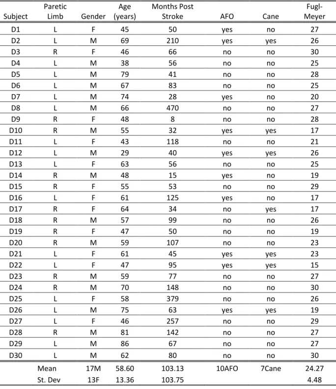
Thirty people post stroke (xage: 58.60±13.36 years, xheight: 170.31±10.34 cm, x
weight: 82.70±17.77 kg, 17M/13F, 18 left limb paretic) were recruited to participate in the study.
All subjects read and signed an IRB approved informed consent document. Confirmation of
ischemic or hemorrhagic stroke was provided by a physician prior to participation. Participants
were at least six months post stroke and used an assistive device or lower extremity bracing (i.e.
ankle/foot orthosis) for walking as needed. Exclusion criteria included: 1) inability to walk
without physical assistance, 2) history of lower extremity joint surgery (e.g., total joint
arthroplasty) or disease that might reduce sensation in the lower extremity (e.g. diabetes), 3)
vision impairments not corrected by lenses, visual neglect, or vestibular deficits, and 4) inability
to follow three step commands. Prior to testing, motor coordination was assessed using the
lower extremity portion of the Fugl-Meyer (Gladstone et al. 2002). Subjects were screened for
unilateral visual neglect (star cancellation test), vision impairments (self-report of visual
20
Table 1. Individual subject demographic data of study participants.
Subject
Paretic
Limb Gender
Age (years)
Months Post
Stroke AFO Cane
Fugl-Meyer
D1 L F 45 50 yes no 27
D2 L M 69 210 yes yes 26
D3 R F 46 66 no no 30
D4 L M 38 56 no no 25
D5 L M 79 41 no no 28
D6 L M 67 83 no no 25
D7 L M 74 28 yes no 20
D8 L M 66 470 no no 27
D9 R F 48 8 no no 28
D10 R M 55 32 yes yes 17
D11 L F 43 118 no no 21
D12 L M 29 40 yes yes 26
D13 L F 63 56 no no 25
D14 R M 48 15 yes no 19
D15 R F 55 53 no no 29
D16 L F 61 125 yes no 17
D17 R F 64 34 no yes 17
D18 R M 57 99 no no 26
D19 R F 47 50 no no 19
D20 R M 59 107 no no 23
D21 L F 61 45 yes yes 23
D22 L F 47 95 yes yes 15
D23 R M 59 77 no no 27
D24 R M 70 148 no no 30
D25 L F 58 379 no no 26
D26 L M 75 63 yes yes 19
D27 L F 46 257 no no 29
D28 R M 81 142 no no 27
D29 L M 86 67 no no 27
D30 L M 62 80 no no 30
Mean 17M 58.60 103.13 10AFO 7Cane 24.27
21 2.2.1 DATA COLLECTION
Overground walking measures were calculated from three walking trials at a self-selected
speed over a 14 foot GaitRite mat (CIR Systems Inc, Havertown, PA). Sensory testing included
cutaneous (Hillier and Dunsford 2006; Lynch et al. 2007; Tyson et al. 2008), vibration (Gin et al.
2011), and proprioceptive sensation (Carey et al. 1996; Lund et al. 2008; Tyson et al. 2008;
Goble 2010) of both lower extremities. Subjects performed all sensory tests with eyes closed.
Cutaneous sensation was assessed at four sites on the plantar surface of the foot (heel, great toe, lateral border of the foot (1/2 the distance from the tip of the 5th toe to the heel), and
base of 1st metatarsal) with a set of five Semmes Weinstein monofilaments (2.83, 3.61, 4.31, 4.56 and 6.65g). Subjects were in a seated position, knees flexed ~90 degrees and feet off the
ground to access the plantar surface of the foot. The threshold of cutaneous sensation at each site
was identified as the minimum gauge of monofilament detected with an accuracy of 80% or
greater (Feng, 2009). A minimum of 15 trials were conducted at each site beginning with the
mid-point monofilament (4.31g). Subjects who correctly identified the monofilament in contact
at the test site were then tested with a smaller gauge monofilament (2.83g) whereas subjects who
did not detect the 4.31g monofilament were then tested with the 6.65g monofilament. Each site
was then tested with the next larger/finer monofilament, dependent upon whether the subject
correctly or incorrectly detected the monofilament. Additionally, 'catch' trials were included,
when no contact was made between the monofilament and surface of the skin, at a rate of
approximately 1 in 5 (Feng, 2009).
Vibration sensation was assessed with a bio-thesiometer (Bio-Medical Instrument
22
femoral condyle. At each site, the threshold of vibration sensation was assessed using two
techniques. Zero to detection threshold testing was conducted by incrementally (approximately
one unit per second) increasing the amplitude of vibration from zero until the subject identified
the site as vibrating with a verbal response. The second technique, maximum to threshold, was
conducted with the bio-thesiometer at the maximum amplitude and then slowly reduced in
frequency until the subject identified the vibration as absent. Each procedure was replicated five
times at each site.
Proprioceptivesensation was assessed at the ankles and knees using a movement
detection task, performed while seated on a Cybex Norm (CSMi, Stoughton, MA). At each site,
the passive range of motion for the joint was determined. To assess movement detection at the
ankle, the hip and knee were flexed approximately 75 and 90 degrees respectively and the motor’s axis of rotation was aligned with the lateral malleolus. To test the knee, subjects were
seated with the hips flexed approximately 75 degrees and the motor’s axis of rotation aligned
with the lateral femoral condyle (van der Esch et al. 2007). Trials consisted of passively moving
the segment to the respective starting position (mid-point of the range of motion for the ankle, 30
degrees of flexion for the knee (Hurkmans et al. 2007; van der Esch et al. 2007)) and then
passively rotating the joint at one degree per second until the participant identified movement at
the joint. Three flexion/dorsiflexion trials and three extension/plantarflexion trials were
conducted at each joint with the angular change in position between the starting position and the
23 2.2.2 DATA MANAGEMENT
The minimum threshold of cutaneous sensation was converted to a rank scale (e.g., 1 to
6) such that the monofilament that produced the least force (2.83 grams) was ranked 1 and each
subsequent monofilament increased in ranked value 3.61 (2), 4.31 (3), 4.56 (4), 6.65 (5). A score
of 6 was provided if the participant was unable to identify when the 6.65 was in contact with the
skin at a specific location.
The average threshold of vibratory sensation for the zero to threshold and maximum to
threshold trials was calculated for each of the three locations on the paretic and non paretic legs for each participant respectively. If a participant could not identify vibration at the machine’s
maximum value (i.e., 50) during a trial, a value of 51 was recorded.
Proprioceptive scores were calculated as angular deviations for each trial of passive
flexion/dorsiflexion and extension/plantarflexion at both the paretic and non-paretic knee/ankle.
An average angular deviation was calculated for each joint direction for each limb (paretic and
non-paretic).
Self-selected overground walking speed, step length, and stance time asymmetry ratios
were calculated as the average of the three walking trials. Step length and stance time were
calculated for each limb (paretic and non paretic) with the larger value divided by the smaller
value to determine the asymmetry ratio (Lewek and Randall 2011).
2.2.3 DATA ANALYSIS
Comparisons between the paretic and non paretic feet for each site of each sensory
24
Multiple comparisons were controlled for by employing the Benjamini-Hochberg False
Discovery Rate (Benjamini and Hochberg 1995). Impairment at the paretic and non paretic limb
for each test was determined by comparison to mean values in existing literature of similarly
performed tests in unimpaired, similarly aged adults. Cutaneous sensation data were compared to
healthy control data from studies that used monofilaments to determine a threshold of perception
at each of the sites tested in the present study (Jeng et al. 2000). Vibratory sensation at each site
was compared to zero to threshold values from studies that included participants with no sensory
impairment (Shakoor et al. 2008). Proprioception impairment in people post stroke was
compared qualitatively to unimpaired similarly aged adults performing movement detection tasks
at similar speeds (0.5 to 1 degree per second movement) and similar starting positions (Xu et al.
2004; van der Esch et al. 2007; Lund et al. 2008). To determine associations among lower
extremity sensation and spatiotemporal walking characteristics, Spearman rank correlations were
conducted with an alpha level of 0.05.
2.3 RESULTS
On the paretic limb 26/30 participants displayed impaired cutaneous sensation (score of >
4.31, (Kokmen et al. 1977; Jeng et al. 2000; Collins et al. 2010)) at one or more of the four sites
tested in comparison with 18/30 of participants displaying a cutaneous score >4.31 at one or
more sites on the non paretic foot . Sensation differed between the paretic and non paretic feet at
each of the four sites tested (Table 2). On the plantar surface of the great toe, the average ranked
cutaneous score was 3.73+1.23 on the paretic foot compared to the average score on the non
paretic foot (2.87+0.73; p=0.001). At the lateral border of paretic foot the average ranked
cutaneous score was 3.50+0.82 compared to 3.03+0.61 (p=0.007) at the non paretic lateral
25
cutaneous score 0.57 greater than the non paretic foot (4.29+0.81 paretic, 3.73+0.87 non paretic;
p=0.008). The base of the first metatarsal head on the paretic foot had an average ranked
cutaneous score of 3.54+1.00 on the paretic limb compared to 2.97+0.76 on the non paretic foot
(p=0.005).
Table 2: Ranked scores of cutaneous sensation of paretic and non paretic plantar surface of foot (mean + standard deviation) with p values.
Paretic Non Paretic P Value
Great Toe 3.73+1.23 2.87+0.73 p=0.001
Lateral Border 3.50+0.84 3.03+0.61 p=0.007
Heel 4.29+0.81 3.73+0.87 p=0.008
Base 1st Metatarsal 3.54+1.00 2.97+0.76 p=0.005
Vibration sensation also differed between the paretic and non paretic limbs in people with
chronic hemiparesis (Table 3). At the paretic heel, the average Zero to Threshold score was
27.35+15.51 while the average Maximum to Threshold score was 24.32+10.15. At the heel of
the non paretic limb, the average Zero to Threshold score was 20.34+13.34 (p<0.001) and
Maximum to Threshold score was 21.12+10.01 (p=0.008) respectively. The Zero to Threshold
scores were 24.59+14.40 and 20.31+14.49 at the great toe for the paretic and non paretic limbs
respectively (p=0.021). Compared to existing literature of unimpaired adults of similar age
(6.40+3.30) (Shakoor et al. 2008), 29/30 participants had impaired zero to threshold vibratory
sensation in the paretic limb at the great toe and 28/30 at the non paretic great toe (Shakoor et al.
2008). Maximum to Threshold scores at the great toe did not significantly differ between paretic
and non paretic limbs (24.93+9.44 and 20.31+14.49, p=0.544). The Zero to Threshold score at
the medial femoral condyle was significantly greater on the paretic limb (32.30+12.68) than the
26
femoral condyle for Maximum to Threshold (27.98+9.57 paretic, 24.97+10.74 non paretic;
p=0.355). 28/30 were identified as displaying impaired sensation at the medial femoral condyle
at the paretic limb compared to existing literature (15.90+7.0) (Shakoor et al. 2008). At the non
paretic limb, 25 of 30 participants were identified as having impaired vibratory sensation at the
medial femoral condyle using the zero to threshold technique (Shakoor et al. 2008).
Table 3: Average vibratory sensation at heel, great toe, and medial femoral condyle (mean + standard deviation, p values) of paretic and non paretic feet of people with chronic hemiparesis.
Site Technique Paretic Non Paretic P Value
Heel
Zero to Threshold 27.35+15.51 20.34+13.34 p<0.001 Max to Threshold 24.32+10.15 21.12+10.01 p=0.008
Great Toe
Zero to Threshold 24.59+14.40 20.31+14.49 p=0.021 Max to Threshold 24.93+9.44 23.92+10.42 p=0.544 Medial Femoral Zero to Threshold 32.30+12.68 23.92+10.42 p<0.001 Condyle Max to Threshold 27.98+9.57 24.97+10.74 p=0.355
Movement detection at the ankle and knee differed between the paretic and non paretic
limbs of people with chronic hemiparesis (Table 4). A greater change from the start position was
necessary for participants to identify movement of the paretic compared to the non paretic ankle
during plantarflexion (4.80+6.55° paretic, 1.98+1.71 non paretic, p=0.009) and dorsiflexion
(3.57+4.76 paretic, 1.98+2.15 non paretic, p=0.035). 17/30 participants were identified as having
impaired movement detection at the paretic ankle (plantar or dorsiflexion) while 8/30 were found
to have impaired sensation at the non paretic ankle compared to existing literature (Xu et al.
2004). Detection of knee flexion required greater flexion in the paretic limb (6.79+11.11) than
the non paretic limb (1.70+1.38, p<0.001), as well as greater extension excursion at the paretic
knee to identify movement compared to the non paretic knee (5.23+8.03 paretic, 1.75+1.14 non
27
at the knee in unimpaired, similarly aged adults to be 1.08-1.57 degrees (Hurkmans et al. 2007;
Lund et al. 2008). With these values as a baseline, 21/30 participants demonstrated impairments
in knee proprioception in the paretic limb and 15/30 with impaired sensation on the non paretic
limb.
Table 4: Average change in position (in degrees) at ankle and knee of paretic and non paretic limbs (mean + standard deviation, p values).
Ankle Knee
Plantarflexion Dorsiflexion Extension Flexion Paretic 4.80+6.55 3.57+4.76 5.23+8.03 6.79+11.11 Non Paretic 1.98+1.71 1.98+2.15 1.75+1.14 1.70+1.38
P Value p=0.009 p=0.035 p=0.043 p<0.001
The comfortable overground walking speed was 0.79+0.21 m/s (range 0.36-1.16 m/s)
with an average step length asymmetry of 1.13+0.13 (range 1.01-1.37) and average stance time
asymmetry of 1.14+0.11 (range 1.01-1.37). Of the 30 participants, ten wore an ankle/foot
orthosis (AFO) during walking trials and seven used a cane (single point or quad point) for
walking. The average Fugl-Meyer lower extremity motor score was 24.27+4.48.
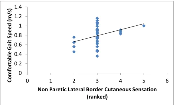
Significant correlations were identified among overground gait measures and lower
extremity sensation. Comfortable walking speed was associated with cutaneous sensation of the
lateral border on the non paretic foot (rs = 0.390, p=0.033). Stance time asymmetry was
correlated with the threshold of vibration on both the paretic and non paretic limbs. On the
paretic limb, stance time asymmetry ratio was associated with the Zero to Threshold and
Maximum to Threshold vibration scores at the medial femoral condyle (rs = -0.423, p=0.020, rs =
-0.380, p=0.038 respectively). We observed significant associations between stance time
28
condyle. At the hallux of the non paretic limb, stance time asymmetry was correlated with both
the Zero to Threshold and Maximum to Threshold vibration scores (rs = -0.441, p=0.015, rs =
-0.365, p=0.047 respectively). At the non paretic calcaneous, the Zero to Threshold vibration
score was correlated with stance time asymmetry ratio (rs = -0.449, p=0.013) while Zero to
Threshold vibration score at the non paretic medial femoral condyle was correlated with stance
time asymmetry ratio (rs = -0.484, p=0.007).
Figure 1 Correlation of cutaneous sensation of the non paretic lateral border (ranked) and comfortable overground gait speed (m/s).
0
0.2
0.4
0.6
0.8
1
1.2
1.4
0
1
2
3
4
5
6
Com
for
tab
le
G
ai
t
Speed
(m/
s)
29
Figure 2 Correlation of vibration sensation of the paretic medial femoral condyle and overground stance time asymmetry ratio.
Figure 3. Correlation of vibration sensation of paretic medial femoral condyle and overground stance time asymmetry ratio.
1
1.1
1.2
1.3
1.4
1.5
0
10
20
30
40
50
60
O
V
er
gr
oun
d
St
an
ce
Time
As
ymme
tr
y
Ra
tio
Paretic Medial Femoral Condyle Zero to Threshold
score (ranked)
1
1.1
1.2
1.3
1.4
1.5
0
10
20
30
40
50
60
O
V
er
gr
oun
d
St
an
ce
Time
As
ymme
tr
y
Ra
tio
30
Figure 4. Correlation of vibration sensation of paretic hallux and overground stance time asymmetry ratio.
Figure 5. Correlation of vibration sensation of non paretic hallux and overground stance time asymmetry ratio.
1
1.1
1.2
1.3
1.4
1.5
0
10
20
30
40
50
60
Ov
er
gr
oun
d
St
an
ce
Time
As
ymme
tr
y
Ra
tio
Non Paretic Great Toe Maximum to Threshold score
(ranked)
1
1.1
1.2
1.3
1.4
1.5
0
10
20
30
40
50
60
Ov
er
gr
oun
d
St
an
ce
Time
As
ymme
tr
y
Ra
tio
31
Figure 6. Correlation of vibration sensation of non paretic heel and overground stance time asymmetry ratio.
Figure 7. Correlation of vibration sensation at non paretic medial femoral condyle and overground stance time asymmetry ratio.
1
1.1
1.2
1.3
1.4
1.5
0
10
20
30
40
50
60
Ov
er
gr
oun
d
St
an
ce
Time
As
ymme
tr
y
Ra
tio
Non Paretic Heel Zero to Threshold score (ranked)
1
1.1
1.2
1.3
1.4
1.5
0
10
20
30
40
50
60
Ov
er
gr
oun
d
St
an
ce
Time
As
ymme
tr
y
Ra
tio
32 2.4 DISCUSSION
The purpose of this study was to determine differences in lower extremity sensation
between the paretic and non-paretic limbs of people with chronic hemiparesis and to identify
associations with spatiotemporal characteristics of overground walking. We hypothesized that
sensation in the paretic limb would be impaired compared to the non-paretic limb of people post
stroke and that impairments in lower extremity sensation would be correlated with increased
spatiotemporal gait asymmetry.
Our results indicate cutaneous, vibration, and proprioceptive sensation is diminished in
the paretic limb compared to the non-paretic limb of people post stroke. Tyson also identified
sensory deficits in the lower extremity in people in the acute phase of rehabilitation (Tyson et al.
2008; Tyson et al. 2013). Tyson et al found impairments in light touch and movement detection
at the plantar surface of the foot and shank (Tyson et al. 2008). Our results suggest that a similar
percentage of people post stroke in the chronic phase of hemiparesis have sensory impairments
in the lower extremity compared to people in the acute phase of rehabilitation (62.75%).
Studies indicating sensory impairments in the lower extremity of people with chronic
hemiparesis are limited, however studies that have investigated lower extremity sensation in
people post stroke have also identified impairments. Movement detection was identified as
impaired at the ankle in the lower extremity of people post stroke in a pilot study completed by
Hillier and Dunsford that explored the effectiveness of sensory retraining in people at least two
years post stroke (Hillier and Dunsford 2006). Lee found impairments in detection of movement
33
study also found impaired movement detection in people post stroke at the ankle when tested at a
movement speed of one degree per second. In the Lee study, six of the eleven participants had
significant impairment of sensation in the paretic limb (Lee et al. 2005). In the current study, we
identified 17 of 30 participants that displayed impairment in movement detection at the paretic
ankle.
To determine the prevalence and persistence of lower extremity sensory impairment in
people post stroke, longitudinal cohort studies are necessary. Sensory assessment of people post
stroke should be initiated in the acute phase of stroke rehabilitation (similarly to (Tyson et al.
2008)) and then assessed periodically (i.e. semi-annually) to identify the prevalence of lower
extremity sensory impairments in people post stroke and the potential recovery of sensation as
they progress to the chronic phase of hemiparesis. Such studies would provide valuable
information regarding sensory changes that may occur as people progress through to the chronic
phase of hemiparesis.
Sensory impairment in people post stroke may influence the ability to appropriately adapt
walking patterns to changes in conditions (i.e. change in surface) or demand (increase/decrease
in walking speed). To identify movement errors and produce coordinated movements, sensory
feedback from the periphery provides information to the central nervous system regarding the
position, movement information, and interaction with the environment (i.e. walking surface). The
central nervous system uses afferent feedback to modify movement patterns, including
adaptation to altered walking conditions (Wei and Kording 2009). Misinterpreted sensory
feedback may result in inappropriate neuromuscular responses to the demands of the
environment to make predictive (feed-forward) adjustments (Reisman et al. 2005; Choi et al.
34
movement to determine if such feedback can be used to modify future movements (Marigold et
al. 2004). In the context of people post stroke, afferent feedback received from the lower
extremity remains unchanged as a result of the stroke, but instead the interpretation of sensory
information and resultant motor response may be altered following stroke. As a result of this
inappropriate interpretation of sensory feedback in the central nervous system, it may be
necessary to augment movement errors to provide a magnitude of sensory feedback that can be
perceived as different from typical movements in the cerebellum.
Interestingly, lower extremity sensory impairments were negatively correlated with
walking function (comfortable gait speed, step length asymmetry, stance time asymmetry). This
suggests that lower extremity sensation as assessed in the present study may not be necessary for
people in the chronic phase of rehabilitation to walk. Although lower extremity sensation has
been identified as necessary for fine motor control such as stepping over an obstacle which has
been identified in cats (Rossignol et al. 1996), people post stroke may utilize other sensory cues,
such as afferent information at the hip (Dietz et al. 2002) to maintain walking function.
There is evidence that suggests lower extremity sensation is not positively correlated with
walking function. Perry et al (1995) examined proprioceptive sensation at the hallux, ankle, knee
and hip of the paretic lower limb in people post stroke and found no association with walking
function (Perry et al. 1995). Reisman et al, reported 11 of 13 subjects had impaired cutaneous
sensation (detection threshold greater than 3.61 grams) at the hallux, however no significant
association between sensory impairment (cutaneous or proprioceptive sensation) and adaptive
ability was found (Reisman et al. 2007). Results of the Reisman et al study may have been
influenced by the single location on the paretic foot (Reisman et al. 2007). Although cutaneous