Ross River virus envelope glycans contribute to disease through activation of
the host complement system
Bronwyn M. Gunn
a,1, Jennifer E. Jones
a, Reed S. Shabman
a,2, Alan C. Whitmore
b, Sanjay Sarkar
b,
Lance K. Blevins
b,3, Thomas E. Morrison
c, Mark T. Heise
a,b,⁎aDept. of Microbiology and Immunology, University of North Carolina at Chapel Hill, 160 Dental Circle, 9024 Burnett Womack, Chapel Hill, NC 27599, USA bDept. of Genetics, University of North Carolina at Chapel Hill, 160 Dental Circle, 9024 Burnett Womack, Chapel Hill, NC 27599, USA
cDept. of Microbiology, University of Colorado School of Medicine, 12800 E. 19th Ave., RC1N 9119, Mail Stop 8333, Aurora, CO 80045, USA
A R T I C L E I N F O
Keywords: Ross River virus Alphavirus
Mannose-binding lectin Complement N-linked glycan
A B S T R A C T
Mannose binding lectin (MBL) generally plays a protective role during viral infection, yet MBL-mediated
com-plement activation promotes Ross River virus (RRV)-induced inflammatory tissue destruction, contributing to
arthritis and myositis. As MBL binds to carbohydrates, we hypothesized that N-linked glycans on the RRV en-velope glycoproteins act as ligands for MBL. Using a panel of RRV mutants lacking the enen-velope N-linked gly-cans, we found that MBL deposition onto infected cells was dependent on the E2 glycans. Moreover, the
glycan-deficient viruses exhibited reduced disease and tissue damage in a mouse model of RRV-induced myositis
compared to wild-type RRV, despite similar viral load and inflammatory infiltrates within the skeletal muscle.
Instead, the reduced disease induced by glycan-deficient viruses was linked to decreased MBL deposition and
complement activation within inflamed tissues. These results demonstrate that the viral N-linked glycans
pro-mote MBL deposition and complement activation onto RRV-infected cells, contributing to the development of RRV-induced myositis.
1. Importance
Mannose binding lectin (MBL), a lectin that can initiate the host complement cascade, generally has a protective role following viral infection. However, in the context of alphavirus-induced disease, MBL-mediated complement activation is pathologic, promoting Ross River virus (RRV)-induced inflammatory tissue destruction within the mus-culoskeletal system. MBL binds to glycosylated proteins, thus we hy-pothesized that the RRV envelope N-linked glycans promote MBL binding, complement activation, and disease. Using a panel of mutant viruses lacking one or more envelope glycans, we found that the RRV E2 glycans are required for MBL binding to infected cells and sub-sequent disease. Mice infected with a virus lacking both E2 glycans exhibited reduced disease and tissue damage, and decreased MBL binding and complement activation compared to mice infected with the wild type virus. These results suggest that interactions between MBL and the viral N-linked glycans play a major role in development of al-phavirus-induced inflammatory disease.
2. Introduction
Arthritogenic alphaviruses such as Ross River virus (RRV) and chi-kungunya virus (CHIKV) are mosquito-borne viruses that cause out-breaks of infectious arthritis and myositis in many regions of the world. Both RRV and CHIKV share similar disease symptoms that are char-acterized by debilitating polyarthritis and myositis that frequently re-sults in myalgia and arthralgia. Studies in both humans and mice have identified a critical role for the host inflammatory response in the de-velopment of disease and immunopathology following infection, with macrophages playing an essential role in damage to the musculoskeletal system (Lidbury et al., 2000; Morrison et al., 2006; Herrero et al., 2013, 2011).
We have previously demonstrated that the host complement system initiated by mannose binding lectin (MBL) also has a critical role in development of disease (Gunn et al., 2012). Interestingly, rather than regulating the infiltration of macrophages and other inflammatory cells into the musculoskeletal system, MBL-mediated complement activation
⁎Corresponding author.
1Present address: Ragon Institute of MGH, MIT, and Harvard, Cambridge, MA 02139, USA. 2Present address: ATCC, Gaithersburg, MD, USA.
3Present address: Center for Integrative Toxicology, Michigan State University, USA.
E-mail addresses:[email protected](B.M. Gunn),[email protected](J.E. Jones),[email protected](A.C. Whitmore),[email protected](S. Sarkar),
[email protected](T.E. Morrison),[email protected](M.T. Heise).
https://doi.org/10.1016/j.virol.2017.12.022
Received 20 July 2017; Received in revised form 24 November 2017; Accepted 20 December 2017 Available online 08 January 2018
24-day-old C57BL/6J mice were inoculated with 103PFU of RRV in following RRV infection contributes to disease by regulating
in-flammatorycellactivationwithintheinflamedtissuethrough comple-mentreceptor3(CR3)(Gunnetal.,2012;Morrisonetal.,2008).The C3cleavageproductiC3bproducedfollowingcomplementactivation can bind toCR3 on inflammatory cells including macrophages and neutrophilstoinducephagocytosisofiC3b-opsonizedpathogens,and can also initiate cytotoxic effector programs through Syk-mediated outside-in signalingthat leadstotissue damage in autoimmune dis-orders(Ross,2000;AbramandLowell,2009;Hirahashietal.,2006).In theabsenceofMBL,C3,orCR3expressionofcytotoxicinflammatory mediators within muscle tissue are reduced in RRV-infected mice, correlatingwithreduceddisease,butviraltropismandviralburdenare unaffected (Gunn et al., 2012; Morrison et al., 2008). Importantly, productionoftheCR3ligandiC3bisreducedin theabsence ofMBL following RRV infection (Gunn et al., 2012), suggesting that MBL-mediated complement activation regulates production of iC3b and subsequent activation phenotype of CR3-bearing inflammatory cells presentwithinthetissues.
MBL-mediatedcomplementactivationisinitiatedbytherecognition of terminalsugars on glycosylated proteinsby the carbohydrate re-cognitiondomain(CRD)ofMBL(reviewedinTakahashietal.,2006). ThealphavirusglycoproteinsE1andE2containthreetofourN-linked glycosylationsitesthatareglycosylatedwithN-linkedglycans(Strauss andStrauss,1994).Duringstructuralproteinsynthesis,theE2andE1 glycoproteinsareprocessedthroughtheERandGolgitogenerate ma-tureE2-E1heterodimers,whichself-assembleintotrimericspikesatthe plasmamembrane.Alphavirusesbudfromtheplasmamembraneand eachbuddingalphavirusvirionincorporates80glycoproteinspikesto make up theviral envelope.TheE2glycoprotein isprominently dis-playedonthesurfaceofthevirusandonthesurfaceofinfectedcells, andformostalphaviruses,twooftheN-linkedglycansarelocatedon E2.TheRRVE2N200glycanislocatedononesideof thetipofthe protrudingE2“petal”ontheglycoproteinspike(E2N200),andtheE2 N262glycanappearstobelocatedbetweenthetrimericspikes(Pletnev etal.,2001).Thus,theRRV E2N-linkedglycansaresurfaceexposed andareinakeypositiontointeractwithhostproteins.TheRRVE2 N-linkedglycans areglycosylatedwithacombinationofhighmannose (E2 N200)andcomplex(E2 N262)glycans whenproducedin mam-malian cells (Shabmanet al., 2008). Therefore,while we have pre-viously demonstrated that MBL does not directly bind to free RRV virions(Gunnetal.,2012),wehypothesizedthatMBLmightinteract with theN-linked glycans on theRRV envelope glycoproteins when theyaredisplayedonthesurfaceofinfectedcells.
Inthisstudy,weusedRRVmutantsthatlackoneorbothofthetwo N-linkedglycosylationsitesonE2todemonstratethattheE2N-linked glycansarerequiredforMBLbindingtoinfectedcellsandsubsequent inductionofvirus-induceddisease.WhileRRV-infectedcellsarereadily boundbyMBL,thisactivitywaslostwhenthecellswereinfectedwith RRV lacking both N-linked glycans on the viral E2 glycoprotein. Furthermore, viruses lacking either E2glycosylation site caused re-ducedRRVdiseaseinmice,andaviruslackingbothsiteswasfurther attenuated.OurresultssupportamodelofRRVpathogenesiswherein theE2N-linkedglycanspromote activationofthelectincomplement pathwaybyMBL,resultinginactivationofCR3-bearinginflammatory cellsandsubsequentdamagewithininflamedtissues.
3. Materialsandmethods
3.1. Ethics statement
All mouse studies were performed in strict accordance with the recommendations in the Guide for theCare andUse of Laboratory Animals oftheNational Institutesof Health.Allmousestudieswere performed at the University of North Carolina (Animal Welfare Assurance#A3410-01)usingprotocolsapprovedbytheUniversityof NorthCarolinaInstitutionalAnimalCareandUseCommittee(IACUC;
Protocol #10–204 and 11–224). All studies were performed in a mannerdesignedtominimizepainandsufferingininfectedanimals, and any animal that exhibited severe disease signs was euthanized immediatelyinaccordancewithIACUCapprovedendpoints.
3.2. Viruses and cells
WTRRVisderivedfromtheinfectiouscloneofRRVT48(pRR64), andtheE2N-linkedglycanmutantsweregeneratedpreviouslybysite directed mutagenesisof N-linked glycosylationsites in E2in pRR64 (Shabmanetal.,2008).Theviralstocksusedinthisstudywere gen-eratedasdescribedin(Morrisonetal.,2006).Primarymyoblastswere generatedfromskeletalmusclefromoneday-oldC57BL/6Jmice,and musclewas dissociatedbytypeIcollagenase(Worthington Biochem-icals)andgrowninDMEMsupplementedwith6%FBS.Todifferentiate cells,themediumwasreplacedwithDMEMcontaining3%FBS.Baby hamster kidney(BHK-21) cellswere culturedin MEM alpha supple-mented with 10% FBS and L-glutamine. Human embryonic kidney (HEK)293TcellswereculturedinDMEMsupplementedwith10%FBS.
3.3. MBL deposition onto myotubes
Differentiated myotubes(C2C12or primary cells from C57BL/6J mice)wereeithermock-infectedorinfectedwithRRVWTorE2DMat anMOIof20.At18hpi,culturemediumwasremovedandcellswere incubatedinmediumcontainingeither10%serumfromWTor MBL-DKOmiceforanadditional30min.CellswerewashedwithPBS con-taining400mMNaClandharvestedinlysisbuffer.Celllysates were analyzedbyimmunoblotanalysisbystandardtechniques.Densitometry wasperformedusingImageJ(NIH),andvalueswerenormalizedeither toactin,RRVE2,orRRVcapsid.
3.4. Immunofluorescence
BHK-21cellswereinfected atMOIof1witheitherdiluent alone (mock),RRVWTorE2DM.At12hpi,mediumwasremovedandcells wereincubatedinHBSSwith5mMCaCl2withorwithout 10µg/ml rhMBL(R&DSystems)for30min.CellswerewashedwithPBS,fixed withPFA,andstainedusingstandardtechniquesusingthefollowing antibodies:α-MBL-C(SCBT1:50);α-RRV(ATCC1:1000);AlexaFluor 488-α mouse (Invitrogen 1:1000); and Alexa Fluor 594 α-rabbit (Invitrogen 1:1000). Slides were mounted with ProLong Gold with DAPI, (Invitrogen) and imaged by fluorescence microscopy. Images wereprocessedusingImageJ(NIH).
3.5. RRV antigen detection by flow cytometry
BHK-21orHEK293TcellswereinfectedatanMOIof5witheither diluent(mock),RRVWT,ortheindicatedRRVE2mutants.At12hpi, mediawasremoved,cellswerewashedwithPBS,andcellswere har-vestedusingcelldissociationbuffer(Gibco).Cellswerethenspundown andstained for extracellular RRV antigen using α-RRV (ATCC) and FITC-conjugatedα-mouse(eBioscience). Cellswerethenfixedin4% PFAandanalyzedbyflowcytometryaspreviouslydescribed(Morrison etal.,2008).
3.6. Mice
Allmiceusedinthisstudyweremaintainedandbredinhouseatthe University of North Carolina (UNC) in accordance with UNC Institutional Animal Care and Use Committee guidelines. C57BL/6J micewerepurchasedfromTheJacksonLaboratories(BarHarbor,ME).
diluent in the left rear footpad. Mice were weighed daily and assigned a clinical score based on hind limb weakness and altered gait on the following scale: 0 = no disease; 1 = mild loss of hind limb grip; 2 = moderate loss of hind limb grip; 3 = severe loss of hind limb grip; 4 = no hind limb grip and mild inability to right; 5 = no hind limb grip and complete inability to right; 6 = moribund.
3.8. Viral burden analysis
At indicated times post infection mice were sacrificed, perfused with 1× PBS, and indicated tissues were dissected and removed, weighed, and homogenized with glass beads in diluent. Viral titers within
infected tissues were determined by plaque assay on BHK-21 cells from tissue homogenates.
3.9. Quantification of RRV genomes
Quadriceps muscle from infected mice were removed, and homo-genized with glass beads in Trizol (Invitrogen). Total RNA was ex-tracted and cDNA was generated from 1 µg of RNA by Superscript III reverse transcriptase. RRV genomes were amplified using a tagged RRV specific primer by qRT-PCR, and absolute numbers of RRV genomes were determined using a standard curve of serial dilutions ranging from 108to 10° copies of RRV genomes.
modulation of MBL production by the different viruses within the in-fected cells. Interestingly, infection with the viruses lacking each glycan individually showed comparable MBL deposition levels to RRV WT (Fig. 1C), indicating that both N-linked glycans contribute to MBL de-position onto RRV-infected cells. To confirm the western blot analysis and to determine if MBL was binding to virally infected cells in a glycan-dependent manner, we evaluated MBL deposition by fluores-cence microscopy. BHK-21 cells were infected with either diluent alone (mock-infected), RRV WT or RRV E2 DM, and subsequently incubated with recombinant human MBL. Cells were washed extensively,fixed, stained without permeabilization to only detect surface localization of MBL and RRV structural proteins, and imaged byfluorescence micro-scopy. As shown inFig. 1Di, we observed enhanced deposition of MBL onto RRV-infected cells compared to mock-infected cells. Within the RRV WT-infected culture, we observed deposition primarily onto the infected cells rather than onto uninfected cells (indicated by white arrow), indicating that viral infection of the cell contributes to MBL deposition. Levels of MBL were reduced on cells infected with E2 DM compared to WT, despite comparable levels of RRV antigen between WT and E2 DM infection. Furthermore, the percentage of infected cells with MBL deposition was significantly reduced in cells infected with RRV E2 DM compared to WT (Fig. 1Dii). These results indicate that N-linked glycans on the E2 glycoprotein are required for MBL deposition on infected cells. However, while images inFig. 1Di indicate equivalent levels of RRV antigen expression on the cell surface, western blotting with this antibody did not efficiently detect the E2 glycoprotein in cells infected with the E2 DM virus, which raised the possibility that the lack of N-linked glycans on the E2 protein may lead to reduced cell surface expression of E2 and decreased MBL binding. Therefore, we quantified RRV glycoprotein expression on the cell surface for each of the mutant viruses byflow cytometry. As shown inFig. 2A and Bi, the E2 DM virus exhibited no defect in surface glycoprotein expression in BHK-21 cells as compared to either of the single mutant viruses, which still bind MBL (Fig. 1c). While we did observe a slight reduction influorescence in-tensity of the E2 mutants as compared to WT (Fig. 2Bii); this difference was less than two-fold. We observed a similar pattern in HEK 293T cells (data not shown). Therefore, it is unlikely that the reduced MBL de-position seen with the E2 DM virus is due to an overall reduction in cell surface glycoprotein expression. Taken together, these data support the hypothesis that MBL recognizes and binds to the E2 N-linked glycans on the surface of infected cells.
4.2. The RRV E2 N-linked glycans contribute to severe disease
Given that the E2 N-linked glycans contributed to MBL binding to infected cells, and since MBL contributes to RRV-induced disease, we hypothesized that the E2 glycans also contributed to development of RRV-induced disease. Mice were infected with either RRV WT or the panel of glycan mutant viruses, and mice were weighed and assigned a clinical score as previously described (Morrison et al., 2006). Consistent with our previous studies, RRV WT-infected mice began to develop severe disease characterized by hind-limb dysfunction by 5 days post infection (dpi), with peak disease severity from 7 to 10 dpi (Fig. 3A), and had reduced weight gain compared to mock-infected mice (Fig. 3B). Consistent with a previous report (Nelson et al., 2016), mice infected with viruses lacking either one of the E2 glycosylation sites (Fig. 3A E2 N200Q, left; E2 N262Q, middle) developed hind-limb dysfunction with approximately the same kinetics as WT-infected mice, but the peak disease severity was reduced in these mice. In addition, these mice had reduced weight loss compared to WT-infected mice (Fig. 3B E2 N200Q, left; E2 N262Q, middle), suggesting that each in-dividual E2 glycan contributes to the severity of RRV-induced disease. Interestingly, mice infected with RRV E2 DM developed a very mild disease with little to no hind-limb dysfunction, and continued to gain weight throughout the course of infection (Fig. 3A-B, right). Notably, the disease in the E2 DM-infected mice was remarkably similar to the 3.10. Histological analysis, immunohistochemistry, and Evans Blue dye
(EBD) analysis
Atindicatedtimespostinfection,miceweresacrificedandperfused with4%PFA.Tissueswereparaffinembeddedand5µmsectionswere generated andstainedwithhematoxylinandeosintoexaminetissue pathologyandinflammationorprobedwithagoata-mouseC3 anti-body (1:500Cappel) forimmunohistochemistryusing theVectastain ABC-APkit (Vector Labs).Tovisualize tissuedamage byEBD, mice wereinjectedwith1%EBD aspreviouslydescribed(Morrisonetal., 2007),andfrozensectionsweregenerated.Sectionswerevisualizedby eitherbrightfieldorfluorescencemicroscopy.
3.11. Analysis of infiltrating inflammatory cells by flow cytometry
At indicated times post infectionmice weresacrificed, andboth quadricepsmuscleswereremoved,minced,anddigestedandcellswere isolatedandanalyzedaspreviouslydescribed(Gunnetal.,2012).
3.12. Statistical analysis
Datawasanalyzedforstatisticallysignificantdifferencesusing ei-ther Mann-Whitney analysis, one-wayANOVA, ort-test(p <0.05 is considered significant). Statistical analyses were performed using GraphPad Prism 5 and the statistical programming language R (v.3.0.2).
4. Results
4.1. RRV E2 N-linked glycans contribute to MBL deposition onto infected cells
WehavepreviouslyshownthatMBLdepositionisenhancedoncells followingRRVinfection,suggestingthatsomeaspectofviralinfection induces MBLbinding toinfected cells(Gunnetal., 2012).MBL can recognizeterminalsugarsonglycansandthuswehypothesizedthatthe RRV E2N-linked glycans mediate MBL binding to infected cells re-sultingincomplementactivation.AlthoughMBLdidnotdirectlybind toRRVvirionparticles(Gunnetal.,2012),wehypothesizedthatthe viral N-linkedglycans maybe moreaccessible whentheviral glyco-proteinsaredisplayedonthecell surfacecomparedtothevirion, al-lowing MBL tobindtheN-linked glycans on theviral glycoproteins locatedonthesurfaceofinfectedcells.
disease induced in MBL-DKO and complement deficient mice (Gunn et al., 2012; Morrison et al., 2007). Taken together, these data indicate while each of the E2 glycans contribute to disease severity individually, both of the E2 N-linked glycans are required for development of max-imal disease, and suggests that the N-linked glycans have a key role in mediating RRV pathogenesis.
4.3. RRV lacking one or both E2 N-linked glycans are attenuated for inflammatory pathology and tissue damage within quadriceps muscle
Infiltration of inflammatory cells into the skeletal muscle is a characteristic of RRV-induced disease, and much of the disease ob-served following RRV infection is due to the pathology mediated by inflammatory infiltrates (Lidbury et al., 2000; Hazelton et al., 1985; Clarris et al., 1975; Fraser et al., 1981). To assess the role of the E2 glycans in promoting inflammatory pathology following RRV infection, we analyzed H&E-stained quadriceps muscle sections from mice in-fected with either RRV WT or the glycan mutants at 10 dpi. As ex-pected, we observed severe inflammatory pathology in the quadriceps of RRV WT-infected mice evidenced by the destruction of thefibrous architecture of the skeletal muscle and the presence of infiltrating cells (Fig. 3C). We observed the presence of infiltrating cells in the
quad-riceps muscle of mice infected with any of E2 glycan mutant viruses. However, consistent with reduced disease scores and previous reports (Nelson et al., 2016), the quadriceps muscles of mice infected with ei-ther E2 N200Q or E2 N262Q had moderate tissue damage that was reduced compared to WT-infected mice, as indicated by the presence of intact musclefibers (Fig. 3C). In contrast, mice infected with E2 DM had intact skeletal muscle fibers with minimal tissue damage despite the presence of infiltrating cells (Fig. 3C).
To confirm that mice infected with the RRV glycan mutants had reduced tissue damage we administered Evans Blue dye (EBD) into ei-ther RRV WT or glycan mutant-infected mice at 10 dpi to visualize damaged musclefibers within the quadriceps muscle. EBD is excluded from healthy cells, but taken up by damaged cells and can be visualized
byfluorescent microscopy; thus, EBD+tissues indicate tissue damage. As shown inFig. 3D, we observed abundant EBD positive musclefibers in quadriceps muscle from RRV WT-infected mice. There were fewer EBD positivefibers observed in mice infected with either of the single E2 glycan mutants compared to RRV WT-infected mice, consistent with the reduced clinical scores and pathology. Furthermore, EBD-positive musclefibers were rare in mice infected with E2 DM, suggesting that both viral N-linked glycans are required for maximal induction of tissue damage.
4.4. The RRV E2 N-linked glycans contribute to complement activation in quadriceps muscle
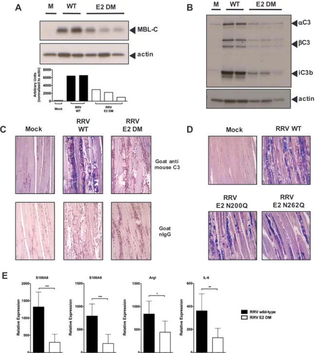
We have previously shown that complement activation following RRV infection is mediated primarily through MBL (Gunn et al., 2012). Given that we observed a decrease in MBL deposition within RRV E2 DM-infected cultures, and since the disease phenotype induced by E2 DM in WT mice was strikingly similar to the disease induced by WT RRV in MBL-DKO mice, we hypothesized that the presence of the E2 glycans contribute to MBL deposition and complement activation in the quadriceps muscle following infection. MBL-mediated complement ac-tivation is initiated by the recognition of terminal sugars on glycosy-lated proteins by the carbohydrate recognition domain of MBL, and binding of MBL to the ligand leads to formation of a C3 convertase by the MBL-associated serine proteases resulting in localized C3 deposition (Ricklin et al., 2010). We infected mice with either RRV WT or E2 DM, harvested quadriceps muscle at 7 dpi and analyzed the muscle homo-genates by western blot for MBL-C levels. As shown inFig. 4A, RRV WT-infected mice had elevated levels of MBL-C in the quadriceps at 7 dpi compared to mock-infected mice, and importantly, we observed re-duced levels of MBL in RRV E2 DM-infected mice compared to RRV WT-infected mice, suggesting that the RRV E2 glycans contribute to MBL deposition within infected tissues.
To determine if the reduced levels of MBL within the quadriceps muscle of RRV E2 DM-infected mice correlated with reduced
complement activation, we analyzed quadriceps muscle homogenates from RRV WT or E2 DM-infected mice for levels of C3 cleavage pro-ducts by western blot. Consistent with our hypothesis, we observed reduced levels of both the alpha and beta chain of C3 in E2 DM-infected mice compared to WT-infected mice (Fig. 4B), indicating that the E2 glycans are required for C3 deposition onto infected tissues. Im-portantly, the levels of the C3 cleavage product and CR3 ligand iC3b was significantly reduced in E2 DM-infected mice compared to WT-infected mice (Fig. 4B), indicating that the RRV E2 glycans are required for robust complement activation following infection.
As western blot analysis of C3 present in quadriceps muscle tissue does not distinguish between C3 produced by infiltrating inflammatory monocytes and C3 that is deposited onto infected tissues, we sought to further confirm that the RRV E2 glycans are required for C3 deposition onto skeletal muscle following infection by immunohistochemical staining of C3 on skeletal muscle sections. We infected mice with either RRV WT or E2 DM, and generated sections of the quadriceps muscle at
7 dpi. Consistent with results inFig. 4B, we observed a reduction in C3 deposition onto the skeletal muscle from mice infected with RRV E2 DM compared to WT (Fig. 4C). Interestingly, we also observed reduced C3 deposition onto quadriceps muscle sections from mice infected with RRV E2 N200Q or E2 N262Q compared to RRV WT (Fig. 4D). The level of C3 deposition correlates with disease severity and is consistent with the role of complement in determining disease severity. As the E2 glycans were required for production of iC3b, we analyzed the ex-pression of MBL, C3, and CR3-dependent pro-inflammatory mediators by real-time PCR to determine if their expression was also dependent on the E2 glycans. Indeed, mice infected with RRV E2 DM had reduced expression of MBL and C3-dependent pro-inflammatory genes com-pared to RRV WT (Fig. 4E), further supporting the hypothesis that the E2 N-linked glycans mediate complement activation following infec-tion.
4.5. RRV glycan mutants retain the ability to replicate in vivo
Although the RRV E2 N-linked glycans are dispensable for replica-tion in cell culture (Shabman et al., 2008), it is possible that the glycans might be required for efficient viral replication and dissemination in vivo. Therefore, we evaluated the viral titer within the quadriceps muscle and the ankle joints from either RRV WT, or glycan mutant-infected mice at multiple times post-infection by plaque assay. There was no difference in viral titer within the quadriceps and ankle joints at
1 dpi between any of the viruses, suggesting that the E2 glycans are not required for initial dissemination and replication within target tissues (Fig. 5A-B). Overall, all of the glycan mutant viruses replicated at or near wild-type levels in target tissues, although there were some dif-ferences in titer observed at intermediate time points. At 3 dpi, we observed reduced viral titer within the quadriceps muscle in mice in-fected with E2 N200Q and E2 DM compared to WT (Fig. 5A). The E2 N262Q virus did not show any difference in titer compared to RRV WT at any time post-infection in either the ankle or the quadriceps muscle.
At 5 dpi, only the RRV E2 DM titer is reduced in the quadriceps muscle compared to RRV WT, but importantly, at time points of severe disease, 7 and 10 dpi, no difference was observed in the quadriceps muscle between any of the viruses (Fig. 5A). Similar results were observed in the ankle joints, although there were significant differences in titer at day 5 in the ankle joint between RRV E2 DM and RRV WT, but these differences were not sustained at time points of peak disease severity (7 or 10 dpi) (Fig. 5B).
In order to confirm the plaque assay results, we also performed quantitative real time RT-PCR to determine the absolute numbers of
RRV genomes per µg of RNA within the quadriceps muscle between WT and E2 DM virus at 3 and 5 dpi. We did not observe any differences in the number of RRV genomes between the two viruses at 3 dpi (Fig. 5C). This suggests that the overall levels of viral replication within the tis-sues are equivalent between the wild type and mutant virus at this time point. However, consistent with reduced viral titer at 5 dpi, we ob-served a similar decrease in viral RNA at 5 dpi in the quadriceps muscle between WT and E2 DM (Fig. 5C). Taken together, these data indicate that the loss of the glycans does not affect the overall ability of the virus disseminate to and replicate within target tissues, however the E2 DM does show reduced replication at intermediate time points.
4.6. The RRV E2 glycan mutants and WT RRV induce equivalent inflammatory responses
RRV infection induces an inflammatory response that results in in-filtration of inflammatory cells into target tissues such as the quadriceps muscle, subsequently leading to tissue damage (Morrison et al., 2006). Prior studies demonstrated that even though RRV-infected C3-/- and MBL-DKO mice were resistant to virus-induced disease, virus induced inflammatory cell recruitment was unaffected (Gunn et al., 2012; Morrison et al., 2007). Therefore, we evaluated the E2 glycan mutants to determine if virus induced inflammation was similar to that observed in C3 or MBL deficient animals by quantifying and characterizing in-flammatory cell populations from whole quadriceps muscle of mice infected with the RRV WT or each of the mutant viruses byflow cy-tometry. At 10 dpi, which is a time point of peak inflammation within tissues, we did not observe any differences in the total magnitude or the composition of the inflammatory cell population between RRV WT and the glycan mutants (Fig. 6). Total numbers of leukocytes in the quad-riceps muscle were equivalent between mice in all groups (Fig. 6), in-dicating that the E2 N-linked glycans do not affect infiltration of in-flammatory cells into skeletal muscle. Furthermore, the numbers of inflammatory macrophages, which are thought to drive much of the tissue damage during RRV infection (Lidbury et al., 2000, 2008), and other cell types including CD4+and CD8+T cells, NK cells, and neu-trophils, were equivalent between RRV WT and the glycan mutants (Fig. 6). These data suggest that loss of the E2 glycans does not sig-nificantly affect the induction of the infiltration or composition of the inflammatory cells into the quadriceps muscle following RRV infection.
5. Conclusions
We report herein that the RRV E2 N-linked glycans bind MBL and contribute to activation of complement, ultimately enhancing RRV-mediated tissue destruction. As MBL binds to carbohydrates, we hy-pothesized that the viral N-linked glycans displayed on RRV-infected cells act as ligands for MBL-mediated complement activation. Thus, to determine whether the viral N-linked glycans contribute to MBL-mediated complement activation during RRV infection, we generated a panel of RRV mutants that lack the E2 N-linked glycans: N200Q, N262Q, or N200;262Q (double mutant, or DM). Consistent with our hypothesis, reduced MBL deposition was observed onto skeletal muscle cells infected with RRV E2 DM compared to RRV wild-type, suggesting that the viral N-linked glycans drive MBL deposition onto infected cells. We next determined whether the E2 N-linked glycans are required for severe disease using a mouse model of RRV-induced myositis. RRV mutants lacking one or both glycans fail to induce severe disease in vivo, with mice infected with the glycan mutants exhibiting sig-nificantly reduced inflammatory pathology and tissue damage in quadriceps muscle compared to mice infected with wild-type RRV, despite similar viral loads within infected quadriceps muscle and ankle joints at times of peak disease. Importantly, MBL deposition and com-plement activation within the skeletal muscle of RRV E2 DM infected mice was reduced compared to wild-type virus, consistent with our hypothesis that the viral E2 glycans drive complement activation within
Fig. 5. RRV lacking E2 N-linked glycans retain ability to replicate in vivo.
Quadriceps muscle(A)and ankle joints(B)from mice infected with RRV WT (solid cir-cles), E2 N200Q (open circir-cles), E2 N262Q (solid squares) or E2 DM (open square) were assayed to determine viral titer at various times post infection. Viral titer was determined by plaque assay on BHK-21 cells. ***p< 0.005 **p< 0.01 by one-way ANOVA.(C)
Quantification of total numbers of RRV RNA genomes per µg of RNA from the quadriceps muscles of mice infected with either RRV WT (solid circles) or E2 DM (open squares). ** p< 0.01 by Student'st-test.
inflamed tissues. Because RRV infection is associated with infiltration of inflammatory leukocytes into the quadriceps muscle, we next tested whether the E2 N-linked glycans modulate recruitment of inflammatory cells into the muscle. However, similar numbers of infiltrating in-flammatory leukocytes within skeletal muscle were observed for all viruses, suggesting that complement activation, rather than in-flammatory cell recruitment, is differentially activated by the E2 N-linked glycan mutants and contributes to disease. Thus, these data de-monstrate that the RRV E2 N-linked glycans present on infected cells contribute to severe RRV-induced disease through MBL-mediated acti-vation of complement.
6. Discussion
Mosquito-borne alphaviruses such as RRV and CHIKV are emerging viruses that are a significant cause of explosive outbreaks of infectious arthralgia and myalgia in humans. The host complement system through MBL plays a critical role in mediating development of disease and tissue damage through activation of inflammatory cells that in-filtrate into the muscle and joints following infection (Gunn et al., 2012; Morrison et al., 2008, 2007). In this study, we demonstrate that the RRV envelope N-linked glycans contribute to MBL deposition and complement activation leading to the development of severe virus-in-duced damage to the musculoskeletal system.
The results presented in this study demonstrate that viral N-linked
glycans promote MBL binding to RRV infected cells and that MBL de-position and subsequent complement activation is reduced in tissues from animals infected with E2 DM (Figs. 1 and 4). Given the central role that MBL, complement activation, and subsequent inflammatory cell activation through CR3 plays in the pathogenesis of RRV-induced dis-ease, it is likely that the major mechanism of attenuation for the E2 DM virus is through reduced MBL binding, complement activation, and production of the CR3 ligand iC3b leading to reduced activation of macrophage within tissues. However, given that the E2 DM virus did show reduced levels of replication at intermediate times post infection (Fig. 5A), we cannot rule out the possibility that at least some of the impact on virus-induced disease with the E2 DM virus is due to tran-sient reduction in viral load. However, it is important to point out that the decrease in viral load at 3 and 5 dpi did not affect the recruitment of inflammatory cells into the sites of viral replication (Fig. 6), and that viral loads were indistinguishable at 7 dpi, which is a time when severe disease is readily apparent with the wild type virus (Fig. 5A-B). Fur-thermore, we and others showed that disease is reduced in mice in-fected with either of the single glycan mutants that exhibit either no difference in viral loads compared to RRV WT (E2 N262Q mutant) or modest differences in viral loads (E2 N200Q mutant) (Nelson et al., 2016), and the reduction in disease in these mice correlates with re-duced C3 deposition within infected tissues. Thus, these data support the hypothesis that the E2 N-linked glycans primarily affect RRV pa-thogenesis through complement activation.
In summary, we have demonstrated that the interaction between the host innate immune protein MBL and the RRV E2 N-linked glycans contribute to the pathogenesis of RRV-induced arthritis and myositis through activation of the host complement system. Our data supports a therapeutic strategy of specifically targeting the interaction between the RRV E2 N-linked glycans and MBL through the use of MBL in-hibitors in the treatment of RRV and other virus-induced inflammatory diseases.
Acknowledgements
We thank the members of the Heise lab for helpful scientific dis-cussions, and in particular would like to thank Martin Ferris for assis-tance with statistical analysis. We would also like to thank Janice Weaver and the LCCC/DLAM histopathology core facility and Bob Bagnell and the UNC microscopy facility.
Funding sources
This work was supported by the National Institutes of Health [grant numbers R01 AR47190, U19 AI 109761, U19 AI100625, and U54 AI 057157].
Appendix A. Supplementary material
Supplementary data associated with this article can be found in the online version athttp://dx.doi.org/10.1016/j.virol.2017.12.022.
References
Abram, C.L., Lowell, C.A., 2009. The ins and outs of leukocyte integrin signaling. Annu. Rev. Immunol. 27, 339–362.
Banda, N.K., Mehta, G., Kjaer, T.R., Takahashi, M., Schaack, J., Morrison, T.E., Thiel, S., Arend, W.P., Holers, V.M., 2014. Essential role for the lectin pathway in collagen antibody-induced arthritis revealed through use of adenovirus programming com-plement inhibitor MAp44 expression. J. Immunol. 193, 2455–2468.
Clarris, B.J., Doherty, R.L., Fraser, J.R., French, E.L., Muirden, K.D., 1975. Epidemic polyarthritis: a cytological, virological and immunochemical study. Aust. N. Z. J. Med. 5, 450–457.
Ezekowitz, R.A., Kuhlman, M., Groopman, J.E., Byrn, R.A., 1989. A human serum man-nose-binding protein inhibits in vitro infection by the human immunodeficiency virus. J. Exp. Med. 169, 185–196.
Fraser, J.R., Cunningham, A.L., Clarris, B.J., Aaskov, J.G., Leach, R., 1981. Cytology of synovial effusions in epidemic polyarthritis. Aust. N. Z. J. Med. 11, 168–173.
Fuchs, A., Lin, T.Y., Beasley, D.W., Stover, C.M., Schwaeble, W.J., Pierson, T.C., Diamond, M.S., 2010. Direct complement restriction offlavivirus infection requires glycan re-cognition by mannose-binding lectin. Cell Host Microbe 8, 186–195.
Gunn, B.M., Morrison, T.E., Whitmore, A.C., Blevins, L.K., Hueston, L., Fraser, R.J., Herrero, L.J., Ramirez, R., Smith, P.N., Mahalingam, S., Heise, M.T., 2012. Mannose binding lectin is required for alphavirus-induced arthritis/myositis. PLoS Pathog. 8, e1002586.
Hart, M.L., Saifuddin, M., Uemura, K., Bremer, E.G., Hooker, B., Kawasaki, T., Spear, G.T., 2002. High mannose glycans and sialic acid on gp120 regulate binding of mannose-binding lectin (MBL) to HIV type 1. AIDS Res. Hum. Retrovir. 18, 1311–1317.
Hazelton, R.A., Hughes, C., Aaskov, J.G., 1985. The inflammatory response in the syno-vium of a patient with Ross River arbovirus infection. Aust. N. Z. J. Med. 15, 336–339.
Herrero, L.J., Nelson, M., Srikiatkhachorn, A., Gu, R., Anantapreecha, S., Fingerle-Rowson, G., Bucala, R., Morand, E., Santos, L.L., Mahalingam, S., 2011. Critical role for macrophage migration inhibitory factor (MIF) in Ross River virus-induced ar-thritis and myositis. Proc. Natl. Acad. Sci. U.S.A. 108, 12048–12053.
Herrero, L.J., Sheng, K.C., Jian, P., Taylor, A., Her, Z., Herring, B.L., Chow, A., Leo, Y.S., Hickey, M.J., Morand, E.F., Ng, L.F., Bucala, R., Mahalingam, S., 2013. Macrophage migration inhibitory factor receptor CD74 mediates alphavirus-induced arthritis and myositis in murine models of alphavirus infection. Arthritis Rheum. 65, 2724–2736.
Hirahashi, J., Mekala, D., Van Ziffle, J., Xiao, L., Saffaripour, S., Wagner, D.D., Shapiro, S.D., Lowell, C., Mayadas, T.N., 2006. Mac-1 signaling via Src-family and Syk kinases results in elastase-dependent thrombohemorrhagic vasculopathy. Immunity 25, 271–283.
Ip, W.K., Chan, K.H., Law, H.K., Tso, G.H., Kong, E.K., Wong, W.H., To, Y.F., Yung, R.W., Chow, E.Y., Au, K.L., Chan, E.Y., Lim, W., Jensenius, J.C., Turner, M.W., Peiris, J.S., Lau, Y.L., 2005. Mannose-binding lectin in severe acute respiratory syndrome cor-onavirus infection. J. Infect. Dis. 191, 1697–1704.
Ji, X., Olinger, G.G., Aris, S., Chen, Y., Gewurz, H., Spear, G.T., 2005. Mannose-binding lectin binds to Ebola and Marburg envelope glycoproteins, resulting in blocking of virus interaction with DC-SIGN and complement-mediated virus neutralization. J. Beyondactivationofthecomplementsystem,theE2N-linked
gly-canslikelymodulateotheraspectsofthedisease(bothprotectiveand pathologic)throughdifferentialinductionofinnateimmuneresponses via interactions with carbohydrate binding proteins such as C-type lectins,andtoll-likereceptors(Shabmanetal.,2008).Inaddition,while we havefocusedontheroleoftheE2glycans inmediating RRV-in-duced disease in this study, the E1 glycan was recently shown to modulateRRVpathogenesis,asamutantviruslackingtheE1glycan (E1N141Q)demonstratedenhancedclearancefromtissues,resultingin reduced inflammation andpathology (Nelson et al., 2016). Interest-ingly,theE1N141QmutantenhancedcirculatingIFNγlevels,whereas viruses lacking the E2 glycans did not affect IFNγ production, sug-gestingthattheE1andE2glycansdifferentiallyinteractwiththe im-munesystem(Nelsonetal.,2016).Futurestudiesaimedatidentifying anddissecting thedifferential interactions ofthe E1andE2glycans withtheinnateimmunesystemmayrevealnovelmechanismsthrough which viral N-linked glycans modulate viral-induced inflammatory disease.
TheinteractionbetweenMBLandviralN-linkedglycanshasbeen demonstratedinanumberofothervirussystemsincludingHIV, SARS-CoV, Ebolavirus,andWestNilevirus(Ezekowitzetal., 1989;Zhou etal.,2010;Ipetal.,2005;Hartetal.,2002;Fuchsetal.,2010;Jietal., 2005).Inthese systems,MBLrecognizesandbindstohighmannose glycansontheenvelopeglycoproteins,andleadseithertodirect neu-tralizationofvirus,oraidsininhibitingvirusinfectionbypreventingor abrogating attachmentand entry into host cells. In contrast to the protectiveroleofMBLforotherviruses,MBLclearlyplaysapathologic roleduringRRVinfectionandthesestudiesprovidenewinsightsinto the roleofviral N-linked glycans indriving this pathologicprocess. Furthermore, studieswith other viruseshave demonstrated that the interactionbetweenMBLandthevirusparticleisimportantfor MBL-mediatedneutralization(Zhouetal.,2010;Fuchsetal.,2010;Jietal., 2005).Incontrasttothosestudies,wehaveseverallinesofevidence thatdemonstrateMBLinteractswithRRV-infectedcellsthroughtheE2 N-linkedglycansonthesurfaceoftheinfectedcellratherthanwiththe virion particle. First,MBLdepositionontoinfectedcells invitroand withininfectedtissuesinvivoareelevatedcomparedtomockinfection (Gunnetal., 2012).Second,MBLdeposition is reducedin cellsand tissuesinfectedwithRRVE2DMcomparedtoWTindependentofviral replication indicating thattheE2glycans promote MBL binding. Fi-nally,weobservedMBLdepositionontoRRV-infectedcellsratherthan uninfected cells,which wasreducedin E2DM-infectedcells,despite surfaceexpressionoftheRRVglycoproteinsonE2DM-infectedcells. Thus,whiletheindividualE2glycansappeartohavelittleimpacton replication in either mammalian or mosquito cells (Shabman et al., 2008;Nelsonetal.,2016),wecannotruleoutthepossibilitythatlossof the N-linked glycans leads to conformational changes in E2, which coulddiminishMBLbinding.OurresultssuggestthattheviralN-linked glycansareessentialforpromotingMBLdepositionontothesurfaceof infected cells.Inthis way,theE2N-linked glycanscontributeto pa-thologyanddiseaseinthemammalianhost.
Tarkowski, A., van Rooijen, N., Fraser, R.J., Mahalingam, S., 2008. Macrophage-derived proinflammatory factors contribute to the development of arthritis and myositis after infection with an arthrogenic alphavirus. J. Infect. Dis. 197, 1585–1593.
Morrison, T.E., Whitmore, A.C., Shabman, R.S., Lidbury, B.A., Mahalingam, S., Heise, M.T., 2006. Characterization of Ross River virus tropism and virus-induced in-flammation in a mouse model of viral arthritis and myositis. J. Virol. 80, 737–749.
Morrison, T.E., Fraser, R.J., Smith, P.N., Mahalingam, S., Heise, M.T., 2007. Complement contributes to inflammatory tissue destruction in a mouse model of Ross River virus-induced disease. J. Virol. 81, 5132–5143.
Morrison, T.E., Simmons, J.D., Heise, M.T., 2008. Complement receptor 3 promotes se-vere ross river virus-induced disease. J. Virol. 82, 11263–11272.
Nelson, M.A., Herrero, L.J., Jeffery, J.A., Hoehn, M., Rudd, P.A., Supramaniam, A., Kay, B.H., Ryan, P.A., Mahalingam, S., 2016. Role of envelope N-linked glycosylation in Ross River virus virulence and transmission. J. Gen. Virol. 97, 1094–1106.
Pletnev, S.V., Zhang, W., Mukhopadhyay, S., Fisher, B.R., Hernandez, R., Brown, D.T., Baker, T.S., Rossmann, M.G., Kuhn, R.J., 2001. Locations of carbohydrate sites on alphavirus glycoproteins show that E1 forms an icosahedral scaffold. Cell 105, 127–136.
Ricklin, D., Hajishengallis, G., Yang, K., Lambris, J.D., 2010. Complement: a key system for immune surveillance and homeostasis. Nat. Immunol. 11, 785–797.
Ross, G.D., 2000. Regulation of the adhesion versus cytotoxic functions of the Mac-1/ CR3/alphaMbeta2-integrin glycoprotein. Crit. Rev. Immunol. 20, 197–222.
Shabman, R.S., Rogers, K.M., Heise, M.T., 2008. Ross River virus envelope glycans con-tribute to type I interferon production in myeloid dendritic cells. J. Virol. 82, 12374–12383.
Strauss, J.H., Strauss, E.G., 1994. The alphaviruses: gene expression, replication, and evolution. Microbiol. Rev. 58, 491–562.
Takahashi, K., Ip, W.E., Michelow, I.C., Ezekowitz, R.A., 2006. The mannose-binding lectin: a prototypic pattern recognition molecule. Curr. Opin. Immunol. 18, 16–23.
Zhou, Y., Lu, K., Pfefferle, S., Bertram, S., Glowacka, I., Drosten, C., Pohlmann, S., Simmons, G., 2010. A single asparagine-linked glycosylation site of the severe acute respiratory syndrome coronavirus spike glycoprotein facilitates inhibition by man-nose-binding lectin through multiple mechanisms. J. Virol. 84, 8753–8764.
Gen.Virol.86,2535–2542.
Lidbury,B.A.,Simeonovic,C.,Maxwell,G.E.,Marshall,I.D.,Hapel,A.J.,2000. Macrophage-inducedmusclepathologyresultsinmorbidityandmortalityforRoss Rivervirus-infectedmice.J.Infect.Dis.181,27–34.