LOOKING INSIDE THE WNT/BETA-CATENIN DESTRUCTION COMPLEX: MECHANISMS AND THE MINIMAL MACHINE
Mira I. Pronobis
A dissertation submitted to the faculty at the University of North Carolina at Chapel Hill in partial fulfillment of the requirements for the degree of Doctor of Philosophy in the Curriculum
of Genetics and Molecular Biology.
Chapel Hill 2016
Approved by:
Mark A. Peifer
Robert J. Duronio
Victoria L. Bautch
M. Ben Major
ABSTRACT
Mira I. Pronobis: Looking inside the Wnt/beta-catenin destruction complex: mechanisms and the minimal machine
(Under the direction of Mark A. Peifer)
ACKNOWLEDGEMENTS
First I want to thank Mark Peifer for being a brilliant mentor. He made me a better scientist.
A big thanks go to the people in the Peifer lab and all the other labs at UNC that I worked with!
Then I would like to thank my committee: Bob, Vicki, Ben and Steve,
who always put me on the right path when I drifted away
To my non-scientific community:
First I like to acknowledge my two dogs Mike and Nala,
who reminded me to relax, and taught me how to live every moment
My friends
who helped to balance my private life with my work life: Josi, Sophie and Josh
My parents and my brother, to
whom I am forever grateful for believing in me and for all the support you gave me.
PREFACE
Chapter 2 describes my early work on APC. This second author paper was published in Molecular Biology of the Cell in 2011 (Molecular biology of the cell 22.11 (2011): 1845-1863). Here we investigated the role of APC’s βcat binding sites using mammalian tissue culture cells and the fruit fly. Dave Roberts conducted the fly experiments and did the cloning of various constructs. John Poulton, Jon Waldmann, Elise Stephenson and Shahnaz Hanna contributed to the fly experiments. This work was completed under the direction of Mark Peifer.
My contribution to the work was the mammalian tissue culture experiments. I measured the βcat levels and Wnt signaling activity, and I generated different APC mutants. Additionally, I found that colocalization of APC to Axin is negatively regulated by 20R2 and region B.
Chapter 3 describes the core of my research during my PhD. This first author paper was published in eLIFE in 2015 (eLife 4 (2015): e08022). Here we investigated the mechanistic role of APC in the destruction complex. We used mammalian tissue culture cells and the fruit fly. Nasser Rusan acquired a subset of the high resolution SIM images, and Mark Peifer analyzed the SIM images and measured the volume. This work was completed under the direction of Mark Peifer.
Chapter 4 describes another project that I started during my PhD. This first author paper is not published yet, but it will be submitted this summer. Here we investigated the cooperative function of APC and Axin in βcat destruction. For this we used mammalian tissue culture cells. Natalie Deuitch conducted the reporter gene assays with help from Vinya Posham. Natalie Deuitch also helped with βcat immunostaining and analysis, and with the cloning of different constructs. This work was completed under the direction of Mark Peifer.
TABLE OF CONTENTS
LIST OF FIGURES ... xiv
LIST OF TABLES ... xvii
LIST OF ABBREVATIONS ... xviii
CHAPTER 1: INTRODUCTION ... 1
Cell signaling pathways in development and disease ... 1
The Wnt signals and cancer ... 2
The Wnt signaling pathway ... 3
Turning the destruction complex off ... 5
Facilitating βcat destruction ... 7
Axin and its role in the βcat destruction complex and disease ... 8
APC and its role in the destruction complex ... 9
APC and colorectal cancer ... 11
Remaining questions in the regulation of Wnt signaling ... 12
References ... 16
CHAPTER 2: DECONSTRUCTING THE BETA-CATENIN DESTRUCTION COMPLEX: MECHANISTICROLES FOR THE TUMOR SUPPRESSOR APC IN REGULATING WNT SIGNALING ... 20
Overview ... 21
Material and Methods ... 28
APC constructs ... 28
Cell culture, transfections, and immunofluorescence ... 29
Quantifying βcat protein levels ... 29
TOP/FOP reporter assays ... 30
Transgenic fly lines, embryonic lethality assay, and cuticle rescue ... 30
IPs and Western blotting ... 31
Immunofluorescence/imaging of Drosophila embryos ... 31
Results ... 32
Model systems to assess APC function in human cells and flies ... 32
Individual high-affinity βcat-binding sites are not essential for βcat destruction ... 34
Multiple βcat-binding sites modulate Wnt signaling regulation via cytoplasmic retention ... 37
20R2 and sequence B are essential for βcat destruction ... 39
Direct binding to Axin via the SAMPs is essential for Wnt regulation in Drosophila but dispensable in SW480 cells ... 41
APC colocalizes with Axin by a second mechanism independent of its SAMP repeats ... 43
Sequence B may regulate APC–Axin interactions ... 45
Discussion ... 46
Individual βcat-binding sites are dispensable for destruction complex function... 46
Cyclic assembly/disassembly of Axin–APC complexes may
regulate destruction complex function ... 59
Cytoplasmic retention of βcat: a regulatory mechanism in normal development and oncogenesis? ... 51
Cytoplasmic retention: helping explain selection of truncated APC proteins in cancer ... 53
References ... 79
CHAPTER 3: A NOVEL GSK3-REGULATED APC:AXIN INTERACTION REGULATES WNT SIGNALING BY DRIVING A CATALYTIC CYCLE OF EFFICIENT BETA-CATENIN DESTRUCTION ... 84
Overviewt ... 85
Introduction ... 85
Material and Methods ... 89
Constructs ... 89
Antibodies ... 90
Cell culture ... 90
Microscopy ... 90
Estimating relative over-expression levels of Axin and APC ... 91
Quantification ... 92
FRAP ... 92
Protein work ... 93
In vitro kinase assay ... 93
Fly work ... 94
Results ... 94
A novel Axin:APC association site regulated by APC motifs R2 and B ... 98
Axin and APC2 form structured macromolecular complexes in vivo ... 99
APC2 stabilizes the Axin complex and promotes
destruction complex throughput of βcat ... 100
Both APC2's Arm rpts and SAMPs are required to stabilize
APC2:Axin complexes and regulate its substructure ... 104
R2 and B regulate APC2 dynamics and APC2's ability
to enhance βcat throughput ... 105
R2 and B regulate APC2:Axin association via the Arm rpts ... 107
APC2:Axin association via the Arm repeats is regulated
by GSK3 kinase ... 108
Mutating putative phosphorylation sites in B disrupts APC2's
function in regulating βcat destruction in SW480 cells ... 111
Mutating putative phosphorylation sites in B severely reduces APC2's function in regulating βcat destruction or cell fate
in vivo in Drosophila ... 113
Discussion ... 115 APC stabilizes the Axin complex and alters
complex assembly, thus increasing destruction complex activity ... 116
Axin and APC cooperate to ensure efficient βcat destruction ... 118
Destruction complex function requires dynamic APC2:Axin
interactions regulated by R2/B ... .119
Phosphorylation of R2 and B may be the trigger for
releasing the APC Arm repeat:Axin interaction ... 121
References ... 148 CHAPTER 4: ENGINEERING THE MINIMAL BETA-CATENIN DESTRUCTION
Overview ... 155
Introduction ... 155
Material and Methods ... 158
Constructs ... 158
Antibodies ... 159
Cell culture ... 159
Microscopy ... 159
Quantifications ... 159
FRAP ... 160
Reporter Gene Assay for Wnt regulated transcription ... 160
Protein work ... 160
Results ... 160
APC and Axin share regions with redundant functions ... 160
APC and Axin have 5 regions that are essential for their individual functions in βcat degradation ... 161
Complementation suggests APC and Axin share regions with redundant functions ... 163
APC’s and Axin’s essential regions each make important but semi-independent contributions to efficiency of βcat destruction ... 165
The essential regions of APC and Axin are sufficient to downregulate Wnt signaling... 166
Covalently linking the essential regions of APC and Axin increases their negative-regulatory activity ... 167
The internal complex structure and the complex size of the Chimera
are similar to those of APC:Axin complexes ... 168
The Chimera mimics dynamics of the APC:Axin complex ... 169
Discussion ... 170
References ... 187
CHAPTER 5: DISCUSSION ... 190
The recruitment of βcat into the APC:Axin complex and the release to the E3-ligase ... 190
The Armadillo repeats in APC and their role in Wnt signaling ... 192
APC and the exclusion from the nucleus ... 193
Wnt regulated transcription and the role of the essential regions in APC and Axin ... 194
The importance of polymerization in the destruction complex ... 194
The ratio of APC and Axin in the destruction complex ... 195
Phosphorylation and dephosphorylation of APC and Axin ... 196
APC and Axin and the combinatorial code of regions that leads to βcat degradation ... 196
Axin and its microtubule binding site ... 197
LIST OF FIGURES
Figure 1.1 - The Wnt pathway... 14 Figure 1.2 - Schematic representation of human APC and Axin proteins ... 15 Figure 2.1 - Diagrams of wild-type APC2 and the mutants used, and summary
of the functions of each ... 58 Figure 2.2 - . Fly and human APC proteins reduce βcat levels in SW480 cells,
and fly APC restores cuticle patterning in APC single and double mutant flies ... 59 Figure 2.3 - Expression levels of transgenes in SW480 cells and in Drosophila embryos ... 60 Figure 2.4 - Individual high-affinity 20Rs are not essential for destruction
but contribute with 15Rs to cytoplasmic retention ... 61 Figure 2.5 - Many βcat-binding sites are dispensable for Arm destruction
but contribute additively to rescue APC2 single mutants ... 62 Figure 2.6 - APC2 mutant transgenes rescue APC2g10 single mutants to adulthood ... 64 Figure 2.7 - Roles of 20Rs and sequence B in targeting Arm for destruction in vivo ... 65 Figure 2.8 - Sequence B and 20R2 are essential for βcat destruction by APC2 in SW480 cells ... 66 Figure 2.9 - Sequence B, 20R2, and the SAMPs are essential for Arm destruction in vivo ... 68 Figure 2.10 - Sequence B and 20R2 are also essential for βcat destruction by human APC ... 70 Figure 2.11 - The SAMPs are necessary for APC2 to coimmunoprecipitate with Axin... 72 Figure 2.12 - APC2 colocalization with Axin is SAMP-independent and is
antagonized by sequence B ... 73 Figure 2.13 - Alignment of the MCR region of APC of different species ... 75 Figure 2.14 - Model: APC regulates βcat signaling through a cycle of
destruction complex assembly and disassembly ... 76 Figure 2.15 -Model: Cytoplasmic retention by APC playing a
Figure 2.16 - Cuticle scoring chart ... 79
Figure 3.1 - APC2's Arm rpts provide a second means of interacting with the Axin complex ... 127
Figure 3.2 - Assessing levels of overexpression of Axin and APC ... 128
Figure 3.3 - Human APC's Arm repeat domain colocalizes with Axin ... 130
Figure 3.4 - Axin and APC2 form structured macromolecular complexes in vivo ... 131
Figure 3.5 - APC2 stabilizes Axin complexes and promotes efficient βcat destruction ... 132
Figure 3.6 - While proteasome inhibition reduces βcat destruction and causes βcat to detectably accumulate in APC2 + Axin puncta, it does not abolish the ability of APC2 to enhance Axin function in this regard ... 134
Figure 3.7 - APC2’s Arm rpts and SAMPs each are required to stabilize APC2:Axin complexes ... 135
Figure 3.8 - Colocalization of APC2's Arm repeat domain with Axin is controlled by R2 and B and APC2 without R2 or region B still stabilizes Axin complexes ... 137
Figure 3.9 - R2 and B regulate APC2 dynamics in the destruction complex, and regulate βcat removal from the destruction complex ... 139
Figure 3.10 - Binding of APC2's SAMP motif to Axin is not regulated by R2 and B ... 140
Figure 3.11 - Association of Axin with APC2's Arm rpts is controlled by R2 and B ... 142
Figure 3.12 - Axin:APC2 Arm rpts association is regulated by GSK3 ... 143
Figure 3.13 - GSK3 regulates association of APC2's Arm repeats with the Axin complex ... 144
Figure 3.14 - Mutating putative phosphorylation sites in B disrupts APC2 function ... 145
Figure 3.15 - Blocking potential phosphorylation at 2 conserved serines in B disrupts APC2 function in the fly ... 147
Figure 3.16 - Speculative Model of APC2's catalytic cycle inside the destruction complex. Figure 3.1 - APC2's Arm rpts provide a second means of interacting with the Axin complex ... 148
Figure 4.2 - APC and Axin each have regions that are essential to facilitate βcat destruction .. 178 Figure 4.3 - APC and Axin mutants can complement each other in βcat degradation ... 180 Figure 4.4 - The APC:Axin Chimera reduces βcat as efficiently as wildtype APC and Axin ... 182 Figure 4.5 - The essential regions in APC and Axin are important for a
functional destruction complex ... 184 Figure 4.6 - All regions in the Chimera are important for its efficient function
in βcat degradation ... 185 Figure 4.7 - The Chimera complex mimics the APC:Axin complex
in the internal structure and size ... 186 Figure 4.8 - The dynamics of the chimera complex are similar to APC2+Axin
LIST OF TABLES
Table 2.1 - Function of APC2 mutants in vivo in Drosophila ... 80 Table 3.1 - Quantitation of relative expression levels of transfected
LIST OF ABBREVATIONS
APC Adenomatous polyposis coli βcat beta-catenin, βcatenin CK1 Casein kinase 1
Frzd Frizzled
CHAPTER 1: INTRODUCTION Cell signaling pathways in development and disease
Every cell in our body has to communicate with the outside environment. The
fundamental process in single and multicellular organisms involves receiving, processing and responding to outside cues. For this cells developed signaling pathways. The text book core principles of cell signaling involves 3 components: (1) A membrane or cytoplasmic receptor, which receives outside cues and transmits information into the cytoplasm, (2) a set of
transducing components, which interacts with the intracellular part of the membrane receptor and transduces the signal into to the cell nucleus, and (3) a transcriptional regulator, which triggers the cell’s response by activating or repressing transcription of target genes.
To this day the number of signaling pathways that are discovered is still increasing; but a subset of these pathways are fundamental in development and disease (Cooper et. al., 2000, Pires-daSilva and Sommer, 2003). The canonical Wnt/β-catenin pathway, Hedgehog pathway, retinoic acid signaling, the FGF pathway, Notch-Delta signaling, and the TGF-β pathway regulate cell proliferation, stem cell pluripotency and cell fate choices during development. They play major roles in axis determination, gastrulation, neural tube formation and other fundamental processes. Interestingly, the same pathways that are essential in development are also the ones that have severe consequences in adulthood when dysregulated (Vogelstein et al., 2013).
tumorigenesis, and that mutations in only two to eight of these promoting genes are sufficient to drive tumorigenesis in a given cell type (Vogelstein et al., 2013). Interestingly, these 140 genes affect only 12 signaling pathways suggesting that dysregulation of these core cellular pathways is the driving force leading to cancer development. These signaling pathways can be divided into those driving cell survival, genome maintenance, and cell fate, such that when altered they allow cells a selective growth advantage. Interestingly, in some signaling pathways one core component is the major target of mutation.
The Wnt signals and cancer
The canonical Wnt signaling pathway is one of these 12 powerful oncogenic pathways. In the early 1980s two independent studies found the first member of the Wnt pathway: The gene Wnt1, the first known ligand of the canonical Wnt receptors, was discovered by studying the Mouse Mammary Tumor Virus, which randomly integrates in the mouse genome (Nusse and Varmus, 1982). The first identified integration site that led to the development of mammary tumors was named Integration site-1 (Int-1). After sequencing Int1, its Drosophila homolog gene wingless was identified. Regulatory mutations in the fly gene generated flies without wings (wingless), while null alleles led to embryonic lethality with defects in embryonic cell fates (Nusslein-Volhard and Wieschaus, 1980). Thus, the pathway was named Wingless-int pathway or Wnt pathway. In 1989, axis duplication studies in Xenopus embryos confirmed the conservation of the canonical Wnt pathway among animals (McMahon and Moon, 1989), and through work in several model organisms its major components were very quickly identified.
Negative regulators of Wnt signaling are frequently mutated in various forms of cancer like colorectal cancer, hepatocellular carcinomas, ovarian cancer, and adenocarcinoma (Vogelstein et al., 2013). In some cancers dysregulation of Wnt signals is the actual primary cause of tumorigenesis, and in others dysregulation occurs later and contributes greatly to tumor development and transition into a metastatic state.
One negative regulator of Wnt signaling, Adenomatous polyposis coli (APC), has been found to be the major cause of intestinal adenomas when mutated. In fact, 85% of sporadic colorectal tumors are caused by APC mutation (Hisamuddin and Yang, 2004). Others are caused by mutations in the DNA Mismatch Repair System, but interestingly they acquire subsequent mutations in genes that are negative regulators of the Wnt pathway; thus dysregulation of Wnt seems to be key in the occurrence of intestinal cancers (Hisamuddin and Yang, 2004). The role of Wnt in the maintenance of the intestinal stem cell niche is very likely an explanation for its leading role in colorectal cancer, and also one reason for its high frequency of dysregulation in other forms of cancer (Nusse, 2008).
The Wnt signaling pathway
target genes, thus βcat plays the role of transducer and activator in the Wnt pathway. When Wnt signal is inactive βcat levels are tightly regulated by a protein complex called the
“destruction complex”. This complex has multiple components which vary among animals. However, there are 4 core players that are found among all animals (Figure 1.1): APC, the scaffold protein Axin and two kinases, glycogen synthase kinase 3 (GSK3) and casein kinase 1 (CK1) (Clevers and Nusse, 2012; Nakamura et al., 1998). APC and Axin recruit βcat into the destruction complex and the kinases phosphorylate specific residues in βcat, creating a binding site for the F-box protein of the SCFbetaTrCP E3-ligase complex (Kitagawa et al., 1999). Once bound to the E3-ligase βcat gets ubiquitinated and is subsequently transferred to the
proteasome for degradation. Thus the destruction complex regulates the intracellular levels of βcat by priming it for destruction.
Turning the destruction complex off
There are 6 major mechanisms that have been proposed to allow Wnt signals to inhibit the function of the destruction complex allowing βcat to accumulate: (1) Axin sequestration via LRP 5/6, (2) Axin-Dvl hetero-polymerization, (3) alterations in the intramolecular conformation of Axin, (4) changes in Axin levels, (5) GSK3 inhibition, and (6) inhibition of βcat transfer to the E3-ligase (Cselenyi et al., 2008; Fiedler et al., 2011; Huang et al., 2009; Li et al., 2012; Luo et al., 2005). I will now discuss these one by one.
(1) Upon Wnt ligand binding, the LRP 5/6 cytoplasmic C-terminus gets phosphorylated, which creates a binding site for Axin (Cselenyi et al., 2008). Axin is a key player and has been proposed to be the limiting factor of the destruction complex. Thus binding to LRP sequesters Axin and inhibits the degradation of βcat (Itoh et al., 1998).
(2) Axin forms the scaffold of the destruction complex by self-polymerization via its DIX domain (Figure 1.2, Fiedler et al., 2011). The Wnt positive regulator Dvl contains also a DIX domain and has been shown to form homo-polymers (Kishida et al., 1999). Axin and Dvl can also hetero-polymerize via their DIX domains and the formation of hetero-polymers interferes with the dynamic properties of Axin molecules in the destruction complex. While Axin dynamics are usually slow in the complex co-expression of Dvl accelerated the dynamics of the scaffold Axin weakening the βcat platform in its stability. Dvl is “activated” upon Wnt ligand binding and could therefore inhibit βcat degradation by interfering with Axin self-polymerization (Schwarz-Romond et al., 2007).
of the DIX domain and thus inhibiting formation of polymers (Luo et al., 2005). This closed conformation is regulated by GSK3. In the presence of GSK3 Axin is in an “open” conformation allowing it to serve as the scaffold of the destruction complex. However with Wnt signaling activation, Axin is dephosphorylated by a phosphatase and this transitions Axin into its closed conformation in which its DIX domain is unavailable to self-polymerize. Thus the destruction complex would lose its scaffolding function and βcat would no longer be targeted for
degradation.
(4) Axin is proposed to be the limiting factor in the destruction complex due to its low protein levels (Salic et al. 2000). Thus degradation of Axin has been suggested as another mechanism for regulation of destruction complex function (Huang et al., 2009). In this study long term treatment with a Wnt ligand reduced Axin levels via Tankyrase-mediated
degradation. However, it remains unclear whether Axin degradation occurs only in tissues treated over many hours with Wnt or whether it occurs also in tissues where Wnt signaling is only activated for a short time.
(5) Phosphorylation of βcat by GSK3 is a key step in the degradation of βcat. Although GSK3 has multiple targets besides βcat, and therefore is highly expressed in cells, a local decrease of GSK3 activity has been shown in experiments and mathematical modeling to be sufficient to interfere with the constitutive degradation of βcat (Cselenyi et al., 2008;
destruction complex function.
(6) The last mechanism proposed to shut down the destruction of βcat targets the hand-over of βcat from the destruction complex to the E3-ligase (Li et al., 2012). This study showed that the destruction complex does not get disassembled (via Dvl or intramolecular “closed” conformation), and also revealed no difference in the phosphorylation activity of GSK3, instead it suggested that βcat transfer to the E3-ligase is reduced and this drives the accumulation of βcat in the cytoplasm.
All these proposed mechanisms affect key steps of βcat destruction, however it remains unclear which step is the most important one, in which order they occur, or whether they are spatially and temporally separate from one another. Nevertheless all these steps could interfere with the function of the destruction complex to allow βcat to transduce Wnt signals into the nucleus and activate Wnt target genes.
Facilitating βcat destruction
Phosphorylation at residues S33 and S37 create a binding site for the F-box protein beta-transducin repeat containing protein (bTrCP), which recognizes phosphorylated βcat (Sadot et al., 2002). It still remains unclear how βcat is transferred to the E3-ligase and whether any core component of the destruction complex, APC or Axin assists or even facilitates the transfer. Further the transfer of ubiquitinated βcat to the proteasome remains largely mysterious.
While the major role of the kinases inside the destruction complex is the
phosphorylation of βcat, studies suggest that they also regulate and impact the interaction of APC and Axin with each other and with βcat (Seo and Jho, 2007; Yamamoto et al., 1999). Thus phosphorylation could be used to regulate the activity of the destruction complex and thus of βcat turnover.
Axin and its role in the βcat destruction complex and disease
Since Axin has the ability to self-polymerize via it’s DIX domain it has been suggested that it is the scaffold of the destruction complex (Figure 1.2, Kishida et al., 1999). However, Axin does much more than that. It is not only a scaffold, but also acts as a hub by recruiting all the components necessary to facilitate βcat destruction. It has binding sites for APC, GSK3, βcat, CK1 and other Axin proteins (Behrens et al., 1998). Thus studies have shown that
overexpressing Axin in an APC-mutant cell line can reduce βcat levels (Nakamura et al., 1998), by stimulating phosphorylation of βcat by both kinases (Ikeda et al., 2000). Its ability to bind GSK3, CK1 and βcat, and bringing them into close proximity to each other might likely be the reason why Axin stimulates βcat phosphorylation.
the destruction complex to serve as a hub is highly conserved throughout animals. The evolutionary conservation of Axin and the role of Wnt signaling in disease is also the reason why Axin is a common target in a wide variety of cancers. Mutations in Axin occur throughout the gene with no apparent hot spot for mutations (Salahshor and Woodgett, 2005). Studies have found mutations in the APC binding site, the regulator of G-protein signaling (RGS)
domain, in the βcat or GSK3 binding site of Axin, all in the same type of cancers, suggesting that no cancer-type specificity exists. Instead all Axin mutations appear to dysregulate βcat
destruction and activate Wnt signaling (Salahshor and Woodgett, 2005). Additionally, since no types of tumors are known that are caused and initiated by Axin mutations, mutations in Axin are very likely late occuring and are acquired during tumorigenesis (Salahshor and Woodgett, 2005). Thus initial loss of Axin function may be too strong of an alteration in βcat levels since high levels of βcat have been associated with apoptosis (Albuquerque et al., 2002). Therefore only if the cell is already in a tumor-like state can Axin mutations drive tumorigenesis.
APC and its role in the destruction complex
only one βcat binding site while APC proteins have up to 10 βcat binding sites it appears more likely that APC is sweeping the cytoplasm and recruiting βcat into the destruction complex rather than Axin. Additionally Axin's role as a scaffold in which Axin’s dynamic in the destruction complex are slow, would also mitigate against the model.
(2) The second model suggests that APC’s role in βcat destruction is indeed the recruitment of βcat into the destruction complex, and that the different affinities of its βcat binding sites reflect the need for recruitment when different levels of βcat are present in the cytoplasm (Ha et al., 2004). When βcat levels are low APC needs phosphorylation of its highest affinity binding sites to sequester the few βcat molecules in the cytoplasm, while after Wnt activation when βcat levels are high APC could use all its βcat binding sites to capture and sequester βcat. Thus this model places APC’s major role in the recruitment of βcat into the destruction complex.
(3) The most recent model suggests that APC protects βcat from dephosphorylation by PP1/PP2A (Su et al., 2008). In this model βcat binding to APC, but interestingly not Axin,
interferes with dephosphorylation. Thus βcat could then be safely handed-over to the E3-ligase for ubiquitination. However, this model contradicts the finding that some APC mutant cell lines exhibit high levels of phosphorylated βcat (Sadot et al., 2002). Thus, although it may be one of APC’s functions to act as a protector for phosphorylated βcat it is very unlikely that this is its major function. Taken together, although a lot of research has been done no study has addressed the mechanistic function of APC in the Axin complex.
APC and colorectal cancer
who are prone to develop colorectal cancer (Kinzler et al., 1991; Soravia et al., 1998). These patients carry a mutation in one of the APC alleles. Although the mechanism remains unclear, patients develop overtime a second deletetion in the other APC allele in a subset of colorectal cells. Since APC is a tumor suppressor losing both copies is necessary to initiate tumor
progression. Loss of APC function leads to formation of colon polyps, which are the initial stage of colorectal cancer progression (Dekker and van Gulik, 2005). Thus different than Axin, APC mutations are not only late occuring mutations, but instead can start tumorigenesis.
Nonsense or frameshift mutations in the APC gene, which truncate the protein, are most frequently detected in colon tumors, while full gene deletions are rarely seen (Kohler et al., 2008). Interestingly, the most frequent truncating mutations that are found in APC are located in a hot spot region in the APC gene where they preserve the N-terminus with a few βcat binding sites, but delete the Axin binding sites (Figure 1.2, Kohler et al., 2008). The partial preservation of the N-terminus of APC could be explained by the ‘just right signaling’ hypothesis in which the preserved βcat binding sites sequester βcat and keep its levels high to activate Wnt signaling but not too high to induce apoptosis (Albuquerque et al., 2002). Thus rapid proliferation is one consequence of APC mutation and this in turn enables cells to acquire additional mutations which benefit tumorigenesis further.
be the disruption of the highly conserved regions 20R2 and region B, which might be involved in APC’s key function. In this regard it remains very curious that mutations in the two APC alleles do not occur randomly, but are interdependent. For instance if the first mutation truncates 20R2 or B, the second allele is very likely to preserve 20R2 and B, and vice versa (Kohler et al., 2009). Thus there seems to be some functional reason for this occurrence which can only be explained by defining the function of both regions. Interestingly, 20R2 and B are critical for APC’s ability to target βcat for degradation, since APC mutants lacking 20R2 and region B cannot destroy βcat, while mutants that preserve both regions can still reduce βcat levels (Roberts et al., 2011).
Remaining questions in the regulation of Wnt signaling
In this work we addressed several questions about how cells maintain low levels of βcat in the absence of Wnt signaling. APC and Axin are the core components of the destruction complex. Without either one βcat levels remain high and Wnt signals are constitutively active. Thus we need to understand APC’s and Axin’s explicit functions in the destruction complex as well as how they function cooperatively together.
In our second study described in Chapter 3, we deepen our understanding of APC’s function further. One former study proposed that APC protects βcat in the destruction complex from dephosphorylation. However, many colon cancer cells with mutated APC have high levels of phosphorylated βcat suggesting that shielding βcat from dephosphorylation cannot be APC’s major function. In Chapter 3 we focused on APC’s mechanic role inside the destruction
complex, its impact on the scaffold Axin, and the role of its essential regions, 20R2/B and their function.
Figure 1.1
Figure 1.1: The Wnt signaling pathway. Left: Wnt off state: βcat is constantly targeted for degradation. (1)Phosphorylation of βcat by the destruction complex initiates transfer to the E3-Ligase complex, (2)which polyubiquitinates βcat and turns it over to the proteasome for
destruction. (4) Groucho, a transcriptional repressor blocks activation of Wnt target genes by binding to Tcf in the absence of βcat.
Figure 1.2
REFERENCES
Albuquerque, C., Breukel, C., van der Luijt, R., Fidalgo, P., Lage, P., Slors, F.J., Leitao, C.N., Fodde, R., and Smits, R. (2002). The 'just-right' signaling model: APC somatic mutations are selected based on a specific level of activation of the beta-catenin signaling cascade. Hum Mol Genet 11, 1549-1560.
Behrens, J., Jerchow, B.A., Wurtele, M., Grimm, J., Asbrand, C., Wirtz, R., Kuhl, M., Wedlich, D., and Birchmeier, W. (1998). Functional interaction of an axin homolog, conductin, with beta-catenin, APC, and GSK3beta. Science 280, 596-599.
Cavallo, R.A., Cox, R.T., Moline, M.M., Roose, J., Polevoy, G.A., Clevers, H., Peifer, M., and Bejsovec, A. (1998). Drosophila Tcf and Groucho interact to repress Wingless signalling activity. Nature 395, 604-608.
Clevers, H., and Nusse, R. (2012). Wnt/beta-catenin signaling and disease. Cell 149, 1192-1205. Cliffe, A., Hamada, F., and Bienz, M. (2003). A role of Dishevelled in relocating Axin to the
plasma membrane during wingless signaling. Curr Biol 13, 960-966.
Cooper GM. The Cell: A Molecular Approach. 2nd edition. Sunderland (MA): Sinauer Associates; 2000. Signaling in Development and Differentiation.
Cselenyi, C.S., Jernigan, K.K., Tahinci, E., Thorne, C.A., Lee, L.A., and Lee, E. (2008). LRP6 transduces a canonical Wnt signal independently of Axin degradation by inhibiting GSK3's phosphorylation of beta-catenin. Proc Natl Acad Sci U S A 105, 8032-8037. Dekker, E., and van Gulik, T. (2005). Colorectal cancer: what the clinician wants to know. Cancer
Imaging 5 Spec No A, S127-132.
Fiedler, M., Mendoza-Topaz, C., Rutherford, T.J., Mieszczanek, J., and Bienz, M. (2011).
Dishevelled interacts with the DIX domain polymerization interface of Axin to interfere with its function in down-regulating beta-catenin. Proc Natl Acad Sci U S A 108, 1937-1942.
Ha, N.C., Tonozuka, T., Stamos, J.L., Choi, H.J., and Weis, W.I. (2004). Mechanism of
phosphorylation-dependent binding of APC to beta-catenin and its role in beta-catenin degradation. Mol Cell 15, 511-521.
Hsu, W., Zeng, L., and Costantini, F. (1999). Identification of a domain of Axin that binds to the serine/threonine protein phosphatase 2A and a self-binding domain. J Biol Chem 274, 3439-3445.
Huang, S.M., Mishina, Y.M., Liu, S., Cheung, A., Stegmeier, F., Michaud, G.A., Charlat, O., Wiellette, E., Zhang, Y., Wiessner, S., et al. (2009). Tankyrase inhibition stabilizes axin and antagonizes Wnt signalling. Nature 461, 614-620.
Ikeda, S., Kishida, M., Matsuura, Y., Usui, H., and Kikuchi, A. (2000). GSK-3beta-dependent phosphorylation of adenomatous polyposis coli gene product can be modulated by beta-catenin and protein phosphatase 2A complexed with Axin. Oncogene 19, 537-545. Itoh, K., Krupnik, V.E., and Sokol, S.Y. (1998). Axis determination in Xenopus involves
biochemical interactions of axin, glycogen synthase kinase 3 and beta-catenin. Curr Biol 8, 591-594.
Kinzler,D., Kenneth, W.(199). Identification of FAP locus genes from chromosome 5q21. Science 253.5020: 661-665.
Kishida, S., Yamamoto, H., Hino, S., Ikeda, S., Kishida, M., and Kikuchi, A. (1999). DIX domains of Dvl and axin are necessary for protein interactions and their ability to regulate beta-catenin stability. Mol Cell Biol 19, 4414-4422.
Kitagawa, M., Hatakeyama, S., Shirane, M., Matsumoto, M., Ishida, N., Hattori, K., Nakamichi, I., Kikuchi, A., and Nakayama, K. (1999). An F-box protein, FWD1, mediates ubiquitin-dependent proteolysis of beta-catenin. EMBO J 18, 2401-2410.
Kohler, E.M., Chandra, S.H., Behrens, J., and Schneikert, J. (2009). Beta-catenin degradation mediated by the CID domain of APC provides a model for the selection of APC mutations in colorectal, desmoid and duodenal tumours. Hum Mol Genet 18, 213-226.
Kohler, E.M., Derungs, A., Daum, G., Behrens, J., and Schneikert, J. (2008). Functional definition of the mutation cluster region of adenomatous polyposis coli in colorectal tumours. Hum Mol Genet 17, 1978-1987.
Li, V.S., Ng, S.S., Boersema, P.J., Low, T.Y., Karthaus, W.R., Gerlach, J.P., Mohammed, S., Heck, A.J., Maurice, M.M., Mahmoudi, T., et al. (2012). Wnt signaling through inhibition of beta-catenin degradation in an intact Axin1 complex. Cell 149, 1245-1256.
tightest binding site of APC for beta-catenin. J Mol Biol 360, 133-144
Luo, W., Zou, H., Jin, L., Lin, S., Li, Q., Ye, Z., Rui, H., and Lin, S.C. (2005). Axin contains three separable domains that confer intramolecular, homodimeric, and heterodimeric interactions involved in distinct functions. J Biol Chem 280, 5054-5060.
McMahon, A.P., and Moon, R.T. (1989). Ectopic expression of the proto-oncogene int-1 in Xenopus embryos leads to duplication of the embryonic axis. Cell 58, 1075-1084. Nakamura, T., Hamada, F., Ishidate, T., Anai, K., Kawahara, K., Toyoshima, K., and Akiyama, T.
(1998). Axin, an inhibitor of the Wnt signalling pathway, interacts with beta-catenin, GSK-3beta and APC and reduces the beta-catenin level. Genes Cells 3, 395-403. Nusse, R. (2008). Wnt signaling and stem cell control. Cell Res 18, 523-527.
Nusse, R., and Varmus, H.E. (1982). Many tumors induced by the mouse mammary tumor virus contain a provirus integrated in the same region of the host genome. Cell 31, 99-109. Nusslein-Volhard, C., and Wieschaus, E. (1980). Mutations affecting segment number and
polarity in Drosophila. Nature 287, 795-801.
Pires-daSilva, A., and Sommer, R.J. (2003). The evolution of signalling pathways in animal development. Nat Rev Genet 4, 39-49.
Pronobis, M.I., and Peifer, M. (2012). Wnt signaling: the many interfaces of beta-catenin. Curr Biol 22, R137-139.
Roberts, D.M., Slep, C.K., Peifer, M. (2007). It takes more than two to tango: Dishevelled polymerization and Wnt signaling.Nature Structural & Molecular Biology 14, 463 - 465 Sadot, E., Conacci-Sorrell, M., Zhurinsky, J., Shnizer, D., Lando, Z., Zharhary, D., Kam, Z.,
Ben-Ze'ev, A., and Geiger, B. (2002). Regulation of S33/S37 phosphorylated beta-catenin in normal and transformed cells. J Cell Sci 115, 2771-2780.
Salahshor, S., and Woodgett, J.R. (2005). The links between axin and carcinogenesis. J Clin Pathol 58, 225-236.
Salic, Adrian, et al. "Control of β-catenin stability: reconstitution of the cytoplasmic steps of the wnt pathway in Xenopus egg extracts." Molecular cell 5.3 (2000): 523-532.
signaling via cytoplasmic retention of beta-catenin. Biochem Biophys Res Commun 357, 81-86. Soravia, C., Berk, T., Madlensky, L., Mitri, A., Cheng, H., Gallinger, S., Cohen, Z., and Bapat, B. (1998). Genotype-phenotype correlations in attenuated adenomatous polyposis coli. Am J Hum Genet 62, 1290-1301.
Srivastava, M., Begovic, E., Chapman, J., Putnam, N.H., Hellsten, U., Kawashima, T., Kuo, A., Mitros, T., Salamov, A., Carpenter, M.L., et al. (2008). The Trichoplax genome and the nature of placozoans. Nature 454, 955-960.
Su, Y., Fu, C., Ishikawa, S., Stella, A., Kojima, M., Shitoh, K., Schreiber, E.M., Day, B.W., and Liu, B. (2008). APC is essential for targeting phosphorylated beta-catenin to the SCFbeta-TrCP ubiquitin ligase. Mol Cell 32, 652-661.
Taelman, V.F., Dobrowolski, R., Plouhinec, J.L., Fuentealba, L.C., Vorwald, P.P., Gumper, I., Sabatini, D.D., and De Robertis, E.M. (2010). Wnt signaling requires sequestration of glycogen synthase kinase 3 inside multivesicular endosomes. Cell 143, 1136-1148. Tamai, K., Semenov, M., Kato, Y., Spokony, R., Liu, C., Katsuyama, Y., Hess, F., Saint-Jeannet, J.P.,
and He, X. (2000). LDL-receptor-related proteins in Wnt signal transduction. Nature 407, 530-535.
Vogelstein, B., Papadopoulos, N., Velculescu, V.E., Zhou, S., Diaz, L.A., Jr., and Kinzler, K.W. (2013). Cancer genome landscapes. Science 339, 1546-1558.
Wehrli, M., Dougan, S.T., Caldwell, K., O'Keefe, L., Schwartz, S., Vaizel-Ohayon, D., Schejter, E., Tomlinson, A., and DiNardo, S. (2000). arrow encodes an LDL-receptor-related protein essential for Wingless signalling. Nature 407, 527-530.
Yamamoto, H., Kishida, S., Kishida, M., Ikeda, S., Takada, S., and Kikuchi, A. (1999).
CHAPTER 2: DECONSTRUCTING THE BETA-CATENIN DESTRUCTION COMPLEX: MECHANISTIC ROLES FOR THE TUMOR SUPPRESSOR APC IN REGULATING WNT SIGNALING1
Overview
Negatively regulating signaling by targeting key effectors for ubiquitination/destruction is essential for development and oncogenesis. The tumor suppressor adenomatous polyposis coli (APC), an essential negative regulator of Wnt signaling, provides a paradigm. APC mutations occur in most colon cancers. Acting in the “destruction complex” with Axin, glycogen synthase kinase 3, and casein kinase, APC targets βcatenin (βcat) for phosphorylation and recognition by an E3 ubiquitin-ligase. Despite 20 years of work, the internal workings of the destruction complex and APC's role remain largely mysterious. We use both Drosophila and colon cancer cells to test hypotheses for APC's mechanism of action. Our data are inconsistent with current models suggesting that high-affinity βcat-binding sites on APC play key roles. Instead, they suggest that multiple βcat-binding sites act additively to fine-tune signaling via cytoplasmic
1 This chapter previously appeared as an article in the Journal of Molecular biology of the cell. The
retention. We identify essential roles for two putative binding sites for new partners— 20-amino-acid repeat 2 and conserved sequence B—in destruction complex action. Finally, we demonstrate that APC interacts with Axin by two different modes and provide evidence that conserved sequence B helps ensure release of APC from Axin, with disassembly critical in regulating βcat levels. Using these data, we suggest a new model for destruction complex action in development, which also provides new insights into functions of truncated APC proteins in cancer.
Introduction
Tumor suppressors that negatively regulate proliferation and other properties of cancer cells play key roles in oncogenesis, emphasizing the built-in negative regulation that keeps powerful signaling pathways in check. One common mechanism of negatively regulating cell signaling is via ubiquitin-mediated destruction. E3 ligases target key components of many critical signaling pathways, including the receptor tyrosine kinase, transforming growth factor-β, Hedgehog, nuclear factor NFκB, and Wnt pathways. In fact, the last three pathways use the same SCF class E3 ligase, with the F-Box protein Slimb/βTrCP as the recognition subunit (Maniatis, 1999 blue right-pointing triangle). In each, a distinct multiprotein complex targets the effector for phosphorylation, triggering recognition by Slimb/βTrCP. This outline provided significant insight into these developmentally critical pathways that also contribute to
Wnt signaling provides a paradigm illustrating these issues. The tumor suppressor adenomatous polyposis coli (APC; Kinzler et al., 1991 blue right-pointing triangle), mutated in most cases of colon cancer, is a key negative regulator of Wnt signaling. It is part of a protein complex that, in the absence of Wnt signals, targets βcatenin (βcat) for phosphorylation, and ultimately ubiquitination and proteasomal destruction (Cadigan and Peifer, 2009 blue right-pointing triangle). In the presence of Wnt signal, or in tumors lacking APC, βcat levels rise, it enters the nucleus, binds T-cell factor (TCF)-family DNA binding proteins, and thus activates Wnt-responsive target genes. This outline of signaling was a major advance, but despite intense interest for more than 20 years in Wnt signaling and APC function in normal development and cancer, the mechanisms by which the destruction complex acts and the mechanistic role that APC plays in regulating βcat stability remain unclear.
Several hypotheses for APC function have been suggested. Some focus on destruction complex–independent roles of APC in nuclear trafficking of βcat or action at promoters of Wnt target genes (Rubinfeld et al., 1996 blue right-pointing triangle; Bienz, 2002 blue right-pointing triangle; Sierra et al., 2006 blue right-pointing triangle). Most studies agree, however, that APC's dominant role is to target βcat for destruction as a part of the destruction complex. Because APC binds βcat and coimmunoprecipitates (co-IPs) with glycogen synthase kinase 3 (GSK3) (Rubinfeld et al., 1993 blue right-pointing triangle), it was initially hypothesized that APC is a scaffold, templating βcat phosphorylation. APC's structure is consistent with this
hypothesis, as APC family members are large, complex proteins with multiple protein
the full repertoire still to be defined. The middle third of APC carries a series of short binding sites for proteins involved in Wnt regulation, including 15- and 20-amino-acid repeats (15Rs and 20Rs) that bind βcat, and SAMP repeats that bind Axin. It also contains the short conserved sequence B (McCartney et al., 1999 blue right-pointing triangle; called sequence B later in the text; also known as the Catenin Inhibitory Domain [CID]; Kohler et al., 2009 blue right-pointing triangle), which we suspect binds an additional unknown partner. The C-terminal third of APC includes binding sites for cytoskeletal proteins (Nathke, 2004 blue right-pointing triangle), but this region is not essential for Wnt regulation, as a truncated mutant lacking it is viable and not tumor prone (Smits et al., 1999 blue right-pointing triangle).
Although initial biochemical data suggested that APC served a scaffolding role in the destruction complex, the subsequent discovery of Axin and the realization that it carries binding sites for βcat, APC, GSK3, CK1, for proteins involved in reception of Wnt signals (LRP5/6,
Dishevelled), and for other putative components of the Wnt pathway, suggested that Axin is the scaffold (Kimelman and Xu, 2006 blue pointing triangle; Polakis, 2007 blue right-pointing triangle). Recent biochemical experiments confirmed this, demonstrating that Axin can accelerate βcat phosphorylation in vitro, in the absence of APC (Ha et al., 2004 blue right-pointing triangle). These findings left the mechanistic role of APC in the destruction complex mysterious.
different types, 15Rs and 20Rs. Important clues came from examining affinities of βcat for both APC and Axin (Xing et al., 2003 blue right-pointing triangle; Ha et al., 2004 blue right-pointing triangle; Choi et al., 2006 blue right-pointing triangle; Liu et al., 2006 blue right-pointing triangle). Under basal conditions, βcat's affinity for Axin is higher than that for APC. APC, however, is a casein kinase 1 (CK1) and GSK3 substrate, with phosphorylation sites within the 20Rs (Rubinfeld et al., 1997b blue right-pointing triangle). Phosphorylated APC has a higher affinity for βcat than Axin (Ha et al., 2004 blue right-pointing triangle; Xing et al., 2004 blue pointing triangle; Choi et al., 2006 blue pointing triangle; Liu et al., 2006 blue right-pointing triangle). These findings led Kimelman and Xu to propose that the destruction complex goes through a catalytic cycle of structural rearrangements to ensure βcat phosphorylation and turnover (Kimelman and Xu, 2006 blue right-pointing triangle). They suggest that the complex assembles with βcat bound to Axin. βcat is then phosphorylated by CK1 and GSK3, and APC is also phosphorylated in the process. In the catalytic cycle model, this triggers transfer of βcat to APC, which is then suggested to facilitate βcat transfer to the E3 ubiquitin ligase for destruction, with presumed dephosphorylation of APC resetting the system, allowing a new βcat to be bound. Consistent with this catalytic cycle model, mammalian APC coimmunoprecipitates with βcat and βTrCP revealing that APC can indeed interact with the E3 ubiquitin ligase (Hart et al., 1999 blue right-pointing triangle). APC can also protect βcat from dephosphorylation during this proposed transfer to βTrCP (Su et al., 2008 blue right-pointing triangle), ensuring ubiquitination once βcat is phosphorylated.
binding to APC protects APC from dephosphorylation, thus precluding the catalytic cycle outlined in the preceding paragraph. Instead, they suggest that APC's ability to bind βcat with different affinities using distinct binding sites is key to modulating destruction of βcat, allowing the destruction complex to accommodate the different intracellular βcat levels found in the presence or absence of Wnt signaling (Ha et al., 2004 blue right-pointing triangle). In this model, high-affinity sites are key to maintain low levels of βcat in the absence of Wnt signal, helping to target any free βcat for destruction. In contrast, low-affinity binding sites capture βcat when βcat levels are elevated by Wnt signaling, facilitating rapid βcat destruction. This results in the pathway being quickly turned off after Wnt ligands are no longer present.
Both the Kimelman and Xu and Weis models suggest an essential role for high-affinity βcat-binding sites. Specifically, in the catalytic cycle model (Kimelman and Xu, 2006 blue right-pointing triangle), 20R3 has a key role in βcat destruction, because, when phosphorylated, its affinity for βcat is 20-fold higher than the other 15Rs or 20Rs, thus exceeding that of Axin (Choi et al., 2006 blue right-pointing triangle; Liu et al., 2006 blue right-pointing triangle). Thus 20R3 may play a key role in transferring βcat to the E3 ligase (βcat affinity of all 20Rs decreases >100× without phosphorylation, but 20R3 still has a 50-fold higher affinity than the next best 20R). Lower affinity binding sites might act as docking sites for transfer of βcat to Axin. In Weis's model, in contrast, high- and low-affinity sites play different roles, with high-affinity sites
recent work suggested that loss of APC alone may not even lead to accumulation of nuclear βcat (Phelps et al., 2009 blue right-pointing triangle), whereas other experiments suggest that APC2 plays an unexpected positive role in Wnt signaling (Takacs et al., 2008 blue right-pointing triangle).
Another puzzling yet critical aspect of APC function is intimately tied to its tumor suppressor role. Colon tumors carry one APC allele encoding a truncated protein retaining the Arm repeats and some of the βcat-binding sites, but lacking Axin binding sites (Polakis, 1997 blue right-pointing triangle; Kohler et al., 2008 blue right-pointing triangle). Data in cultured colon cancer cells (Albuquerque et al., 2002 blue right-pointing triangle), mouse mutants (Gaspar et al., 2009 blue right-pointing triangle), and Drosophila embryos (McCartney et al., 2006 blue right-pointing triangle) suggest that these truncated proteins retain residual ability to target βcat for destruction, but whether this suggests that APC can play an Axin-independent role in βcat destruction or that the complex still targets βcat for destruction without direct APC–Axin interactions (Peterson-Nedry et al., 2008 blue right-pointing triangle) remains
unknown. Interestingly, in cultured colon cancer cells expressing truncated APC, overexpressing a central fragment of APC carrying 20Rs and SAMP repeats restores βcat destruction
Drosophila provides a powerful system to test different hypotheses for APC's function in the destruction complex during development and to explore function of truncated APC
proteins. Flies have two APC proteins sharing human APC's conserved core domains. Both fly APCs regulate Arm (Arm = fly βcat) stability and thus Wnt signaling (the primary Wnt in most fly tissues is Wingless [Wg]). Each APC plays individual roles in tissues where it is primarily
expressed (Ahmed et al., 1998 blue pointing triangle; McCartney et al., 1999 blue pointing triangle; Yu et al., 1999 blue pointing triangle; Takacs et al., 2008 blue right-pointing triangle); for example, APC2 plays an essential role in regulating Wnt signaling in the embryonic epidermis. Many tissues requiring Wg signaling, however, are not affected in either single mutant, due to functional redundancy (Ahmed et al., 2002 blue right-pointing triangle; Akong et al., 2002a blue right-pointing triangle). In Drosophila we have null mutations in both APC family members and thus can produce animals completely lacking APC function. We combined the power of this system with parallel assays in the simpler colon cancer cell line system, giving us the ability to test which features of APC function are conserved between flies and mammals. Together, these studies allowed us to test different hypotheses for APC
function, using mutants altering its structure in specific ways.
Material and Methods APC constructs
mutagenesis, which was accomplished using a combination of standard restriction cloning, PCR stitching, and site-directed mutagenesis. The sites/domains deleted in the various APC
constructs are included in Table 1. APC constructs were then recombined into expression vectors modified for Gateway cloning, using Gateway vectors provided by Terence Murphy (Carnegie Institution for Science, Baltimore MD). For expression in mammalian cells, dAPC2 and hAPC constructs were recombined into a modified ECFP-N1 vector (Clontech, Mountain View, CA) with an EGFP-Gateway-3X STOP cassette restriction cloned downstream of the
cytomegalovirus promoter. A similar ECFP-N1 vector with a 3X Flag-Gateway-3X STOP cassette was generated for Axin expression in mammalian cells. To generate transgenic flies, dAPC2 constructs were Gateway cloned into a modified pUAStattB vector (Basler lab, GenBank accession number EF362409) that added the endogenous dAPC2 promoter (McCartney et al., 2006 blue right-pointing triangle) and an EGFP-Gateway-3X STOP cassette. Details of the
cloning are available upon request.
Cell culture, transfections, and immunofluorescence
SW480 cells were cultured in L15 medium (Cellgro, Mediatech, Manassas, VA)
M2 antibody (cat# F1804; Sigma, St. Louis, MO) at 1:1000 vol/vol. Secondary antibodies were Alexa 568 or 647 (Invitrogen) used at 1:1000 vol/vol. Quantifying βcat protein levels
To quantify βcat protein levels, SW480 cells were transfected with GFP–APC constructs overnight. Twenty-four hours later, cells were fixed and antibody stained for βcat and 4’,6-diamidino-2-phenylindole (DAPI). Individual cells were defined by DAPI, and the average total cellular intensity of βcat was determined for GFP-positive cells using an Array Scan V (Thermo Scientific Cellomics, Pittsburgh, PA) and the vHCS View software (Thermo Scientific Cellomics). Images of 1000–5000 cells per construct from ≥3 independent experiments were acquired and
analyzed.
TOP/FOP reporter assays
The TOP/FOP Flash Luciferase reporter constructs and the pRL Renilla transfection control were gifts from Hans Clevers (Hubrecht Institute, Utrecht, The Netherlands). TOP/FOP reporter assays were conducted using the Dual Glow Luciferase System (Promega, Madison, WI) per the manufacturer's instructions. Briefly, TOP Flash or alternately FOP Flash (1 μg) constructs were transiently cotransfected into SW480 cells together with the pRL transfection control (1 μg) and the appropriate APC construct (2 μg). Transcriptional activity was measured 24 h posttransfection and was defined as the ratio of TOP Flash normalized to Renilla. None of the APC constructs significantly affected FOP Flash values.
Transgenic fly lines, embryonic lethality assay, and cuticle rescue
APC2g10APC1Q8 double mutant backgrounds (McCartney et al., 2006 blue right-pointing triangle). For analysis in the APC2g10 single mutant background, embryos expressing the transgene but maternally/zygotically mutant for APC2 were the progeny of dAPC2 transgene; APC2g10 females and males. In the double mutant background, embryos expressing the transgene but maternally/zygotically mutant for both APCs were generated using the
FRT/FLP/DFS technique (Chou and Perrimon, 1996 blue right-pointing triangle). Heat-shocked dAPC2 transgene/+; FRT82B APC2g10 APCQ8/FRT82B ovoD females were crossed to dAPC2 transgene; FRT82B APC2g10 APCQ8/TM3 males. Heat shocks were performed on day 3 after egg laying for 3 h at 37°C. All crosses were performed at 25°C. Embryonic lethality assays and cuticle preparations were performed as previously described (Wieschaus and Nüsslein-Volhard, 1986 blue right-pointing triangle). The level of embryonic cuticle rescue was assessed using previously established scoring criteria (McCartney et al., 2006 blue right-pointing triangle). IPs and Western blotting
anti-dAPC2 (McCartney et al., 1999 blue right-pointing triangle). Signal was detected with ECL-Plus (Amersham, Piscataway, NJ).
Immunofluorescence/imaging of Drosophila embryos
Embryos were prepared and imaged as in Fox and Peifer (2007 blue right-pointing triangle). Briefly, embryos were dechorionated and fixed in 4% formaldehyde before
devitelizination. Embryos were then blocked in NGS and sequentially incubated with anti-Arm (N27A1, DSHB used at 1:50) and Alexa 568 secondary antibody (Invitrogen) used at 1:500. Images were collected on either a Zeiss LSM 510 or Zeiss Pascal scanning confocal microscope (Carl Zeiss, Thornwood, NY). Adobe Photoshop 7.0 was used to adjust input levels to span entire output grayscale, and to adjust brightness and contrast. When protein levels were compared, images were equally adjusted.
Results
Model systems to assess APC function in human cells and flies
reporter TOPFLASH (Figure 2.2E; Korinek et al., 1997 blue right-pointing triangle). βcat
destruction is rescued by full-length human APC, fragments of its middle region (Figure 2.2, C– E; e.g., Munemitsu et al., 1995 blue pointing triangle; Rubinfeld et al., 1997a blue right-pointing triangle), or by fly APC1 (Hayashi et al., 1997 blue right-right-pointing triangle). We found that green fluorescent protein (GFP)-tagged APC2 restored βcat destruction as effectively as human APC (Figure 2.2, B vs. C and D). We also developed a method to quantify this, measuring βcat fluorescence intensity in hundreds of transfected versus nontransfected cells with the Cellomics ArrayScan VTI, providing unbiased assessment of βcat down-regulation (Figure 2.2F). Fly APC2 also reduced TCF-regulated transcription (Figure 2.2E).
In parallel, we tested APC function in vivo during Drosophila embryonic development. In contrast to SW480 cells, in which the truncated APC may retain function, and which also
Individual high-affinity βcat-binding sites are not essential for βcat destruction
To understand how βcat is targeted for destruction, we must understand how the destruction complex works. In the catalytic cycle model (Kimelman and Xu, 2006 blue right-pointing triangle), the destruction complex assembles with βcat bound to Axin. GSK3 phosphorylation of both βcat and APC creates higher affinity βcat-binding sites on APC, and thus βcat is transferred from Axin to APC, facilitating final transfer to the E3 ligase. In the most explicit versions of this hypothesis, the highest affinity βcat-binding site, 20R3, should be
essential—its affinity for βcat is 20-fold higher than the other 20Rs when phosphorylated, and it is the only 20R with significant βcat affinity when not phosphorylated. In the Weis model (Ha et al., 2004 blue right-pointing triangle), high-affinity binding sites like 20R3 should be essential for baseline regulation of the low βcat levels present in the absence of Wnt signal. Both hypotheses suggest that 20R3 will be critical for proper βcat destruction.
To test these hypotheses, we deleted 20R3 from full-length Drosophila APC2 (APC2ΔR3; Figure 2.1B). In contrast to the predictions of these models, this had no effect on APC2's ability to down-regulate βcat levels (Figure 2.4, A, B, and G) or TCF-regulated transcription in SW480 cells (Figure 2.4H). Furthermore, when introduced into flies, APC2ΔR3 fully rescued embryonic Wnt signaling defects of APC2 single mutants (Figure 2.5, A and D) and restored adult viability (Figure 2.6A). Finally, APC2ΔR3 also rescued embryonic viability, effects on cell fate choices (Figure 2.5, B and M), and destruction of Arm (Compare Figure 2.7, A, B, C, and F) in APC2 APC1 double mutants (in assessing Arm levels, paternally rescued embryos were eliminated from analysis using a GFP-marked Balancer chromosome; e.g., Figure 2.7, E, I, and N). Thus,
To further explore the hypothesis that high- and medium-affinity 20Rs play key roles in βcat destruction, we generated several other mutants. Deleting the medium-affinity βcat-binding site, 20R1 (APC2ΔR1; Figure 2.1B) left APC2 fully functional in down-regulating βcat levels (Figure 2.4, C and G) and TCF-regulated transcription (Figure 2.4H) in SW480 cells. Even more striking, two mutants lacking different subsets of three of the highest affinity binding sites, 20R3, 20R4, and 20R5 (APC2ΔR3-R5; Figure 2.1B), or 20R1, 20R4, and 20R5 (APC2ΔR1,R4-R5; Figure 2.1B), were also fully functional in down-regulating βcat levels in SW480 cells and in substantially reducing TCF-regulated transcription (Figure 2.1, D, E, G, and H). We next assessed APC2ΔR3-R5 and APC2ΔR1,R4-R5 in vivo in Drosophila. Both retained substantial function in vivo; both rescued cell fate choices in APC2 single mutant embryos (Figure 2.5, A, E, and F); and rescued embryos could survive to adulthood (Figure 2.6, B and C). Embryonic viability, however, was reduced relative to wild-type APC2 or APC2ΔR3 (Figure 2.5, D–F), suggesting that they do not retain full function. These data refute models in which the highest affinity βcat-binding sites play essential individual roles in βcat destruction, and instead suggest 20Rs contribute
additively to function in vivo.
If high-affinity βcat binding either mediates transfer of βcat from Axin to APC for handoff to the E3 ligase or helps deliver βcat for destruction when βcat levels are low, one might predict that the highest affinity βcat-binding site, 20R3, would be sufficient for targeting βcat for destruction. We tested this using an APC2 mutant with all other 20Rs deleted
(APC2KeepR3; Figure 2.1B). This provided two interesting surprises. In SW480 cells,
Thus βcat binding provided by 20R3 is not sufficient for βcat destruction.
We next examined APC2KeepR3 function in Drosophila. Consistent with the idea that APC2KeepR3 cannot rescue βcat destruction, it had no ability to rescue the cell fate phenotype or the destruction of Arm in APC2 APC1 double mutants (Figure 2.5, B and N; compare Figure 2.7, A, B, and G). When we tested this mutant in APC2 single mutants, however, to our surprise, it retained significant ability to rescue cell fates, although it did not substantially rescue
embryonic or adult viability (Figure 2.5, A and G). To explore this further, we examined βcat localization in SW480 cells expressing APC2KeepR3. Strikingly, although βcat in SW480 cells is usually significantly enriched in nuclei (e.g., Figure 2.4B′ arrowhead), APC2KeepR3 was able to reduce nuclear βcat enrichment (Figure 2.4F′, arrowhead).
In Drosophila APC2 single mutants, the remaining APC1 protein provides residual activity in regulating Arm levels (Ahmed et al., 2002 blue right-pointing triangle; Akong et al., 2002a blue right-pointing triangle). Based on these data, we hypothesized that cytoplasmic retention by APC2KeepR3 attenuates signaling by sequestering the only slightly elevated Arm levels in APC2 single mutants, thus partially rescuing cell fates, but that it cannot sequester the highly elevated Arm levels seen in APC2 APC1 double mutants. We next discuss our reasons for favoring cytoplasmic retention over alternative models.
Multiple βcat-binding sites modulate Wnt signaling regulation via cytoplasmic retention
APC2Δ20 behaved like APC2KeepR3—it could not rescue βcat destruction (Figure 2.4, I and L) or reduce TCF-regulated transcription by a statistically significant amount (Figure 2.4M). We were surprised to find, however, that APC2Δ20 could still retain βcat in the cytoplasm (Figure 2.4I′, compare arrowheads). In Drosophila APC2Δ20 provided no rescue of Arm levels (Figure 2.7J) or cell fates in APC2 APC1 double mutants (Figure 2.5, B and P), demonstrating that at least one of the 20Rs plays an essential role in destruction complex function. APC2Δ20, however, still largely rescued cell fates in APC2 mutants (Figure 2.5, A and I), although it did not rescue embryonic or adult viability, and in the cell fate assay it was not as effective as APC2KeepR3 (Figure 2.5A). Thus an APC2 protein lacking all 20Rs retains a small degree of function in vivo, correlating with its residual ability for cytoplasmic retention of βcat, but importantly at least one 20R is essential for the destruction complex to mediate βcat destruction.
We next hypothesized that the 15Rs might provide the ability to bind and retain βcat in the cytoplasm, allowing APC2Δ20 to reduce Wnt signaling even though it was nonfunctional in βcat destruction. Previous work explored the function of the 15Rs in βcat regulation in the context of APC fragments (Kohler et al., 2010 blue right-pointing triangle), but not in the context of full-length APC. We thus first deleted them alone (APC2Δ15; Figure 2.1B). APC2Δ15 was fully functional in regulating βcat destruction (Figure 2.4, J and L) and reducing
These results, however, did not rule out the hypothesis that 15Rs, by binding βcat, might participate in cytoplasmic retention. To test this hypothesis, we generated a mutant lacking all 15Rs and 20Rs (APC2Δ15Δ20; Figure 2.1B); this mutant should be completely unable to bind βcat. Strikingly, APC2Δ15Δ20 was fully inactivated in all contexts, in contrast to
APC2Δ20. It provided no rescue of βcat destruction (Figure 2.4, K and L) or TCF-mediated transcription (Figure 2.4M) in SW480 cells, and could not retain βcat in the cytoplasm (Figure 2.4K, arrowhead). APC2Δ15Δ20 was also completely inactive in the animal, failing to provide any rescue activity in either APC2 single mutants (Figure 2.5, A and J) or in APC2 APC1 double mutants (Figure 2.5, B and Q; Figure 2.7K). In fact, in double mutants there was indication of dominant-negative activity, as APC2Δ15Δ20 substantially reduced the ability of paternal APC2 and APC1 to rescue embryonic viability of APC2 APC1 maternal mutants (Figure 2.5, Q and R). Together, these data demonstrate that the 20Rs play an essential role in the ability of the destruction complex to target βcat for destruction. They also suggest that cytoplasmic retention of βcat by APC2 can play an important secondary role in negatively regulating βcat
transcriptional activity, and that both 15Rs and 20Rs contribute in an additive way to this ability.
20R2 and sequence B are essential for βcat destruction
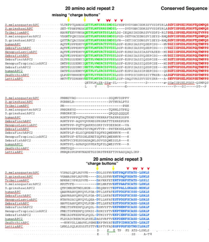
without phosphorylation. Despite the inability of 20R2 to bind βcat, the divergent sequence of 20R2 is as or better conserved between flies and mammals than are high-affinity binding sites like 20R3 (Figure 2.7). This prompted us to explore whether 20R2 is important for βcat
regulation. Given its inability to bind βcat, we hypothesized it would be dispensable. To our surprise, however, removing 20R2 (APC2ΔR2; Figure 2.1B) completely blocked APC2's ability to rescue βcat destruction in SW480 cells (Figure 2.8, A and D). APC2ΔR2 could retain βcat in the cytoplasm (Figure 2.8A, compare arrowheads), however, consistent with the presence of numerous βcat-binding sites, and thus it partially reduced TCF-regulated transcription (Figure 2.8E). In this it contrasted with APC2Δ15Δ20, which could not retain βcat in the cytoplasm (Figure 2.8C, arrowhead) or reduce TCF-regulated transcription (Figure 2.8E).
We next assessed APC2ΔR2 in the animal. Consistent with our observations in SW480 cells, APC2ΔR2 could not rescue Arm destruction in APC2 APC1 double mutants (Figure 2.7L), and it also provided no cell fate rescue there (Figure 2.9, B and K). APC2ΔR2 retained
substantial ability, however, to rescue cell fates, embryonic lethality, and adult viability of APC2 single mutants (Figure 2.9, A and D), correlating with it retaining function in cytoplasmic
retention. Thus 20R2 is essential for the ability of the destruction complex to target βcat for destruction, but is dispensable for βcat retention.
full-length APC2 (APC2ΔB; Figure 2.1B) also abolished its ability to target βcat for destruction in SW480 cells (Figure 2.8, B and D). Deleting sequence B, however, did not disrupt cytoplasmic retention (Figure 2.8B, compare arrowheads), and thus APC2ΔB retained the ability to partially reduce βcat-dependent transcription (Figure 2.8E). When tested in the animal, APC2ΔB
behaved similarly to APC2ΔR2. It could not rescue Arm destruction (Figure 2.7M), cell fates, or embryonic lethality in APC2 APC1 double mutants (Figure 2.9, B and L). APC2ΔB retained, however, substantial ability to rescue cell fates, embryonic lethality, and adult viability of APC2 single mutants (Figure 2.9, A and E; Figure 2.6E), consistent with it retaining function in
cytoplasmic retention. Thus these two adjacent conserved APC motifs, 20R2 and sequence B, play essential roles in βcat destruction.
Sequence B and 20R2 are among the most highly conserved sequences in APC proteins. We thus tested whether they play important roles in human APC, deleting them from a minimal fragment that rescues βcat regulation in SW480 cells (hAPC2.8kb; Rubinfeld et al., 1997a blue right-pointing triangle). This fragment spans from 20R1 through the end of the SAMPs (Figure 2.10A), and rescues βcat destruction in SW480 cells (Figure 2.10B). We generated variants lacking 20R2, 20R3, or sequence B (Figure 2.10A). Deleting either 20R2 (hAPC2.8kbΔR2; Figure 2.10C) or sequence B (hAPC2.8kbΔB; Figure 2.10D; see also Kohler et al., 2009 blue