Ozone-derived Oxysterols Affect Liver X Receptor (LXR)
Signaling
A POTENTIAL ROLE FOR LIPID-PROTEIN ADDUCTS
*Received for publication, April 18, 2016, and in revised form, September 14, 2016 Published, JBC Papers in Press, October 4, 2016, DOI 10.1074/jbc.M116.732362
Adam M. Speen‡, Hye-Young H. Kim§, Rebecca N. Bauer‡,XMegan Meyer‡,X Kymberly M. Gowdy¶, Michael B. Fessler储, Kelly E. Duncan‡, Wei Liu§, Ned A. Porter§, andX Ilona Jaspers‡1
From the‡Curriculum in Toxicology, Departments of Pediatrics and Microbiology and Immunology, Center for Environmental Medicine, Asthma, and Lung Biology, University of North Carolina, Chapel Hill, North Carolina 27599, the§Department of Chemistry and Center for Molecular Toxicology, Vanderbilt University, Nashville, Tennessee 37235, the¶Department of Pharmacology and Toxicology, Brody School of Medicine, East Carolina University, Greenville, North Carolina 27834, and the储Immunity, Inflammation, and Disease Laboratory, NIEHS, National Institutes of Health,
Research Triangle Park, North Carolina 27709
Edited by Dennis Voelker
When inhaled, ozone (O3) interacts with cholesterols of air-way epithelial cell membranes or the lung-lining fluid, generat-ing chemically reactive oxysterols. The mechanism by which O3-derived oxysterols affect molecular function is unknown. Our data show thatin vitroexposure of human bronchial epi-thelial cells to O3results in the formation of oxysterols, epoxy-cholesterol-␣and -and secosterol A and B (Seco A and Seco B), in cell lysates and apical washes. Similarly, bronchoalveolar lavage fluid obtained from human volunteers exposed to O3 contained elevated levels of these oxysterol species. As expected, O3-derived oxysterols have a pro-inflammatory effect and increase NF-B activity. Interestingly, expression of the choles-terol efflux pump ATP-binding cassette transporter 1 (ABCA1), which is regulated by activation of the liver X receptor (LXR), was suppressed in epithelial cells exposed to O3. Additionally, exposure of LXR knock-out mice to O3enhanced pro-inflamma-tory cytokine production in the lung, suggesting LXR inhibits O3-induced inflammation. Using alkynyl surrogates of O3 -de-rived oxysterols, our data demonstrate adduction of LXR with Seco A. Similarly, supplementation of epithelial cells with alky-nyl-tagged cholesterol followed by O3exposure causes observ-able lipid-LXR adduct formation. Experiments using Seco A and the LXR agonist T0901317 (T09) showed reduced expression of ABCA1 as compared with stimulation with T0901317 alone,
indicating that Seco A-LXR protein adduct formation inhibits LXR activation by traditional agonists. Overall, these data demonstrate that O3-derived oxysterols have pro-inflammatory functions and form lipid-protein adducts with LXR, thus lead-ing to suppressed cholesterol regulatory gene expression and providing a biochemical mechanism mediating O3-derived for-mation of oxidized lipids in the airways and subsequent adverse health effects.
Currently, the oxidant gas ozone (O3) is the most widespread air pollutant found in the United States and contributes to a growing variety of adverse health outcomes (1). O3exposure causes decreased lung function and increased airway inflamma-tion, which exacerbates pre-existing diseases such as asthma and may contribute to certain cardiovascular diseases, all potentially increasing the risk of premature death (2). Although the inflammatory response and adverse health effects of O3 exposure have been documented for decades, the biochemical and cellular mechanisms by which O3mediates adverse health effects remain poorly understood. As a very potent oxidant gas, O3reacts readily with the surface components of the airway and causes cellular modification through reactions with the airway-lining fluid and epithelial cellular membranes (2– 4). The lung-lining fluid and epithelial cell membranes are rich in cholesterol and other lipids, which can be directly oxidized by O3(5, 6). Previous studies have determined the impact that ozonization of lipids, particularly polyunsaturated fatty acids (PUFA), may have on O3-associated toxic effects. These studies demon-strated that ozonization of PUFAs and the formation of lipid ozonization products can mimic many of the adverse health effects observed after exposure to O3(5, 7–9). However, the involvement of cholesterol ozonization products requires fur-ther study. The cholesterol 5,6-double bond and concomitant vinylic methylene group moieties are particularly susceptible to oxidation, resulting in the formation of oxysterols (5, 10 –12).
Many oxysterols are electrophiles capable of reacting with nucleophilic residues on proteins and other biological macromolecules. They are formed endogenously through enzy-*This work was supported in part by the United States Environmental
Pro-tection Agency through cooperative agreement CR83346301 with the Center for Environmental Medicine, Asthma and Lung Biology at the Uni-versity of North Carolina at Chapel Hill (to I. J. and K. E. D.) and National Institutes of Health Grants T32ES007126 (to A. M. S. and R. N. B.) and R21ES024666 (to H. Y. K., N. A. P., and I. J.). The authors declare that they have no conflicts of interest with the contents of this article. This article has not been formally reviewed by EPA. The views expressed in this document are solely those of the authors and do not necessarily reflect those of the Agency. EPA does not endorse any products or commercial services men-tioned in this publication. The content is solely the responsibility of the authors and does not necessarily represent the official views of the National Institutes of Health.
1To whom correspondence should be addressed: Dept. of Pediatrics,
Microbiology and Immunology, and Environmental Sciences and Engi-neering, University of North Carolina at Chapel Hill, 104 Mason Farm Rd., Chapel Hill, NC 27599. Tel.: 919-966-8657; Fax: 919-966-9863; E-mail: [email protected].
crossmark
matic and non-enzymatic reactions. P450 cytochrome enzymes metabolize endogenous cholesterol into oxysterol species such as 27-hydroxycholesterol, an essential oxysterol for cellular cholesterol homeostasis (13). Studies show that endogenous oxysterols can act as both agonists and antagonists for tran-scription factors such as the liver X receptor (LXR).2Within the LXR protein, a ligand binding domain has been characterized and shown to bind to synthesized oxysterol speciesin vitro, which is the same region that is the target for various post-translational modifications such as acetylation and SUMOyla-tion (14). LXR regulates the synthesis of cholesterol trans-port proteins, such as ATP-binding cassette transtrans-porter A1 (ABCA1). ABCA1 transports cholesterol across the cell mem-brane and shuttles it onto apolipoproteins, resulting in the for-mation of high density lipoproteins (HDL). Systemically, failure to produce adequate ABCA1 reduces the ability to generate and transport HDL and is associated with early onset cardiovascular disease, supporting the protective role of ABCA1 in cholesterol metabolism and atherosclerosis (15). In addition to lipid metab-olism, both LXR and ABCA1 have been implicated in anti-in-flammatory functions (16 –19). Moreover, exogenously derived oxysterols can cause adverse cellular effects and have been shown to modulate host immune cell and inflammatory responses (20 –22). Oxidation of endogenous cholesterols by various reactive oxygen species results in the formation of oxy-sterols associated with various pathological processes driven by interaction with the nucleophilic domains of key proteins (23). Elevated levels of specific oxysterol species have been linked to a variety of adverse biological activities, including cytotoxicity, increased inflammation, and amplified infection (24 –26).
Rodent exposure studies andin vitrostudies using human pulmonary surfactant have demonstrated the formation of O3-derived oxidized cholesterol products in various chemical
compositions (5, 6, 10, 27, 28). Fig. 1 depicts the primary reac-tive O3-derived oxysterol products secosterol A (Seco A) along with its aldol condensation product secosterol B (Seco B), and epoxycholesterols ␣ and  (␣-EpCh and -EpCh) (11, 29). Some of these O3-derived oxysterols have been found in ath-erosclerotic tissue and cause foam cell formation, a hallmark of plaque buildup (26). Despite increased interest in the role of oxysterols in human health, especially cardiovascular diseases, information about O3-derived oxysterols in the airway, their cellular targets, and their biochemical interactions is very limited.
We hypothesize that exposure to O3generates lipid-derived electrophiles in the human airway, including reactive species of oxysterols, at or near the epithelial surface that can adduct to cellular proteins, thus ultimately affecting cellular function and inflammatory response. Our study is designed to uncover elec-trophilic interactions between O3-derived oxysterols and pro-teins as a new mechanistic paradigm mediating adverse health effects induced upon inhalation of O3.
Results
Identification of O3-induced Oxysterol Formation in Vitro
and in Vivo—Primary differentiated HBEC cells and 16HBE cells were chosen to evaluate the presence of oxysterol species
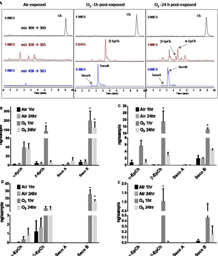
in vitrofollowing exposure to O3. Representative HPLC-MS profiles reflected increased levels of oxysterol species in O3 -ex-posed 16HBE cells (Fig. 2A) compared with filtered air-exposed cells. 16HBE cells exposed to O3exhibited elevated concentra-tions of␣-EpCh,-EpCh, and Seco B in cell lysates (Fig. 2B) and in the apical washes (Fig. 2C) collected both 1 and 24 h post-exposure compared with the air-exposed control. A similar trend was observed in primary HBEC lysates (Fig. 2D) and api-cal washes (Fig. 2E) at both 1 and 24 h post-exposure compared with air-exposed controls. Overall, the cholesterol concentra-tion did not significantly change due to O3exposure or sample collection time in either 16HBE (4870⫾597 ng/sample air to 5168 ⫾ 138 ng/sample O3) or primary HBE (50121⫾ 1554 ng/sample air to 5506⫾772 ng/sample O3) cells.
To test the formation of O3-derived oxysterols in humansin vivo, we determined the levels of oxysterols identified in BALF obtained from healthy volunteers exposed to filtered air or 0.3 ppm O3for 2 h. The BALF was collected by bronchoscopy at 1 and 24 h post-exposure, and various oxysterol species were 2The abbreviations used are: LXR, liver X receptor;␣-EpCh and-EpCh,
epoxycholesterol-␣ and -; BisTris, 2-[bis(2-hydroxyethyl)amino]-2-(hy-droxymethyl)propane-1,3-diol; qPCR, quantitative PCR; ANOVA, analysis of variance; LXRE, LXR response element; BALF, bronchoalveolar lavage fluid; HBEC, human bronchial epithelial cell; PPAR, peroxisome proliferator-acti-vated receptor; HBBS, Hanks’ balanced salt solution; EPA, Environmental Protection Agency; Seco, secosterol; BHT, butylated hydroxytoluene; TPP, triphenylphosphine; T09, T0901317;a-Chol, alkynyl-tagged cholesterol; qPCR, quantitative PCR;a-Seco, alkynyl-Seco; AMBIC, ammonium bicarbonate; MEM, minimal essential medium; FAM, 6-carboxyfluorescein; TAMRA,
6-carboxy-N,N,N⬘,N⬘-tetramethylrhodamine; 7-DHC,d7-7-dehydroxycholesterol.
FIGURE 1.Cholesterol and the major oxysterols formed in the reaction with ozone.
quantified by HPLC-MS. Exposure to O3significantly elevated the concentrations of-EpCh (Fig. 3B) and Seco A (Fig. 3C) compared with individuals exposed to filtered air controls at 1 h
post-exposure withpvalues of less than 0.001 and 0.05 respec-tively. A moderately convincing increase (p⬍0.1) was observed for ␣-EpCh (Fig. 3A) and no difference in Seco B (Fig. 3D).
FIGURE 2.Oxysterol concentrations measured in cells and apical washes exposed to filtered air or O3and their HPLC-MS profiles.All cells were grown in 24-mm Transwell membrane plate until confluency followed by removal of the apical medium and exposure to filtered air or 0.4 ppm O3for 4 h. Cell lysates
and apical washes were collected at 1 and 24 h post-exposure.A,representative reverse phase-HPLC-multiple reaction monitoring chromatograms of epithe-lial cells exposed to air or 0.4 ppm ozone. MS for each panel is selected reaction monitoring of them/zindicated in the air exposure.B,16HBE cell lysate.C,16HBE apical wash.D,primary HBEC cell lysate.E,primary HBEC apical wash. Data are presented as means⫾S.E. Statistical analysis was performed with a one-way ANOVA and Fisher’s LSD post hoc test comparing observed means against the respective air-exposed control. *,p⬍0.05.n⫽3.
Unlike the results observed in thein vitrosamples, we observed that oxysterol concentrations returned to baseline levels 24 h post-exposure. Again, there was no significant change in overall BALF cholesterol concentration in subjects exposed to air (6639⫾1556 ng/ml) compared with O3(6698⫾2245 ng/ml).
Effects of O3on Cholesterol Efflux and Cytokine Gene
Ex-pression—As expected and previously described by us (30, 31), exposure to O3caused an inflammatory response as marked by increased gene expression (Fig. 4,AandB) and protein con-centration (Fig. 4,CandD) of pro-inflammatory cytokines IL6 and IL8. Treatment with synthetic LXR agonist T0901317 (T09) alone did not affect the levels ofIL6orIL8 expression (Fig. 4,AandB). Interestingly, exposure to O3in the presence of T09 significantly decreased the expression of cholesterol efflux pump proteinsABCG1(Fig. 4E) andABCA1(Fig. 4F), suggest-ing O3-induced inhibition of the LXR pathway. Additionally, ABCA1 protein levels were significantly reduced in cells exposed to O3compared with air as shown in relative densi-tometry from three separate experiments (Fig. 4G) and repre-sentative immunoblot (Fig. 4H).
LXR-␣⫺/⫺Mice Are More Susceptible to O
3-induced
Inflam-mation—Female LXR␣⫺/⫺ were compared with WT mice. Each were exposed to filtered air or 2 ppm O3for 3 h. BALF was collected from both conditions and evaluated for concentra-tions of IL6. The LXR-␣⫺/⫺mice showed significantly higher concentrations of IL6 compared with the WT (Fig. 5A). At necropsy, the lungs were removed and homogenized for RNA isolation and qPCR analysis. In a similar result, IL6 mRNA lev-els were significantly higher in the LXR-␣⫺/⫺mice exposed to
O3compared with the WT control mice exposed to O3(Fig. 5B). As seen in the human BALF samples (Fig. 3,CandD), exposure to O3increases the amount of Seco A (Fig. 5C) and Seco B (Fig. 5D) in the mouse BALF samples with significantly higher amounts of Seco A present in the LXR-␣⫺/⫺
com-pared with the WT mice. Interestingly, unlike the human
samples, total cholesterol levels decreases in O3-exposed animals (Fig. 5E).
Exposure to a-Seco A Reveals LXR-␣Protein Adducts—Based on recent studies demonstrating oxysterol-induced lipid-pro-tein adducts, we hypothesized that O3-derived oxysterols can adduct to the LXR-␣and LXR-proteins, which contain reac-tive lysine residues (14, 29). Seco A, a primary ozonide and one of the most reactive O3-derived oxysterols, reacts with proteins and causes covalent modifications leading to lipid-protein adduct formation (32). 16HBE cells were treated with alkynyl-tagged Seco A (a-Seco A), and the adducted proteins were iden-tified by first adding biotin to the alkynyl tag utilizing click cyclo-addition (Fig. 6A) followed by immunoaffinity purifica-tion with streptavidin beads and photo-release (Fig. 6B), which frees cellular proteins adducted bya-Seco A. This mixture of proteins was subsequently subjected to Western blotting anal-yses using antibodies against HSP90, LXR-␣, LXR-, PPAR-␥, and-actin (Fig. 6C). HSP90 was used as a positive control based on previous studies (33). Our data show that treatment of bronchial epithelial cells witha-Seco A results in the formation of Seco A-LXR adducts.
Alkynyl Cholesterol-supplemented Cells Exposed to O3Reveal
the Formation of LXR-␣Adducts—16HBE cells were supple-mented with 20Malkynyl-tagged cholesterol (a-Chol) for 6 days. Media containinga-Chol changed daily with washes on the apical side 24 h prior to exposure (Fig. 7B). The 16HBE cells were exposed to filtered air or 0.4 ppm O3for 4 h and harvested 1 h post-exposure. Our data show thata-Chol, a surrogate of endogenous cholesterol, is incorporated into the cells and, in response to O3,generates a host of alkynyl-oxysterols identical to the oxysterol species generated via oxidation of endogenous cholesterol (Fig. 7A). Based on streptavidin analysis, cells exposed to O3exhibit a higher overall level of protein adduction with oxidized cholesterol species than cells exposed to air (Fig. 7C). Similar to cells treated witha-Seco A, HSP90 and LXR-␣
FIGURE 3.Oxysterol concentrations measured in airway BALF.Healthy individuals were exposed to either filtered air (control) or 0.3 ppm O3for 2 h. Cell-free
BALF was collected by bronchoscopy at 1 and 24 h post-exposure. Oxysterols were quantified by HPLC-MS spectrometry.A,␣-EpCh.B,-EpCh.C,Seco A.D,
Seco B. Data are presented as means⫾S.E. Statistical analysis was performed with paired Student’sttest (two-tailed distribution, pairing based on subject). *,
p⬍0.05; **,p⬍0.001.n⫽9 –11.
were found to be targets of alkynyl-tagged oxysterols generated endogenously in cells exposed to O3(Fig. 7D). The broad band of LXR antibody-reactive proteins shown in Fig. 7D(Elute⫹O3) suggests that a set of O3-derived adducts are formed under conditions of endogenous oxysterol genesis. This is in contrast to the homogeneous protein adduct observed after exposure to a single exogenous electrophile, as shown in Fig. 6C for the experiment with a-Seco A. Fig. 7E shows the densitometric analysis of O3-derived adducts of LXR-␣from three separate experiments.
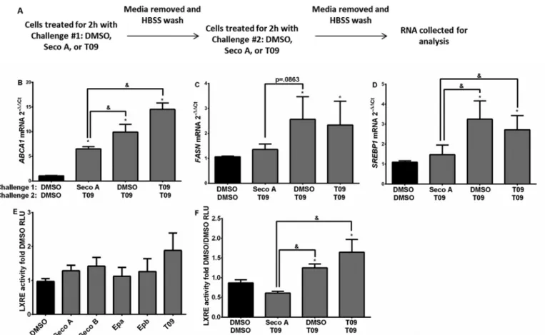
Oxysterol Exposure Alters ABCA1, FASN, and SREBP1 Gene Transcription—Considering the effects of oxysterols on adduct formation with LXR, we evaluated whether exposure to oxyste-rols can alter expression levels of genes controlling cholesterol biosynthesis, fatty acid synthesis, and cholesterol efflux in 16HBE cells. To determine whether oxysterol adduct formation with LXR inhibits subsequent activation of LXR, we sequen-tially treated 16HBE cells with DMSO, Seco A, or T09 for 2 h, removed the first treatment, and followed with a second chal-lenge for 2 h (Fig. 8A). Treatment with Seco A followed by T09 significantly suppressedABCA1(Fig. 8B),FASN(Fig. 8C), and
SREBP1(Fig. 8D) gene expression compared with cells exposed to DMSO followed by T09 and T09 followed by T09, suggesting that initial treatment with Seco A reduces the ability of T09 to enhance cholesterol synthesis and efflux gene expression. Chal-lenge with individual oxysterol species yielded no significant change in LXRE activity when compared with the DMSO vehi-cle control (Fig. 8E). However, LXRE activity was significantly reduced in cells sequentially treated with Seco A followed by T09 compared with cells sequentially treated with DMSO fol-lowed by T09 or T09 folfol-lowed by T09 (Fig. 8F), again suggesting that initial treatment with Seco A reduces the ability of T09 to activate LXR.
Exposure to Individual Oxysterols Activates the Inflamma-tory Gene Transcription Pathway—To determine whether O3-derived oxysterols modify expression of inflammatory genes alone and in relation to known agonists, 16HBE cells were sequentially treated with DMSO, Seco A, or T09 for 2 h fol-lowed by a second challenge for 2 h as described above (Fig. 8A). Exposure to Seco A followed by T09 increased the expression of
IL6 (Fig. 9A) and IL8 (Fig. 9B) compared with the DMSO-treated control. Additionally, individual O3-derived oxysterol
FIGURE 4.Cholesterol efflux pump protein and pro-inflammatory cytokine gene expression levels in 16HBE cells.Cells were exposed to filtered air or 0.4 ppm ozone for 4 h with and without 10MT09. RNA and apical wash samples were collected 1 h post-exposure and analyzed. Inflammatory gene expression
levelsIL6(A),IL8(B), and protein levels IL6 (C) and IL8 (D) as well as gene expression levels forABCG1(E) andABCA1(F) were measured. Additionally, ABCA1 protein levels were evaluated by immunoblot (G) and relative densitometry (H) from three separate experiments. Data are presented as means⫾S.E. Statistical analysis was performed with a one-way ANOVA and Fisher’s LSD post hoc test comparing observed means against the respective treatment/air-exposed control, *,p⬍0.05, or against the DMSO/air-exposed vehicle control, #,p⬍0.05,n⫽3.
species significantly increased the NF-B activity compared with the DMSO control (Fig. 9C), suggesting that O3-derived oxysterols enhance pro-inflammatory pathways.
Discussion
Despite a large body of research on O3-induced toxicity and adverse health effects, the biochemical and cellular signaling cascades induced by O3exposure in the human airways are not fully understood. O3is a known oxidant of the macromolecules in the airway-lining fluid, and the formation of many oxidized lipid products has been previously described along with their biochemical effects. For example, instillation of 5,6 -epoxy-cholesterol (-EpCh) and 1-palmitoyl-2-(9⬘ -oxo-nonanoyl)-glycerophosphocholine, which are both formed during O3 exposure, causes neutrophilic influx, which in turn was regu-lated by class A scavenger receptors, known to bind oxidized lipids (3). Ozonization of phospholipids generates products with strong pro-inflammatory effects (8, 9). Additionally, O3is known to react with lung surfactant proteins, modifying and inhibiting their normal biological functions (34 –36). O3 -in-duced oxidized cholesterol products, however, have only par-tially been described in their effects on the biological properties of the human airway. Through this study, our results address the knowledge gap between O3-induced cholesterol oxidation in the airway and downstream cellular signaling events. Our data demonstrate that O3-derived oxysterols can form lipid-protein adducts with cellular signaling molecules, revealing a new paradigm that lipid-protein adduct formation provides a
central mechanism for O3-derived oxidized lipids to modify cellular responses.
Lipid protein adduction as a means to alter normal signaling has been shown to occur during exposure to other oxidative stressors, including the free radical-induced formation of 4-hy-droxynoneal (37, 38). Although formation of oxysterols follow-ing exposure to O3has been shown before (28, 35), whether and how these O3-derived oxysterols can form oxysterol-protein adducts in the context of O3exposure in the human airway has not been examined. Assays based on “click” cyclo-addition methods make it possible to visualize protein-lipid adducts via immunostaining and to determine protein adductions with specific signaling molecules, thus providing novel insight into the mechanisms behind O3-induced adverse health effects (39). Using this approach, our results demonstrate the formation of oxysterol-protein adducts in airway epithelial cells as a result of exposure to O3. The synthetic O3-derived oxysterol Seco A has previously been described as a highly reactive ozonolysis prod-uct capable of addprod-ucting with the lysine amino acid residues on various proteins (29). Throughout our study, we determined that O3-derived oxysterols, including Seco A, can effectively form adducts with proteins LXR-␣, LXR-, and PPAR-␥, which all regulate lipid metabolism (40, 41). Additionally, supplemen-tation with alkynyl-tagged cholesterol reveals that in the human airway exposure to O3generates reactive oxysterols capable of forming adducts with the LXR-␣ and HSP90 proteins. The comparatively diffuse band observed in Fig. 7Dand
quantita-FIGURE 5.IL6 expression and production in the lung of O3-exposed LXR␣-deficient mice.Wild type (WT) and LXR-␣⫺
/⫺female mice were exposed to filtered air (Air) or 2 ppm O3for 3 h. Mice were necropsied 6 h after start of exposure.A,bronchoalveolar lavage was collected and evaluated for IL6 cytokine
concentrations by ELISA.B,after necropsy, lung tissues were collected for RNA isolation and evaluated for gene expression of IL6 and 18S. Bronchoalveolar lavage was also evaluated for total Seco A (C), total Seco B (D), and total cholesterol (E). Data were presented as means⫾S.E. Statistical analysis was performed with Mann-Whitney test comparing LXR-␣⫺/⫺to WT, *,p⬍0.05,n⫽5 animals per group.
tively analyzed in Fig. 7Eis most likely indicative of a heteroge-neous mixture of O3-derived oxysterols being formed that can adduct LXR and of a variety of post-translational modifications to LXR-␣ in addition to a-Seco A adduction resulting in a mixture of proteins recognizable by the anti-LXR-␣antibody. Hence, data presented here reveal a novel biochemical mecha-nism involving the conversion of cellular cholesterol by O3into reactive oxysterol species, a process that results in the adduc-tion of key proteins and modificaadduc-tion of cellular signaling.
The formation of adducts between O3-derived oxysterols and LXR provides a model signaling mechanism, which results in modifications of airway inflammatory and cholesterol home-ostasis signaling. Endogenous oxysterols are known to be formed during normal cholesterol regulation and metabolism. These compounds play a role in normal LXR activation, pro-moting cholesterol homeostasis. Mouse model systems with LXR and cholesterol efflux deficiencies have higher cholesterol accumulation, potentiated atherosclerosis, and are more sus-ceptible to systemic infection (42). These effects occur partially via the endogenous oxysterol-induced activation of LXR and transcriptional activation of genes such asABCA1, which medi-ate cholesterol shuttling and ultimmedi-ately packaging into HDL (43, 44). In contrast, our data show that in human bronchial epithelial cells, O3 exposure decreases the expression of
ABCA1, suggesting that O3or its oxidation products have an
inhibitory effect on the transcriptional activation ofABCA1. DecreasedABCA1expression and subsequent decreased lesterol efflux may result in accumulation of intracellular cho-lesterol leading to dyslipidemia and propagation of respiratory, metabolic, and cardiovascular health problems (13, 45). Whether the observations related to O3-induced suppression of
ABCA1expression affect systemic cholesterol metabolism and potentially contribute to the enhanced atherosclerosis and car-diovascular events associated with O3exposure remain to be established. In addition to cholesterol homeostasis, previous studies have indicated that normal LXR activation is necessary for immune health. LXR activation by synthetic agonists leads to decreased neutrophil recruitment and increased bacterial burden, indicating that LXR activation balance contributes to host immune defense (46). Recent studies also suggest that LXR agonists show therapeutic promise in treating lung disorders such as dyslipidemia and asthma (43, 47, 48). Furthermore, our data show that O3exposure of LXR␣⫺
/⫺mice results in
enhanced pro-inflammatory response, indicating the impor-tance of normal LXR␣activity to regulate O3-induced inflam-mation (Fig. 5,AandB). The enhanced concentration of O3 -de-rived oxysterols in the bronchoalveolar lavage sampled from the LXR␣⫺/⫺mice further link airway cholesterol ozonolysis and LXR␣activity. It is interesting that overall cholesterol levels were reduced in these mice exposed to O3, a change we did not
FIGURE 6.Simplified steps of protein catch and photo-release usinga-Seco A probe in 16 HBE cells.Supplementation witha-Seco A (20M) in 16HBE cells for 4 h at 37 °C followed by cell lysis and click reaction to introduce biotin for immunoaffinity purification. A single aliquot was taken to probe the extent of alkyne labeling via biotin-streptavidin interaction. The rest of the clicked cells were immobilized onto streptavidin beads to catch only adducted proteins and remove un-adducted proteins. Subsequent photo-release permitted us to collect adducted proteins.A,IRDye姞800CW streptavidin visualization exhibits the extent of protein labeling witha-Seco A.B,SDS-polyacrylamide gel of photo-released proteins. An equal amount of total protein was loaded. Thea-Seco A-treated cells (right 2 lanes) exhibited significant protein adduction compared with the input lanes (left 2 lanes).C,selective antibody analyses indicate adduction on HSP90, LXR-␣, LXR-, and PPAR-␥proteins. Anti-actin of the input lane provides another confirmation of equal loading. Representative blots are from three separate experiments (n⫽3).
observe in our human subjects. Taken together, LXR dysfunc-tion caused by O3-derived oxysterols may contribute to enhanced respiratory inflammation and adverse cardiovascular health effects in humans exposed to O3.
Based on our observation of adduct formation between O3-derived oxysterols and LXR, we hypothesize that electro-philic O3-induced oxysterols and their derived lipid protein adducts inhibit cholesterol signaling pathways that are nor-mally activated by endogenous oxysterols. Our data in Fig. 8E
show that O3-derived oxysterols alone are not themselves potent activators of LXR but that treatment with Seco A prior to activation of LXR by the agonist T09 reduces T09-induced
LXRE activation as well as expression of ABCA1, FASN,and
SREBP1. These data suggest that Seco A-induced adduct for-mation with LXR may prevent its activation with known ago-nists, such as T09, or endogenous oxysterols, such as 27-hy-droxycholesterol (49). Our previous studies have demonstrated that Seco A readily adducts to nucleophilic lysine amino acid residues in human serum albumin and other proteins, and we hypothesize a similar binding site in LXR (29). LXR contains eight lysines in a ligand binding domain (amino acids 215– 434) that are capable of binding both oxysterol and T09 ligands (14). Based on our results, we propose that O3-derived oxysterols block T09-mediated LXR activation either by non-competitive
FIGURE 7.Protein adduct formation is observable in 20Ma-Chol-supplemented 16HBE cells exposed to O3.Followinga-Chol supplementation (20M), apical medium was removed, and the cells were exposed to clean air or 0.4 ppm O3for 4 h. Cells were harvested after 1 h of postincubation.A,lipids were
extracted as described in experiment. LC-selected reaction monitoring profile demonstrates thata-cholesterol was incorporated into the cells and generated ozone-derived oxysterols as those of endogenous cholesterol.B,schematic depictinga-Chol supplementation prior to O3exposure.C,cells harvested in lysis
buffer containing inhibitors were clicked with azido-biotin. IRDye姞800CW streptavidin probe displays significant extent of protein adduction upon O3
-ex-posed cells, whereas the background levels of adduction with air-ex-ex-posed cells were observed. Anti-actin is served as a loading control.D,HSP90 and LXR-␣
Western blots of click and photo-released proteins. 16HBE cell lysates were loaded as a positive control.E,relative densitometry analysis of LXR-␣protein levels from three separate immunoblots (n⫽3).
binding to the same site or by modifying the LXR structure to inhibit normal ligand binding. Additional studies are necessary to clarify the nucleophilic peptide site of O3-derived oxysterol LXR adduct formation.
In contrast to expression ofABCA1, expression ofIL6and
IL8as well as activation of NF-B were enhanced by exposure to Seco A and other O3-derived oxysterols. O3is known to activate NF-B leading to increased transcription of inflammatory cyto-kines such as IL6 and IL8(31, 50, 51). Similarly, other lipid ozonization products have been shown to activateIL6andIL8
in human airway epithelial cells (9). Our study shows that O3-derived oxysterols enhance activation of NF-B and poten-tiate the transcription ofIL6andIL8. LXR signaling has been shown previously to have an inhibitory effect on NF-B and inflammatory signaling in epithelial cells (52, 53), making it a reasonable target for O3-derived oxysterol-induced modifica-tion of inflammatory signaling. All the O3-derived oxysterol species we tested increased NF-B activity, revealing that in their presence airway epithelial cells may experience increased inflammatory signaling leading to adverse health effects. In the
FIGURE 8.Seco A-altered cholesterol efflux pump gene expression when treated with Seco A and the T09 LXR agonist in sequence.A,experimental design depicting how 16HBE cells were challenged for 2 h to 20MSeco A, 10MT09, or DMSO control for first challenge; media were removed and followed by 2 h of second challenge. Samples were evaluated forABCA1(B),FASN(C),andSREBP1(D) gene expression. LXRE activity was measured in relative luciferase units (RLU) compared with the respective vehicle control in the individual oxysterol challenges at 20Mfor 4 h (E) and sequential treatment in 16HBE cells (F). Data are presented as mean⫾S.E. of fold change compared with the DMSO control. Statistical analysis was performed with a one-way ANOVA and Fisher’s LSD post hoc comparison test, * significantly different from the DMSO control;&significantly different from the indicated conditions,p⬍0.05,n⫽3.
FIGURE 9.Oxysterol altered inflammatory signaling.IL6(A) andIL8(B) gene expression in sequential challenge of 2 h with first challenge followed by another 2 h of second challenge on 16HBE cells exposed to 20Mof Seco A, 10MT09, or DMSO control on collagen-coated plates.C,relative light units of NF-B
promoter reporter activity in 16HBE cells exposed to 20Mof various oxysterols, 20 ng/ml TNF-␣, or DMSO negative control for 4 h on collagen-coated plastic.
Data are presented as mean⫾S.E. of fold-change compared with the DMSO control. Statistical analysis was performed with a one-way ANOVA and Fisher’s LSD post hoc comparison test, *,p⬍0.05,n⫽3.
experiments using sequential stimulation with Seco A and T09, stimulation with Seco A alone, regardless of the secondary stimulus, increases IL6 and IL8 expression, indicating that O3-derived oxysterols potentiate the transcription of pro-in-flammatory cytokines. In addition to directly activating the NF-B pathway, we hypothesize that this effect may be linked to inactivation of LXR. Activation of LXR induces a number of anti-inflammatory responses, marked by inhibition of NF-B activity and decreased production of pro-inflammatory media-tors (54). Previous rodent studies show the use of LXR agonists to inhibit NF-B activation to reduce lung injury and inflam-mation after hemorrhage and resuscitation (55). In this role, LXR is not recruited to LXR response elements in genes related to lipid metabolism, but it does participate in a transrepression mechanism of NF-B, resulting in inhibition of inflammatory genes such asIL6(42, 56, 57). LXR activity is controlled by a post-translational SUMOylation modification and interaction with nuclear receptor co-repressors, reducing NF-B activity (58 – 60). The proposed SUMOylation occurs at the lysine res-idues of LXR ensuring retinoid X receptor dimerization and nuclear translocation (60). Thus, lipid-adduct formation at or near these lysine residues may prevent SUMOylation of LXR, interaction with co-repressors, and consequently activation of NF-B. This proposed signaling mechanism provides a link between our observed O3-derived oxysterol-induced changes to LXR signaling and a model for O3-induced NF-B activation. To enhance our understanding of O3-induced cardiovascular events and atherosclerosis, the biochemical mechanisms involving O3-derived oxysterols, adduct formation with LXR, and reducedABCA1expression that are reflected in systemic changes need to be examined. Interestingly, epidemiological studies repeatedly demonstrate the beneficial effects of choles-terol-modifying drugs, such as statins, on air pollutant-induced adverse health effects (61– 65). Yet the mechanisms mediating these effects are unknown and have often been ascribed to the anti-inflammatory effects of statins. The findings presented in this study provide an additional explanation for the beneficial effects of statins on the response to inhaled O3. Therapeutic treatment with lipid-targeting agents such as statins may reduce cholesterol availability in the airways, thus reducing oxysterol formation, and lead to changes in O3-induced inflammation.
Collectively, our results suggest that reactive oxysterols formed in the airways following O3exposure interact with the LXR signaling pathway, alter expression of genes that regulate cholesterol efflux, fatty acid synthesis, and cholesterol regula-tion, and enhance pro-inflammatory signaling pathways known to be associated with O3exposure. Our findings indicate that cholesterol availability is paramount to the formation of O3 -de-rived oxysterols in the airway and may provide a novel thera-peutic target to reduce the adverse effects associated with ambi-ent O3 exposure. In addition to LXR examined here, other proteins are also likely targets for adduction by O3-derived oxy-sterols and warrant future study into their implications in O3-induced airway inflammation. Together, our data describe a novel mechanistic concept linking O3reactions with choles-terol moieties in the human airway and its impact on health outcomes. Our data also highlight the use of samples derived
from humanin vivoO3exposure as well as primary HBECs and 16HBE cells to provide clinical and biological relevance for O3-derived oxysterol and their role in O3-induced inflamma-tion in the human airway.
Experimental Procedures
Reagents—Alkynyl probes were synthesized following the procedures published elsewhere (29, 32). DTT and iodoacet-amide were purchased from Sigma. Streptavidin beads were purchased from GE Healthcare. The following reagents were purchased from their respective companies: 10% NuPAGE Novex BisTris威precast mini gel (Invitrogen); PVDF membrane and Simply Blue (Bio-Rad); IRDye威800CW streptavidin (925-32230, Li-Cor, Lincoln, NE); Blocking buffer (Rockland, Gilbertsville, PA, or Odyssey Blocking buffer, Li-Cor); and sequencing grade trypsin (Promega V5111, Madison, WI). Antibodies of HSP90, LXR-, PPAR-␥, and actin were from Santa Cruz Biotechnology (Dallas, TX), and LXR-␣was pur-chased from Abcam威(Cambridge, MA).
Cell Culture—16HBE14o (16HBE) cells, an SV-40-trans-formed human bronchial epithelial cell line, were a gift from Dr. D. C. Gruenert (University of California at San Francisco). 16HBE cells were plated on fibronectin-coated (LHC Basal Medium (Life Technologies, Inc.), 0.01% BSA (Sigma), 1% Vit-ricol (Advanced Bio Matrix, San Diego), and 1% human fibronectin (BD Biosciences)) 0.4-m Transwell威 plates (Costar, Corning, NY) and grown submerged in minimal essen-tial media (Gibco) with 10% FBS, 1% penicillin-streptomycin, and 1%L-glutamine (Life Technologies, Inc.) until confluent for 6 days and 1 day at air-liquid interface before use. Primary HBECs were obtained from healthy donors in collaboration with the Environmental Protection Agency (EPA) using a pro-tocol approved by the University of North Carolina at Chapel Hill Institutional Review Board (Chapel Hill, NC), as described previously (66) The HBECs were cultured in PneumaCult medium for 21 days to differentiate according to previously described methods (67, 68).
In Vitro O3 Exposure—Cultures at the air-liquid interface were exposed to filtered air or 0.4 ppm O3for 4 h in exposure chambers operated by the United States EPA, as described pre-viously (69). The dose was selected for maximal innate immune response to O3with minimal cytotoxicity and has been used previously by our group (68). At 1 or 24 h after exposure, apical sides of all cultures were washed with 100l of Hanks’ balanced salt solution (HBSS) (Life Technologies, Inc.) and saved for LC-MS analysis. The remaining cells were collected in 200l of PBS, centrifuged at 500⫻gfor 5 min, and stored at⫺80 °C until LC-MS analysis.
In Vivo Exposure of Healthy Volunteers to O3—Written informed consent was provided by each participant. Healthy volunteers were randomly exposed to air and (in a separate exposure) 0.3 ppm O3for 2 h with exercise, with a minimum 2-week separation between exposures in collaboration with the EPA using a protocol approved by the University of North Car-olina Chapel Hill Institutional Review Board, as described pre-viously (70). Bronchoscopy was performed 1 and 24 h after exposure. Cell-free BALF was stored at⫺80 °C pending analy-sis (30).
Real Time qPCR—Total RNA was isolated from 16HBEs and HBECs with the use of the Pure Link RNA mini kit (Life Tech-nologies, Inc.). First strand cDNA preparation and real time qPCR were performed as described previously (71, 72). The following primers and probes for ABCA1, ABCG1, FASN, SREBP1, andACTBwere commercially available (Applied Bio-systems, Foster City, CA) and were prepared in-house: human
IL8, 5⬘ -FAM-CCTTGGCAAAACTGCACCTTCAC-TAMR-A-3⬘(probe), 5⬘-TTGGCAGCCTTCCTGATTTC-3⬘ (sense), and 5⬘-TATGCACTGACATCTAAGTTCTTTAGCA-3⬘ (antisense); andIL6,5⬘ -FAM-CCAGCATCAGTCCCAAGAA-GGCAACT-TAMRA-3⬘(probe), 5⬘ -TATGAAGTTCCTCTC-TGCAAGAGA-3⬘ (sense), and 5⬘ -TAGGGAAGGCCGTG-GTT-3⬘(antisense). Differences in expression were determined with the⌬⌬Ctmethod andACTBfor normalization. Briefly, threshold cycle (Ct) value for the housekeeping geneACTB) was subtracted from theCtvalue for the gene of interest to determine the⌬Ctvalue. For each pairwise set of samples to be compared, the difference in⌬Ctvalues between the two sam-ples were calculated for the genes of interest to determine the
⌬Ctvalue. The fold-change in gene expression was calculated as 2⫺⌬⌬Ct.
Sterol Profiling Using PTAD Derivatization and LC-selected Reaction Monitoring Analysis—Cells were scraped into 400l of cold PBS. 200l of the 400l were taken and internal stan-dards added (13 ng ford7-7-dehydroxycholesterol (d7-7-DHC), 97 ng for13C3-desmosterol, 99 ng for
13
C3-lanosterol, and 342 ng ford7-cholesterol/sample), 10l of butylated hydroxytolu-ene (BHT)/triphenylphosphine (TPP) solution (2.5 mg of TPP and 1 mg of BHT in 1 ml of MeOH), 400l of 1% NaCl, and 500 l of Folch solution (2:1⫽CHCl3/MeOH). Cells and standards were vortexed vigorously and centrifuged at 3099⫻gfor 5 min. CHCl3layer was removed and added to PTAD-predeposited tube (200g/tube). The sample tubes were vortexed and ana-lyzed by LC-MS using the following conditions: 10 l was injected onto the column (Acquity UPLC BEH C18, 1.7m, 2.1⫻50 mm) with 100% MeOH (0.1% acetic acid) mobile phase for a 1 min runtime at a flow rate of 300l/min. The monitored transitions included 7-DHC m/z5603365, d7-7-DHC m/z 567 3372, desmosterol m/z 592 3365, 13C
3-desmosterol
m/z5953368, lanosterolm/z6343602,13C
3-lanosterolm/z 637 3 605 with retention times of 0.8, 0.5, and 0.6 min, respectively.
Oxysterol Extraction from 16HBE or HBEC Cells—To the cell pellets 10l ofa-Chol (25 ng/l), 500l of NaCl (0.9%), 10l of TPP and BHT (25 mg of TPP and 10 mg of BHT in 10 ml of MeOH), and 1 ml of Folch solution (2:1⫽CHCl3/MeOH) were added. The mixture was mixed vigorously by vortex for 2 min and separated by centrifugation with 2300⫻gfor 3 min. The collected organic layer (bottom layer) was evaporated to dry-ness in a SpeedVacTMconcentrator and resuspended in 100l of MeOH for LC-MS analysis.
Oxysterol Extraction from Cell-free BALF—To 1 ml of BALF 10l of alkynyl-Seco B (a-Seco B) (50 ng/l), 1 ml of NaCl (0.9%), 10l of TPP and BHT (25 mg of TPP and 10 mg of BHT in 10 ml of MeOH), 2 ml of MeOH, and 3 ml of iso-octane were added. The mixture was vortexed vigorously for 2 min and sep-arated by centrifugation. The collected organic layer was
evap-orated to dryness in a SpeedVacTM concentrator and resus-pended in 100l of MeOH for LC-MS analysis.
LC-MS Analysis—The resuspended samples were chromato-graphed by reverse phase-HPLC using a UPLC BEH C18 col-umn (1.7m, 2.1⫻100 mm) in Waters Acquity UPLC system equipped with an autosampler (Waters, Milford, MA) and either electrospray ionization or atmospheric pressure chemi-cal ionization in positive ion mode. For electrospray ionization, the oxysterols were separated by 95% solvent B in an isocratic method with a flow rate of 200l/min, and the mobile phase solvents consisted of 2 mMNH4OAc (solvent A) in water and 2 mMNH4OAc in MeOH (solvent B). The injection volume was 10l using a partial loop with needle overfill mode. MS detec-tions were done using a TSQ Quantum Ultra tandem mass spectrometer (ThermoFisher, Waltham, MA), and data were acquired and analyzed using a Thermo XcaliburTM2.2 software package. The cholesterol and oxysterols form [M⫹NH4]⫹ions in positive ion mode (10). The transitions monitored werem/z
4363383 for Seco A/B,m/z4183365 fora-Seco A/B,m/z
4043369 for Chol,m/z3863351 fora-Chol,m/z4203385 for␣/EpCh, andm/z4023367 for␣/a-EpCh. For atmo-spheric pressure chemical ionization, 95% MeOH in H2O con-taining 0.01% acetic acid was used as a mobile phase. The cho-lesterol and oxysterols form [M ⫹ H]⫹ ions in positive ion mode. The transitions werem/z3693369 for Chol,m/z3833 383 for Seco A/B,m/z3513351 fora-Chol, andm/z3853 385 for␣/EpCh. The transitions of cholesterol and choles-terol esters werem/z3653365 monitored by HPLC-MS fol-lowing the method described elsewhere (73). The amount of the cholesterol esters were found to be less than 2% of free cholesterol in the cells studied.
Cytokine Analysis—Concentration of IL6 and IL8 in the api-cal wash of 16HBE cells exposed to O3 was determined by enzyme-linked immunosorbent assay (ELISA) according to the manufacturer’s instructions (BD Biosciences).
Western Blotting—Cell lysates from 16HBE cells exposed to air/O3or O3-derived oxysterols were separated by 10% SDS-PAGE and transferred to nitrocellulose. Proteins were detected using specific antibodies (Santa Cruz Biotechnology) to ABCA1 (1:500) or-actin (1–2000), which served as a loading control. Antigen-antibody complexes were incubated with horseradish peroxidase-conjugated secondary antibody and were detected using chemiluminescence.
Murine Whole Body O3Exposure, BAL, Cytokine and RNA
Analysis—C57BL/6J (WT) female mice, 8 –12 weeks old and weighing 15–20 g, were purchased from The Jackson Labora-tory (Bar Harbor, ME). LXR␣⫺/⫺female mice, 8 –12 weeks old
a humidified air supply to the chamber. Temperature and humidity of chamber air were monitored continuously, as was the O3concentration with a Teledyne T400 ultraviolet light photometer. BALF was collected immediately following sacri-fice, and cell counts were performed as described previously (74). Protein analysis was performed using BCA protein assay (Pierce). Cytokine analysis was performed using a Bioplex assay for IL6 on the BALF (Bio-Rad). After necropsy, lung tissue sam-ples were snap-frozen in liquid nitrogen and stored at⫺80 °C until RNA isolation. RNA was extracted and cDNA transcribed (Applied Biosystems, Foster City, CA). 50 ng of cDNA was used for qPCR for IL6 (Mm00446190_m1; Applied Biosystems) and endogenous 18S (4319413) (Applied Biosystems). Ct values were determined using ABI 7500 Real Time PCR System with SDS software version 1.3.1. Change in expression was calcu-lated using the 2⫺⌬⌬Ctmethod normalized to 18S expression and expressed as fold-change compared with the control group.
Cell Culture and Whole Cell Labeling with a-Seco A in 16HBE—16HBE cells were plated 2⫻106in 10-cm plates using the conditions described above and then allowed to settle and grow for 24 h. The cells were then incubated in the presence of
a-Seco A (20M) in reduced FBS (2%) MEM for 4 h. Cells were harvested in lysis buffer (50 mMHEPES, 150 mMNaCl, 0.1% Triton X-100, pH 7.0) containing protease inhibitors (Sigma P8340) on ice. The lysate was cleared by centrifugation at 10,000⫻gfor 10 min at 4 °C to remove cellular debris. The total protein concentration was determined using standard BCA assay (Pierce, ThermoFisher) for further click reaction.
Cell Culture and Whole Cell Labeling with a-Cholesterol Fol-lowed by Ozone Exposure in Human Bronchial Epithelial Cells (16HBE)—16HBE cells were grown in 24-mm transwells sub-merged in MEM with reduced FBS containing 20Ma-Chol for 6 days. The apical media were removed 24 h before exposure. Next, the plate was exposed to filtered air or O3(0.4 ppm) for 4 h and allowed to continue for an additional 1 h of incubation without O3(1 h postincubation). Cells were harvested in 300l of lysis buffer/well on ice, and all wells were then combined for each condition. The lysate was cleared by centrifugation at 10,000⫻gfor 10 min at 4 °C to remove cellular debris. The total protein concentration was determined using standard BCA assay (Pierce, ThermoFisher) for further click reaction.
Click Biotinylation of a-Seco A/a-Oxysterols Adducted Pro-teins in 16HBE and Streptavidin Affinity Purification—1 ml of cell lysates (2 mg/ml) was reduced with sodium borohydride, 5 mMfinal concentration, for 1 h on ice to stabilize the adducts. Excess sodium borohydride was deactivated by acidification of the mixture by adding 1MHCl. Subsequently, all click reagents were added to the reduced cell lysates, including the photo-cleavable azido-biotin (39) (0.2 mMfinal concentration, tris(3-hydroxypropyltriazoylmethyl)amine (75) (0.2 mM), CuSO4(1 mM), and sodium ascorbate (1 mM)), and the reaction mixture was vortexed and allowed to react at room temperature for 1 h. 50l of the reaction mixture was saved for streptavidin visual-ization. The rest was precipitated using cold methanol (3:1⫽ MeOH/H2O, v/v) to remove excess biotin linker. The precipi-tated protein pellets were reconstituted in 1 ml of 0.1% SDS in PBS, including 200l of streptavidin (GE Healthcare) slurry. The slurry was rotated in the dark for 2 h at room temperature
to capture the adducted proteins. After 2 h, the tube was spun at 95 ⫻ g for 1 min, and the supernatant was removed (flow-through). The beads were then washed with 1 ml of 1% SDS (two times), 4Murea (two times), and 1MNaCl (two times, and 25 mMammonium bicarbonate (AMBIC, two times), respec-tively. The slurry was transferred to a 0.2-m cellulose acetate spin filter (Costar) in 500l of 25 mMAMBIC and spun at 95⫻
g for 1 min. The slurry was resuspended with AMBIC and photolyzed under hand-held UV light (365 nm) for 2 h with gentle stirring at room temperature. The spin filter was spun at 95⫻gfor 1 min to recover photo-released proteins. The beads were washed twice with 500l of 25 mMAMBIC, and the combined filtrates were evaporated to dryness in a SpeedVacTMconcentrator.
Visualization of Biotinylated Proteins Adducted with a-Seco A/a-Oxysterols—The saved 50l of click reaction mixture was mixed with SDS sample loading buffer and resolved using 10% NuPAGE Novex BisTris威gel (Invitrogen). The proteins were electrophoretically transferred to a polyvinylidene difluoride (PVDF) membrane (Bio-Rad) and probed with IRDye威800CW streptavidin (Li-Cor). The extent of adduction was visualized using Odyssey Infrared Imaging SystemTM.
Immunoblotting Analysis of Photo-released Proteins—The photo-released and dried proteins were reconstituted in 70l of PBS, 25l of LDS buffer, and 5l of 1MDTT and resolved by 10% NuPAGE Novex BisTris威 gel and then transferred to PVDF membrane. The transferred proteins were incubated with antibodies of HSP90 (anti-rabbit, Santa Cruz Biotechnol-ogy), LXR-␣(anti-rabbit, Abcam威), LXR-(anti-rabbit, Santa Cruz Biotechnology), or PPAR-␥(anti-mouse, Santa Cruz Bio-technology) (1:1000) overnight in the cold room at 4 °C. Alexa Fluor 680威-labeled secondary anti-rabbit or anti-mouse was used to detect target proteins. Immunoreactive proteins were visualized using Odyssey Infrared Imaging SystemTM(Li-Cor).
Modification of Gene Expression by Sequential Seco A-T09 Challenges—16HBE cells were grown submerged in MEM and repeatedly challenged with 20MSeco A, 10MT09 or DMSO in a crossover design. Briefly, cells were challenged with the first stimulus for 2 h, washed with HBSS, and challenged again with either 20MSeco A, 10MT09 or DMSO for 2 h. The cells were washed again with HBSS, and RNA was collected for anal-ysis as described above.
WI). Data were normalized to total protein levels and expressed as relative luciferase units.
Statistical Analysis—Allin vitrodata were performed in at least three separate experiments. Data shown are means⫾S.E. and significance indicated asp⬍0.05. See figure legends for further information on the specific statistical analysis used for each experiment.
Author Contributions—A. M. S. conducted the biological experi-ments, analyzed the results, and summarized the findings in the manuscript. H. Y. K. performed the biochemical analysis and helped with writing the manuscript. W. L. performed the preliminary bio-chemical analysis. R. N. B. performed preliminary biological experi-ments and replicate cell culture experiexperi-ments for this study. M. M. conducted lentiviral transfection assays and luciferase promoter/re-porter analysis. K. M. G. and M. B. F. conducted the mouse model exposure experiment, sample acquisition, and analysis. K. E. D. obtained and processed BALF samples for analysis. N. A. P. provided expertise in cholesterol chemistry, conceived the biochemical anal-ysis, and helped write the manuscript. I. J. conceived the idea for the project and wrote and edited the manuscript with A. M. S.
Acknowledgments—We thank Drs. Ghio, Carraway, Devlin, and Diaz-Sanchez as well as J. S. Soukup and L. D. Dailey for obtaining and processing the bronchoalveolar lavage samples.
References
1. United States Environmental Protection Agency (2014) National Ambient Air Quality Standards (NAAQS)-Ozone (O3). Office of Air and Radiation, www.epa.gov/naaqs/ozone-o3-air-quality-standards
2. Hollingsworth, J. W., Kleeberger, S. R., and Foster, W. M. (2007) Ozone and pulmonary innate immunity.Proc. Am. Thorac. Soc.4,240 –246 3. Dahl, M., Bauer, A. K., Arredouani, M., Soininen, R., Tryggvason, K.,
Kleeberger, S. R., and Kobzik, L. (2007) Protection against inhaled oxi-dants through scavenging of oxidized lipids by macrophage receptors MARCO and SR-AI/II.J. Clin. Invest.117,757–764
4. Kirichenko, A., Li, L., Morandi, M. T., and Holian, A. (1996) 4-Hydroxy-2-nonenal-protein adducts and apoptosis in murine lung cells after acute ozone exposure.Toxicol. Appl. Pharmacol.141,416 – 424
5. Pryor, W. A., Wang, K., and Bermúdez, E. (1992) Cholesterol ozonation products as biomarkers for ozone exposure in rats.Biochem. Biophys. Res. Commun.188,618 – 623
6. Uppu, R. M., Cueto, R., Squadrito, G. L., and Pryor, W. A. (1995) What does ozone react with at the air/lung interface? Model studies using hu-man red blood cell membranes.Arch. Biochem. Biophys.319,257–266 7. Kafoury, R. M., Pryor, W. A., Squadrito, G. L., Salgo, M. G., Zou, X., and
Friedman, M. (1999) Induction of inflammatory mediators in human air-way epithelial cells by lipid ozonation products.Am. J. Respir. Crit. Care Med.160,1934 –1942
8. Kafoury, R. M., Pryor, W. A., Squadrito, G. L., Salgo, M. G., Zou, X., and Friedman, M. (1998) Lipid ozonation products activate phospholipases A2, C, and D.Toxicol. Appl. Pharmacol.150,338 –349
9. Kafoury, R. M., Hernandez, J. M., Lasky, J. A., Toscano WAJr, and Fried-man, M. (2007) Activation of transcription factor IL-6 (NF-IL-6) and nu-clear factor-B (NF-B) by lipid ozonation products is crucial to interleu-kin-8 gene expression in human airway epithelial cells.Environ. Toxicol. 22,159 –168
10. Pulfer, M. K., Taube, C., Gelfand, E., and Murphy, R. C. (2005) Ozone exposure in vivo and formation of biologically active oxysterols in the lung.
J. Pharmacol. Exp. Ther.312,256 –264
11. Murphy, R. C., and Johnson, K. M. (2008) Cholesterol, reactive oxygen species, and the formation of biologically active mediators.J. Biol. Chem. 283,15521–15525
12. Smith, L. L., and Johnson, B. H. (1989) Biological activities of oxysterols.
Free Radic. Biol. Med.7,285–332
13. Fessler, M. B. (2008) Liver X receptor: crosstalk node for the signaling of lipid metabolism, carbohydrate metabolism, and innate immunity.Curr. Signal. Transduct. Ther.3,75– 81
14. Jakobsson, T., Treuter, E., Gustafsson, J. Å., and Steffensen, K. R. (2012) Liver X receptor biology and pharmacology: new pathways, challenges and opportunities.Trends Pharmacol. Sci.33,394 – 404
15. Fitzgerald, M. L., Mujawar, Z., and Tamehiro, N. (2010) ABC transporters, atherosclerosis and inflammation.Atherosclerosis211,361–370 16. Ito, A., Hong, C., Rong, X., Zhu, X., Tarling, E. J., Hedde, P. N., Gratton, E.,
Parks, J., and Tontonoz, P. (2015) LXRs link metabolism to inflammation through Abca1-dependent regulation of membrane composition and TLR signaling.Elife4,e08009
17. Bochem, A. E., van der Valk, F. M., Tolani, S., Stroes, E. S., Westerterp, M., and Tall, A. R. (2015) Increased systemic and plaque inflammation in ABCA1 mutation carriers with attenuation by statins. Arterioscler. Thromb. Vasc. Biol.35,1663–1669
18. Tang, C., Houston, B. A., Storey, C., and LeBoeuf, R. C. (2016) Both STAT3 activation and cholesterol efflux contribute to the anti-inflamma-tory effect of apoA-I/ABCA1 interaction in macrophages.J. Lipid Res.57,
848 – 857
19. Wu, C. H., Chen, C. C., Lai, C. Y., Hung, T. H., Lin, C. C., Chao, M., and Chen, S. F. (2016) Treatment with TO901317, a synthetic liver X receptor agonist, reduces brain damage and attenuates neuroinflammation in ex-perimental intracerebral hemorrhage.J. Neuroinflammation13,62 20. Björkhem, I. (2009) Are side-chain oxidized oxysterols regulators alsoin
vivo?J. Lipid Res.50,S213–S218
21. Björkhem, I. (2013) Five decades with oxysterols.Biochimie95,448 – 454 22. Björkhem, I. (2002) Do oxysterols control cholesterol homeostasis?J. Clin.
Invest.110,725–730
23. Janowski, B. A., Grogan, M. J., Jones, S. A., Wisely, G. B., Kliewer, S. A., Corey, E. J., and Mangelsdorf, D. J. (1999) Structural requirements of li-gands for the oxysterol liver X receptors LXR␣and LXR.Proc. Natl. Acad. Sci. U.S.A.96,266 –271
24. Nieva, J., Shafton, A., Altobell, L. J., 3rd, Tripuraneni, S., Rogel, J. K., Went-worth, A. D., Lerner, R. A., and WentWent-worth, P., Jr. (2008) Lipid-derived aldehydes accelerate light chain amyloid and amorphous aggregation. Bio-chemistry47,7695–7705
25. Stewart, C. R., Wilson, L. M., Zhang, Q., Pham, C. L., Waddington, L. J., Staples, M. K., Stapleton, D., Kelly, J. W., and Howlett, G. J. (2007) Oxi-dized cholesterol metabolites found in human atherosclerotic lesions pro-mote apolipoprotein C-II amyloid fibril formation. Biochemistry 46,
5552–5561
26. Wentworth, A. D., Song, B. D., Nieva, J., Shafton, A., Tripurenani, S., and Wentworth, P., Jr. (2009) The ratio of cholesterol 5,6-secosterols formed from ozone and singlet oxygen offers insight into the oxidation of choles-terolin vivo.Chem. Commun.2009, 3098 – 4100
27. Pulfer, M. K., and Murphy, R. C. (2004) Formation of biologically active oxysterols during ozonolysis of cholesterol present in lung surfactant.
J. Biol. Chem.279,26331–26338
28. Almstrand, A. C., Voelker, D., and Murphy, R. C. (2015) Identification of oxidized phospholipids in bronchoalveolar lavage exposed to low ozone levels using multivariate analysis.Anal. Biochem.474,50 –58
29. Windsor, K., Genaro-Mattos, T. C., Miyamoto, S., Stec, D. F., Kim, H. Y., Tallman, K. A., and Porter, N. A. (2014) Assay of protein and peptide adducts of cholesterol ozonolysis products by hydrophobic and click en-richment methods.Chem. Res. Toxicol.27,1757–1768
30. Bauer, R. N., Müller, L., Brighton, L. E., Duncan, K. E., and Jaspers, I. (2015) Interaction with epithelial cells modifies airway macrophage response to ozone.Am. J. Respir. Cell Mol. Biol.52,285–294
31. Jaspers, I., Flescher, E., and Chen, L. C. (1997) Ozone-induced IL-8 expres-sion and transcription factor binding in respiratory epithelial cells.Am. J. Physiol.272,L504 –L511
32. Windsor, K., Genaro-Mattos, T. C., Kim, H. Y., Liu, W., Tallman, K. A., Miyamoto, S., Korade, Z., and Porter, N. A. (2013) Probing lipid-protein adduction with alkynyl surrogates: application to Smith-Lemli-Opitz syn-drome.J. Lipid Res.54,2842–2850
33. Connor, R. E., Marnett, L. J., and Liebler, D. C. (2011) Protein-selective capture to analyze electrophile adduction of Hsp90 by 4-hydroxynonenal.
Chem. Res. Toxicol.24,1275–1282
34. Hemming, J. M., Hughes, B. R., Rennie, A. R., Tomas, S., Campbell, R. A., Hughes, A. V., Arnold, T., Botchway, S. W., and Thompson, K. C. (2015) Environmental pollutant ozone causes damage to lung surfactant protein B (SP-B).Biochemistry54,5185–5197
35. Uhlson, C., Harrison, K., Allen, C. B., Ahmad, S., White, C. W., and Mur-phy, R. C. (2002) Oxidized phospholipids derived from ozone-treated lung surfactant extract reduce macrophage and epithelial cell viability.Chem. Res. Toxicol.15,896 –906
36. Oosting, R. S., van Greevenbroek, M. M., Verhoef, J., van Golde, L. M., and Haagsman, H. P. (1991) Structural and functional changes of surfactant protein A induced by ozone.Am. J. Physiol.261,L77–L83
37. Sayre, L. M., Lin, D., Yuan, Q., Zhu, X., and Tang, X. (2006) Protein ad-ducts generated from proad-ducts of lipid oxidation: focus on HNE and one.
Drug Metab. Rev.38,651– 675
38. Uchida, K., Shiraishi, M., Naito, Y., Torii, Y., Nakamura, Y., and Osawa, T. (1999) Activation of stress signaling pathways by the end product of lipid peroxidation. 4-Hydroxy-2-nonenal is a potential inducer of intracellular peroxide production.J. Biol. Chem.274,2234 –2242
39. Kim, H. Y., Tallman, K. A., Liebler, D. C., and Porter, N. A. (2009) An azido-biotin reagent for use in the isolation of protein adducts of lipid-derived electrophiles by streptavidin catch and photorelease.Mol. Cell. Proteomics8,2080 –2089
40. Forman, B. M., Tontonoz, P., Chen, J., Brun, R. P., Spiegelman, B. M., and Evans, R. M. (1995) 15-Deoxy-⌬12,14-prostaglandin J2 is a ligand for the adipocyte determination factor PPAR␥.Cell83,803– 812
41. Ulven, S. M., Dalen, K. T., Gustafsson, J. A., and Nebb, H. I. (2005) LXR is crucial in lipid metabolism.Prostaglandins Leukot. Essent. Fatty Acids73,
59 – 63
42. Joseph, S. B., Bradley, M. N., Castrillo, A., Bruhn, K. W., Mak, P. A., Pei, L., Hogenesch, J., O’connell, R. M., Cheng, G., Saez, E., Miller, J. F., and Ton-tonoz, P. (2004) LXR-dependent gene expression is important for macro-phage survival and the innate immune response.Cell119,299 –309 43. Gowdy, K. M., and Fessler, M. B. (2013) Emerging roles for cholesterol and
lipoproteins in lung disease.Pulm. Pharmacol. Ther.26,430 – 437 44. Azzam, K. M., and Fessler, M. B. (2012) Crosstalk between reverse
cho-lesterol transport and innate immunity.Trends Endocrinol. Metab.23,
169 –178
45. Draper, D. W., Gowdy, K. M., Madenspacher, J. H., Wilson, R. H., White-head, G. S., Nakano, H., Pandiri, A. R., Foley, J. F., Remaley, A. T., Cook, D. N., and Fessler, M. B. (2012) ATP binding cassette transporter G1 deletion induces IL-17-dependent dysregulation of pulmonary adaptive immunity.J. Immunol.188,5327–5336
46. Smoak, K., Madenspacher, J., Jeyaseelan, S., Williams, B., Dixon, D., Poch, K. R., Nick, J. A., Worthen, G. S., and Fessler, M. B. (2008) Effects of liver X receptor agonist treatment on pulmonary inflammation and host defense.
J. Immunol.180,3305–3312
47. Gong, H., He, J., Lee, J. H., Mallick, E., Gao, X., Li, S., Homanics, G. E., and Xie, W. (2009) Activation of the liver X receptor prevents lipopolysaccha-ride-induced lung injury.J. Biol. Chem.284,30113–30121
48. Crisafulli, C., Mazzon, E., Paterniti, I., Galuppo, M., Bramanti, P., and Cuzzocrea, S. (2010) Effects of liver X receptor agonist treatment on signal transduction pathways in acute lung inflammation.Respir. Res.11,19 49. Fu, X., Menke, J. G., Chen, Y., Zhou, G., MacNaul, K. L., Wright, S. D.,
Sparrow, C. P., and Lund, E. G. (2001) 27-Hydroxycholesterol is an endog-enous ligand for liver X receptor in cholesterol-loaded cells.J. Biol. Chem. 276,38378 –38387
50. Alexis, N. E., Lay, J. C., Hazucha, M., Harris, B., Hernandez, M. L., Brom-berg, P. A., Kehrl, H., Diaz-Sanchez, D., Kim, C., Devlin, R. B., and Peden, D. B. (2010) Low-level ozone exposure induces airways inflammation and modifies cell surface phenotypes in healthy humans.Inhal. Toxicol.22,
593– 600
51. Jörres, R. A., Holz, O., Zachgo, W., Timm, P., Koschyk, S., Müller, B., Grimminger, F., Seeger, W., Kelly, F. J., Dunster, C., Frischer, T., Lubec, G., Waschewski, M., Niendorf, A., and Magnussen, H. (2000) The effect of repeated ozone exposures on inflammatory markers in bronchoalveolar
lavage fluid and mucosal biopsies.Am. J. Respir. Crit. Care Med.161,
1855–1861
52. Wang, D., Liu, M., Wang, Y., Luo, M., Wang, J., Dai, C., Yan, P., Zhang, X., Wang, Y., Tang, C., and Xiao, J. (2011) Synthetic LXR agonist T0901317 attenuates lipopolysaccharide-induced acute lung injury in rats.Int. Im-munopharmacol.11,2098 –2103
53. Kim, H. J., Yoon, K. A., Yoon, H. J., Hong, J. M., Lee, M. J., Lee, I. K., and Kim, S. Y. (2013) Liver X receptor activation inhibits osteoclastogenesis by suppressing NF-B activity and c-Fos induction and prevents inflamma-tory bone loss in mice.J. Leukocyte Biol.94,99 –107
54. Zelcer, N., and Tontonoz, P. (2006) Liver X receptors as integrators of metabolic and inflammatory signaling.J. Clin. Invest.116,607– 614 55. Solan, P. D., Piraino, G., Hake, P. W., Denenberg, A., O’Connor, M.,
Lentsch, A., and Zingarelli, B. (2011) Liver X receptor␣activation with the synthetic ligand T0901317 reduces lung injury and inflammation after hemorrhage and resuscitation via inhibition of the nuclear factorB path-way.Shock35,367–374
56. Joseph, S. B., Castrillo, A., Laffitte, B. A., Mangelsdorf, D. J., and Tontonoz, P. (2003) Reciprocal regulation of inflammation and lipid metabolism by liver X receptors.Nat. Med.9,213–219
57. Ghisletti, S., Huang, W., Jepsen, K., Benner, C., Hardiman, G., Rosenfeld, M. G., and Glass, C. K. (2009) Cooperative NCoR/SMRT interactions establish a corepressor-based strategy for integration of inflammatory and anti-inflammatory signaling pathways.Genes Dev.23,681– 693 58. Venteclef, N., Jakobsson, T., Ehrlund, A., Damdimopoulos, A., Mikkonen,
L., Ellis, E., Nilsson, L. M., Parini, P., Jänne, O. A., Gustafsson, J. A., Stef-fensen, K. R., and Treuter, E. (2010) GPS2-dependent corepressor/SUMO pathways govern anti-inflammatory actions of LRH-1 and LXRin the hepatic acute phase response.Genes Dev.24,381–395
59. Treuter, E., and Venteclef, N. (2011) Transcriptional control of metabolic and inflammatory pathways by nuclear receptor SUMOylation.Biochim. Biophys. Acta1812,909 –918
60. Ghisletti, S., Huang, W., Ogawa, S., Pascual, G., Lin, M. E., Willson, T. M., Rosenfeld, M. G., and Glass, C. K. (2007) Parallel SUMOylation-depen-dent pathways mediate gene- and signal-specific transrepression by LXRs and PPAR␥.Mol. Cell25,57–70
61. Miyata, R., Bai, N., Vincent, R., Sin, D. D., and Van Eeden, S. F. (2013) Statins reduce ambient particulate matter-induced lung inflammation by promoting the clearance of particulate matter,⬍10m from lung tissues.
Chest143,452– 460
62. Ostro, B., Malig, B., Broadwin, R., Basu, R., Gold, E. B., Bromberger, J. T., Derby, C., Feinstein, S., Greendale, G. A., Jackson, E. A., Kravitz, H. M., Matthews, K. A., Sternfeld, B., Tomey, K., Green, R. R., and Green, R. (2014) Chronic PM2.5 exposure and inflammation: determining sensitive subgroups in mid-life women.Environ. Res.132,168 –175
63. O’Neill, M. S., Veves, A., Sarnat, J. A., Zanobetti, A., Gold, D. R., Econo-mides, P. A., Horton, E. S., and Schwartz, J. (2007) Air pollution and in-flammation in type 2 diabetes: a mechanism for susceptibility.Occup. Environ. Med.64,373–379
64. Zanobetti, A., Schwartz, J., and Ridker, P. M. (2004) Air pollution and markers of cardiovascular risk.Epidemiology15,S22
65. Fessler, M. B. (2009) Simvastatin as a potential therapeutic for acute res-piratory distress syndrome.Am. J. Respir. Crit. Care Med.180,1031 66. Devlin, R. B., McDonnell, W. F., Becker, S., Madden, M. C., McGee, M. P.,
Perez, R., Hatch, G., House, D. E., and Koren, H. S. (1996) Time-dependent changes of inflammatory mediators in the lungs of humans exposed to 0.4 ppm ozone for 2 hr: a comparison of mediators found in bronchoalveolar lavage fluid 1 and 18 hr after exposure.Toxicol. Appl. Pharmacol.138,
176 –185
67. Lopez-Souza, N., Avila, P. C., and Widdicombe, J. H. (2003) Polarized cultures of human airway epithelium from nasal scrapings and bronchial brushings.In Vitro Cell. Dev. Biol. Anim.39,266 –269
68. Müller, L., Brighton, L. E., and Jaspers, I. (2013) Ozone exposed epithelial cells modify cocultured natural killer cells.Am. J. Physiol. Lung Cell. Mol. Physiol.304,L332–L341
69. Kesic, M. J., Meyer, M., Bauer, R., and Jaspers, I. (2012) Exposure to ozone modulates human airway protease/antiprotease balance contributing to increased influenza A infection.PLoS ONE7,e35108
70. Kim, C. S., Alexis, N. E., Rappold, A. G., Kehrl, H., Hazucha, M. J., Lay, J. C., Schmitt, M. T., Case, M., Devlin, R. B., Peden, D. B., and Diaz-Sanchez, D. (2011) Lung function and inflammatory responses in healthy young adults exposed to 0.06 ppm ozone for 6.6 hours.Am. J. Respir. Crit. Care Med. 183,1215–1221
71. Jaspers, I., Zhang, W., Fraser, A., Samet, J. M., and Reed, W. (2001) Hy-drogen peroxide has opposing effects on IKK activity and IB␣breakdown in airway epithelial cells.Am. J. Respir. Cell Mol. Biol.24,769 –777 72. Jaspers, I., Ciencewicki, J. M., Zhang, W., Brighton, L. E., Carson, J. L.,
Beck, M. A., and Madden, M. C. (2005) Diesel exhaust enhances influ-enza virus infections in respiratory epithelial cells. Toxicol. Sci.85,
990 –1002
73. Liu, W., Xu, L., Lamberson, C. R., Merkens, L. S., Steiner, R. D., Elias, E. R., Haas, D., and Porter, N. A. (2013) Assays of plasma dehydrocholesteryl
esters and oxysterols from Smith-Lemli-Opitz syndrome patients.J. Lipid Res.54,244 –253
74. Madenspacher, J. H., Azzam, K. M., Gowdy, K. M., Malcolm, K. C., Nick, J. A., Dixon, D., Aloor, J. J., Draper, D. W., Guardiola, J. J., Shatz, M., Menendez, D., Lowe, J., Lu, J., Bushel, P., Li, L., Merrick, B. A.,et al.(2013) p53 Integrates host defense and cell fate during bacterial pneumonia.J. Exp. Med.210,891–904
75. Hong, V., Presolski, S. I., Ma, C., and Finn, M. G. (2009) Analysis and optimization of copper-catalyzed azide-alkyne cycloaddition for biocon-jugation.Angew. Chem. Int. Ed. Engl.48,9879 –9883
76. Tal, T. L., Simmons, S. O., Silbajoris, R., Dailey, L., Cho, S. H., Ramabhadran, R., Linak, W., Reed, W., Bromberg, P. A., and Samet, J. M. (2010) Differential transcriptional regulation of IL-8 expression by human airway epithelial cells exposed to diesel exhaust particles.Toxicol. Appl. Pharmacol.243,46–54