Identification of Targeting Factors and Substrates of the Mycobacterial SecA2 Protein Export System
Meghan Elizabeth Feltcher
A dissertation submitted to the faculty of the University of North Carolina at Chapel Hill in partial fulfillment of the requirements for the degree of Doctor of Philosophy in the
Department of Microbiology and Immunology, School of Medicine.
Chapel Hill 2013
Approved by:
ii © 2013
iii ABSTRACT
MEGHAN ELIZABETH FELTCHER: Identification of Targeting Elements and Substrates of the Mycobacterial SecA2 Protein Export System
(Under the direction of Miriam Braunstein)
Tuberculosis disease is a major global health crisis, as nearly one-third of the world’s population is infected with Mycobacterium tuberculosis, resulting in 1.4 million deaths annually. The essential general Sec export pathway is the most widely conserved system for exporting proteins in bacteria. Central to Sec export is the SecA ATPase, which powers translocation of unfolded preproteins containing Sec signal peptides through the SecYEG membrane channel. Mycobacteria have two non-redundant SecA homologs: SecA1 and SecA2. While the essential SecA1 handles housekeeping export, the nonessential SecA2 exports a subset of proteins and is required for M. tuberculosis
iv
and Tat export systems, suggesting that some mycobacterial Sec signal peptides might be compatible for export by the Tat pathway. We also performed a proteomic analysis of the
M. tuberculosis secA2 mutant cell wall, which led to the identification of several candidate SecA2-dependent exported effectors that could explain the attenuation of the secA2 mutant. Additionally, our proteomic analysis revealed that export of several
v
ACKNOWLEDGMENTS
I would like to thank the following people that contributed to the work described in this dissertation. Lauren S. Ligon constructed and confirmed by Southern blotting both the M. smegmatis secA2 blaS double mutant used in Chapter 2 and the secA2 tatC
double mutant used in Chapter 4. Lauren also tested the azide sensitivity of the secA2
tatC mutant. Dr. Henry S. Gibbons assisted in construction of some of the signal
sequence swap chimeric proteins used in Chapter 2. Dr. Harsha P. Gunawardena performed the LC-MS/MS work described in Chapter 3. Dr. Ellen Young constructed a
M. tuberculosis tatC plasmid from which the pMF253 plasmid used in Chapter 4 was
derived.
vi
TABLE OF CONTENTS
LIST OF TABLES ... vii LIST OF FIGURES ... viii LIST OF ABBREVIATIONS AND SYMBOLS ...x Chapter
I. Introduction ...1 II. Protein Export by the Mycobacterial SecA2 System is
Determined by the Preprotein Mature Domain ...57 III. Identification of M. tuberculosis Proteinsthat Depend on SecA2
for Cell Wall Localization ...107 IV. Export of a Mycobacterial SecA2 Substrate is Influenced by the
Twin-arginine Translocation Pathway ...183 V. Discussion ...222
vii
LIST OF TABLES
Table
2.1 Plasmids used in this study ...86
2.2 Oligonucleotides used in this study ...88
3.1 Proteins with 2-fold or greater abundance differences between the M. tuberculosis H37Rv and secA2 cell wall fractions ...157
3.2 Proteins identified only the M. tuberculosis H37Rv (wild type) cell wall fraction by at least 2 peptides ...162
3.3 Proteins identified only the M. tuberculosis mc23112 (secA2) cell wall fraction by at least peptides ...163
3.4 Solute-binding lipoproteins of M. tuberculosis ...165
3.5 DosR-regulated protein of M. tuberculosis ...166
4.1 Plasmids used in this study ...208
viii
LIST OF FIGURES
Figure
1.1 General Sec export ...30
1.2 Doman organization of SecA2 proteins ...31
1.3 Organization of secA2 genomicloci ...32
1.4 SecA2/SecY2-targeting features ... 33
1.5 Model of SecA2–SecY2 export ... 34
1.6 Model of SecA2-only export ... 35
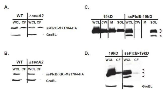
1.7 The Twin-arginine translocation (Tat) pathway ... 36
2.1 Schematic of signal sequence chimeras ... 89
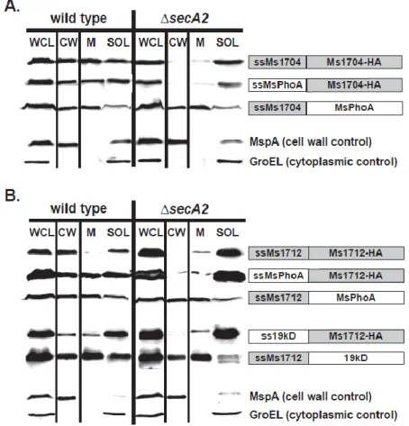
2.2 The mature domains of Ms1704 and Ms1712 require SecA2 for export to the cell wall ... 90
2.3 SecA2-dependent export occurs regardless of lipidation ... 91
2.4 Periodic acid-Schiff staining fails to detect glycosylation of the Ms1704 mature domain... 92
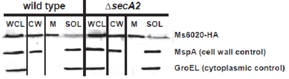
2.5 Ms6020-HA is exported to the cell wall independent of SecA2 ... 93
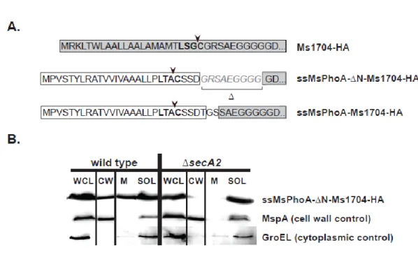
2.6 The extreme N-terminus of the Ms1704 mature domain is not required for SecA2-mediated export ... 94
2.7 An Ms1704-‘BlaTEM1 fusion is exported to the cell wall independently of SecA2 ... 95
2.8 Ms1704 is compatible with export by the twin-arginine translocation (Tat) pathway ... 96
2.9 Models for mycobacterial SecA2 export ... 97
ix
3.2 SDS-PAGE of M. tuberculosis cell wall fractions for
proteomic analysis ...150
3.3 Functional categories and export signals of cell wall proteins identified ...151
3.4 Label-free quantitation of M. tuberculosis wild type and secA2 cell wall proteins ...152
3.5 Functional categories of proteins with different relative abundances between wild type M. tuberculosis and the secA2 cell wall ...153
3.6 Export signals of proteins with differential abundance ...154
3.7 Components of two Mce transporters are reduced in the M. tuberculosis secA2 mutant cell wall ...155
3.8 Immunoblot conformation of relative PhoS1 protein abundance between the M. tuberculosis wild type and secA2 cell wall ...156
4.1 Export of Ms1704 and Ms6020 is dependent on TatC ...209
4.2 Export of Ms1704-HA requires both SecA2 and TatC, and deletion of secA2 rescues reduced Ms1704 protein levels of the tatC mutant ...210
4.3 Signal peptides of SecA2-dependent SBPs...211
4.4 Model for shared export of Ms1704 by SecA2 and Tat pathways ...212
x
LIST OF ABBREVIATIONS AND SYMBOLS deletion
2D Two-dimensional ABC ATP binding cassette ADP adenosine diphosphate
AIDS Acquired Immune Deficiency Syndrome ATP adenosine triphosphate
BCG bacillus Calmette-Guerin CW cell wall fraction
E. Escherichia
ESX ESAT secretion system HA hemagglutinin
HIV Human Immunodeficiency Virus
hyg hygromycin resistance gene
K lysine
kan kanamycin resistance gene
kDa kilodalton
L. Listeria
M. Mycobacterium
Mtb Mycobacterium tuberculosis
MDR Multi-drug resistant MEM membrane fraction
xi
min minute
ml milliliter mM millimolar
MS mass spectra
ng nanogram
N-terminal amino terminal
OD600 optical density at 600 nanometers ORF open reading frame
PAGE polyacrylamide gel electrophoresis PCR polymerase chain reaction
R arginine
S. Streptococcus
SBP solute-binding protein SDS sodium dodecyl sulfate Sec secretion
SOL soluble fraction
SRP signal recognition particle Tat Twin-arginine translocation TB tuberculosis
CHAPTER 1
INTRODUCTION1
Mycobacterium tuberculosis, the causative agent of tuberculosis (TB), is a serious global health threat accounting for nearly two million deaths every year (169). There is no effective mycobacterial vaccine available and increased prevalence of drug-resistant
M. tuberculosis strains seriously undermine current therapies (148). M. tuberculosis is spread through aerosols released from infected individuals and inhaled bacilli are engulfed by alveolar macrophages inside the lung. Instead of being killed, M.
tuberculosis survives within the phagosomal compartment of the macrophage, blocks
phagosome maturation, and replicates intracellularly (129). Another significant feature of
M. tuberculosis pathogenesis is the ability of the bacteria to persist long-term in a latent state in the host and later reactivate to cause disease, particularly in individuals who become immune-compromised.
The ability of M. tuberculosis to reside in a latent state has contributed to an ongoing HIV-TB co-epidemic, where TB is the leading killer of those infected with HIV (169). The ability of M. tuberculosis to survive and replicate within macrophages is ________________________
1Adapted for this dissertation from:
Feltcher, M. E., and M. Braunstein. 2012. Emerging themes in SecA2-mediated protein export. Nature Reviews Microbiology 10:779-789.
2
essential for M. tuberculosis pathogenesis, but our understanding of this process is incomplete. A better understanding of M. tuberculosis biology will facilitate the discovery of novel mycobacterial targets for new TB treatment strategies.The research described in this thesis is directed at elucidating the mechanisms of the SecA2-dependent protein export pathway that contributes to the success of M. tuberculosis as an
intracellular pathogen. Bacterial protein export
Over 20% of bacterial proteins have functions outside the cytoplasm and are exported to their proper locations by protein export systems (137). All bacteria, including mycobacteria, have systems for exporting these specific proteins out of the cytoplasm and into the cell envelope or extracellular environment, where they have roles in cell wall synthesis, nutrient acquisition, and other vital physiological processes (23, 30, 99). For bacterial pathogens like M. tuberculosis, some exported proteins are virulence factors that are required for modulation of the host environment to promote bacterial survival and growth.
3
systems include five ESX pathways (ESX-1 through ESX-5) and the SecA2 export pathway (1, 48). Both ESX-1 and the SecA2 system are essential for M. tuberculosis
virulence, presumably because they export effectors that modulate the host immune response or promote bacterial growth in the intracellular environment. The research described in this thesis is aimed at elucidating the mechanisms of SecA2-dependent export and how SecA2-dependent exported effectors promote intracellular M. tuberculosis growth.
The general Sec export pathway
Although best studied in Escherichia coli, all bacteria have a general secretion (Sec) pathway, which performs the bulk of protein export [for extensive reviews of Sec export, see references (97, 114)]. The Sec pathway translocates unfolded proteins through a heterotrimeric protein complex composed of the SecY, SecE, and SecG proteins (19). SecY forms the channel through which the unfolded proteins pass the cytoplasmic membrane (159). SecE is thought to stabilize an open SecY conformation necessary for translocation while SecG increases export efficiency (37, 63, 101, 102, 153, 155).
The SecYEG channel is used in two types of Sec export: post-translational and co-translational. In post-translational Sec export, proteins translocate completely across the cytoplasmic membrane through SecYEG with energy provided by the cytoplasmic SecA motor protein. In co-translational export, SecYEG works with the signal
4
emerge from the ribosome during translation and targets them as ribosome-mRNA-nascent protein complexes to FtsY for delivery to SecYEG (158). A lateral gate in SecY is thought to then open and allow passage of transmembrane domains into the membrane (41).
In post-translational Sec export, the proteins destined for translocation across the cytoplasmic membrane are synthesized as preproteins that are distinguished from the larger pool of cytoplasmic proteins by the presence of N-terminal Sec signal peptides. Sec signal peptides have a positively charged N-terminus, hydrophobic core, and polar C-terminus containing a signal peptidase cleavage site (164). In addition to the signal peptide, another requirement for Sec export is that the mature portion of the preprotein remains unfolded for competent passage through SecY. Some proteins are recognized and kept unfolded by cytoplasmic chaperones, such as SecB (6, 51), although other
preproteins may be unfolded as they are translocated (3, 104).
A central component of the post-translational Sec pathway is the cytosolic SecA ATPase motor protein (77), which has a vital role in targeting and powering preprotein transport through SecYEG (19, 40). Since the discovery of SecA in 1981, Sec export has been the focus of extensive study (108). A combination of genetic, structural and
5
SecY channel conformation (58, 67, 82, 147). SecA then undergoes cycles of
conformational changes coupled to ATP-binding and hydrolysis to drive preproteins through the SecY channel (40, 57, 161). Several models have been proposed to explain how SecA powers preprotein insertion through SecY (77). Nonetheless, most of these models propose that portions of SecA, including the IRA-1 (intramolecular regulator of ATP hydrolysis 1) two-helix finger, insert into SecY to promote forward preprotein motion through the channel (42, 168). During or shortly after translocation through the SecYEG channel, the signal peptide is removed. This cleavage event takes place on the periplasmic side of the membrane by one of two possible peptidases: the Type I signal peptidase (LepB) or the lipoprotein Type II signal peptidase (LspA) (110). After signal peptide cleavage, the protein folds into a mature conformation.
SecA, SecY, and SecE all have essential roles in Sec export and are consequently essential for cell viability (95, 107). The SecG component of SecYEG is not essential but increases translocation efficiency, possibly by stabilizing the SecY/E complex or
assisting the conformational changes of SecA (100-102). Other non-essential membrane-bound proteins that increase Sec export efficiency are SecD, SecF and YajC (38, 100, 117).
The Sec pathway of mycobacteria
Mycobacteria have homologs of all the Sec export factors reviewed above except the SecB chaperone (Figure 1.1). However, detailed studies have focused on only a few components of the mycobacterial Sec pathway. The M. tuberculosis Type I signal
6
of the general Sec pathway (109). The M. tuberculosis Type II signal peptidase (LspA) removes signal peptides from lipoproteins (133), but this lipoprotein processing is not required for in vitro growth. However, an lspA mutant of M. tuberculosis is attenuated in both macrophages and mice, illustrating the importance of functional lipoproteins to M. tuberculosis virulence (120, 133). The other components of the mycobacterial Sec pathway to receive attention are the SecA proteins. Mycobacteria are unusual in having two homologs of SecA: SecA1 and SecA2. SecA1 is the “housekeeping” SecA protein of mycobacteria responsible for exporting the majority of proteins while SecA2 is an
accessory SecA, which is discussed in detail below. As is the case for housekeeping SecA proteins of other bacteria, SecA1 of mycobacteria is essential. The secA1 gene cannot be deleted from M. tuberculosis or the nonpathogenic M. smegmatis unless an exogenous copy of secA1 is provided (16, 61, 135). The contribution of SecA1 to protein export in mycobacteria can be examined by conditional silencing of secA1 in M.
smegmatis (61). Under the control of a tetracycline repressor, SecA1 depletion leads to growth inhibition and decreased Sec export as evidenced by reduced export of the cell wall porin, MspA (61, 125).
SecA2 Systems
7
including Listeria, Staphylococcus,and some Streptococcus species (48, 124). In bacteria with two SecAs, the two proteins are not interchangeable and each SecA has unique functions (10, 16). SecA1 is the name given to the SecA with higher sequence similarity to the well-studied SecA proteins of Escherichia coli and Bacillus subtilis. SecA1 is essential and is responsible for canonical Sec export, as described above (16, 24, 43, 61, 125). Unlike SecA1, SecA2 is responsible for exporting a smaller set of proteins and often dispensable. Notably, proteins exported by SecA2 are linked to virulence in many bacterial pathogens including M. tuberculosis (17, 76, 150), Streptococcus gordonii
(175), Streptococcus parasanguinis (172), Staphylococcus aureus (146), and Listeria monocytogenes (80).
Currently, there are two types of SecA2 systems known to exist. Some bacteria with a SecA2 also have an accessory SecY2 protein. As a consequence, these SecA2– SecY2 systems appear to function largely independent of the canonical Sec machinery to export a set of proteins that are highly glycosylated and incompatible with the canonical SecA1/SecYEG (12, 26). There are also SecA2-only systems, so named because they lack a SecY2 or an obvious accessory membrane channel. SecA2-only systems likely function as part of the canonical Sec pathway, utilizing SecYEG (43, 125). Furthermore, the repertoire of proteins exported by SecA2-only systems is more diverse than that of SecA2–SecY2 systems.
8
conserved between SecA (SecA1) and SecA2 (Figure 1.2). In addition, all SecA2 proteins have two nucleotide-binding domains (NBD1 and NBD2) which together
constitute the DEAD (Asp-Glu-Ala-Asp)-like motor domain. The motor domain contains two ATP-binding Walker boxes and is responsible for ATP hydrolysis, suggesting that SecA2 proteins are functional ATPases (69, 73, 143). In fact, SecA2 from S. gordonii and
M. tuberculosis have demonstrated endogenous ATPase activity in vitro (10, 68).
Furthermore, SecA2 ATPase activity is shown to be required for accessory SecA2 protein export in mycobacteria and C. difficile (43, 68, 125).
Even though SecA2 proteins of SecA2–SecY2 and SecA2-only systems likely function differently, it is interesting that all SecA2 proteins are smaller than their SecA1 counterparts due to a carboxyl-terminal domain (CTD) truncation, although the boundary of this truncation varies (Figure 1.2). In E. coli, portions of the SecA CTD binds
phospholipids, SecB, and Zn+ (18, 45-47, 162, 180). One area of the CTD missing in all SecA2 proteins is the C-terminal linker (CTL), which lies within the preprotein-binding cleft and in E. coli, has been shown to influence substrate binding (53). In addition to the CTD truncation, the helical wing domain (HWD) is absent in the mycobacterial SecA2 and truncated in other SecA2 proteins. However, even in the canonical SecA1, the function of the HWD is not clear. The significance of the CTD and HWD truncations in SecA2 proteins awaits further studies.
SecA2–SecY2 systems
9
addition to genes encoding SecA2 and SecY2, each locus contains a gene that encodes a large serine-rich glycosylated protein that is exported by the SecA2–SecY2 system, as well as glycosylation factors that modify this substrate, and additional export machinery with unknown functions. The SecA2–SecY2 systems that are found in a subset of Gram positive species include pathogenic Streptococcus gordonii (9), Streptococcus agalactiae
(91), Streptococcus parasanguinis (27), Streptococcus pneumoniae (105), and
Staphylococcus aureus (145), although it should be noted that not all streptococcal species possess SecA2–SecY2 systems (124).
The current model of SecA2–SecY2 export suggests that these specialized systems exist to export the large serine-rich protein encoded in the secA2–secY2 locus. The serine-rich substrates have cleavable N-terminal signal peptides that are unusually long and mature domains that are heavily glycosylated (124). Examples of
experimentally confirmed SecA2–SecY2-exported substrates include GspB of S. gordonii
(9), Fap1 of S. parasanguinis (27), Srr1 of S. agalactiae (91), and SraP of S. aureus
(145). These substrates have roles related to bacterial adhesion to host tissues and/or biofilm formation (9, 91, 132, 146, 171, 172). Consequently many of these exported glycoproteins, and presumably their respective SecA2–SecY2 systems, are required for virulence (91, 105, 146, 175). In S. parasanguinis, the FimA adhesin is a second protein whose export is reported to depend on SecA2 (27). However, FimA is not a serine-rich glycoprotein and the fimA gene is not at the S. parasanguinis secA2–secY2 locus. It is currently unknown whether FimA is a true SecA2–SecY2 substrate.
10
substrates, suggesting a lack of functional redundancy with canonical SecA1. Mutations in secA2 abolish export of the serine-rich glycosylated substrates of S. gordonii (9), S. parasanguinis (27), S. agalactiae (91), and S. aureus (145). Another notable Gram positive pathogen with a putative SecA2–SecY2 pathway is Bacillus anthracis. However, the B. anthracissecA2 is phylogenetically more distant from those of Streptococcus and
Staphylococcus (124), which is also reflected by the dissimilar organization of the B. anthracissecA2 locus(Figure 1.3a). B. anthracis SecA2 is required for optimal export of two glycosylated proteins, Sap and EA1, but it appears that SecY2 is not required for this export (98). Furthermore, the Bacillus SecA2–SecY2 system lacks the additional export machinery found in the other SecA2–SecY2 systems, which suggests this system may function differently than those of streptococcal and staphylococcal species.
Below, we discuss other genes in the secA2–secY2 loci that have also been analyzed for roles in glycosylation and/or export.
Glycosylation factors of SecA2–SecY2 systems
In export-defective SecA2–SecY2 mutants, the serine-rich substrate retained in the cytoplasm is glycosylated, indicating that the protein is modified by cytoplasmic glycosylation factors prior to export (7, 27). There are two core glycosyltransferases conserved in all SecA2–SecY2 systems, GtfA and GtfB (Gtf1 and Gtf2) (Figure 1.3a). Some SecA2–SecY2 systems include additional glycosylation factors that further modify the substrate prior to export, including Gly and Nss of S. gordonii (151), Nss (Gtf3) and GalT1-2 of S. parasanguinis (173, 182), and the GtfC-GtfH proteins of S. agalactiae
11 Export machinery of SecA2–SecY2 systems
In S. gordonii (9) and S. aureus (145), secY2 mutations result in a loss of substrate export that is equivalent to the export defect exhibited by secA2 mutations, demonstrating that SecY2 is essential for accessory SecA2–SecY2 export in these systems. However, in
S. parasanguinis (170), deletion of secY2 has only a modest effect on Fap1 export and the residual exported Fap1 species is incorrectly glycosylated (170). This result suggests that in the absence of SecY2 and full glycosylation, Fap1 export defaults to the canonical SecA1/SecYEG pathway. This result also suggests that in the S. parasanguinis SecA2– SecY2 system, export and glycosylation of Fap1 are coupled (as discussed further below).
There are additional proteins encoded by SecA2–SecY2 loci that are referred to as accessory secretion proteins (Asps) in S. gordonii or glycosylation accessory proteins (Gaps) in S. parasanguinis. All SecA2–SecY2systems include the Asp1, Asp2, and Asp3 proteins (Gap1-3). While Asp1 and Asp3 are primarily cytosolic proteins, Asp2 may be membrane localized (140, 178). Some organisms, including S. gordonii, have the
additional Asp4 and Asp5 (Figure 1.3). Asp4 and Asp5 are both required for GspB export in S. gordonii (152) and are predicted integral membrane proteins with sequence
12
gap3 in S. parasanguinis (the asp1 and asp3 homologs) has only a modest effect on export of Fap1, and the residual exported Fap1 protein has altered glycosylation (83, 115). On the basis of this result, the Asp1 and Asp3 homologs of S. parasanguinis were named Gap1 and Gap3 to reflect a proposed function in glycosylation. However, a secY2
deletion in S. parasanguinis results in a phenotype similar to that of the gap1 and gap3 mutants – export of an aberrantly glycosylated Fap1 (83, 115, 170). It seems highly unlikely that SecY2 would have a direct role in protein glycosylation. Additionally, the S. gordonii and S. aureus studies of Asp1-3 in SecA2–SecY2 export are compelling. It is possible that the discrepancy in S. parasanguinis results is because Fap1 glycosylation and export are highly coupled processes. In this case, export defects of gap1, gap3 and
secY2 mutants (83, 115, 170) would indirectly affect Fap1 glycosylation to such an extent that the resulting altered Fap1 species is then compatible for export by the canonical SecA1/SecYEG system. However, at this time, a more direct role for Gaps in
13
substrate to an export/glycosylation complex (179). In S. parasanguinis, interactions between Gap1 (Asp1), Gap3 (Asp3), and SecA2 have also been identified (83, 181, 183).
Targeting proteins to the SecA2–SecY2 pathway
The serine-rich glycoproteins exported by SecA2–SecY2 systems have features that not only prevent their routing to the canonical SecA1/SecYEG pathway but promote their targeting to the SecA2–SecY2 pathway (Figure 1.4). The characteristic
glycosylation of these exported substrates is one such element (7, 27, 91). In addition to being important for protein stability (12, 91), glycosylation of these proteins blocks their export by the canonical Sec pathway in both S. gordonii and S. parasanguinis (12, 26). For example, in the absence of secA2, the canonical SecA1/SecYEG pathway can export a stable, truncated GspB variant that is non-glycosylated. However, a glycosylated GspB protein cannot utilize the canonical Sec pathway and instead requires SecA2–SecY2 for export (12).
As mentioned above, SecA2–SecY2 serine-rich proteins are glycosylated in the cytoplasm prior to export. This is in contrast to many other glycosylated Sec substrates in bacteria (103, 163), as well as the analogous eukaryotic Sec pathway where glycosylation occurs only after proteins are translocated from the cytosol into the endoplasmic
reticulum lumen (138). However, there is evidence that some bacterial proteins in addition to SecA2–SecY2 substrates share the unusual property of being glycosylated prior to Sec export (25, 28, 59), such as the HMW1 adhesin of Haemophilus influenza
14
the degree and structure of glycosylation modifications that are incompatible with the SecA1/SecYEG.
In addition to glycosylation, there are other features of SecA2–SecY2 substrates that dictate export by the accessory SecA2 pathway. The distinctive long signal peptides of GspB, Fap1, and presumably other SecA2–SecY2 substrates, are absolutely required for export. Furthermore, three glycine residues in the hydrophobic core of the GspB signal peptide promote SecA2–SecY2 -dependent export (8). However, these same glycine residues also act along with glycosylation to block export by the canonical SecA1 (8). The mechanisms by which these glycine residues act in preprotein targeting are currently unknown. Interestingly, these glycine residues are conserved in the signal peptides of most SecA2–SecY2 substrates.
Finally, there is also a region of approximately 20 amino acids at the start of the mature domain of GspB that is required for targeting this protein to the SecA2–SecY2 system (11). This accessory Sec transport (AST) domain can interact with SecA2 (but not SecA1), which suggests that in S. gordonii the AST is required for GspB docking to the accessory SecA2 system (13). It was initially hypothesized that the AST domain may interact directly with SecY2 to stabilize an open channel conformation. But it is possible that stabilization of the SecY2 channel is promoted by a high affinity interaction
15 Model for SecA2–SecY2 export
The current model of SecA2–SecY2 export is as follows (Figure 1.5). The distinctive glycosylation of the serine-rich proteins of SecA2–SecY2 systems is incompatible with export via the canonical SecA1/SecYEG pathway and demands a specialized export system. In a signal peptide-dependent manner, the SecA2–SecY2 preproteins are targeted to the SecA2–SecY2 machinery (8, 12, 26). Features of the mature domain, such as the AST (11, 13), may also be involved in targeting. In addition, Asp2 and Asp3 could contribute to translocase-targeting by binding the unmodified substrate (179).
Analogous to canonical Sec export, SecA2 likely uses cycles of ATP hydrolysis to drive glycosylated preproteins through the SecY2 channel. In some bacteria, Asp4 and Asp5 may function like SecE and SecG, whereas SecA2–SecY2 systems lacking Asp4 and Asp5 could utilize the canonical SecE or SecG for export. In fact, there is some genetic evidence that SecY2 and SecG may function together in S. aureus (144). Also akin to canonical Sec export, experiments using a slow-folding model protein suggest that SecA2–SecY2 preproteins must remain unfolded for passage through the SecY2 channel (11).
SecA2-only systems
16
channel (10, 16). The emerging model is that the SecA2 proteins of these systems work with the canonical SecA1/SecYEG translocase. Unlike secA2–secY2 loci, there is no conservation of gene content or organization at the secA2 genomic region for SecA2-only systems (Figure 1.3). In addition, there is a greater variety in the types of proteins
exported by SecA2-only systems when compared to the category of glycosylated serine-rich proteins exported by SecA2–SecY2 systems of Streptococcus and Staphylococcus. SecA2-only systems exist in all mycobacteria, including the human pathogen M.
tuberculosis (16), as well as some Gram positive bacteria such as L. monocytogenes (81),
Corynebacterium glutamicum (24), and Clostridium difficile (43).
In mycobacteria (16, 17, 130) and Listeria (20, 62, 81, 85, 90), SecA2 is not essential for growth in liquid media but secA2 mutants are defective in the export of specific proteins. However, secA2 mutants of both M. tuberculosis and L. monocytogenes
are attenuated for growth in infection models, indicating the importance of the respective SecA2 systems for exporting virulence factors (17, 76, 80). Additionally, secA2 mutants of both M. tuberculosis and L. monocytogenes elicit aberrant immune responses during infection, which has led to the use of these mutants in vaccination studies (65, 66, 76, 119). By contrast, SecA2 is essential for growth of Corynebacterium glutamicum (24), and Clostridium difficile (43).
SecA2-only exported substrates
17
Ms1712) with predicted N-terminal Sec signal peptides that are exported to the cell wall in a SecA2-dependent manner (54, 125). Ms1704 and Ms1712 are homologous solute-binding proteins, specifically predicted to bind sugars as part of two ABC-type
transporters (54). However, it is important to note that not all mycobacterial lipoproteins require SecA2 for export (54).
In M. tuberculosis and L. monocytogenes, several proteins are reduced in exported fractions of secA2 mutant bacteria analyzed by 2D-PAGE, 3 and 17 respectively (17, 80). Of these proteins, only a few have been studied further and confirmed to be
SecA2-dependent. In M. tuberculosis, one of these proteins is superoxide dismutase SodA, which notably lacks a predicted cleavable Sec signal peptide.
In L. monocytogenes the p60 autolysin, which is a cell wall amidase that cleaves peptidoglycan, is a confirmed SecA2 substrate (80). The gene for p60 is positioned adjacent to secA2 in the genome (Figure 1.3); although, other SecA2-dependent proteins of Listeria are encoded elsewhere. An additional peptidoglycan-hydrolyzing autolysin NamA (MurA) of Listeria is also SecA2-dependent (80, 85, 90). However, unlike p60, NamA lacks a typical Sec signal peptide. MnSod superoxide dismutase is another protein lacking a predicted Sec signal peptide that is exported in a SecA2-dependent manner in L.
monocytogenes (2). This particular finding parallels the SecA2-dependence of SodA
18
solute-binding proteins is predicted to bind a sugar, similar to the Ms1704 and Ms1712 substrates of M. smegmatis (54, 80).
In C. difficile the S-layer protein SlpA, which constitutes a proteinaceous lattice structure surrounding the Clostridium cell, has been identified as being exported in a SecA2-dependent fashion (44) (22). SlpA is a member of a larger family of 29 clostridial cell wall proteins (Cwp) that are implicated in host-pathogen interactions (21, 44, 71, 166). CwpV is another protein shown to require SecA2 for export (43), suggesting that additional members of this Cwp family may be SecA2-dependent as well. In C. difficile, the secA2 gene is adjacent to slpA and the larger secA2 genomic region includes genes encoding 12 Cwps (Figure 1.3) (43). However, the gene encoding CwpV is notably located elsewhere in the genome (43). In C. difficile, both of the demonstrated SecA2-dependent proteins (SlpA and CwpV) contain predicted N-terminal Sec signal peptides (44).
Export machinery of SecA2-only systems
SecA2-only systems lack an obvious alternative membrane channel and accessory export factors. An attractive idea is that SecA2 works with the canonical SecA1/SecYEG machinery either through cooperation with SecA1 or by sharing SecYEG. In support of this model, depletion of the essential SecA1 protein in M. smegmatis abolishes export of the SecA2 substrate Ms1712 (125). The simplest interpretation of this experiment is that mycobacterial SecA2 export requires the canonical SecA1. However, it remains possible that SecA1 depletion in this experiment has an indirect effect on SecA2 export.
19
canonical Sec machinery (43, 125). Dominant negative proteins often exert their effect by forming nonfunctional complexes with their normal binding partners. In mycobacteria, over-expression of the dominant negative SecA2 inhibits growth (125). This result
implies an interaction between SecA2 and proteins important to an essential process, with the essential SecA1/SecYEG machinery being a leading candidate. In C. difficile,
expression of the corresponding dominant negative SecA2 also inhibits growth and over shorter time frames is shown to impact protein export (43). Importantly, over-expression of a dominant negative SecA1 in C. difficile reduces export of SecA2 substrates, possibly by blocking accessibility of the SecA2 substrates to the SecYEG channel (43). However, unlike in mycobacteria, depletion of SecA1 in C. difficile does not influence export of SecA2 substrates (43) suggesting that in Clostridium SecA2 works with SecYEG but not SecA1.
There has not been a similar investigation for a relationship between SecA2 and SecA1/SecYEG in Listeria, but recently it was shown that secretion of the SecA2-dependent proteins p60 and NamA depends on the DivIVA protein (62). The DivIVA protein is involved in localizing proteins to the cell poles and septa of bacteria (15). Interestingly, GFP fusions to DivIVA, SecA2, p60, and NamA all localize to the septum in Listeria (62). Thus, it is possible that the SecA2-only system is specifically localized and DivIVA is required to either establish that localization pattern or deliver the SecA2-dependent proteins to the SecA2 machinery.
Targeting proteins to the SecA2-only pathway
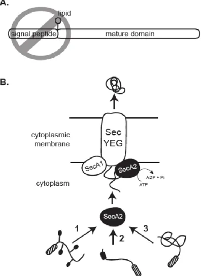
20
SecA2-targeting factors of export in the mycobacterial SecA2-only system. The two M. smegmatis lipoproteins that require SecA2 for export contain typical N-terminal Sec signal peptides. While these signal peptides are required for export (54), they are not specific for targeting these proteins to SecA2 (49). Therefore, there appears to be one or more features of the mature domains that determine SecA2-dependent export in
mycobacteria. The mature domain of these SecA2-dependent substrates could possess post-translational modifications, as seen with the SecA2–SecY2 substrates, although there is currently no evidence for this possibility.
Another unresolved issue is whether preproteins without recognizable Sec signal peptides, like the Sod proteins of M. tuberculosis and L. monocytogenes, are true SecA2 substrates or if the export of these proteins is indirectly affected by SecA2. For example, SecA2 could export a currently unknown protein with a signal peptide that is itself required for export of proteins like Sod. Still, it is also plausible that proteins without signal peptides are recognized by SecA2 and exported directly through the SecYEG channel. In support of this model, SodA of Rhizobium leguminosarum is exported in a SecA-dependent manner despite lacking a recognizable signal peptide (75). However, at this time an indirect role for the Sec system in exporting SodA of R. leguminosarum
cannot be ruled out (17, 43).
Model for SecA2-only Export
21
that make them incompatible for Sec export without the assistance of SecA2. SecA2 of both mycobacteria and Clostridium primarily localize to the cytoplasm, while much of SecA1 is found at the membrane in these bacteria (43, 125). Thus, SecA2 could possibly function in the cytoplasm to recognize and target for export a specific subset of proteins that are otherwise overlooked or incompatible with the canonical SecA1. Alternatively, SecA2 could serve as an alternate motor protein that is necessary for translocation of certain proteins through SecYEG. In either case, SecA2 ATPase activity is required for export.
Contribution of SecA2 export to M. tuberculosis pathogenesis
As previously mentioned, the SecA2 system is important to virulence. The secA2
mutant of M. tuberculosis is attenuated for growth during the acute phase of mouse infection when it is thought that M. tuberculosis is actively proliferating in macrophages (17). Consistent with this mouse phenotype, the secA2 mutant is also unable to grow in bone-marrow derived murine macrophages (17, 150). Because SodA is an antioxidant, the identification of SodA as a SecA2-dependent protein suggested that the role of the SecA2 system might be to protect M. tuberculosis against reactive oxygen intermediates (ROI) produced by macrophages. However, the M. tuberculosis secA2 mutant is
22
In further support of this possibility, it has been shown that SecA2 is required to block phagosome maturation, which is a vital step in allowing M. tuberculosis to grow inside host macrophages during infection (150). The secA2 mutant more readily localizes to acidified phagosomal compartments than wild type M. tuberculosis, and chemical inhibitors of phagosome acidification rescue growth of the secA2 mutant (150). These results suggest that SecA2 is required for exporting one or more protein effectors that block phagosome acidification. As very little is currently known about proteins involved in M. tuberculosis phagosomal maturation arrest, elucidation of SecA2-dependent exported proteins that contribute to this process would be extremely informative.
The SecA2-dependent export pathway of M. tuberculosis also has affects on host immune responses. Macrophages infected with the M. tuberculosis secA2 mutant produce higher levels of pro-inflammatory cytokines and exhibit more apoptosis than wild type infected macrophages (66, 76). The secA2 mutant of M. tuberculosis also
elicits better protective immunity in mice and guinea pigs to M. tuberculosis challenge than vaccination with the M. bovis Bacille Calmette-Guérin (BCG) vaccine (66). The identification of SecA2-dependent exported proteins that contribute to these processes would also be valuable.
The Tat Export Pathway
23
not present in all bacteria (34). Also, the Tat system differs from the Sec system because it only exports proteins that are pre-folded in the cytoplasm. Substrates requiring export by the Tat system include those that must acquire metal co-factor insertion prior to translocation out of the cytoplasm (156), as well as hetero-oligomeric protein complexes that form before export (136). It has also been proposed that Tat export systems are important for handling export of proteins that are difficult to keep unfolded in the cytoplasm prior to export. This rationale might explain why halophilic archaea, which live in high salt environments that interfere with periplasmic protein folding, rely extensively on the Tat system for protein export of proteins folded in the cytoplasm (14, 127).
The N-terminal signal peptides of Tat preproteins are similar to Sec signal
peptides (79). However, a distinguishing feature of Tat signal peptides is the presence of a pair of arginine residues that are contained within the Tat motif, R-R-X-Φ-Φ, where Φ is a hydrophobic residue. The twin-arginine pair is nearly invariant and replacement of both arginines with lysine residues abolishes Tat-dependent export (149).
The Tat export machinery consists of two core components: the TatA and TatC integral membrane proteins (126). Most Gram positive bacteria possess this minimal Tat translocase, while Gram negative and some Gram positive species (including
mycobacteria and other actinomycete species) possess an additional protein, TatB (111). TatB is similar to TatA in amino acid sequence but functionally distinct. The mechanisms of Tat export are less understood than those of Sec export, but there is a growing
24
The current model is that a Tat signal peptide first targets a folded preprotein to the TatBC complex in the cytoplasmic membrane (52, 78, 87, 123, 154). With energy supplied by the proton motive force, TatA is then recruited to the TatBC complex and forms a homo-oligomeric translocase channel (94, 176). There is evidence that the size of the TatA pore can vary, which may explain how the pore can handle folded proteins of different shapes and sizes (56, 167). The preprotein is then translocated across the cytoplasmic membrane through the TatA channel and the signal peptide is removed by a Type I signal peptidase (84). Type II signal peptidases may also act on Tat precursors since some Tat signal peptides contain lipobox motifs. For example, in Haloferax
volcanii a lipoprotein is exported by the Tat pathway (55). There is also a category of Tat substrates that become integrally-embedded in the membrane (64).
A folded conformation prior to export is not only a characteristic of Tat
substrates, but is actually a requirement for Tat export. Proteins are only exported by the Tat system when conditions are favorable for cytoplasmic folding (33). Therefore, in addition to the Tat signal peptide there are features of the mature domain of Tat substrates that promote folding and thereby dictate Tat export (157). Some Tat
25
The Tat pathway is present and linked to virulence in a number of bacterial pathogens (31). In Pseudomonas aeruginosa, the Tat pathway exports multiple virulence factors, and a mutant defective in Tat export is attenuated in the rat model of infection (106). Two of the Tat-dependent virulence factors in P. aeruginosa are secreted
phospholipase C enzymes (106). In Legionella pneumophila, Tat export is required for replication in the amoeba host as well as in macrophages (32, 128). Furthermore, the L.
pneumophila phospholipase C enzyme also requires the Tat pathway for export (128).
The Mycobacterial Tat Pathway
The Tat pathway is functional in both M. tuberculosis and M. smegmatis. Both species contain genes encoding TatA, TatB, and TatC. Tat export is essential for growth of M. tuberculosis, at least under standard laboratory conditions, as shown by the inability to delete tatA, tatB, or tatC unless exogenous copies of the tat genes are provided (131). However, deletion mutants of tatA, tatB, and tatC can be made in M. smegmatis. These mutantshave growth defects in vitro; nonetheless, M. smegmatis tat
mutants can be utilized to study Tat export in mycobacteria (88, 118).
Another phenotype of M. smegmatis tat mutants is increased sensitivity to
26
BlaC signal peptide is changed to KK, BlaC export in M. smegmatis is abolished, indicating that the twin-arginine motif is required for Tat export in mycobacteria as expected (88).
In silico analysis of the M. tuberculosis genome using several Tat prediction programs predicts a total of 108 proteins with Tat signal peptides (86, 89). Some of these predicted Tat substrates have demonstrated or suggested roles in M. tuberculosis
pathogenesis or essential physiologic processes. However, relying exclusively on Tat prediction programs to identify Tat substrates is risky. The current Tat prediction programs are built on Tat consensus sequences defined in bacteria other than
mycobacteria. There is also little overlap in the predictions of the currently available Tat prediction programs (89). Furthermore, there is an increasing list of unusual Tat exported proteins that lack a cleavable signal peptide with a twin arginine motif, and are missed by the current programs (50).
27
virulence of M. tuberculosis in mice, providing strong support for the Tat pathway
contributing to M. tuberculosis pathogenesis (121). Another protein identified as having a functional Tat signal peptide is Rv2525c, which is suggested to have a role in infection by the demonstration of increased virulence of a M. tuberculosisrv2525c mutant in macrophages and mice (89, 131).
Summary
Our understanding of the mechanisms of canonical Sec export has reached an impressive level of detail (35), but by comparison our understanding of SecA2 export is limited (48). In addition to mechanistic unknowns, our understanding of how the SecA2 export pathway contributes to M. tuberculosis remains incomplete, as we do not fully appreciate the repertoire of proteins exported by SecA2 in this pathogen. In the following chapters, we describe experiments to determine what features define mycobacterial SecA2 substrates. We also examine a potential relationship between preproteins export by the SecA2 and Tat export systems in mycobacteria. Finally, we identify new SecA2-dependent exported proteins of M. tuberculosis.
28
may share folding characteristics of Tat substrates, as the mature domain a SecA2 preprotein is compatible for export by the Tat pathway.
In Chapter 3, we describe a semi-quantitative comparative mass spectrometry study aimed at defining the SecA2-dependent exported proteome of M. tuberculosis. We identified several proteins underrepresented in the cell wall of the M. tuberculosis secA2
mutant, as expected for proteins that require SecA2 for their export. Interestingly, we observed that the majority of predicted solute-binding proteins of M. tuberculosis (the same family of proteins represented by the two known SecA2 substrates of M.
smegmatis) are dependent on SecA2. We also identified several predicted Tat substrates under-represented in the M. tuberculosis secA2 mutant. Because the Tat pathway only exports proteins that are folded, this supports a model where SecA2 assists in export of a subset of proteins with cytoplasmic folding tendencies. Further, it suggests that the SecA2 and Tat systems of mycobacteria may share a common pool of preprotein substrates. Our proteomic analysis also resulted in several candidate SecA2-dependent exported effectors that could help explain the requirement of SecA2 for M. tuberculosis
virulence.
In Chapter 4, we demonstrate that export of Ms1704 is not only dependent on SecA2 but additionally influenced by the Tat pathway of M. smegmatis. Or data suggest portions of the cytoplasmic pool of Ms1704 preprotein can be exported by
29
model where the defining feature of mycobacterial SecA2 substrates is cytoplasmic folding, a property shared with preproteins of the Tat export system.
30 Figure 1.1 General Sec export
Post-translational Sec export is powered by the essential SecA ATPase. SecA can be divided into two main structural domains: a motor domain that drives ATP hydrolysis and a specificity domain that interacts with the preprotein destined for export. Step 1: Preproteins synthesized with N-terminal Sec signal peptides (grey) are bound by
cytoplasmic SecA along a cleft between the two domains. Cytoplasmic chaperones, such as SecB, aid in keeping some preproteins unfolded prior to export and can directly deliver these preproteins to SecA. Step 2: SecA delivers the preprotein to a membrane-spanning complex composed of SecY, SecE, and SecG. Here, the signal peptide inserts into SecY to help keep an open channel conformation. Step 3: SecA goes through rounds of
31
Figure 1.2 Domain organization of SecA2 proteins
Sequence alignments and structural modeling suggest that most functional domains are conserved between SecA1 and SecA2 proteins. The crystal structure of the M.
tuberculosis SecA1 protein depicted here represents a typical SecA1 protein with the corresponding colored domains outlined below (141). *The C-terminal domain (CTD) was not resolved in the M. tuberculosis crystal structure but is shown in the domain graphic. For comparison to SecA1, the predicted domain organization of M. tuberculosis
SecA2 and Streptococcus gordonii SecA2 are included. SecA1 can be divided into two main structural domains, which are both composed of several subdomains (77, 134). The DEAD (Asp-Glu-Ala-Asp)-like motor domain is responsible for the ATP hydrolysis (69, 73, 143) and consists of two nucleotide-binding folds: NBD1 and NBD2. NBD1 contains the two ATP-binding Walker boxes (92, 177). The helical scaffold domain (HSD)
32 Figure 1.3 Organization of secA2 genomicloci
a. SecA2–SecY2 systems are found in a diverse set of Streptococcus and Staphylococcus
spp. (including Streptococcus gordonii, Streptococcus parasanguinis, Streptococcus agalactiae and Staphylococcus aureus), in which they function in the biogenesis of surface glycoproteins (the genes encoding which are shown in blue). All of these loci contain the core secA2 and secY2 genes (shown in red). Some Bacillus spp., including
Bacillus anthracis, have putative SecA2–SecY2 systems, although the exported
substrates of these systems are unknown, and secA2 and secY2 are separated. Genes encoding putative export machinery components are shown in yellow, and those encoding the glycosylation machinery are shown in green. b. Examples of SecA2-only systems are found in Mycobacterium tuberculosis, Listeria monocytogenes and
Clostridium difficile. The secA2 loci of SecA2-only systems are not conserved and export a diverse set of substrates. In some cases, the genes encoding the exported substrates (blue) are found at the secA2 locus. However, this is not always the case, and substrate-encoding genes that are located elsewhere in the genome are not depicted. In addition to
slpA (encoding the protein that constitutes the S-layer (surface layer) of bacteria and is a SecA2-only substrate), the C. difficile secA2 locus contains genes encoding 11 cell wall proteins (Cwps) that are putative SecA2 substrates, of which three are shown in light blue. asp, accessory Sec system protein gene; gap, glycosylation-associated protein gene;
33 Figure 1.4 SecA2/SecY2-targeting features
SecA2–SecY2 preproteins have features both for targeting to the SecA2–SecY2
34 Figure 1.5 Model of SecA2–SecY2 Export
The biogenesis of surface glycoproteins by SecA2–SecY2 systems involves both glycosylation factors (green) and export machinery (red) that are distinct from the canonical Sec machinery. Serine-rich proteins are synthesized with N-terminal signal peptides. The accessory Sec system proteins (Asps) promote SecA2–SecY2-mediated export by unknown mechanisms, but could target preproteins to the translocase and/or serve as a scaffold for the export complex. Asp4 and Asp5 are putative accessory
components of the SecY2 channel, but they are not present in all SecA2–SecY2 systems. SecA2–SecY2-mediated export and glycosylation are likely to be coupled processes. As glycosyl groups (orange hexagons) are added to the preprotein by cytoplasmic
35 Figure 1.6 Model of SecA2-only Export
36
Figure 1.7 The Twin-arginine translocation (Tat) Pathway
37 References
1. Abdallah, A. M., N. C. Gey van Pittius, P. A. Champion, J. Cox, J. Luirink, C. M. Vandenbroucke-Grauls, B. J. Appelmelk, and W. Bitter. 2007. Type VII secretion--mycobacteria show the way. Nat Rev Microbiol 5:883-891.
2. Archambaud, C., M. A. Nahori, J. Pizarro-Cerda, P. Cossart, and O.
Dussurget. 2006. Control of Listeria superoxide dismutase by phosphorylation. J Biol Chem 281:31812-31822.
3. Arkowitz, R. A., J. C. Joly, and W. Wickner. 1993. Translocation can drive the unfolding of a preprotein domain. Embo J 12:243-253.
4. Auclair, S. M., J. P. Moses, M. Musial-Siwek, D. A. Kendall, D. B. Oliver, and I. Mukerji. 2010. Mapping of the signal peptide-binding domain of
Escherichia coli SecA using Forster resonance energy transfer. Biochemistry 49:782-792.
5. Bashiri, G., E. F. Perkowski, A. P. Turner, M. E. Feltcher, M. Braunstein, and E. N. Baker. 2012. Tat-dependent translocation of an F420-binding protein
of Mycobacterium tuberculosis. PLoS One 7:e45003.
6. Bechtluft, P., N. Nouwen, S. J. Tans, and A. J. Driessen. 2010. SecB--a chaperone dedicated to protein translocation. Mol Biosyst 6:620-627.
7. Bensing, B. A., B. W. Gibson, and P. M. Sullam. 2004. The Streptococcus gordonii platelet binding protein GspB undergoes glycosylation independently of export. J Bacteriol 186:638-645.
8. Bensing, B. A., I. R. Siboo, and P. M. Sullam. 2007. Glycine residues in the hydrophobic core of the GspB signal sequence route export toward the accessory Sec pathway. J Bacteriol 189:3846-3854.
9. Bensing, B. A., and P. M. Sullam. 2002. An accessory sec locus of
38
10. Bensing, B. A., and P. M. Sullam. 2009. Characterization of Streptococcus
gordonii SecA2 as a paralogue of SecA. J Bacteriol 191:3482-3491.
11. Bensing, B. A., and P. M. Sullam. 2010. Transport of preproteins by the
accessory Sec system requires a specific domain adjacent to the signal peptide. J Bacteriol 192:4223-4232.
12. Bensing, B. A., D. Takamatsu, and P. M. Sullam. 2005. Determinants of the streptococcal surface glycoprotein GspB that facilitate export by the accessory Sec system. Mol Microbiol 58:1468-1481.
13. Bensing, B. A., Y. T. Yen, R. Seepersaud, and P. M. Sullam. 2012. A Specific interaction between SecA2 and a region of the preprotein adjacent to the signal peptide occurs during transport via the accessory Sec system. J Biol Chem 287:24438-24447.
14. Bolhuis, A. 2002. Protein transport in the halophilic archaeon Halobacterium sp. NRC-1: a major role for the twin-arginine translocation pathway? Microbiology 148:3335-3346.
15. Bramkamp, M., and S. van Baarle. 2009. Division site selection in rod-shaped bacteria. Curr Opin Microbiol 12:683-688.
16. Braunstein, M., A. M. Brown, S. Kurtz, and W. R. Jacobs, Jr. 2001. Two nonredundant SecA homologues function in mycobacteria. J Bacteriol 183:6979-6990.
17. Braunstein, M., B. Espinosa, J. Chan, J. T. Belisle, and W. R. J. Jacobs. 2003. SecA2 functions in the secretion of superoxide dismutase A and in the virulence
of Mycobacterium tuberculosis. Mol Microbiol 48:453-464.
18. Breukink, E., N. Nouwen, A. van Raalte, S. Mizushima, J. Tommassen, and B. de Kruijff. 1995. The C terminus of SecA is involved in both lipid binding and SecB binding. J Biol Chem 270:7902-7907.
39
20. Burkholder, K. M., K. P. Kim, K. K. Mishra, S. Medina, B. K. Hahm, H. Kim, and A. K. Bhunia. 2009. Expression of LAP, a SecA2-dependent secretory protein, is induced under anaerobic environment. Microbes Infect 11:859-867.
21. Calabi, E., F. Calabi, A. D. Phillips, and N. F. Fairweather. 2002. Binding of
Clostridium difficile surface layer proteins to gastrointestinal tissues. Infect Immun 70:5770-5778.
22. Calabi, E., S. Ward, B. Wren, T. Paxton, M. Panico, H. Morris, A. Dell, G. Dougan, and N. Fairweather. 2001. Molecular characterization of the surface layer proteins from Clostridium difficile. Mol Microbiol 40:1187-1199.
23. Calva, E., and R. Oropeza. 2006. Two-component signal transduction systems, environmental signals, and virulence. Microb Ecol 51:166-176.
24. Caspers, M., and R. Freudl. 2008. Corynebacterium glutamicum possesses two
secA homologous genes that are essential for viability. Arch Microbiol.
25. Charbonneau, M. E., and M. Mourez. 2008. The Escherichia coli AIDA-I autotransporter undergoes cytoplasmic glycosylation independently of export. Res Microbiol 159:537-544.
26. Chen, Q., B. Sun, H. Wu, Z. Peng, and P. M. Fives-Taylor. 2007. Differential roles of individual domains in selection of secretion route of a Streptococcus parasanguinis serine-rich adhesin, Fap1. J Bacteriol 189:7610-7617.
27. Chen, Q., H. Wu, and P. M. Fives-Taylor. 2004. Investigating the role of secA2
in secretion and glycosylation of a fimbrial adhesin in Streptococcus parasanguis
FW213. Mol Microbiol 53:843-856.
28. Choi, K. J., S. Grass, S. Paek, J. W. St Geme, 3rd, and H. J. Yeo. 2010. The
Actinobacillus pleuropneumoniae HMW1C-like glycosyltransferase mediates
N-linked glycosylation of the Haemophilus influenzae HMW1 adhesin. PLoS One 5:e15888.
40
Rajandream, J. Rogers, S. Rutter, K. Seeger, J. Skelton, R. Squares, S. Squares, J. E. Sulston, K. Taylor, S. Whitehead, and B. G. Barrell. 1998. Deciphering the biology of Mycobacterium tuberculosis from the complete genome sequence. Nature 393:537-544.
30. Crick, D. E., L. Quadri, and P. J. Brennan. 2008. Biochemistry of the cell envelope of Mycobacterium tuberculosis, p. 1-20. In K. S.H.E and E. J. Rubin (ed.), Handbook of Tuberculosis: Molecular Biology and Biochemistry. WILEY-VCH Verlag GmbH & Co.
31. De Buck, E., E. Lammertyn, and J. Anne. 2008. The importance of the twin-arginine translocation pathway for bacterial virulence. Trends Microbiol 16:442-453.
32. De Buck, E., L. Maes, E. Meyen, L. Van Mellaert, N. Geukens, J. Anne, and E. Lammertyn. 2005. Legionella pneumophila Philadelphia-1 tatB and tatC affect intracellular replication and biofilm formation. Biochem Biophys Res Commun 331:1413-1420.
33. DeLisa, M. P., D. Tullman, and G. Georgiou. 2003. Folding quality control in the export of proteins by the bacterial twin-arginine translocation pathway. Proc Natl Acad Sci U S A 100:6115-6120.
34. Dilks, K., R. W. Rose, E. Hartmann, and M. Pohlschroder. 2003. Prokaryotic utilization of the twin-arginine translocation pathway: a genomic survey. J Bacteriol 185:1478-1483.
35. du Plessis, D. J., N. Nouwen, and A. J. Driessen. 2011. The Sec translocase. Biochim Biophys Acta 1808:851-865.
36. Dubini, A., and F. Sargent. 2003. Assembly of Tat-dependent [NiFe]
hydrogenases: identification of precursor-binding accessory proteins. FEBS Lett 549:141-146.
37. Duong, F., and W. Wickner. 1999. The PrlA and PrlG phenotypes are caused by a loosened association among the translocase SecYEG subunits. EMBO J
41
38. Duong, F., and W. Wickner. 1997. The SecDFyajC domain of preprotein translocase controls preprotein movement by regulating SecA membrane cycling. EMBO J 16:4871-4879.
39. Economou, A. 1999. Following the leader: bacterial protein export through the Sec pathway. Trends Microbiol 7:315-320.
40. Economou, A., and W. Wickner. 1994. SecA promotes preprotein translocation by undergoing ATP-driven cycles of membrane insertion and deinsertion. Cell 78:835-843.
41. Egea, P. F., and R. M. Stroud. 2010. Lateral opening of a translocon upon entry of protein suggests the mechanism of insertion into membranes. Proc Natl Acad Sci U S A 107:17182-17187.
42. Erlandson, K. J., S. B. Miller, Y. Nam, A. R. Osborne, J. Zimmer, and T. A. Rapoport. 2008. A role for the two-helix finger of the SecA ATPase in protein translocation. Nature 455:984-987.
43. Fagan, R. P., and N. F. Fairweather. 2011. Clostridium difficile has two parallel and essential Sec secretion systems. J Biol Chem 286:27483-27493.
44. Fagan, R. P., C. Janoir, A. Collignon, P. Mastrantonio, I. R. Poxton, and N. F. Fairweather. 2011. A proposed nomenclature for cell wall proteins of
Clostridium difficile. J Med Microbiol 60:1225-1228.
45. Fekkes, P., J. G. de Wit, A. Boorsma, R. H. Friesen, and A. J. Driessen. 1999. Zinc stabilizes the SecB binding site of SecA. Biochemistry 38:5111-5116.
46. Fekkes, P., J. G. de Wit, J. P. van der Wolk, H. H. Kimsey, C. A. Kumamoto, and A. J. Driessen. 1998. Preprotein transfer to the Escherichia coli translocase requires the co-operative binding of SecB and the signal sequence to SecA. Mol Microbiol 29:1179-1190.
47. Fekkes, P., C. van der Does, and A. J. Driessen. 1997. The molecular
42
48. Feltcher, M. E., and M. Braunstein. 2012. Emerging themes in SecA2-mediated protein export. Nat Rev Microbiol 10:779-789.
49. Feltcher, M. E., H. S. Gibbons, L. S. Ligon, and M. Braunstein. 2013. Protein export by the mycobacterial SecA2 system Is determined by the preprotein mature domain. J Bacteriol 195:672-681.
50. Ferrandez, Y., and G. Condemine. 2008. Novel mechanism of outer membrane targeting of proteins in Gram-negative bacteria. Mol Microbiol 69:1349-1357.
51. Fisher, A. C., and M. P. DeLisa. 2004. A little help from my friends: quality control of presecretory proteins in bacteria. J Bacteriol 186:7467-7473.
52. Frobel, J., P. Rose, F. Lausberg, A. S. Blummel, R. Freudl, and M. Muller. 2012. Transmembrane insertion of twin-arginine signal peptides is driven by TatC and regulated by TatB. Nat Commun 3:1311.
53. Gelis, I., A. M. Bonvin, D. Keramisanou, M. Koukaki, G. Gouridis, S. Karamanou, A. Economou, and C. G. Kalodimos. 2007. Structural basis for signal-sequence recognition by the translocase motor SecA as determined by NMR. Cell 131:756-769.
54. Gibbons, H. S., F. Wolschendorf, M. Abshire, M. Niederweis, and M. Braunstein. 2007. Identification of two Mycobacterium smegmatis lipoproteins exported by a SecA2-dependent pathway. J Bacteriol 189:5090-5100.
55. Gimenez, M. I., K. Dilks, and M. Pohlschroder. 2007. Haloferax volcanii twin-arginine translocation substates include secreted soluble, C-terminally anchored and lipoproteins. Mol Microbiol 66:1597-1606.
56. Gohlke, U., L. Pullan, C. A. McDevitt, I. Porcelli, E. de Leeuw, T. Palmer, H. R. Saibil, and B. C. Berks. 2005. The TatA component of the twin-arginine protein transport system forms channel complexes of variable diameter. Proc Natl Acad Sci U S A 102:10482-10486.
43
58. Gouridis, G., S. Karamanou, I. Gelis, C. G. Kalodimos, and A. Economou. 2009. Signal peptides are allosteric activators of the protein translocase. Nature 462:363-367.
59. Grass, S., A. Z. Buscher, W. E. Swords, M. A. Apicella, S. J. Barenkamp, N. Ozchlewski, and J. W. St Geme, 3rd. 2003. The Haemophilus influenzae
HMW1 adhesin is glycosylated in a process that requires HMW1C and
phosphoglucomutase, an enzyme involved in lipooligosaccharide biosynthesis. Mol Microbiol 48:737-751.
60. Graubner, W., A. Schierhorn, and T. Bruser. 2007. DnaK plays a pivotal role in Tat targeting of CueO and functions beside SlyD as a general Tat signal binding chaperone. J Biol Chem 282:7116-7124.
61. Guo, X. V., M. Monteleone, M. Klotzsche, A. Kamionka, W. Hillen, M. Braunstein, S. Ehrt, and D. Schnappinger. 2007. Silencing Mycobacterium smegmatis by using tetracycline repressors. J Bacteriol 189:4614-4623.
62. Halbedel, S., B. Hahn, R. A. Daniel, and A. Flieger. 2012. DivIVA affects secretion of virulence-related autolysins in Listeria monocytogenes. Mol Microbiol 83:821-839.
63. Harris, C. R., and T. J. Silhavy. 1999. Mapping an interface of SecY (PrlA) and SecE (PrlG) by using synthetic phenotypes and in vivo cross-linking. J Bacteriol 181:3438-3444.
64. Hatzixanthis, K., T. Palmer, and F. Sargent. 2003. A subset of bacterial inner membrane proteins integrated by the twin-arginine translocase. Mol Microbiol 49:1377-1390.
65. Hinchey, J., B. Y. Jeon, H. Alley, B. Chen, M. Goldberg, S. Derrick, S. Morris, W. R. Jacobs, Jr., S. A. Porcelli, and S. Lee. 2011. Lysine auxotrophy combined with deletion of the secA2 gene results in a safe and highly
immunogenic candidate live attenuated vaccine for tuberculosis. PLoS One 6:e15857.
44
67. Hizlan, D., A. Robson, S. Whitehouse, V. A. Gold, J. Vonck, D. Mills, W. Kuhlbrandt, and I. Collinson. 2012. Structure of the SecY complex unlocked by a preprotein mimic. Cell Rep 1:21-28.
68. Hou, J. M., N. G. D'Lima, N. W. Rigel, H. S. Gibbons, J. R. McCann, M. Braunstein, and C. M. Teschke. 2008. ATPase activity of Mycobacterium
tuberculosis SecA1 and SecA2 proteins and its importance for SecA2 function in
macrophages. J Bacteriol 190:4880-4887.
69. Hunt, J. F., S. Weinkauf, L. Henry, J. J. Fak, P. McNicholas, D. B. Oliver, and J. Deisenhofer. 2002. Nucleotide control of interdomain interactions in the conformational reaction cycle of SecA. Science 297:2018-2026.
70. Jack, R. L., G. Buchanan, A. Dubini, K. Hatzixanthis, T. Palmer, and F. Sargent. 2004. Coordinating assembly and export of complex bacterial proteins. Embo J 23:3962-3972.
71. Janoir, C., S. Pechine, C. Grosdidier, and A. Collignon. 2007. Cwp84, a surface-associated protein of Clostridium difficile, is a cysteine protease with degrading activity on extracellular matrix proteins. J Bacteriol 189:7174-7180.
72. Karamanou, S., E. Vrontou, G. Sianidis, C. Baud, T. Roos, A. Kuhn, A. S. Politou, and A. Economou. 1999. A molecular switch in SecA protein couples ATP hydrolysis to protein translocation. Mol Microbiol 34:1133-1145.
73. Keramisanou, D., N. Biris, I. Gelis, G. Sianidis, S. Karamanou, A. Economou, and C. G. Kalodimos. 2006. Disorder-order folding transitions underlie catalysis in the helicase motor of SecA. Nat Struct Mol Biol 13:594-602.
74. Kimura, E., M. Akita, S. Matsuyama, and S. Mizushima. 1991. Determination of a region in SecA that interacts with presecretory proteins in Escherichia coli. J Biol Chem 266:6600-6606.
75. Krehenbrink, M., A. Edwards, and J. A. Downie. 2011. The superoxide dismutase SodA is targeted to the periplasm in a SecA-dependent manner by a novel mechanism. Mol Microbiol 82:164-179.
45
growth in macrophages and inhibits the host immune response. Infect Immun 74:6855-6864.
77. Kusters, I., and A. J. Driessen. 2011. SecA, a remarkable nanomachine. Cell Mol Life Sci 68:2053-2066.
78. Lausberg, F., S. Fleckenstein, P. Kreutzenbeck, J. Frobel, P. Rose, M. Muller, and R. Freudl. 2012. Genetic evidence for a tight cooperation of TatB and TatC during productive recognition of twin-arginine (Tat) signal peptides in
Escherichia coli. PLoS One 7:e39867.
79. Lee, P. A., D. Tullman-Ercek, and G. Georgiou. 2006. The Bacterial Twin-Arginine Translocation Pathway. Annu Rev Microbiol 60:373-395.
80. Lenz, L. L., S. Mohammadi, A. Geissler, and D. A. Portnoy. 2003. SecA2-dependent secretion of autolytic enzymes promotes Listeria monocytogenes
pathogenesis. Proc Natl Acad Sci U S A 100:12432-12437.
81. Lenz, L. L., and D. A. Portnoy. 2002. Identification of a second Listeria secA
gene associated with protein secretion and the rough phenotype. Mol Microbiol 45:1043-1056.
82. Li, W., S. Schulman, D. Boyd, K. Erlandson, J. Beckwith, and T. A.
Rapoport. 2007. The plug domain of the SecY protein stabilizes the closed state of the translocation channel and maintains a membrane seal. Molecular Cell 26:511-521.
83. Li, Y., Y. Chen, X. Huang, M. Zhou, R. Wu, S. Dong, D. G. Pritchard, P. Fives-Taylor, and H. Wu. 2008. A conserved domain of previously unknown function in Gap1 mediates protein-protein interaction and is required for
biogenesis of a serine-rich streptococcal adhesin. Mol Microbiol 70:1094-1104.
84. Luke, I., J. I. Handford, T. Palmer, and F. Sargent. 2009. Proteolytic processing of Escherichia coli twin-arginine signal peptides by LepB. Arch Microbiol 191:919-925.