Int. J. Adv. Res. Sci. Technol. Volume 2, Issue1, 2013, pp 40-45
International Journal of Advanced Research in
Science and Technology
journal homepage: www.ijarst.com
ISSN 2319 – 1783 (Print)
ISSN 2320 – 1126 (Online)
MRI Image Retrieval Using Gabor Wavelet Based Texture Features
N. Kumaran 1*, R. Bhavani 1
1
Department of Computer Science & Engineering, Annamalai University, Annamalainagar, Tamil Nadu, INDIA. *Corresponding Author’s Email: [email protected]
A R T I C L E I N F O A B S T R A C T
Article history:
Received 24 July 2013 Accepted 08 Aug. 2013 Available online 12 Aug. 2013
Due to vast number of medical technologies and equipments the medical images are growing in rapid rate. This directs to retrieve efficient medical images based on visual contents. This paper proposed the content based medical image retrieval system by means of Gabor Wavelet to extract texture features of MRI images. Then the K-means clustering and Euclidean distance measure are used to retrieve related images for the query image in medical diagnosis. The experimental results demonstrate the efficiency of this system in clustering and MRI image retrieval against Haralick’s and Texture Spectrum based texture features.
© 2013 International Journal of Advanced Research in Science and Technology (IJARST).
All rights reserved. Keywords:
CBMIR,
Euclidean Distance, Gabor Wavelet, K-means Clustering
Introduction
The Content Based Medical Image Retrieval (CBMIR) systems are search engines for medical image databases, which indexing and retrieving medical images according to their visual contents like colour, texture and shape information. [1-3]
Today, there are huge numbers of medical images being created in hospitals around the world. It is estimated that the amount of such images will further increase exponentially in the future. The significance of new technologies such as X-Ray radiography, Ultrasound, Computed Tomography (CT), Magnetic Resonance Imagining (MRI) and Picture Archiving and Communication Systems (PACS) have resulted in an explosive growth in the number of images stored in the database. This will lead various new systems for storage, organization, indexing and retrieval of the medical images in different fields like medical diagnosis, research and teaching.
Generally, the medical image database contains a lot of texture based information capable for retrieval purpose. This paper, proposed the concept of spine, brain,
knee and abdomen MRI image retrieval using Gabor Wavelet [GW] [4] based texture features and the performance of this system is compared with Haralick’s [5] and Texture Spectrum [6] based texture features.
Related Works:
There are many existing systems that provide different methods and algorithms for CBMIR. The most important intention of all these systems is to prove the improvement of results so as to give support to the doctors and radiologists in diagnosis of treatments.
In [7], the authors presented an idea by combining low level content features and high level semantic features to retrieve medical images. Gabor wavelet [8] was one of the methods for texture feature extraction in content based medical image retrieval. In this approach texture feature vector was computed according to the multi-scale and multi-direction fuzzy set which is calculated based on all energy co-efficient.
Int. J. Adv. Res. Sci. Technol. Volume 2, Issue1, 2013, pp 40-45
structure [10] was consisting of a continuous and probabilistic image representation process using Gaussian Mixture Modelling (GMM) for image matching via the Kullback-Leibler (KL) measure. For matching and categorizing X-ray images, the GMM-KL framework was used.
Horsthemke et. al. [11] explained two different texture features based CBMIR systems. The first system can be used to provide context-sensitive tools for computer-aided diagnosis with pixel-level co-occurrence matrices. The second system can be used directly as a computer-aided diagnosis system for case-based and evidence-based medicine with pixel level and global level co-occurrence matrices. Muller et. al. [12] compared several texture analysis methods, colour (grey level) quantization and other useful features to extract include image intensity, region size, shape and statistical moments.
Sanghavi et. al. [13] focused on recent advances in CBIR system in medical domain. It also focused on various feature extraction techniques and algorithms implemented for CBIR systems in different cases of medical domain. B. G. Prasad et. al. [14] evaluated the performance of two statistical methods of texture features proposed by Haralick’s and Tamura for retrieving similar cases for CT scan brain images. In [15] the author demonstrated CBMIR system using canny edge based shape detection and k-means clustering algorithms.
S. Nagendram et. al. [16] gave an overview in the field of content based access to medical image data and on the tools used in the field. They also gave about generic content based image retrieval for medical images and described various methods of CBIR implementing. In [17] the importance of medical visual information search and the major challenges were detailed and proposed a flexible framework for indexing medical visual information of any dimension and any modality based on texture information.
Xiang Sean Zhou et. al. [18] took a critical look at the semantic medical CBIR problem and provided their perspective regarding the gaps and opportunities present in this domain. They listed today’s medical imaging modalities, along with an almost exhaustive list of existing medical CBIR systems. Katarina Trojacanec et. al. [19] applied the edge histogram and region-based shape descriptors standardized by MPEG-7 standard to
MRIs. The analysis showed that the edge histogram descriptor achieves higher precision.
In our previous work [20-21] the performance measures for spine MRI image retrieval proved that Texture Spectrum based texture features (black-white symmetry, geometric symmetry, degree of direction, orientation features and central symmetry) were somewhat good compared to Haralick’s texture features (contrast, angular second moment, coarseness, entropy) and the combination of both features of image retrieval was the best.
This paper is organized such that brief discussion on proposed work and Gabor Wavelet Transform in section 3 and 4. In sections 5 and 6, we explain about Gabor Wavelet based texture features and K-means clustering. In section 7, we deal with image retrieval. Section 8 shows experiments and results. In section 9, we conclude our work with feature prospects.
Proposed Work:
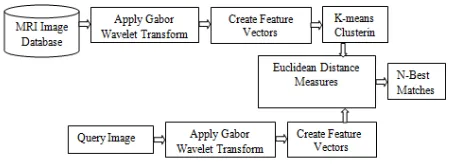
The block diagram of the proposed Gabor Wavelet and texture features based MRI medical image retrieval system is shown in Fig 1.
Fig. 1 The configuration of the proposed retrieval system
In our work, applying Gabor Wavelet Transform (GWT) to the collection of MRI image database and feature vector is constructed using mean, standard deviation, skew and kurtosis as feature components and the database are created. Then with K-means clustering [22] the database images are clustered. After that Euclidean distance measure [23] is using to retrieve N-best matches for the query image.
Gabor Wavelet Transform:
Int. J. Adv. Res. Sci. Technol. Volume 2, Issue1, 2013, pp 40-45
(1)
Where, x, y denote the pixel position in the spatial domain, is the radial centre frequency of the complex exponential, is the orientation of the GW, and is the standard deviation of the Gaussian function. By selecting different centre frequencies and orientations, we can obtain a family of Gabor kernels, which can be used to extract features from an image.
The GW forms complex valued function like real and imaginary parts. Other wavelets are shifted, rotated and dilated versions of these two. Given an image f(x, y), GW features are extracted by
convolving f(x, y) with as follows:
(2)
Where, denotes the convolution operator. For a neighbourhood window of size W × W for W = 2t + 1, the discrete convolutions operation of the image f(x, y) with GW we get
(3)
We can write down,
(4)
With,
(5)
(6)
Where, is a complex conjugate of
.
Gabor Wavelet Based Texture Features:
The feature vectors µ(x, y), STD(x, y), Skewness and Kurtosis are constructed as feature components using equation (7) to (10).
(7)
(8)
(9)
(10)
Where, (X, Y) is the image dimension. Table 1 shows the sample values of the feature vectors for a MRI spine and brain images.
Table. 1 Extracted sample values of feature vectors
M R I Im a g es 0 4 0 .2 2 1 3 7 ,2 0 3 2 .0 6 2 3 .5 5 4 3 5 .1 3 2 3 2 .2 6 2 1 .2 2 1 2 .4 5 5 4 5 1 3 .1 2 0 1 6 ,3 3 6 3 .0 6 0 1 2 .6 0 2 1 2 .5 0 1 1 0 .4 5 1 3 .0 1 1 1 6 .8 3 0 9 0 4 1 .0 1 0 3 8 ,2 5 1 1 .3 0 3 2 .4 2 2 3 3 .4 3 5 3 1 .5 4 7 2 .0 0 4 3 .5 0 8 1 3 5 2 6 .0 3 1 1 9 ,1 6 2 1 .4 3 1 4 .0 2 3 1 4 .2 0 1 1 2 .5 3 4 2 .2 2 0 1 1 .0 3 1 µ ( x , y ) S T D ( x ,y ) S k ew K u rt o si s µ ( x , y ) S T D ( x ,y ) S k ew K u rt o si s K-means Clustering:
In our problem, the K-means clustering algorithm can be formally stated as follows: Given 1850 MRI images in a 16-dimensional metric space, determine a partition of the images into maximum 10 clusters and 20 iterations, such that the images in a cluster are more similar to each other than to images in different clusters.
Int. J. Adv. Res. Sci. Technol. Volume 2, Issue1, 2013, pp 40-45
Fig. 2 Sample Output of fifth cluster
Image Retrieval:
We use Euclidean distance measure in order to balance between computational complexity and retrieval accuracy. The Euclidean distance is calculated between the query image and the selected precise images. If xi and yi are 2D feature vectors of selected precise images and query image respectively then the distance measure is
defined as, . The
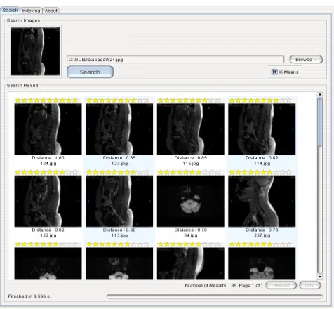
calculated distances are sorted in increasing order and display the first N images as the best matching images. The sample output screens are shown in Fig. 3 and 4.
Fig. 3 Best retrieved spine MRI images
Fig. 4 Best retrieved brain MRI images
Experiments and Results:
This method is implemented on a computer system using JAVA as the programming language and MySQL as the backend. In this work, we used around 1850 MRI scan images in BMP format with the size of 256 x 256 as a database. Different parts of human body MRI scan images such as 850 spine, 400 brain, 300 abdomen and 300 knee images are used.
Int. J. Adv. Res. Sci. Technol. Volume 2, Issue1, 2013, pp 40-45
We did many experiments and recorded the feature extraction time and response time for a query image on different capacity of database. After a large number of experiments the Table 2 showed the feature extraction time and the response time when retrieval of 140 MRI images. Along with growth of the database, the extraction time and response time increases proportionally. These results compared to Haralick’s and Texture Spectrum based texture features.
Table. 2 Comparison of feature extraction time and the response time
Methods
Number of Features
Feature Extraction
Time ( in Seconds)
Response Time ( in Seconds)
Gabor Wavelet 16 581 3.742
Texture Spectrum 8 321 4.85
Haralick’s Features
14 used
only 4 258 5.23
Texture Spectrum + Haralick’s
Features
8 + 4 =12 433 4.205
Our proposed Gabor Wavelet based texture features like mean, standared diviation, Skewness and Kurtosis are extracted for four different orientations with the extraction time of 581 seconds which is higher compare to other techniques. But it’s response time is 3.742 seconds which is lesser compare to other techniques.
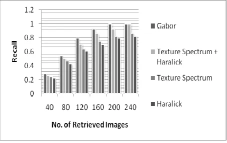
Fig. 5 Performance measures based on Recall
The effectiveness of the proposed method can be measured by Recall and Precision, which are often referred to together since they measure the different aspects of the system performance. Recall measures the system’s ability to retrieve relevant images from the database. It is defined as the ratio between the number of retrieved relevant images and the total number of relevant
images in the database. Thus, the recall rate demonstrates the power of a learning system by revealing its level of false negatives. The performance measures of recall are given in Fig. 5 which proved that our proposed method has highest recall rate than methods.
Precision measures the retrieval accuracy and is defined as the ratio between the number of retrieved relevant images and the number of total retrieved images. This measure demonstrates the efficiency of a learning system and is closely related to its level of false positives. The performance measures of precision are given in Fig. 6 which proved that our proposed method has highest precision rate than other methods.
Fig. 6 Performance measures based on Precision
Conclusion:
In this paper, we have proposed an efficient MRI image retrieval method using Gabor Wavelet based texture features. Our Experimental results demonstrate that the proposed method has best clustering accuracy and retrieval efficiency than other conventional methods such as Haralick’s and Texture Spectrum based MRI image retrieval methods. Further, Euclidean distance measure to reduce the execution time whereas maintaining a reasonable level of retrieval performance. We have planned to extend our work via Curvelet Transform based texture features with all types of human body scan images.
References:
1. X. S. Zhou, S. Zillner, M. Moeller,M. Sintek, Y. Zhan, A. Krishnan, A. Gupta “Semantics and CBIR: A Medical Imaging Perspective”, In Proceedings of the ACM International Conference on Content-based Image and Video Retrieval, pp. 571-580, July 2008.
2. V. Vijaya Kumar, N. Gnaneswara Rao, and
Int. J. Adv. Res. Sci. Technol. Volume 2, Issue1, 2013, pp 40-45 Images”, International Journal of Future Generation
Communication and Networking, Vol. 2, No. 4, pp. 39-48, December 2009.
3. Thomas M. Lehmann, Mark O. Güld, Daniel Keysers, Thomas Deselaers, Henning Schubert, Berthold B. Wein, Klaus Spitzer, “Similarity of Medical Images Computed from Global Feature Vectors for Content-Based Retrieval”, In the Proceedings of KES ‘2004, Vol. 3214, pp. 989–995, Springer-Verlag, Berlin, Heidelberg, 2004.
4. B. S. Manjunath and W. Y. Ma, “Texture features for browsing and retrieval of image data,” IEEE Trans. Pattern Analysis and Machine Intelligence, 18(8): 837-842, 1996
5. Robert M. Haralick, `` Statistical and structural approaches to texture '', Proceedings of the IEEE, Vol. 67, No. 5. pp. 786-804, 1979.
6. D. He and L. Wang, “Texture features based on texture spectrum” Pattern Recognition, Vol. 24(5), pp. 391–399,
Elsevier Ltd., 1991.
7. A. Grace Selvarani, Dr. S. Annadurai, “Medical Image Retrieval By Combining Low Level Features and Dicom Features ”, IEEE International Conference on Computational Intelligent and Multimedia Applications, Vol. 1, pp. 587 - 589 , Dec. 2007.
8. Gang Zhang, Zong-Min Ma,“ Texture Feature Extraction and Description Using Gabor Wavelet in Content-Based Medical Image Retrieval ”, Proceedings of the International Conference on Wavelet Analysis and Pattern Recognition, Vol..1, pp. 2-4, Nov. 2007.
9. Dah-Jye Lee, Sameer Antani, Yuchou Chang, Kent Gledhill, L. Rodney Long, Paul Christensen, “CBIR of spine X-ray images on inter-vertebral disc space and shape profiles using feature ranking and voting consensus”, Data &Knowledge Engineering Vol. 68, pp. 1359–1369, Elsevier B.V., 2009.
10. Hayit Greenspan, AdiT.Pinhas, “Medical Image
Categorization and Retrieval for PACS Using the GMM-KL Framework”, IEEE Transactions on Information Technology in Biomedicine, Vol.11, No.2, pp. 190 – 202, March 2007.
11. William Horsthemke , Daniela Raicu , Jacob Furst, “Task-Oriented Medical Image Retrieval”, work shop proceedings, citeseerx, MICCAI 2007, pp 31-44. 12. H. Müller, N. Michoux, D. Bandon, A. Geissbuhler, “A
review of content-based image retrieval systems in medical applications: clinical benefits and future directions”, International journal of medical informatics, Volume 73, pp 1-23, 2004.
13. Sanghavi J.B., Bhoyar K.K.,Gawande U.H., “ Review of Content Based Image Retrieval Systems of Medical Domain”, Advances in Medical Informatics, Volume 2, Issue 1, pp..22-24, March 2012.
14. Prasad, B.G., Krishna, A.N., “Statistical texture feature-based retrieval and performance evaluation of CT brain images “,International Conference on Electronics Computer Technology (ICECT), IEEE, Volume 6, , pp. 1 – 4, April 2011.
15. B. Ramamurthy, and K.R. Chandran, “CBMIR:Shape-based Image Retrieval using Canny Edge Detection and K-means Clustering Algorithms for Medical Images”, International Journal of Engineering Science and Technology, Vol. 3, No. 3, pp. 209-212, 2011.
16. S.Nagendram, Ch.Radhika Rani , G.S.Sharma, “A Study on Content Based Image Retrieval and Storage Methods for Medical Images”, International Journal of research in science and technology, Volume 01, No. 1, pp. 1-108, Feb 2012.
17. Adrien Depeursinge, Henning M¨uller “Medical visual information retrieval based on multi–dimensional texture modeling” The European Future Technologies Conference and Exhibition, Procedia Computer Science, Elsevier, Volume 7, Pages 127–129, 2011. 18. Xiang Sean Zhou, Sonja Zillner, Manuel Moeller ,
Michael Sintek , “semantics and CBIR: a medical imaging perspective”, Proceedings of the 2008 international conference on Content-based image and video retrieval, ACM, pp. 571-580, 2008.
19. Katarina Trojachanec, Mirceva, Danco Davcev,
“Application of edge histogram descriptor and region shape descriptor to MRIs”, ICT Innovations, Springer, Macedonia, 2010.
20. N.Kumaran and Dr.R.Bhavai “Spine MR Image
Retrieval using Co-occurrence Matrix and Texture Spectrum,” CiiT International Journal of Digital Image Processing, vol. 3, no.12, pp. 766-772, August 2011.
21. N.Kumaran and Dr.R.Bhavai “Spine MRI Image
Retrieval using Texture Features” International Journal of Computer Applications, vol. 46, no.24, pp. 1-7, May 2012.
22. Junguk Baek, Sangwook Shin, Minhyuk Chang and Jongan Park, “Classification of Feature Set Using K-means Clustering from Histogram Refinement Method” ”, Proceedings of the Fourth International Conference on Networked Computing and Advanced Information Management, vol. 2, pp. 320-324, IEEE, Sep. 2008. 23. Hai Wang, Shuwu Zhang, Wei Liang, Fangyuan Wang