This is an open access journal, and articles are distributed under the terms of the Creative Commons Attribution-Non Commercial-ShareAlike 4.0 License, which allows others to remix, tweak, and build upon the work non-commercially, as long as appropriate credit is given and the new creations are licensed under the identical terms.
Interdisciplinary management of large periapical lesion: a case
report
Swati Mohanty
1, Sindhu Ramesh
2*
1Postgraduate Student, Department of Conservative Dentistry and Endodontics, Saveetha Dental College, Saveetha University, Chennai, India.2Professor and Head
(Admin), Department of Conservative Dentistry and Endodontics, Saveetha Dental College, Saveetha University, Chennai, India.
Correspondence: Sindhu Ramesh, Professor and Head, Department of Conservative Dentistry and Endodontics, Saveetha Dental College, Saveetha University, 162, Poonamallee High Road, Chennai 600077, Tamil Nadu, India. Email: [email protected].
ABSTRACT
Periapical inflammatory lesion is the response of bone around the apex of tooth that occurs after the necrosis of the pulp tissue or due to some peri-radicular diseases. Regeneration is the reproduction of a lost or an injured part of the body in such a way that the architecture and function of the lost or injured tissues are completely restored. Bone graft allows faster regeneration and remodeling of osseous defects. PRF, on the other hand is a 2nd generation platelet rich growth factor that acts both as a scaffold and as center for release of various growth factors that further improves bone healing. This case report, shows the bone regeneration ability of combined use of platelet rich fibrin (PRF) and bio-resorbable Demineralized Bone Matrix (DMBM) – Osseo graft in the treatment of a large periapical lesion.
Keywords: Periapical lesion, Platelet rich fibrin, Bone graft, Regeneration.
Introduction
Periapical pathology occurs as sequelae of microbial activity from within the root canal. If the infection within the canal are contained, it will progress to the periapical region leading into excessive osteoclastic bone resorption circumscribing the root
[1]. Long standing lesions eventually form well defined
radiolucent area around the root apex of the involved tooth. This is evident radiographically as a radiolucent lesion and can be determined histologically as a cyst or granuloma [2]. The
initial treatment for such pathology is root canal treatment. This is followed by the apical surgical procedure which removes the pathology, cystic lining and the granulation tissues surrounding the tooth. This procedure creates a surgical defect in the area. To accelerate the healing of the bony defect, PRF [3] and bone
grafts [4] have been documented. Here a case with failed root
canal pathology was treated by Apicectomy followed by filling osseous defect with Platelet Rich Fibrin and bone graft.
Case Report
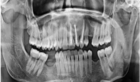
25-years-old female patient came to the Department with a chief complaint of swelling in the upper front teeth region teeth. The patient had no medical contraindication to dental treatment. Dental history revealed an incident of trauma to the upper front teeth region 12 years ago. Clinical examination revealed discolored tooth. The teeth were non-tender to percussion test. Upon radiographic examination, a large periapical defect involving root apices of 21 and 22, and lateral border of 23 was seen with complete loss of labial cortical plate. CBCT revealed its close proximation to the nasal floor.
Access this article online
Website: www.japer.in E-ISSN: 2249-3379
How to cite this article: Swati Mohanty, Sindhu Ramesh.Interdisciplinary
management of large periapical lesion: a case report. J Adv Pharm Edu Res 2017;7(3):303-307.
Figure 1: Pre-Operative Clinical Photograph
Figure 2:Palatal View- No Evident Lesion
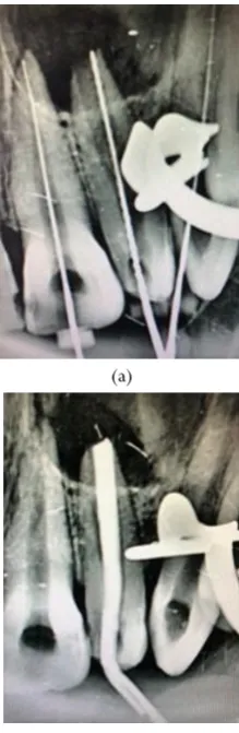
Figure 3: Pre-operative Radiograph Showing Periapical Lesion
in 21, 22 and Lateral Border of 23
(a)
(c)
Figure 4(a-c): Endodontic Treatment Was Carried Out For
21, 22 and 23
Figure 5: Pre-Operative CBCT Depicting Close Proximity of
the Lesion with The Nasal Floor
5.25% sodium hypochlorite was used to irrigate the canals during the canal preparation. Calcium hydroxide was used an intracanal medicament for 2 sittings before completing the root canal treatment. The root canals were obturated using gutta percha (Dentsply maillefer Ballaigues) and AH 26 sealer (Dentsply DeTrey GmbH, Philadelphia, USA) by the lateral condensation technique. Due to presence of immature Apex, Roll Cone Technique of Obturation was done in 22.
gluconate solution as mouth rinse for a period of 5 days. Suture removal was done 1 week later. Patient was reviewed at 3 months and 6 months during which there were no symptoms of pain, inflammation, or discomfort. Radiographically, hydroxyapatite particles were almost resorbed and replaced with new bone at the end of 12 months.
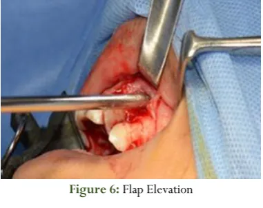
Surgical Management
Figure 6: Flap Elevation
Figure 7: Exposure of Defect
Figure 8: Apicectomy in 21
Figure 9: Retrograde Filling with MTA
Figure 10: PRF Obtained
Figure 11: PRF + Bone Graft
Figure 12: Sutures Placed
Figure 13:Pathological Tissue Sent For
Figure 14:1 Year Post-Operative
Discussion
Periapical lesions are usually composed of solid soft tissue (granulomas) or they have a semisolid, liquefied cystic area (bay cyst or true cyst). Therefore, to diagnose these lesions the least dense area of the radiographic lesion should be measured [5-9].
The combination of PRF in platelet gel form along with bone graft promoted wound healing, bone growth, maturation, graft stabilization and homeostasis, leading to an overall improvement in the handling properties of graft materials. PRF is a concentrated suspension of growth factors found in platelets which are involved in wound healing and are known to be promoters of tissues regenerations [4, 10]. Many authors had
concluded that, combination of growth factors in PRF along with bone graft had increased the bone density in many clinical trials [11-13]. PRF is a rich source of PDGF, TGF and IGF.TGF
known to stimulate biosynthesis of type-1 collagen, which induces deposition of bone matrix in vitro. PGDF is known to increase bone regeneration in calvarias defect when used along with bio-absorbable membrane as carrier [14]. IGF-1 is
synthesized and secreted by osteoblast. It stimulates bone formation by proliferation and differentiation, all these factors along with epidermal growth factor, increases the growth factor of human osteoblast. [15-17] DMBM is believed to act as an
osteo-conductive and osteo-inductive material and also as a bone growth promotor [11]. The DMBM was used in this study
because the bone morphogenetic proteins (BMPs) present in it are osteo-inductive that is, they induce differentiation of mesenchymal cells into cartilage and bone [13]. Deug et al in his
study evaluated histologically that there was enhanced new bone formation, cementum regeneration, new improved connective tissue growth and improved adhesion capacity with the decalcified freeze-dried bone grafted on the intrabony graft [18].
In this case report the role of both PRF and DMBM was placed in the bony defect, the benefit being superior proliferation of human periosteal cells thereby enhancing bone regeneration [19].
Progressive proliferation mode of PRF coagulation results in increased incorporation of circulating cytokines into the fibrin mesh which further augments wound healing [19-24]. The use of
bone graft material along with PRF might have accelerated the resorption of graft and would have induced the rapid rate of bone formation. However histologically studies are required to examine the nature of the newly formed tissues in the defect
and controlled long term clinical trials will be required to know the effect of this combination.
Conclusion
In this case report, there was radiographic evidence of almost complete bone healing of the periapical bone defect using PRF and DMBM in the lesion site after 1-year post surgery. Thus, this combination has the potential to accelerate bone healing and regeneration.
Conflict of Interest:
The authors stated no conflict of interest.References
1. Balaram Naik, Role of Platelet rich fibrin in wound healing: A critical review J Conserv Dent. 2013 Jul-Aug; 16(4): 284–293.
2. Boyapati L, Wang HL. The role of stress in periodontal disease and wound healing. Periodontol 2000.2007; 44:195–210.
3. Bashutski JD, Wang HL. Periodontal and endodontic regeneration. J Endod. 2009; 35:321–8.
4. Sunitha Raja V, Munirathnam Naidu E. Platelet-rich fibrin: Evolution of a second-generation platelet concentrate. Indian J Dent Res. 2008; 19:42–6. 5. Simon JHS. Incidence of periapical cysts in relation to
the root canal. J Endod 1980; 6:845– 8.
6. Nair PNR. New perspectives on radicular cysts: do they heal? Int Endod J 1998; 31:155–160.
7. McCall JO, Wald SS. Clinical dental radiology, 4th ed. Philadelphia: Saunders, 1954:234 –51.
8. Cunningham CJ, Penick EC. Use of a roentgenographic contrast medium in the differential diagnosis of periapical lesions. Oral Surg Oral Med Oral Pathol 1968; 26:96 –102.
9. Howell FV, De la Rosa VM. Cytologic evaluation of cystic lesions of the jaws: a new diagnostic technique. J Calif Dent Assoc 1968; 36:161– 6.
10. Chung CP, Kim DK, Park YJ, Nam KH, Lee SJ. Biological effects of drug-loaded biodegradable membranes for guided bone regeneration. J Periodontal Res. 1997; 32:172–5.
11. Ahmad Mogharehabed, Reza Birang, Nakisa Torabinia, Saman Nasiri, and Parichehr Behfarnia. Socket preservation using demineralized freezed dried bone allograft with and without plasma rich in growth factor: A canine study. Dent Res J (Isfahan). 2014; 11(4): 460–468.
13. Sonal Mishra.R K Singh, Shadab Mohammad, R Pradhan, U S Pal. A comparative evaluation of decalcified frieze dried bone allograft, hydroxyapatite and their combination in osseous defects of the jaw. J. Maxillofac. Oral Surg. 2010; 9(3):236–240.
14. Arnaud E, Morieux C, Wybier M, de Vernejoul MC. Potentiation of transforming growth factor (TGF-beta 1) by natural coral and fibrin in a rabbit cranioplasty model. Calcif Tissue Int. 1994; 54:493–8.
15. Pfeilschifter J, Oechsner M, Naumann A, Gronwald RG, Minne HW, Ziegler R. Stimulation of bone matrix apposition in vitro by local growth factors: A comparison between insulin-like growth factor I, platelet-derived growth factor, and transforming growth factor beta. Endocrinology. 1990; 127:69–75. 16. Hock JM, Centrella M, Canalis E. Insulin-like growth
factor I has independent effects on bone matrix formation and cell replication. Endocrinology. 1988; 122:254–60.
17. Baker NL, Carlo Russo V, Bernard O, D’Ercole AJ,
Werther GA. Interactions between bcl-2 and the IGF system control apoptosis in the developing mouse brain. Brain Res Dev Brain Res. 1999; 118:109–18. 18. Deug Han Kim, Ji Youn Hong, Eun Kyoung Pang.The
effect of freeze dried bone allograft and gell /putty type demineralized bone matrix on osseous bone
regeneration in rat calvarias defects. J Korean adac periodontal 2009; 39,349,358.
19. Gassling V, Douglas T, Warnke PH, Açil Y, Wiltfang J, Becker ST. Platelet-rich fibrin membranes as scaffolds for periosteal tissue engineering. Clin Oral Implants Res. 2010; 21:543–9.
20. Morse DR, Patnik JW, Schacterle GR. Electrophoretic differentiation of radicular cysts and granulomas. Oral Surg Oral Med Oral Pathol 1973; 35:249 – 64. 21. Trope M, Pettigrew J, Petras J, Barnett F, Tronstad L.
Differentiation of radicular cyst and granulomas using computerized tomography. Endod Dent Traumatol 1989; 5:69 –72.
22. Shrout MK, Hall JM, Hildebolt CE. Differentiation of periapical granulomas and radicular cysts by digital radiometric analysis. Oral Surg Oral Med Oral Pathol 1993; 76:356 – 61.
23. Camps J, Pommel L, Bukiet F. Evaluation of periapical lesion healing by correction of gray values. J Endod 2004; 30:762– 6.