Łukasz Szylberg
1, A–C, E, Marlena Janiczek
1, B–D, Aneta Popiel
1, B–D,
Andrzej Marszałek
2, E, FLarge Bowel Genetic Background and Inflammatory
Processes in Carcinogenesis – Systematic Review
1 Department of Clinical Pathomorphology, Collegium Medicum in Bydgoszcz, Nicolaus Copernicus University
in Toruń, Poland
2 Department of Oncological Pathology, Poznan University of Medical Sciences and Greater Poland Oncology
Center, Poland
A – research concept and design; B – collection and/or assembly of data; C – data analysis and interpretation; D – writing the article; E – critical revision of the article; F – final approval of article; G – other
Abstract
Colorectal cancer (CRC) has become the third most common cancer in developed countries. Each year more and more people die from CRC. CRC is also one of the most effectively studied topics in recent years. It has been found that the key phenomena in CRC development are genetic and inflammatory processes. Well-known genetic bases for the carcinogenesis of CRC include chromosomal changes characteristic of the chromosomal instability pathway which correlates with specific and well-defined genetic alterations (such as APC, K-RAS, DCC and p53) and genomic instability characteristics for the mutator pathway focused on KRAS and BRAF mutations. Recent studies have highlighted the impact of inflammation in CRC, especially elevated levels of pro-inflammatory cyto-kines. Among important risk factors of colon carcinogenesis are colorectal polyps, which are currently the subject of intense research. Recent studies have shown that different adenomas are characterized by different pathways of carcinogenesis as well as diverse COX-2 expression in various polyps. Understanding the mechanism of inflam-matory processes in CRC parallel to basic genetic alterations might allow for effective and targeted treatment (Adv
Clin Exp Med 2015, 24, 4, 555–561).
Key words: colorectal cancer, inflammation, carcinogenesis, genetic alterations.
EDITORIAL
Adv Clin Exp Med 2015, 24, 4, 555–561
DOI: 10.17219/acem/31239 © Copyright by Wroclaw Medical University ISSN 1899–5276
cancer and chronic inflammation [3]. Recently, the effects of the inflammatory process on neopla-sia development were demonstrated in endometri-al, cervicendometri-al, ovarian, breast, prostate and colon tu-mors [4]. Innate immune system cells are included in the microenvironment of tumors. These cells secrete proinflammatory cytokines, chemokines, growth factors and reactive oxygen species that could cause DNA damage [5, 6]. The inflammato-ry process most likely has a key role in the patho-genesis of CRC, especially in its promotion. This is an important factor in cancer staging and progno-sis. The aim of currently research is to discover the factor initiating the inflammatory process, which potentially could be used effectively for preventive activities. Another important aspect of research is focused on the determination of the exact time and Colorectal cancer (CRC) has become the third
most common cancer in developed countries [1]. CRC is also one of the most effectively studied topics in recent years. The pathogenesis of CRC is still not fully understood. Initially it was thought that the key role in its pathogenesis involved ge-netic mutations. A number of studies have been performed to confirm this hypothesis. Karoliina Stefanius’ study shows two distinct ways of CRC carcinogenesis, depending on the existing muta-tion [2]. Although there is a well-known genetic basis for the carcinogenesis of colorectal cancer, this knowledge does not allow us to introduce new, effective CRC treatment modalities.
location of the inflammation that leads to carcino-genesis. Most likely, understanding the mecha-nisms of inflammatory processes in CRC and com-bining it with a genetic background should allow for a reduction in the number of cases and for ef-fective treatment.
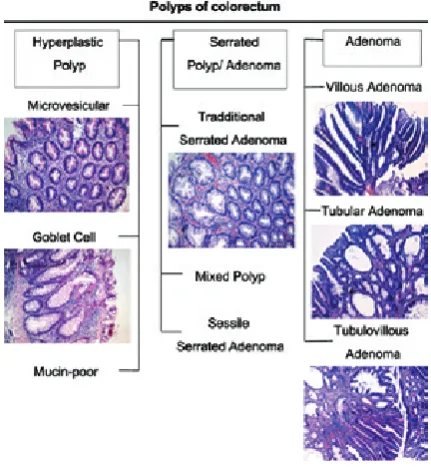
Studies of CRC have been focused on sporad-ic and familial cases. Hereditary colon cancer pre-disposition is confirmed in less than 5% of all CRC patients. Detailed information in such cases pro-vides the opportunity for isolation of the genetic, environmental, and epigenetic factors which deter-mine the phenotypes of the cancer. An important phenomenon of colon carcinogenesis is its devel-opment preceded by colorectal adenomas, which are currently under intense research. According to the data published, almost half of the population will develop at least one benign adenomatous co-lon polyp during his or her lifetime [7, 8]. Three percent of these patients will develop CRC. Ade-nomas are considered an important risk factor in CRC and the removal of adenomatous polyps has been shown to reduce the risk of development of CRC. The polyps of colon cancer are divided into 3 subgroups according to their malignant poten-tial (Fig. 1). Namely, there are hyperplastic polyps (HPP) with the least malignant potential, serrat-ed polyps/adenoma (SA) and adenoma with a high malignant potential. Adenomas are classified into 3 types according to their morphological nature. These are villous (VA), tubular (TA) and tubu-lovillous (TVA). An interesting subgroup of le-sions are serrated adenomas, which are discussed in conjunction with specific conditions such as ad-enoma and hyperplastic polyps. The term serrated
adenoma was introduced by Longacre and Feno-glio-Preiser [9]. Serrated adenoma are divided in-to 3 subtypes. These are traditional serrated ade-noma, sessile serrated adenoma (SSA) and mixed polyps (MP).
Hyperplastic Polyps
HP, typically are small, smooth, sessile le-sions which are usually located in the distal colon and rectum and are diagnosed in patients above 40 years of age. The hyperplastic polyp has a lot of crypts with a convoluted luminal pattern and im-mature proliferative cells on the lower portion. Su-perficial serration could be observed. Regarding their cellular composition, polyps can be classi-fied as microvesicular, goblet cells or mucin-poor variants.
Traditional Serrated
Adenoma
These were described for the first time in 1984 as a group of lesions with features of both hyperplastic and adenomatous polyps and were thought as “mixed hyperplastic adenomatous polyps” [10]. Traditional serrated adenoma have a serrated architecture with characteristic cy-tological features of central, elongated nuclei, mild pseudostratification and eosinophilic cyto-plasm and with microscopic impression of serra-tion in crypts. In contrast to convenserra-tional adeno-mas, traditional serrated adenomas might present more hyperchromatic nuclei in the lower crypts than those on the surface and more basophilic cy-toplasm. The common feature of TSAs and con-ventional adenomas is location. Eighty percent of TSAs are located in the left colon, mostly the rec-tosigmoid region [10, 11].
Sessile Serrated Adenoma
This term was recently introduced by Torla-kovic and colleagues [10]. SSA is the premalignant sessile lesion of the colon with crypts without tra-ditional dysplasia in contrast to adenomas or tradi-tional serrated adenomas. The basal crypts are not compact and with architecture different from the hyperplastic polyps. The crypts usually are dilated on the bottom and narrowed in the middle part. The area of proliferation is not symmetrical and mitoses could be present in the upper part of the crypts. Generally SSA can be found in the right co-lon of middle-aged females [10, 11].
Traditional Adenoma
This is a benign neoplasm of glandular ori-gin lined by columnar epithelium without chief and parietal cells. Serration is not the dominant feature of traditional adenomas. The incidence of TA increases with age. According to published da-ta, about 20–30% of patients with polyps lesions developed them under 40 years of age, while the occurrence of colorectal adenomas after 60 years of age increases to 40–50%. There is no difference between the incidence between women and men. There is a family predisposition to the occurrence of adenomas. All such lesions arise from adeno-matous proliferation of epithelial cells. Addition-ally, dysplasia could be found, which range from low to high grade, reflecting neoplastic transfor-mation potential.
Mixed Polyp
This change has the elements of the structure of a typical hyperplastic polyp, and focal AT, AV, ATV or TSA.
Influence of Genetic
Mutation in Neoplasia
of Colorectal Cancer
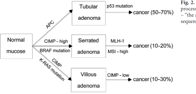
Based on Fearon and Vogelstein’s research, we can define the main multi-step process for CRC development as “the adenoma-carcinoma sequence” [12] (Fig. 2). Today we also call it the CIN (chromosomal instability) pathway, which correlates to specific genetic events within the in-volved tissue. The other two main pathways, in contrast to chromosomal changes, show genom-ic instability called MSI (mgenom-icrosatellite instability) – the process characteristic for mutator pathways.
It has been well proven in HNPCC (hereditary non-polyposis colorectal cancer) syndrome, in which the cancer is caused by a germ-line mu-tation of genes engaged in the mismatch repair (MMR) system. The mutator pathway it is also called the “serrated pathway” and related to spo-radic CRCs, in which an aberration is present in MMR and in other DNA repair systems. These events are caused by hypermethylation. Tumors caused by the aforementioned pathway also show MSI [12]. In a classic “adenoma-carcinoma se-quence”, every step from the normal mucosa to-wards the carcinoma was attributed to specif-ic and well-defined genetspecif-ic alterations such as APC (adenomatous polyposis coli), oncogenes K-RAS, DCC (deleted in CRC) and p53. The first step includes a loss of APC suppressor gene func-tion. APC contains 15 exons and it is mutated in 60% and 82% of colon and rectal cancers, re-spectively [13]. APC function is closely linked to β-catenin. Normally, it stimulates β-catenin de-struction in proteasomes. If this gene is disrupted, it leads to β-catenin accumulation and transloca-tion to the cell nucleus, where it activates tran-scription of several genes such as MYC and cy-clin D, which stimulate proliferation [2].
The next genetic alteration is RAS mutation. As K-RAS, it leads to disturbances in the genet-ic material, giving rise to cancerogenesis. Proto-oncogene located at 12p12.1, which encodes the 21-kDa GTP-binding protein, plays an important role in transmitting extracellular signals into an in-tracellular signal. Transduction mediates cellular responses to growth signals by switching between active GTP and inactive GTP forms of RAS mu-tation. Mutated RAS in its active form stimulates cells to mitosis and inhibits apoptosis of cells. RAS mutations are present with the same frequency in large adenomas and carcinomas, but with lower numbers in small adenomas. This might lead to the conclusion that RAS mutation is a late event in the “adenoma-carcinoma sequence” [14].
Fig. 2. The main multistep
Another genetic alteration leading to can-cer is DCC, located at 18q21.1. About 60–70% of colorectal cancers show allelic losses in DCC. It is not fully understood how this gene is linked to cancerogenesis [15]. The study results point out that the wild-type DCC, but not the mutant one, induces apoptosis and activates caspase-3, and that DCC expression induces a rapid G2/M cell cycle arrest in some cell lines [2].
The most common mutation, present in 70– –80%, is loss of the function of TP 53. Mutation of TP 53 usually occurs at the time of the transition from adenoma to cancer [15]. The frequency of TP 53 mutations have a propensity to increase with the progression of the lesion [13]. Thus changes are found in 4–26% of adenomas, 50% of adeno-mas with invasive foci, and in 50–75% of CRCs. The P53 protein induces G1 cell-cycle inhibition to facilitate DNA repair during the replication of cells exposed to environmental or oncogenic stress [2].
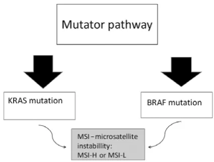
In recent years, several scientific projects have focused on the mutator pathway, which enclos-es 2 subtypenclos-es (Fig. 3), one characterized by K-RAS and the other one by BRAF mutation. However, the same feature is present, MSI, but at different lev-els. Microsatellite instability is a result of a defec-tive MMR (mismatch repair) system that leads to slippage DNA polymerase during DNA replication. According to the level of MSI, the tumors differ in clinical and pathological features. There is MSI (microsatellite instability) at high (MSI-H) or low (MSI-L) levels. MSI-H is a result of a loss of DNA mismatch repair activity. MSI-L is a consequence of MMR deficiency. Of note is the correlation between the occurrence K-RAS mutation and MSI-L or BRAF mutation and the presence of MSI-H. The mutator pathway has two variants. One uses K-RAS mutations and the other BRAF mutations. A com-mon feature for both is the presence of MSI. The mu-tator pathway presents a phenomen of the CpG-is-land methylator pathway (CIMP). Normally, DNA
methylation takes place at cytosine located 5’ to guanosine of the dinucleotide sequence CpG. Then cancer-related DNA hypermethylation is present in CG-rich areas called CpG islands. It might result in transcriptional silencing of the gene. CIMP is sub-divided into CIMP- high, CIMP-low and CIMP- -negative, according to the amount of methyla-tion. CIMP-high exhibits hypermethylation of the promoter region the hMLH-1 (human mutL ho-molog 1) gene and silences this gene, leading to MSI-H [16]. Additionally to CpG island methyl-ation and MSI-H, a typical feature of the mutator pathway is mutation that activates the BRAF gene. On the other hand, an active RAS-gene stimulates the cell to mitosis and inhibits apoptosis. The next group of mutator pathways are tumors with K-RAS mutation and low levels of CpG island methylation, e.g. the MSI-L phenotype and some features sim-ilar to carcinomas developing through the adeno-ma-carcinoma sequences [2].
There are much larger unresolved questions in this pathway than in the BRAF mutation pathway. The substantial MGMT (methylguanine-DNA) in-activation by hypermethylation can support a fail-ure to repair G:C to A:T transition. This highlights the DNA MMR systems and increases the muta-tion level [2].
Recent studies have shown that the mucosa in a healthy population and in patients with hy-perplastic polyps have a normal expression of hMLH1 and hMSH2. A decrease in the expression of hMLH1 and hMSH2 occurs in patients with ad-enomas and adenocarcinomas. The decrease in the expression of hMLH1 was observed in patients with microsatellite instability (MSI) in CRC [28]. So, inactivation of the genes taking part in MMR lead to MSI in CRC. A reduction of hMLH1 activi-ty is also related to DNA methylation, as shown by Herman et al. [17].
Inflammatory Processes
in Colorectal Cancer
Inflammatory processes play an important role in the development and progression of inva-sive colorectal tumors, which additionally are the basis by which we can determine their progres-sion and outcome [18]. The inflammatory pro-cess is connected to elevated levels of anti-inflam-matory cytokine such as interleukin-4 (IL-4) and pro-inflammatory cytokines such as interleukin-6 (IL-6), interleukin-17 (IL-17), tumor necrosis fac-tor (TNF), interleukin-23 (IL-23), interleukin-12 (IL-12) and interleuk8 (IL-8). There is also in-terleukin-10 (IL-10), but researchers’ opinions on its role are diverse. Some authors consider it as
a factor in pro-inflammatory processes and others say it has a suppressive action. IL-6 has a lot very important functions in cancer. It is produced by both tumor and normal cells [19]. It is pleiotropic and demonstrates both a pro- and anti-inflamma-tory role [20]. IL-6 is regarded as correlated with colon cancer increased incidence. Its level in se-rum in patients with cancer is higher in compari-son to healthy controls. Recent studies have shown that it has a significant but not primary role in the pathogenesis of CRC [19, 20]. According to pub-lished data, macrophages are activated by the tu-mor cells to produce consistently large amounts of IL-6, which affects the production of IL-10 via the STAT3 signaling pathway [19]. Currently, IL-10 still has a controversial dual role in carcinogenesis. On one hand, it is regarded as an immunosuppres-sive cytokine that affects tumor growth. On the other hand, there are many studies that confirm IL-10’s effectiveness in the host response against cancer [21]. However, all researchers agree that its serum levels are significantly elevated in patients with CRC compared to healthy people and post-operative patients. The role of IL-10 is significant in cancer development and prognosis, which is confirmed by several studies [21–24]. Cecev et al. shows that IL-10 has an effect on the differentia-tion of cancer. Immunohistochemical studies have shown that the level of IL-10 is higher in well-dif-ferentiated and moderately difwell-dif-ferentiated tumors compared to poorly differentiated ones. There-fore, such findings support the protective role of IL-10 in the development and progression of CRC [23]. On the other hand, in recent research based on the level of IL-10 in all of the 4 stages of the TNM classification of CRC, significant differences were found between patients and healthy individ-uals. Moreover, colon cancer patients in the fourth stage of the clinical advancement of the disease have a higher level of IL-10 in serum compared to the other 3 stages. The summation of such studies indicates that IL-10 has a pro-inflammatory role that is linked to tumor growth [21, 22].
Nayko et al. shows that the sex of the patient and the tumor location within the colon vs. the rectum did not affect the differences in IL-10 se-rum levels [22]. The effects of IL-10 are affected by other pro-inflammatory cytokines such as IL-12. Their activity is described as antagonistic. The in-crease in IL-10 is associated with dein-creased pro-duction of IL-12. This behavior was observed in CRC. Recent studies have shown that patients with CRC have decreased levels of IL-12. This cytokine level decreases respectively in the individual clini-cal stages of the disease. In the fourth stage, the lev-els are lowest when compared to the other 3 on the TNM scale. IL-12 is produced mainly by Th1 cells,
and to a lesser extent by B cells, mast cells and mac-rophages. IL-12 affects the production of cytokines such as TNF-alpha and IFN-gamma. Additional-ly, IL-10 inhibits the production of the latter by suppression of IL-12 transcription. This last men-tioned phenomenon is important, as IL-12, when it is reduced, can be used as a marker predicting the presence of cancer metastases in lymph nodes [24]. The bioactive form of IL-12 is IL-12p70, which consists of 2 subunits, namely p35 and p40. The p40 subunit can combine with a p19 subunit to form IL-23, which, like IL-12p70, affects the ac-tivation of memory T cells to produce IFN-gamma and the production of pro-inflammatory cytokines such as IL-17 by Th17. Recent studies compared the serum levels of IL-23 and IL-17 in patients with CRC who were divided into the 4 stages according to the TNM scale and their post-operative levels. It showed that the level of IL-23 is elevated in pa-tients with CRC compared to healthy people. Ad-ditionally, the authors observed that there were no differences between level of IL-23 and IL-17 in dif-ferent clinical stages and they do not changed af-ter surgery [22].
Another important cytokine in the inflammato-ry process is IL-4. This anti-inflammatoinflammato-ry cytokine is produced mainly by Th-2 cells and its level is sig-nificantly higher in patients with CRC compared to healthy subjects [25]. IL-4 inhibits the expression of E-cadherin and carcinoembryonic antigen (CEA). This affects the inhibition of cell-cell adhesion of co-lon cancer cells. A study conducted by Kanai et al. shows that cancer cells treated with IL-4 levels de-creased E-cadherin and CEA molecules compared to untreated cells. Abnormal adhesion is responsi-ble for metastasis and invasion in CRC. However, studies haven’t shown that inhibition of adhesion of cancer cells has an effect on metastasis and inva-sion. Additionally, an imbalance between Th1 and Th2 cytokine levels is important in prognosis. It was found that patients with CRC whose cytokines were produced mainly by Th2 cells predisposes them to liver metastases [26].
Expression
of Cyclooxygenase-2
in Serrated Adenomas,
Non-Serrated Adenomas,
Hyperplastic Polyps
and Colorectal Cancers
The inflammatory process is reflected by the amount of cytokines and cyclooxygenase-2 (COX-2) protein [27]. COX-2 is a prostaglan-din synthetase enzyme. It is involved in the con-version of arachidonic acid to prostaglandin H2 (PGH2), an important precursor of prostacyclin
and thromboxane A2. The expression level of COX-2 is elevated in many cancers (e.g. in gall-bladder carcinomas along with increased angio-genesis) [28]. PGH2, the product of COX-2, is
converted by prostaglandin E2 synthase into pros-taglandin E2 (PGE2), which can stimulate cancer
progression. Expression of COX-2 is regulated by IL-1β [26]. IL-1β is a pro-inflammatory cytokine produced by activated macrophages. This inter-leukin affects the activity of cells including cell proliferation, differentiation and apoptosis.
Many studies have shown a significant in-crease in COX-2 in patients with CRC [29–31]. It was proved that COX-2 plays an important role in CRC, highlighted by the fact that the reg-ular use of COX-2 inhibitors, such as aspirin, re-duces the risk of development of CRC [31]. Im-munoexpression of COX-2 does not emerge in normal mucosa and mucosa with hyperplas-tic polyps, but there is an increase in the expres-sion of COX-2 in adenomas and adenocarcino-mas. In these studies, no correlation between the expression of COX-2 and age, sex, location and degree of dysplasia or adenoma size was found. Further studies on the expression of COX-2 in colon neoplasia conducted by Kawasaki and Bal-binotti have demonstrated that increased expres-sion of COX-2 is not related to serrated leexpres-sions. They showed that COX-2 expression was higher in non-serrated adenomas in comparison to HPP, MP with SSA and adenoma and SSA. The sub-types of non-serrated adenoma such as TVA, VA and TA showed no significant differences in the expression of COX-2. Later it was shown that in-creased expression of COX-2 in non-serrated ad-enomas is similar to that of traditional serrated adenomas. Over-expression of COX-2 in colorec-tal adenocarcinomas did not show any differenc-es related to cancers with or without serrations, but was still higher in comparison to non-serrat-ed adenomas [30, 31].
The Role of Anti-
-Inflammatory Agents
in Carcinogenesis
It has been observed that using inhibitors of the COX-2 protein might have benefits in the prevention and treatment of gastrointestinal cancers [32, 33]. COX-2 is the target for non-steroidal anti-inflam-matory drugs (NSAIDs) and selective cyclooxygen-ase-2 (COX-2) inhibitors (COXIBs). The risk of ma-lignancies among the elderly (especially over 75 years of age) significantly increases [34]. Elderly patients al-so often have several parallel diseases such as arthritis, diabetes, cardiovascular disease or psoriasis. In all of them, chronic inflammation plays a major role, thus anti-inflammatory agents are the basic method of treatment. However, anti-inflammatory therapy may affect the results of cancer therapy [32, 35]. Low doses of NSAIDs have a favorable impact on many disease treatments, but it can also have negative effects, such as bleeding from the upper and lower parts of the gas-trointestinal tract. The risk also increases with simul-taneous use of drugs such as NSAIDs, selective cyclo-oxygenase-2 inhibitors, and corticosteroids or other anticoagulants. Of note, low doses of aspirin (< 325 mg per day) are recommended for patients at high risk of developing blood clots, to help prevent heart attacks, strokes, and blood clot formation. In addi-tion, there is growing evidence that long-term aspi-rin therapy reduces the risk of colon cancer. High doses of aspirin (> 325 mg per day) have analgesic and strong anti-inflammatory effects. Ultimately, in-dividual assessment of the benefits and risks of using NSAIDs is the key to effective treatment [33, 36–38].
Kim et al., in their study on mouse mod-els, introduced a new family member, namely COXIB CG100649. It inhibits the activity of both COX-2 and carbonic anhydrase-II-I (I-II-CA). CG100649 inhibits the development of premalig-nant and maligpremalig-nant colorectal lesions via inhibi-tion of tumor cell proliferainhibi-tion. A long-term safety profile of CG100649 has not yet been established [39]. The use of COX-2 inhibitors also causes se-rious consequences in the form of cardiovascular events such as hypertension and kidney toxicity. The risk increases in patients with pre-existing risk factors for cardiovascular disease [40].
The Problem of Treatment
of Colorectal Cancer
in Geriatric Patients
reaches its peak in people over 65 years [42]. Cancer is a leading cause of death in older women and men aged 60–79 years [43]. Geriatric patients are special as, in comparison to younger ones, they may have comorbidities, polypharmacy and reduced physio-logical tissues and organs reserves. Their treatment is a challenge for the oncologist in selecting appropriate treatment and predicting the long-term treatment results. Older age is also associated with increased toxicity of treatment and its side effects [44, 45]. Yet another problem for oncologists in the man-agement of such patients is the low number of old-er patients in large clinical trials for the treatment of colorectal cancer, even though they constitute the majority of those suffering. However, in recent years there has been a change in the approach to geriatric patients and they are also included in clinical trials [46]. In addition, nowadays in developed countries, it has been noted that older people with cancer are often in better physical form and they are able to tol-erate even the most intensive treatment. However, in early stages of the disease, clinical dilemmas ap-pear regarding the ability of elderly patients to un-dergo successful surgical and therapeutic treatment and the evaluation of the risk-benefit ratio of adju-vant chemotherapy giving the benefit of prolonged life free from disease. According to Foster et al., on-cologists planning the treatment of older people suf-fering from colon cancer are more likely to choose conservative treatment in these patients instead of the standard treatment of cancer, despite the pa-tient’s good condition [44]. The author also draws attention to the need for a more profound look at ge-riatric patients’ oncologists when choosing a cancer treatment in which age is not a key determinant of therapy. For clinical practice, applying a method of comprehensive geriatric assessment (CGA) may fa-cilitate this process and improve longevity estimates, which may have an impact on the treatment of the patient [44]. Geriatric assessment (CGA) is a mul-tidisciplinary assessment of the functional status of the patient, comorbidities, mental status, social sup-port, cognitive function and nutritional status. Nev-ertheless, this is a very time-consuming method, so the alternative simplified method of geriatric assess-ment is often used by oncologists, which is based on the division of patients into “fit” and “frail” catego-ries [44, 45]. A patient referred to as “fit” is in good condition and has limited comorbidities and geri-atric syndromes. They are at increased risk of can-cer incidence and mortality due to cancan-cer. Patients referred to as “frail” are in poor general condition and have many comorbidities and geriatric syn-dromes. These patients have a limited life expectan-cy due to other diseases and are more susceptible to morbidity and mortality from other comorbidi-ties, and not only the cancer. And an approach to
therapy has been proposed with these two different categories, with more aggressive treatment for “fit” patients and more conservative therapies for “frail” patients. Dotan et al. gave his analysis of the benefits of surgery, adjuvant chemotherapy and aggressive surveillance in “fit” older patients with early stag-es of colorectal cancer [45]. The rstag-esults were com-parable to younger patients. The decision is more complicated for older patients assigned to the “frail” category, for whom a treatment strategy should be determined by a joint decision of the patient and on-cologist. Treatment of elderly patients with metastat-ic colorectal cancer requires careful rethinking of the strategy of treatment, overall, the demonstrated clin-ical benefit at the expense of increased toxicity when the agents are approved for use in the elderly pop-ulation. The coefficient of risk/benefit of treatment should be considered prior to treatment [45]. Ad-ditional clinical trials targeting the older population are needed to increase our knowledge of the opti-mal management of elderly patients with colorectal cancer, and to be able to know the impact of treat-ment of comorbidities on oncological treattreat-ment for “frail” patients.
Final Remarks
References
[1] Spann S, Levin B, Rozen P, Young G: Colorectal cancer: How big is the problem, why prevent it and how might
it present? In: Colorectal cancer in clinical practice: prevention, early detection and management. Eds.: Rozen P, Young G, Levin B. Martin Dunitz, London 2002, 1–13.
[2] Stefanius K: Colorectal carcinogenesis via serrated route. Acta Univ Oul D 2011, 1091.
[3] Balkwill F, Mantovani A: Inflammation and cancer: back to Virchow? Lancet 2001, 357, 539–545. [4] Morrison WB: Inflammation and Cancer: A Comparative View. J Vet Intern Med 2012, 26, 18–31.
[5] Goswami B, Rajappa M, Sharma M: Inflammation: its role and interplay in the development of cancer, with
spe-cial focus on gynecological malignancies. Int J Gynecol Cancer 2008, 18, 591–599.
[6] Coussens LM, Werb Z: Inflammation and cancer. Nature 2012, 420, 860–867.
[7] Jemal A, Siegel R, Ward E, Murray T, Xu J, Smigal C, Thun MJ: Cancer Statistics. CA Cancer J Clin 2006, 56,
106–130.
[8] Kwong LN, Dove WF: APC and its modifiers in colon cancer. Adv Exp Med Biol 2009, 656, 85–106.
[9] Longacre TA, Fenoglio-Preiser CM: Mixed hyperplastic adenomatous polyps/serrated adenomas. Am J Surg
Pathol 1990, 14, 524–537.
[10] Torlakovic EE, Gomez JD, Driman DK: Sessile Serrated Adenoma (SSA) vs. Traditional Serrated Adenoma
(TSA). Am J Surg Pathol 2008, 32, 21–44.
[11] Valerie P, Bauer MD, Harry T: Management of Serrated Adenomas and Hyperplastic Polyps. Clin Colon Rectal
Surg 2008, 21, 273–279.
[12] Fearon ER, Vogelstein B: A Genetic model for Colorectal Tumorigenesis. Cell 1990, 61, 759–767.
[13] Sandmeier D, Benhattar J, Martine P: Serrated polyps of the large intestine: a molecular study comparing sessile
serrated adenomas and hyperplastic polyps. Histopathology 2009, 55, 206–213.
[14] Krasinskas AM: EGFR Signalling in colorectal carcinoma. Patholog Res Int 2011.
[15] Itoh F, Imai K: Genetic diagnosis of colorectal cancer. Hokkaido Iqaku Zasshi 1996, 71, 9–14.
[16] Weisenberger HT: Genome-scale analysis of aberrant Dna methylation in colorectal cancer. Genome Res 2012,
22, 271–282.
[17] Herman JG, Umar A, Polyak K, Graff JR, Ahuja N, Issa JP, Markowitz S, Willson JK, Hamilton SR, Kinzler KW, Kane MF, Kolodner RD, Vogelstein B, Kunkel TA, Baylin SB: Incidence and functional consequences of hMLH1
promoter hypermethylation in colorectal carcinoma. Proc Natl Acad Sci U S A 1998, 95, 6870–6875.
[18] McLean MH, Murray GI, Stewart KN, Norrie G, Mayer C: The Inflammatory Microenvironment in Colorectal
Neoplasia. PLoS ONE 2011, 6, e15366.
[19] Herbeuval JP, Lelievre E, Lambert C, Dy M, Genin C: Recruitment of STAT3 for Production of IL-10 by Colon
Carcinoma Cells Induced by Macrophage-Derived IL-6. J Immunol 2004, 172, 4630–4636.
[20] Becker C, Fantini MC, Wirtz S: IL-6 Signaling Promotes Tumor Growth in Colorectal Cancer. Cell Cycle 2005, 4, 217–220. [21] Stanilov N, Stankova N, Miteva L, Jovchev J, Deliyski T, Stanilova S: Role of IL-12P40 and IL-10 in progression
of colorectal cancer. TJS 2010, 8, 132–136.
[22] Stanilov N, Miteva L, Deliysky T, Jovchev J, Stanilova S: Advanced Colorectal Cancer Is Associated With
Enhanced IL-23 and IL-10 Serum Levels. Labmedicine 2010, 41, 159–163.
[23] Cacev T, Radosevic S, Krizanac S, Kapitanović S: Influence of interleukin-8 and interleukin-10 on sporadic colon
cancer development and progression. Carcinogenesis 2008, 29, 1572–1580.
[24] O’Hara R J, Greenman J, MacDonald AW, Gaskell KM, Topping KP, Duthie GS, Kerin MJ, Lee PW, Monson JR:
Advanced colorectal cancer is associated with impaired interleukin-12 and enhanced interleukin-10 production. Clin Cancer Res 1998, 4, 1943–1948.
[25] Galon J, Costes A, Sanchez-Cabo F, Kirilovsky A, Mlecnik B, Lagorce-Pagès C, Tosolini M, Camus M, Berger A, Wind P, Zinzindohoué F, Bruneval P, Cugnenc PH, Trajanoski Z, Fridman WH, Pagès F: Type, density, and
location of immune cells within human colorectal tumors predict clinical outcome. Science 2006, 313, 1960–1964.
[26] Kanai T, Watanabe M, Hayashi A, Nakazawa A, Yajima T, Okazawa A, Yamazaki M, Ishii H, Hibi T: Regulatory
effect of interleukin-4 and interleukin-13 on colon cancer cell adhesion. Br J Cancer 2000, 82, 1717–1723.
[27] Wiiliam CS, Mann M, Dubois RN: The role of cycloxygenases in inflammation, cancer and development.
Oncogene 1999, 18, 7906–7916.
[28] Legan M: Cyclooxygenase-2, p53 and glucose transporter-1 as predictors of malignancy in the development of
gallbladder carcinomas. Bosn J Basic Med Sci 2010, 10, 192–196.
[29] Gustafsson Asting A, Caren H, Andersson M, Lönnroth C, Lagerstedt K, Lundholm K: COX-2 gene expression
in colon cancer tissue related to regulating factors and promotor methylation status. BMC Cancer 2011, 11, 238.
[30] Balbinotti RA, Ribeiro U, Saka P, Safatle-Ribeiro AV, Balbinotti SS, Scapulatempo C, Alves VA, Corbett CE, Carrilho FJ: hMLH1, hMSH2 and Cyclooxygenase-2 (Cox-2) in Sporadic Colorectal Polyps. Anticancer Res 2007,
27, 4465–4472.
[31] Kawasaki T, Nosho K, Ohnishi M, Suemoto Y, Glickman JN, Chan AT, Kirkner GJ, Mino-Kenudson M, Fuchs CS, Ogino S: Cyclooxygenase-2 overexpression is common in serrated and non-serrated colorectal adenoma, but
uncommon in hyperplastic polyp and sessile serrated polyp/adenoma. BMC Cancer 2008, 8, 33.
[32] Menter DG, Schilsky RL, DuBois RN: Cyclooxygenase-2 and cancer treatment: understanding the risk should be
worth the reward. Clin Cancer Res 2010, 16, 1384–1390.
[33] Romagnolo DF, Papoutsis AJ, Selmin O: Nutritional targeting of cyclooxygenase-2 for colon cancer prevention.
[34] Pelzer O, Kielan W: Risk analysis for the surgical treatment of colorectal cancer in elderly patients undergoing
scheduled and urgent interventions. Pol Przegl Chir 2014, 86, 61–67.
[35] Rayburn ER, Ezell SJ, Zhang R: Anti-Inflammatory Agents for Cancer Therapy. Mol Cell Pharmacol 2009, 1,
29–43.
[36] Peura DA, Wilcox CM: Aspirin and proton pump inhibitor combination therapy for prevention of cardiovascular
disease and Barrett’s esophagus. Postgrad Med 2014, 126, 87–96.
[37] Sostres C, Lanas A: Gastrointestinal effects of aspirin. Nat Rev Gastroenterol Hepatol 2011, 8, 385–394.
[38] Lanas A: Gastrointestinal bleeding associated with low-dose aspirin use: relevance and management in clinical
practice. Expert Opin Drug Saf 2011, 10, 45–54.
[39] Kim SH, Margalit O, Katoh H, Wang D, Wu H, Xia D, Holla VR, Yang P, DuBois RN: CG100649, a novel
COX-2 inhibitor, inhibits colorectal adenoma and carcinoma growth in mouse models. Invest New Drugs 2014, PMID: 25085205.
[40] Cooper K, Squires H, Carroll C, Papaioannou D, Booth A, Logan RF, Maguire C, Hind D, Tappenden P:
Chemoprevention of colorectal cancer: systematic review and economic evaluation. Health Technol Assess 2010, 14, 1–206.
[41] Lin LL, Hahn SM: Combined modality therapy in the elderly population. Curr Treat Options Oncol 2009, 10,
195–204.
[42] U.S. Census Bureau: National Population Projections. Percent Distribution of the Projected Population by
Selected Age Groups and Sex for the United States: 2010 to 2050. http://www.census.gov/population/www/projec-tions/summarytables.html, accessed 2009.
[43] Jemal A, Siegel R, Xu J, Ward E: Cancer statistics 2010. CA Cancer J Clin 2010, 60, 277–300.
[44] Foster JA, Salinas GD, Mansell D, Williamson JC, Casebeer LL: How Does Older Age Influence Oncologists’
Cancer Management? The Oncologist 2010, 15, 584–592.
[45] Dotan E, Browner I, Hurria A, Denlinger C: Challenges in the Management of Older Patients with Colon Cancer.
J Natl Compr Canc Netw 2012, 10, 213–225.
[46] Lewis JH, Kilgore ML, Goldman DP, Trimble EL, Kaplan R, Montello MJ, Housman MG, Escarce JJ:
Participation of patients 65 years of age or older in cancer clinical trials. J Clin Oncol 2003, 21, 1383–1389.
Address for correspondence:
Łukasz Szylberg
Department of Clinical Pathomorphology Collegium Medicum in Bydgoszcz Nicolaus Copernicus University in Toruń Skłodowskiej-Curie 9
85-094 Bydgoszcz Poland
Tel.: +48 52 58 54 200 E-mail: [email protected]
Conflict of interest: None declared