Turkish Journal of Fisheries and Aquatic Sciences 16: 513- 522 (2016)
www.trjfas.org ISSN 1303-2712 DOI: 10.4194/1303-2712-v16_3_03
RESEARCH PAPER
© Published by Central Fisheries Research Institute (CFRI) Trabzon, Turkey in cooperation with Japan International Cooperation Agency (JICA), Japan
Glycoprotein Attributed Effect of Flatfish Extract on 3T3-L1 Preadipocyte
Differentiation
Introduction
Fish is one of the major marine products in Hokkaido, Japan. Although the Japanese eat various kinds of fish species, most of the bones, fins, and scales are discarded. Since great quantities of these waste materials are produced in fish-processing factories and fish shops daily, the effective utilization of the waste via recycling is desirable.
Obesity is caused by not only an increase in the adipocyte cell number, but also the size of individual adipocyte cells due to increased lipid accumulation. Lipid accumulation in adipocyte cells and an increase in adipocyte cell numbers are closely related to the process by which an undifferentiated preadipocyte is converted to a fully differentiated adipocyte (Kawada
et al., 2001; Spiegelman and Flier, 2001). Recent evidence has demonstrated that accelerated adipocyte differentiation causes the excessive accumulation of adipose tissue (Gregoire et al. 1998): therefore, components that inhibit adipogenesis could be important tools in preventing obesity.
To understand the preadipocyte differentiation process better, 3T3-L1 preadipocyte cells have been widely used (Green and Meuth, 1971; Gregoire et al. 1998). Subsequent stimulation of 3T3-L1 preadipocyte cells with dexamethasone,
1-methyl-3-isobutylxanthine, and insulin causes them to undergo approximately ~two rounds of mitosis, known as mitotic clonal expansion after initiation of differentiation (Cornelius et al., 1994; MacDougald and Lane, 1995). During and after mitotic clonal expansion, transcription factors such as CCAAT/enhancer-binding protein (C/EBP) and peroxisome proliferator-activated receptor (PPAR) are activated. These transcription factors induce various genes determining the adipocyte phenotype, one of which is fatty acid binding protein (FABP) (MacDougald and Lane, 1995; Rangwala and Lazar, 2000; Rosen et al., 2000; Rosen and Spiegelman, 2000). It has been reported that many substances suppressing the expression of PPAR-and C/EBPinhibit adipocyte differentiation (Lai et al., 2016; Kim et al., 2015; Cho et al., 2008).
Bioactive substances with antihypertensive, anti-diabetic, antioxidant, and anti-microbial activities have been investigated in marine-processing wastes in order to recycle it (Harnedy and FitzGerald, 2012; Kim and Mendis, 2006). We have also studied the in vitro activities of components in the scallop shell which are generated as marine-processing waste and discovered several biological activities (Takahashi et al, 2014; Liu and Hasegawa, 2006). In this study, we investigated the biological activities of the flatfish tail
Yasushi Hasegawa
1,*, Masahiro Nakaya
1, Chihiro Hasegawa
11 College of Environmental Technology, Muroran Institute of Technology, 27-1 Mizumoto, Muroran 050-8585, Japan.
* Corresponding Author: Tel.: 81.143 46-5745; Fax: 81.143 46-5701; E-mail: hasegawa @mmm.muroran-it.ac.jp
Received 06 January 2016 Accepted 02 May 2016
Abstract
The biological activities of water-soluble extracts obtained from the flatfish (Pleuronectes obscurus) tail fin which is a waste material generated by the fish processing industry were investigated in this study. The flatfish extract inhibited lipid accumulation in 3T3-L1 preadipocyte cells during their differentiation in a concentration-dependent manner. Furthermore, it downregulated the expression of peroxisome proliferator-activated receptor (PPAR) andCCAAT/enhancer-binding protein (C/EBP) , which are the key transcription factors, involved in adipocyte differentiation. Additionally, a differentiation-inhibiting glycoprotein with a molecular weight of approximately 30-kDa from the flatfish extract was isolated. These results suggest that several glycoproteins in the flatfish extract, particularly purified 30-kDa glycoprotein, inhibit adipocyte differentiation by suppressing the expression of PPAR andC/EBP
514
fin discarded by fish-processing factories and found that the flatfish extract inhibits adipogenesis of 3T3-L1 preadipocyte cells by suppressing the expression of PPAR and C/EBP- . In addition, we report that a glycoprotein in the flatfish extract contributes to the inhibition of the differentiation of 3T3-L1 preadipocyte cells.
Materials and Methods
Materials
Tail fins of flatfish (Pleuronectes obscurus) were collected from fish-processing factories, in Hokkaido, Japan. Antibodies against -actin and PPAR were purchased from Sigma Aldrich (MO, USA), and antibodies against C/EBP- and FABP were purchased from Cell Signaling Technology (Tokyo, Japan).
Preparation of the Flatfish Extract
The flatfish tail fin (about 50 g) was homogenized in 250 ml of deionized water and centrifuged at 12,000 × g for 15 min at 4°C. Protein concentration of the supernatant was determined using the bicinchoninic acid assay (Smith et al.,1985) and used as the flatfish extract. Additionally, the bone, scales, skin, and muscle tissues of the tail fin were separated and prepared as described above.
Cell Culture
3T3-L1 preadipocyte cells were obtained from the Japanese Collection of Research Bioresources Cell Bank (Osaka, Japan) and were cultured as described previously (Takahashi et al., 2012). Briefly, 3T3-L1 preadipocyte cells were maintained in Dulbecco’s modified Eagle’s medium (DMEM) supplemented with 10% fetal bovine serum (FBS) and 1 mM ascorbic acid. 3T3-L1 cells were seeded at a density of 2 × 10⁴ cells per well in a 24-well plate. To
induce differentiation, 3T3-L1 preadipocytes were stimulated for 2 days with DMEM containing 0.25
M dexamethasone, 0.5 mM
1-methyl-3-isobutylxanthine, and 10 g/ml insulin (on day 0). On day 2, the culture medium was exchanged for DMEM containing 5 g/ml insulin and maintained for 5 to 7 days. Sample extracts were added on day 0 or 2.
Oil Red-O dye Staining
Intracellular lipid accumulation was evaluated by oil red-O dye staining during adipocyte differentiation as described previously (Takahashi et al., 2012; Takahashi et al., 2014). Differentiated 3T3-L1 cells were fixed in 3.7% formaldehyde -containing phosphate buffered saline (PBS) at 4°C for 5 min, washed twice with PBS and then stained with 0.16% oil red-O solution for 20 min. After the excess oil
red-O dye was washed away, the cell-incorporated dye was dissolved with 60% isopropanol and then quantified by measuring the absorbance at 490 nm.
Proliferation Assay
The 3T3-L1 cells were differentiated in the absence or presence of flatfish extract on day 0. After
48 h,
3-(4,5-dimethylthiazol-2-yl)-2,5-diphenyltetrazolium bromide (MTT) (0.5 mg/ml in PBS) was added to each well and the cells were incubated for 4 h at 37°C (Manthorpe et al., 1986). The medium was then carefully removed, and 200 L of 20% sodium dodecyl sulfate (SDS) was added to solubilize the MTT-formazan complex. The absorbance was measured at 570 nm.
Assessment of Cytotoxicity
The cytotoxic effect of the flatfish extract was evaluated by measuring the release of lactate dehydrogenase (LDH). Various concentrations of flatfish extracts were added to the 3T3-L1 preadipocytes on day 2 after the initiation of differentiation. On day 10, the culture medium was recovered, and measured using an LDH cytotoxicity test kit (Wako, Osaka, Japan).
Glycerol-3-Phosphate Dehydrogenase Activity
Glycerol-3-phosphate dehydrogenase (GPDH) activity was measured spectrophotometrically using a commercial GPDH measuring kit (Wako, Osaka, Japan). After 3T3-L1 cells were differentiated, the flatfish extract was added on day 2. On day 10, the cells were washed 3 times with PBS and dissolved in deionized water. The cell lysate was centrifuged at 12,000 × g for 10 min and the supernatant was used for the measurement of GPDH activity.
Electrophoresis
A solution containing 2% SDS, 20 mM Tris-HCl (pH 7.5), 1 mM 2-mercaptoethanol, 10% glycerol, and bromophenol blue (SDS sample buffer) was added to samples and SDS-polyacrylamide gel electrophoresis (SDS-PAGE) was performed (Leammli, 1970). Silver staining was used to visualize proteins on the gel.
Western Blotting
515
blocked in 10% skim milk in Tris-buffered saline containing 0.5M NaCl, 20mM Tris HCl (pH 7.5), and 0.05% Tween 20. After incubation of the membrane with -actin, PPAR , C/EBP- , or FABP primary antibodies, the alkaline phosphatase-conjugated goat anti-rabbit IgG secondary antibody was added. The color was developed by adding nitroblue tetrazolium and 5-bromo-4-chloro-3-indoyl phosphate. Protein band intensities were quantified using the Image J software.
Separation of Glycoproteins in the Flatfish Extract
Glycoprotein in the flatfish extract was separated using three types of lectin-affinity column: concanavalin A (Con A), wheat germ agglutinin (WGA), and lens culinaris agglutinin (LCA). The affinity column was equilibrated with a solution containing 0.15M NaCl, 20 mM Tris-HCl (pH 7.5), 1 mM CaCl2, and 1 mM MgCl2 (TBS). After the flatfish
extract was subjected to each column, the adsorbed glycoproteins were eluted using a TBS solution containing 0.2 M methyl-α-D-glucopyranoside in the Con A and LCA affinity column or 0.2 M D-glucosamine in the WGA affinity column. Eluted glycoproteins were then dialyzed against 20 mM Tris-HCl (pH 7.5) ready for further experimentation.
Treatment of the Flatfish Extract with Trifluoromethanesulfonic Acid (TFMS)
The flatfish extract was deglycosylated as described previously (Edge, 2003; Takahashi et al., 2014). Briefly, lyophilized flatfish extract (approximately 50 mg) was mixed with 900 μl TFMS containing 10% anisole and the mixture was incubated at 4ºC for 1 h. After the TFMS was neutralized using 1 M Tris, the flatfish extract was dialyzed against deionized water to completely remove the TFMS and then lyophilized. The deglycosylated flatfish extract was employed as the TFMS-treated flatfish extract.
Isolation of the 30-kDa Glycoprotein
The fraction bound to the LCA-affinity column was subjected to DEAE-5PW ion exchange column chromatography equilibrated with 20 mM Tris-HCl (pH7.5). The adsorbed proteins were eluted with a concentration gradient of NaCl ranging from 0 - 500 mM. The peak fraction containing the 30-kDa glycoprotein was pooled and its purity was evaluated using SDS-PAGE.
Monosaccharide Composition Analysis
The monosaccharide composition of the 30-kDa glycoprotein was analyzed with an ABEE labeling kit plus S (Honen Corp., Tokyo, Japan) according to the manufacturer’s instructions.
Amino acid Composition and Partial Amino Acid Sequence Analyses
The amino acid composition was determined as described previously (Takahashi et al., 2014). Briefly, the 30-kDa glycoprotein was hydrolyzed at 110°C with 6 M HCl for 24 h. The hydrolysate was then analyzed on a JLC-500V Amino Tac™ Amino Acid Analyzer (JEOL, Tokyo, Japan).
The partial amino acid sequence was analyzed using matrix-assisted laser desorption/ionization time of flight (MALDI-TOF) spectrometry. After conducting SDS-PAGE, the 30-kDa glycoprotein was excised and treated with 0.1 mg/ml trypsin at 37°C for 12 h. Mass spectra of the proteolytic sample were acquired on an Ultraflex TOF/TOF (Bruker Daltonics, MA, USA) in the positive reflection mode and analysed as described previously (Takahashi et al., 2012).
Statistical Analysis
Each experiment was performed two to three times. Data were combined from at least four data and analyzed by one-way ANOVA followed by Tukey’s multiple-comparison test.
Results
Flatfish Extract Inhibits Differentiation of 3T3-L1 Preadipocytes
Following incubation of 3T3-L1 cells with the flatfish extract, we examined adipocyte differentiation. A significant decrease in lipid accumulation in 3T3-L1 cells was observed as the concentration of flatfish extract increased (Figure 1).At a concentration of 1.0 mg/ml, the flatfish extract reduced lipid levels to approximately 60% relative to the control. Therefore, this suggests that the flatfish extract inhibits the differentiation of adipocytes.
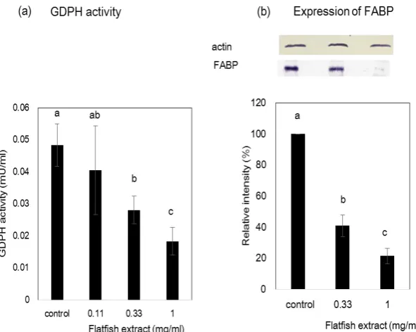
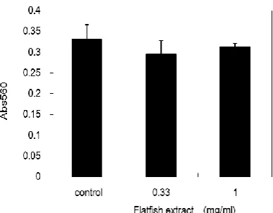
GPDH activity and the expression of FABP were also examined as indicators of 3T3-L1 adipocyte differentiation. GPDH activity in 3T3-L1 adipocyte cells treated with flatfish extract decreased in a dose-dependent manner (Figure 2). The activity was reduced to approximately 40% at a concentration of 1.0 mg/ml of flatfish extract when compared to that of the control. The expression of FABP was also downregulated following the treatment (Figure 2). These results further suggest that the flatfish extract inhibit the differentiation of 3T3-L1 preadipocytes. On the other hand, LDH activity did not increase, even with a treatment concentration of 1.0 mg/ml (Figure 3). This suggests that the inhibition of differentiation is not due to the toxicity of the extract.
516
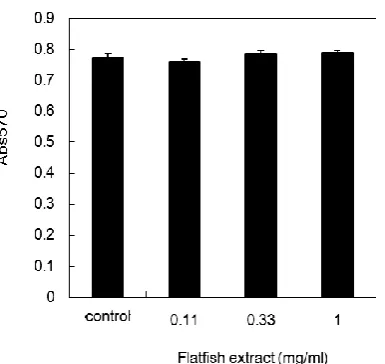
After differentiation was induced in 3T3-L1 preadipocytes, they underwent one or two rounds of cell division, known as mitotic clonal expansion, before initiating the differentiation process. We examined the effect of the flatfish extract on mitotic clonal expansion (Figure 4). As shown in Figure 4, the extract did not affect cell proliferation even at a
concentration of 1.0 mg/ml that was shown to inhibit the differentiation.
Effect of Flatfish Extract on the Expression of
PPAR andC/EBP
To investigate the mechanism underlying the Figure 1. Effect of flatfish extract on lipid accumulation during the differentiation of 3T3-L1 preadipocyte cells.
(a) The extent of differentiation was estimated by staining intracellular lipid droplets with oil red-O dye. The flatfish extract was added to 3T3-L1 preadipocyte cells at the indicated protein concentrations. Data represent the mean ± SEM. Mean values without common letters is significantly different (P<0.05). (b) Phase contrast microscopy in the absence or presence of flatfish extract. The accumulated lipid was stained with oil red-O (red color).
517
flatfish extract-induced inhibition of differentiation, we examined the expression levels of transcription factors (PPAR- and C/EBP- using western blotting (Figure 5). The usual increases of PPAR and
C/EBP- expression were attenuated by treatment with the flatfish extract. Therefore, this demonstrates that the flatfish extract suppresses the adipocyte differentiation by inhibiting PPAR and C/EBP- expression.
Identification of the Tissue Inhibiting the Differentiation
The flatfish tail fin comprises of skin, muscle, bone, and scale tissue. We examined which type of tissue inhibits the differentiation. The extracts from
scale and bone significantly inhibited the differentiation. In particular, extracts from the scales showed greater inhibition of differentiation when compared to extracts from other tissues (Figure 6). Lipid accumulation was reduced to approximately 60% after treatment with 1.0 mg/ml scale extract.
Identification of a Differentiation-Inhibiting Substance
We previously found that a glycoprotein from scallop shell inhibited the differentiation of 3T3-L1 preadipocytes (Takahashi et al., 2014). To examine whether the differentiation-inhibiting substance in the flatfish extract is also a glycoprotein, we separated glycoproteins from the extract using Con A, WGA, Figure 3. Effect of flatfish extract on mitotic clonal expansion. The flatfish extract was added to 3T3-L1 preadipocyte cells at the indicated protein concentrations immediately after induction of differentiation; viable cells were estimated with the MTT assay after 48 h. Data represent the mean ± SEM.
Figure 4. Effect of flatfish extract on the expression of PPAR and C/EBP-.
518
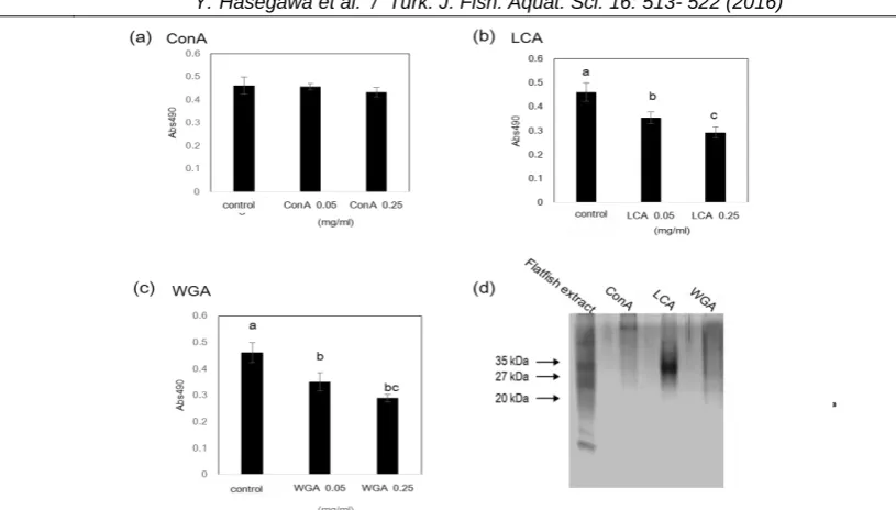
and LCA affinity columns. We then examined the differentiation-inhibitory activity of Con A-, LCA-, and WGA-binding fractions (Figure 7). These results showed that the LCA- and WGA-binding fractions significantly inhibited the differentiation of 3T3-L1 preadipocytes. On the other hand, the Con A-binding fraction did not inhibit differentiation. These findings demonstrate that several glycoproteins in the flatfish extract inhibit 3T3-L1 preadipocyte differentiation. Next, we investigated whether the sugar chains present in the flatfish extract are responsible for the inhibition of adipocyte differentiation. The flatfish extract was treated with TFMS, which is known to cleave protein-linked sugar chains. When the TFMS-treated flatfish extract was added to the culture medium during the differentiation of 3T3-L1 preadipocyte cells, the differentiation-inhibiting activity of the flatfish extract was completely
abolished (Figure 8), suggesting that sugar chains in the flatfish extract were responsible for the inhibitory activity.
SDS-PAGE analysis of the LCA-binding fraction revealed a major band with a molecular weight of approximately 30-kDa and several minor bands, while the WGA-binding fraction showed smear bands. To examine whether the 30-kDa glycoprotein inhibited differentiation, we purified the 30-kDa protein from the LCA-binding fraction using DEAE-5PW ion exchange column chromatography. The isolated 30-kDa glycoprotein showed a single band after silver staining post-SDS-PAGE (Figure 9). When 3T3-L1 preadipocytes were treated with 0.07 mg/ml purified 30-kDa glycoprotein, lipid accumulation was reduced to 65% of that of the control (Figure 9).
Figure 5. Cytotoxicity of flatfish extract
On day 10 after the induction of differentiation, the amount of LDH released into the culture medium was measured. The flatfish extract was added to 3T3-L1 preadipocyte cells at the indicated protein concentrations. Data represent the mean ± SEM.
519
Structure Analysis of the 30-kDa Glycoprotein
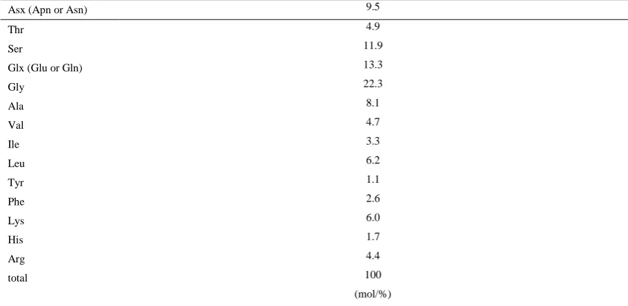
Amino acid composition analysis of the 30-kDa glycoprotein revealed the presence of high concentrations of Asx (Asp or Asn), Glx (Glu or Gln), Gly, and Ser residues (Table 1). Partial amino acid sequence analysis of the glycoprotein by MALDI-TOF mass spectrometry identified one sequence: TR(Q or K)NAAVVDHSF. MS Blast and Blast searches showed that the 30-kDa glycoprotein is a novel protein.
The results of monosaccharide composition analysis showed that glucose, mannose, galactose, and fucose are the dominant monosaccharides present
with relatively lower amounts of N-acetylglucosamine (Table 2).
Discussion
We previously reported that LCA- and ConA-binding glycoproteins in scallop shells inhibit the differentiation of 3T3-L1 preadipocyte cells. This differentiation-inhibiting activity was attributed to the sugar chain in the glycoproteins (Takahashi et al., 2014). In this study, TFMS-treated flatfish extract did not inhibit the differentiation of 3T3-L1 preadipocyte cells. The differentiation-inhibiting activity of the flatfish extract may be also attributable to the sugar Figure 7. Isolation of glycoproteins from the flatfish extract and their differentiation-inhibitory activity.
(a – c) The effects of Con A, LCA and WGA-binding fractions on the differentiation of 3T3-L1 preadipocyte cells were estimated. Each fraction was added to 3T3-L1 preadipocyte cells at the indicated protein concentrations Data represent the mean ± SEM. Mean values without common letters is significantly different (P<0.05). (d) Con A, LCA, and WGA-binding fractions were analyzed using SDS-PAGE.
520
chain, as in the case of the glycoproteins in the scallop shell. Several studies have reported that sugar or sugar chains inhibits the differentiation of 3T3-L1 preadipocytes (Konga et al., 2010; Karagozlu et al.,
2011; Kim et al., 2009; Cho et al. 2008). Therefore, it is necessary to determine the structures of the sugar chains in the isolated 30-kDa glycoproteins in order to compare them with other sugar chains that inhibit the Figure 9. Isolation of a 30-kDa glycoprotein in the LCA-binding fraction and its differentiation-inhibiting activity.
(a) The 30-kDa glycoprotein was purified by DEAE-5PW ion exchange column chromatography, analyzed using SDS-PAGE. Silver staining was carried out in order to visualize the bands. (b) The effect of the 30-kDa glycoprotein on the differentiation of 3T3-L1 preadipocyte cells was estimated. The 30-kDa glycoprotein was added at the indicated protein concentrations. Data represent the mean ± SEM. Mean values without common letters is significantly different (P<0.05).
Table 1. Amino acid composition of the 30-kDa glycoprotein
Asx (Apn or Asn) 9.5
Thr 4.9
Ser 11.9
Glx (Glu or Gln) 13.3
Gly 22.3
Ala 8.1
Val 4.7
Ile 3.3
Leu 6.2
Tyr 1.1
Phe 2.6
Lys 6.0
His 1.7
Arg 4.4
total 100
(mol/%)
Table 2. Monosaccharide analysis of the 30 kDa glycoprotein
Gal 19.1
Man 20.7
Glc 32.5
GlcNAc 2.7
Fuc 25.0
Total 100
521
differentiation.
We demonstrated that the extracts of scale and bone from the flatfish tail fin possess differentiation-inhibitory activity. Scales and bone are biomineralized tissues which are composed of hydroxyapatite and organic matrix substances containing type I collagen fibers (Olson and Watabe, 1980). Scallop shell is also a biomineralized tissue which is composed of organic substances and calcium carbonate. Glycoproteins in biomineralized tissues may have a similar structure that inhibits the differentiation of 3T3-L1 preadipocytes. We previously reported that the 16-kDa glycoprotein with differentiation-inhibiting activity isolated from scallop shells contained high levels of Asx (Asp or Asn), Glx (Glu or Gln), Gly, and Ser residues (Takahashi et al., 2014). The 30-kDa glycoprotein was also rich in Asx (Asp or Asn), Glx (Glu or Gln), Gly, and Ser residues. The monosaccharide composition of both the 16-kDa and 30-kDa glycoprotein was also similar, in that glucose and mannose were the dominant monosaccharides present along with fucose. These results suggest that the 16-kDa and 30-16-kDa glycoproteins have similar structures.
We showed that a 30-kDa glycoprotein in the flatfish extract inhibited the differentiation of 3T3-L1 preadipocyte cells. The extract appears to contain several other differentiation-inhibiting substances, as inhibition of the differentiation was also observed with the WGA-binding fraction and bone extract, which are not thought to contain the 30-kDa glycoprotein. Attempt to identify other differentiation-inhibiting substances is ongoing.
In this study, we showed that the glycoproteins in the flatfish extract inhibited the differentiation of 3T3-L1 preadipocyte by suppressing the expression of PPAR and C/EBP . In order to utilize the flatfish tail fin effectively, it is necessary for future studies to investigate whether the consumption of flatfish extract or the 30-kDa glycoprotein is able to induce a decrease in fat mass.
References
Cho, E.J., Rahman, M.A., Kim, S.W., Baek, Y.M., Hwang, H.J., Oh, J.Y., Hwang, H.S., Lee, S.H., Yun, J.W. 2008. Chitosan oligosaccharides inhibit adipogenesis in 3T3-L1 adiocytes. Journal of Microbiology and Biotechnology, 18: 80-87.
Cornelius, P., MacDougald, O. A., Lane, M. D. 1994. Regulation of Adipocyte Development. Annual Review of Nutrition, 14:99–129. doi:10.1146/annurev.nu.14.070194.000531
Edge, A.S.B. 2003. Deglycosylation of glycoproteins with trifluoromethanesulphonic acid: elucidation of molecular structure and function. Biochemical Journal, 376: 339-350. doi:10.1042/bst0170737 Green, H., Meuth, M. 1974. An established pre-adipose cell
line and its differentiation in culture. Cell, 3: 127-133. doi:10.1016/0092-8674(74)90116-0
Gregoire, F.M., Smas, C.M., Sul, H.S. 1998. Understanding
adipocyte differentiation. Physiological Reviews, 78: 783-809.
Harnedy, P.A., FitzGerald, R.J. 2012. Bioactive peptides from marine processing waste and shellfish: A review. Journal of Functional Foods, 4: 6-24. doi:10.1016/j.jff.2011.09.001
Karadeniz, F., Karagozlu, M. Z., Pyun, S.Y. Kim, S.K. 2011. Sulfation of chitosan oligomers enhances their anti-adipogenic effect in 3T3-L1 adipocytes. Carbohydrate Polymers, 86: 666-671. doi:10.1016/j.carbpol.2011.05.005
Kawada, T., Takahashi, N., Fushiki, T. 2001. Biochemical and physiological characteristics of fat cell. Journal of Nutritional Science and Vitaminology, 47: 1-12. doi:org/10.3177/jnsv.47.1
Kim, M.J., Lee, J.S. 2009. Inhibitory effects of fucoidan in 3T3-L1 adipocyte differentiation. Marine Biotechnolgy, 11: 557-562. doi:10.1007/s10126-008-9170-1
Kim, S.K. Mendis, E. 2006. Bioactive compounds from marine processing byproducts – A review. Food Research International, 39: 383-393. doi:10.1016/j.foodres.2005.10.010
Kim, Y.M., Kim, E.Y., Kim, I.H., Nam, T.J. 2015. Peptide derived from desalinated boiled tuna extract inhibits adipogenesis through the downregulation of C/EBP-α and PPAR-γ in 3T3-L1 adipocytes. International Journal of Molecular Medicine, 35: 1362-1368. doi: 10.3892/ijmm.2015.2127
Konga, C.S., Kimb, J.A., Eomb,T.K., Kima,S.K. 2010. Phosphorylated glucosamine inhibits adipogenesis in 3T3-L1 adipocytes. Journal of Nutritional
Biochemistry, 21: 438-443.
doi:10.1016/j.jnutbio.2009.01.018
Laemmli, U.K. 1970. Cleavage of structural proteins during the assembly of the head of bacteriophage T4. Nature, 227: 680-685. doi:10.1038/227680a0
Lai, C.S., Chen, Y.Y., Lee, P.S., Kalyanam, N., Ho, C.T., Liou, W.S., Yu, R.C., Pan, M.H. 2016. Bisdemethoxycurcumin inhibits adipogenesis in 3T3-L1 preadipocytes and suppresses obesity in high-fat diet-fed C57BL/6 Mice. Journal of Agricultural and Food Chemistry, 64: 821-830. doi: 10.1021/acs.jafc.5b05577
Liu, Y.C., Hasegawa, Y. 2006. Reducing effect of feeding powdered scallop shell on the body fat mass of rats. Bioscience Biotechnology, and Biochemistry, 70: 86-92. doi.org/10.1271/bbb.70.86
MacDougald, O. A., Lane, M. D. 1995. Transcriptional Regulation of Gene Expression during Adipocyte Differentiation. Annual Review of Biochemistry, 64: 345–373. doi:10.1146/annurev.bi.64.070195.002021 Manthorpe, M., Fagnani, R., Skaper, S.D., Varon, S. 1986.
An automated colorimetric microassay for neuronotrophic factors. Brain Research,390: 191-198. doi:10.1016/0165-3806(86)90208-7
Olson, O.P., Watabe, N. 1980. Studies on formation and resorption of fish scales. IV. Ultrastructure of developing scales in newly hatched fry of the sheepshead minnow, Cyprinodon variegatus (Atheriniformes: Cyprinodontidae). Cell and Tissue Research, 211:303-316. doi:10.1007/BF00236451 Rangwala, S.M., Lazar, M.A. 2000. Transcriptional control
of adipogenesis. Annual Review of Nutrition, 20: 539–559. doi:10.1016/S0955-0674(98)80138-5 Rosen, E.D., Spiegelman, B.M. 2000. Molecular regulation
522
Development Biology, 16: 145–171. doi:10.1146/annurev.cellbio.16.1.145
Rosen, E.D., Walkey, C.J., Puigserver, P., Spiegelman, B.M. 2000. Transcriptional regulation of adipogenesis. Genes and Development, 14:1293– 1307. doi:10.1101/gad.14.11.1293
Smith, P.K., Krohn, R.I., Hermanson, G.T., Mallia, A.K., Gartner, F.H., Provenzano, M.D., Fujimoto, E.K., Goeke, N.M., Olson, B.J., Klenk, D.C. 1985. Measurement of protein using bicinchoninic acid. Analytical Biochemistry, 150: 76-85. doi:10.1016/0003-2697(85)90442-7
Spiegelman, B.M., Flier, J.S. 2001. Obesity and the regulation of energy balance. Cell, 104: 531-543. doi:10.1016/S0092-8674(01)00240-9
Takahashi, K., Hasegawa, Y. 2014. Glycoproteins isolated from scallop shell inhibit differentiation of 3T3-L1 preadipocyte cells. Fisheries Science, 80: 1301-1310. doi;10.1007/s12562-014-0801-3