Shuo-Ming Wu
1, B–D, Wen-Xiong Zhang
1, B–D, Ming-Hui Wang
1, B, C,
Hui-Zhong Zhang
1, A, E, F, Duo-Guang Wu
1, C, Zhi-Juan Zhou
2, C, Li-Hua Xiong
1, B, CProteomic Analysis of the Immunosuppressive Effects
of Mesenchymal Stem Cells in a Rat Heart
Transplantation Model
Analiza proteomiczna działania immunosupresyjnego
mezenchymalnych komórek macierzystych na modelu zwierzęcym
z wykorzystaniem serca szczurów
1 Department of Heart Surgery, Sun Yat-sen Memorial Hospital, Sun Yat-sen University, China 2 Department of Heart Surgery,Fifth Affiliated Hospital of Sun Yat-sen University, China
A – research concept and design; B – collection and/or assembly of data; C – data analysis and interpretation;
D – writing the article; E – critical revision of the article; F – final approval of article; G – other
Abstract
Background. Some reports suggest mesenchymal stem cells (MSCs) have immunosuppressive properties. However,
conflicting evidence regarding the role of MSCs has emerged.
Objectives. To gain a better understanding of the immunosuppressive properties of mesenchymal stem cells
(MSCs)in a rat heart transplantation model.
Material and Methods. MSCs were obtained from the femoral and tibial bone marrow of Sprague-Dawley rats and
cultured. Heart-transplanted rats were allocated into a MSC-treated group and 2 control groups. On postoperative day 7, 1 rat was sacrificed and the pathological changes of heart tissues were assessed. Serum proteomic spectra were generated by surface-enhanced laser desorption/ionization-time-of-flight mass spectrometry (SELDI-TOF-MS).
Results. Rat MSCs displayed the typical spindle-shaped morphology in culture and significantly prolonged the
graft survival up to 33.25 ± 2.54 days compared with controls (19.75 ± 1.56 and 11.16 ± 1.34 days, respectively). Pathological analysis showed the inflammatory cell infiltration in the MSC-treated group was significantly reduced. SELDI analysis showed that 5 protein/peptide peaks with M/Z 1272.33, 1986.65, 2323.42, 5375.59 and 12968.11 were up-regulated in the MSC-treated group (P < 0.001).
Conclusions. Donor-derived MSCs clearly alleviate acute rejection following heart transplantation in rats and
significantly prolong the isograft survival time (Adv Clin Exp Med 2013, 22, 6, 785–794).
Key words:ratheart transplantation, immunosuppression, mesenchymal stem cells, SELDI ProteinChip, decision
tree.
Adv Clin Exp Med 2013, 22, 6, 785–794 ISSN 1899–5276
ORIGINAL PAPERS
© Copyright by Wroclaw Medical University
Mesenchymal stem cells (MSCs) are found in a variety of sources, including the skin, bone mar-row and adipose tissue, and can be differentiated into chondrocytes, osteoblasts, and adipocytes in vitro; therefore, MSCs was considered to be poten-tial candidates for cellular and genetic therapy.
Emerging evidence suggests the immunosup-pressive function is one of the most important pro-files of MSCs. It has been successfully applied in animal models and clinical trials. Different studies
3) MSCs highly express MHC I, but do not express the FAS ligand, the MHCm and other costimula-tory molecules. These characteristics make MSCs promising for the application in solid organ trans-plantation to prolong the graft survival.
To clarify the possible modulation mechanism of MSCs on the transplantat rejection, a preclini-cal model of heart transplantation in rats was used and serum proteomic pattern was analyzed using the SELDI ProteinChip technology. Our results showed that donor-derived MSCs could signifi-cantly decrease the acute rejection following heart transplantation, prolong the survival time, and represent a promising candidate in clinical tissue transplantation.
Material and Methods
Animals
Healthy Sprague-Dawley (SD) and Wistar rats (160–200 g) were obtained from the Experimen-tal Animal Center of Sun Yat-sen University, Chi-na. All animals were held under standard labora-tory conditions at constant temperature, humidity, and light/dark cycles. They were fed with standard diet and had free access to tap water. All animals received care in compliance with the Principles of Laboratory Animal Care and the experimental protocol was approved by the local Animal Care and Research Committee.
Isolation and Culture of MSC
Male SD rats (3–4 weeks old) were sacri-ficed by cervical dislocation. Bone marrow cells were flushed out of the femoral and tibial cavities, washed twice with PBS buffer and re-suspended in DMEM medium (Gibco, Grand Islands, N.Y., USA) containing 10% fetal calf serum (FCS, Gib-co). Following the determination of cell viabili-ty and numbers, bone marrow mononuclear cells were transferred to culture flasks with control me-dium consisting of DMEM with fetal bovine serum (10%), penicillin (100 U/mL) and streptomycin (100 µg/mL). The re-suspended cells were incu-bated at 37°C with 5% CO2, and the unattachedcells were removed by medium replacement. The medium was changed every 24 h. When the cul-tures reached 50–70% confluence, the cells were harvested by centrifugation and washed twice with PBS buffer, then trypsinized by 0.25% trypsin and passaged. The cells atpassage 3 were re-suspended in basal DMEM at a density of 1 × 10⁶ cells/mL and
used for transplantation.
Flow Cytometric
Analysis of MSCs
The 3rd passage cells were collected by cen-trifugation at 600 g for 5 min at 25°C, washed twice with PBS buffer, digested with trypsin (0.25%) – EDTA (0.02%) and washed 3 times with PBS. Cell pellets were obtained by the centrifuga-tion at 1000 rpm for 10 min, then re-suspended in PBS with 10% FBS. CD44-FITC and CD45-FITC antibodies (Pharmacia Corporation, USA) were respectively added and incubated in the dark at 4°C for 30 min. FITC-conjugated IgG antibodies served as controls. Then cells were centrifuged at 1400 rpm for 5 min, washed with PBS and fixed by 1% formaldehyde in PBS buffer for 10–30 min. Fi-nally, cells were analyzed in a FACSCanto II Flow Cytometer.
Application of MSCs in a Rat
Heart Transplantation Model
Rats were allocated to 3 groups: isograft group (group I, n = 8), from SD rats to SD rats; allograft group, from SD rats to Wistar rats and then divided into a control group (II, n = 8) and a MSC-treated group (III, n = 8). Abdominal cardiac transplanta-tion was performed using the modified method as described. In brief, ketamine mixed with xylazine was given intraperitoneal injection for anesthesia. Donor hearts were perfused with chilled, heparin-ized saline via the vena cava and harvested after li-gation of the vena cava and pulmonary veins. The aorta and pulmonary artery of donor hearts were anastomosed to the abdominal aorta and inferiorvena cava of recipients using a microsurgical tech-nique. Before the anesthesia recovery period, re-cipients (Group III) were injected with donor-de-rived MSCs (1.0 × 10⁶ cells/rat) via the tail vein,
followed by 3 additional doses each day for 3 con-secutive days’ post-transplantation. Group I and II were injected with PBS and used as control. All ex-perimental groups were checked by daily abdomi-nal palpation. 1 microliter of blood was obtained from the tail vein of each group (I, II and III) at Day 1, 3 and 5 after transplantation, respective-ly. The samples were stored at –20°C and used for protein-chip analysis.
SELDI-TOF-MS Analysis
Mass-spectrum analysis was performed as pre-viously describedwith some modifications. Briefly, all blood samples were thawed and centrifuged at 10.000 rpm for 5 min at 4°C. 10-microliter super-natant was diluted with 10 µL U9 buffer (9 mol/L urea, 2% CHAPS, and 1% DTT). Then, the sam-ples were continuously diluted 20-fold in binding buffer (50 mmol/L NaAc, pH 4). The weak cation exchange protein chips (CM10) (Ciphergen Bio-systems) were put into a bioprocessor, and 200 µL binding buffer was added to each well with gentle agitation at 600 rpm at 25°C for 5 min; this step was repeated once. Next, 100 µL diluted sample was loaded on each spot. The processor was sealed and shaken at 600 rpm at 4°C for 1 hour to re-move unbound samples. The chips were rinsed twice with 200 µL binding buffer and 1 time with 200 µL HPLC grade water, covered with 1 µL satu-rated sinapinic acid (SPA) solution (prepared in 0.5% trifluoroacetic acid (TFA) and 50% acetoni-trile) and air-dried at room temperature.
SELDI ProteinChip Analysis
The chips were scanned by the ProteinChip (Model PBS IIc) reader (Ciphergen). The parame-ters were as follows: mass range (0 to 50.000 Da), optimized mass-to-charge ratio (1.000 to 50.000 Da), laser intensity (240), laser shots (112), and sensitivity (8). All-in-one protein marker contain-ing arg 8-vasopressin (1084.2 Da), somatostatin(1637.9 Da), sovine insulin beta-chain (3495.9 Da), human insulin (5807.6 Da) and Hirudin BHVK (7033.61 Da) (Ciphergen Biosystems, USA) (Fig. 7) was used as the calibration.The peak inten-sities were normalized by the background subtrac-tion. Protein peaks were calculated using the bio-marker detection software (Ciphergen Biobio-marker Wizards, Ciphergen Biosystems, Inc). Spectra were analyzed using the ProteinChip (Version 3.2.1) in Biomarker Wizard software (Ciphergen Biosys-tems) as previously described. The baselines were normalized, subtracted with the signal-noise ratio threshold of 5. Protein expression patterns were clustered with GENE-E (http://www.broadinsti-tute. org/cancer/software/GENE-E/index.html).
Decision Tree Construction
The decision tree was constructed by marker Patterns Software 5.0 (BPS; Ciphergen Bio-systems). The splitting decisions were made based on the normalized peak intensities. The process was continued until terminal nodes were created. After cross-validation in test model, the decision tree was further confirmed using the test data that was independent of the training set.Statistical Analysis
The data was expressed mean ± standard de-viation (SD). Significant differences in the peaks intensity of proteins between each group were
A
R 1
C
B D
Fig. 1. Measenchymal stem cells from adult rat bone marrow were examined under microscopy and stained by
calculated. Statistical analysis among groups was performed using one-way analysis of variance (ANOVA) followed by the Student-Newman- -Keuls (SNK) multiple range test. The data that did not fit a normal distribution was analyzed by Krus-kal-Wallis H test. All of the statistical analysis was carried out using SPSS version 16.0. Statistical sig-nificant difference was defined as p < 0.01.
Results
Characterization of Rat MSCs
After 48 hours culture, attached cells displayed an oval-, polygon- or short spindle-shaped fibro-blastic morphology. One week later, on most of the cells there distinctly appeared a fibroblast- -like, spindle-shaped morphology and these cellsexhibited an enlarged cell size, heterogeneous cell projection in different length, a large round nu-cleus with an unclear outline, and visible nucleo-li. On the 12th day, the attached monolayer cells were harvested as the 1st passage. As the culture proceeded, the cells were distributed evenly with the same growth pattern, and presented a typical fibroblast-like morphology (Fig. 1A), which was consistent with the morphology of bone marrow-derived stem cells. Cell proliferation speed was in-creased to about 9 days. MSCs were still in latent phase after being subculture for 2 days, followed by logarithmical proliferation from day 3, and reached the growth platform at day 7.
Flow cytometry analysis confirmed that the cell population were consistent between samples and then were purified and homogeneous. Most of the cultured MSCs were positive for CD44 and negative for hematopoietic stem cell marker CD45 (Fig. 1C and 1D), which were in good agreement with the surface marker phenotype of MSCs.
Application of MSCs
to an Allogeneic Heart
Transplantation Model
To elucidate whether the immunomodulative effects of rat MSCs in vivo, MSCs were applied to a heart transplantation model. The process of do-nor-preparation and heart-harvesting took about 8 ± 2 min, and reception or vascular preparation took 4 ± 2 min. Abdominal aorta anastomosis time was 12 ± 2 min and inferior vena cava anastomo-sis time was 9 ± 2 min. The average cold ischemia time was 36 ± 3 min. The recipient rats usually took 2 ± 1 min to restart the blood supply of the heart and start ventricular fibrillation after opera-tion. Taken together, the whole surgical procedure lasted 53 ± 7 min.
The mean allograft survival of control group I and II were 19.75 ± 1.56 days and 11.16 ± 1.34 days, 0
10 20 30 40
I II III
treatment
survival day after transplantation
**
**
**
Fig. 2. Survival time of heart transplanted rats in
dif-ferent groups. Group I: The transplanted SD rats were treated with PBS buffer (SD rats as donors and recipi-ents, n = 16); Group II and III: the SD rats were used as donor (n = 8/each group) and Wistar rats as recipients (n = 8/each group). The control group II was injected with PBS, while the experimental group III was injected with MSCs. ** P < 0.01
A B C
Fig. 3. Histological examination of heart tissue of the transplanted rats from different groups (HE stainm ×400).
1250 1500 1750 2000
1250 1500 1750 2000
? ? ? 0 10 20 30 40 1986.6+H 0 10 20 1986.6+H 0 5 10 15 1986.6+H
1000 1200 1400 1600
1000 1200 1400 1600 ? ? ? 0 20 40 60 1293.0+H 0 20 40 60 1293.0+H 0 20 40 60 1293.0+H
5000 5500 6000
5000 5500 6000
? ? ? 0 10 20 30 5375.6+H 0 10 20 30 5375.6+H 0 10 20 30 5375.6+H
1200 1400 1600
1200 1400 1600
? ? ? 0 20 40 1272.3+H 0 20 40 1272.3+H 0 20 40 1272.3+H
10000 12000 14000 16000
10000 12000 14000 16000 ? ? ? 0 5 10 15 20 12968.1+H 0 5 10 15 20 12968.1+H 0 5 10 15 20 12968.1+H
1500 1750 2000 2250
1500 1750 2000 2250 ? ? ? 0 10 20 30 40 2323.4+H 0 10 20 30 40 2323.4+H 0 10 20 30 40 2323.4+H E D B A C F
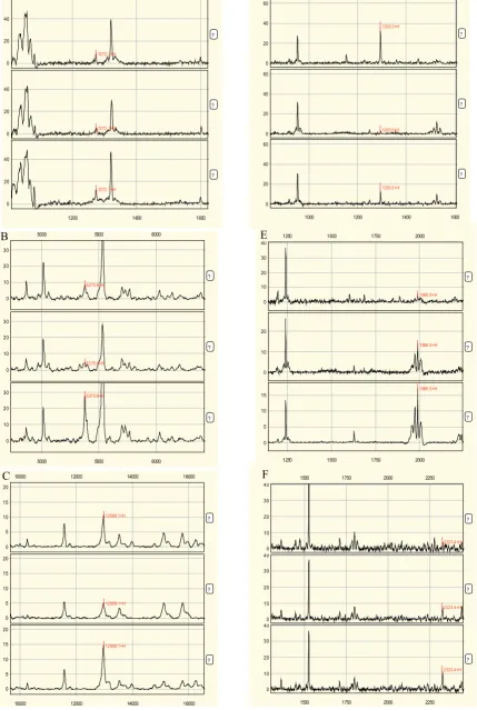
Fig. 4. Representative overview of protein profiling of serum from the heart transplanted rats. Group I: The
trans-planted SD rats (SD rats as donors and recipients, n = 16) were treated with PBS; Group II and III: the SD rats were used as donor (n = 8/each group) and Wistar rats as recipients (n = 8/each group). The control group II was injected with PBS, while the experimental group III was injected with MSCs. Differentially expressed proteins were (A) N107, (B) N224, (C) N257,
(D) N112, (E) N176 and (F) N184. The x-axis represents the molecular mass calculation (mass-to-change ratio [m/z]) and the
respectively. Donor-derived MSCs significantly pro-longed graft survival up to 33.25 ± 2.54 days (Fig. 2). These results showed that rat donor-derived MSCs
effectively prolonged the graft survival compared to control group I and II (p < 0.01). Furthermore, graft rejections were significantly different between con-trol group I and II treated with PBS (p < 0.01).
Histopathological Examination
The representative heart tissues of 7-day trans-plantation rats from different groups were shown in Fig. 3A–3C. Pathological examination revealed that in the control group I there was moderate disorder in myocardial tissue with moderate cel-lular swelling and inflammatory cell infiltration (Fig. 3A). In group II, there were cardiac cells necrosis, cellular swelling, sever acute rejection and inflammatory cell infiltration in myocardi-al interstitimyocardi-al (Fig. 3B). In contrast to these histo-pathological observations, in group III, patholog-ical examination showed that there were no mild or moderate acute rejections and the transplanted heart has normal myocardial tissue structure with significantly alleviated inflammatory cell infiltra-tion (Fig. 3C).7500 10000 12500 15000
7500 10000 12500 15000
1d
3d
5d 0
5 10 15
12968.1+H
0 5 10 15
12968.1+H
0 5 10
15 12968.1+H
20
20
20
Fig. 5. Differential expression profile of protein N257
with m/z 12968.1 at different day after MSCs treatment for heart transplantation rats. The x-axis represents the molecular mass calculation (mass-to-change ratio [m/z]) and the y-axis represents the relative intensity. The peaks were indicated by red line and markered with m/z
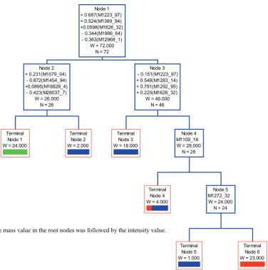
The mass value in the root nodes was followed by the intensity value. Terminal
Node 1 W = 24.000
Terminal Node 2 W = 2.000 Node 2
+ 0.231(M1079_04) - 0.872(M1454_94) +0.0895(M18828_4) - 0.423(M28037_7)
W = 26.000 N = 26
Terminal Node 3 W = 18.000
Terminal Node 4 W = 4.000
Terminal Node 5 W = 1.000
Terminal Node 6 W = 23.000 Node 5
M1272_32 W = 24.000
N = 24 Node 4
M1109_18 W = 28.000
N = 28 Node 3
- 0.151(M1223_97) + 0.548(M1283_14) + 0.791(M1292_95) + 0.225(M1626_32)
W = 46.000 N = 46 Node 1
+ 0.687(M1223_97) + 0.524(M1389_94) +0.0598(M1626_32) - 0.344(M1986_64) - 0.363(M12968_1)
W = 72.000 N = 72
Fig. 6.Diagram of a decision tree for the classification of the PBS and MSCs treated heart transplanted
2500 5000 7500
2500 5000 7500
all-in-1 peptide
0 20 40
1084.2+H
1638.0+H
3495.4+H
5808.5+H
7033.1+H
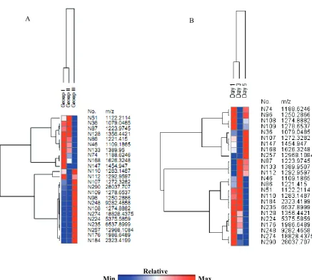
Red represents maximal expression level and blue represents minimum expression level. Relative
Max Min
A B
Fig. 7. All-in-one peptide reference standard used for optimization of the experimental conditions and evaluation of
the reproducibility. Arg8-vasopressin (1084.2 Da), somatostatin (1637.9 Da), sovine insulin beta-chain (3495.9 Da) human insulin (5807.6 Da) and Hirudin BHVK (7033.61 Da)
Fig. 8. A hierarchical clustering analysis of twenty four differentially expressed proteins/peptides in group-model (A)
Serum SELDI Profiles of MSCs
Treated Rats vs. Controls
After noise filtering, clustering analysis re-vealed that 313 proteins were differentially ex-pressed among different groups. Further analysis resulted in a subset of 83 proteins which were dif-ferentially expressed between MSCs treated sam-ples and controls (p < 0.05). In this subset, 24 pro-teins were detected to be significantly different at p < 0.01 level. To search for the classification and comparability information of those differen-tial protein/peptide peaks, a hierarchical clustering analysis among group I, II and III was performed. The results indicated that the relative expression level of some proteins was not significant differ-ence between group I and II samples (Fig. 8). Fur-ther analysis showed 6 proteins were significant-ly difference among groups (p < 0.001) (Fig. 4). They were N107 with m/z 1272.33 (p = 3.36 × 10⁻⁴),
N112 with m/z 1292.96 (p = 2.44 × 10⁻⁴); N176
with m/z 1986.65 (p = 3.09 × 10⁻⁴), N184 with
m/z 2323.42 (p = 4.08 × 10⁻⁴), N224 with m/z
5375.59 (p = 1.55 × 10⁻⁴) and N257 m/z 12968.11
(p = 3.25 × 10⁻⁴).
To study the protein expression variations in MSCs treated groups, SELDI-TOF-MS data for day 1, 3 and 5 post-postoperative were analyzed by ANOVA. The results showed the expression pattern of N107 was day 5 > day 1 > day 3. For N112, the
ex-pression profile was day 1 > day 5 > day 3. N257 was
up-regulated gradually from day 1–day 5 (Fig. 5). The expression of N176, N224 and N248 were higher at
day 1 and then declined with time (Fig. 8).
Decision Tree
After discriminatory analysis, 13 of 24 protein mass peaks (Table 1) were chosen by optimization to distinguish the differentially expressed serum proteins. The most optimal decision tree with the highest accuracy was established. (Fig. 6). The de-cision tree used 5 splitters with distinct masses and classified the samples into 6 nodes. The optimal de-cision tree manifested 95.8% (23/24), 100% (24/24) and 100% (24) accuracy for classifying group I, II and III, respectively.
Discussion
Immunologic rejection of a grafted organ re-mains a common problem after transplantation. Usually acute rejection is alleviated by using high doses of immunosuppressive drug with side-ef-fects and severe toxicity. Therefore, it is impera-tive to find a safe and effecimpera-tive way to reduce im-munologic rejection.
MSCs are considered a promising option for improving graft survival and achieving immuno-logical tolerance without pharmacoimmuno-logical immu-nosuppression. Several studies reported that MSCs can evade immune recognition and modulate im-mune responses. One of the first in vivo studies showed that systemic infusion of MSCs isolated from bone marrow prolonged the survival of al-logeneic skin graft from 7 to 11 days in baboon. In other studies, Bartholomew et al. reported that a single infusion (1–3 × 10⁶ cells/kg) of
allogene-ic or third-part MSC prolonged skin graft surviv-al in a baboon. In contrast, similar MSCs dosing (2 × 10⁶ cells/rat) was applied before and
repeated-ly after the heart transplantation and significant-ly prolonged the survival of rat cardiac allograft. Further analysis based on real-time PCR showed that the induction of allograft tolerance through changing the Th1/Th2 balance. In a more recent study, Biancamaria et al. reported MSCs alone were able to prolong lung-graft survival time in rat mo del. On the contrary, Inoue S et al. report-ed MSCs alone could not prolong graft survival in a rat heart transplantation model. The different ef-fects observed among different applications may be caused by the source, purity, site, dose and time of MSCs application, and the state of activation of cells for maximal toleragenic effects.
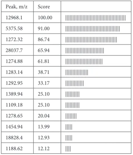
Table 1. Important peaks selected by the biomarker pat-tern software and used for decision tree analysis in group-ing model
Peak, m/z Score
12968.1 100.00 |||||||||||||||||||||||||||||||||||||||||| 5375.58 91.00 |||||||||||||||||||||||||||||||||||||| 1272.32 86.74 |||||||||||||||||||||||||||||||||||| 28037.7 65.94 ||||||||||||||||||||||||||| 1274.88 61.81 |||||||||||||||||||||||||| 1283.14 38.71 |||||||||||||||| 1292.95 33.17 ||||||||||||| 1389.94 25.10 |||||||||| 1109.18 25.10 |||||||||| 1278.65 20.04 |||||||| 1454.94 13.99 ||||| 18828.4 12.93 ||||| 1188.62 12.12 ||||
To develop MSC-based therapies for tolerance induction in clinical organ transplantation, the cel-lular mechanisms of their action must be under-stood thoroughly. Therefore, donor-derived MSCs (1 × 10⁶ cells/rat) were evaluated in
heart-trans-planted rat model in the present study. Consistant with previous reports, donor-derived MSCs are able to alleviate inflammatory cell infiltration and significantly prolong graft survival.
83 proteins differentially expressed between experimental and control groups were identi-fied by SELDI-TOF MS technology. Five of them were significantly up-regulated in the experimen-tal group (p < 0.001). As serum protein profile al-ternates with immune rejection, we deemed that the up-regulated expression pattern of N257 would
play an important role in immunosuppression or anti-inflammation. On the contrary, the expres-sion pattern of N176, N224 and N248 implied that they
negatively function in heart transplantation sur-vival. Although biomarkers generated with SELDI- -TOF technology were anonymous, the technology is very reliable in generating biomarkers signatures for the estimation of the total treatment effect on the serum proteome.
In conclusion, our results supported that do-nor-derived MSCs have immunosuppressive and/or anti-inflammatory properties in vivo and be able to prolong graft survival, opening new insights in the prevention and treatment of graft rejection in tis-sue and organ transplantation. However, further studies are needed to investigate the immunosup-pressive mechanisms of MSCs based on the iden-tification and characterization of these potential protein biomarkers, and the optimal conditions for handling stem cells. A careful evaluation of the harmful immunosuppressive effects of MSCs is al-so necessary.
Acknowledgments. The authors would like to thank all healthcare workers from Heart Surgery Department and ICU for their expert technical assistances and suggestions. We also thank the staff from the Experimental Animal Center of Sun Yat-sen University for the help with the animal work.
References
[1] Toma JG, McKenzie IA, Bagli D, Miller FD: Isolation and characterization of multipotent skin-derived
precur-sors from human skin. Stem Cells 2005, 23, 727–737.
[2] Zuk PA, Zhu M, Ashjian P, De Ugarte DA, Huang JI, Mizuno H, Alfonso ZC, Fraser JK, Benhaim P,
Hedrick MH: Human adipose tissue is a source of multipotent stem cells. Mol Biol Cell 2002, 13, 4279–4295.
[3] Pittenger MF, Mackay AM, Beck SC, Jaiswal RK, Douglas R, Mosca JD, Moorman MA, Simonetti DW, Craig S,
Marshak DR: Multilineage potential of adult human mesenchymal stem cells. Science 1999, 284, 43–47.
[4] Kim YH, Wee YM, Choi MY, Lim DG, Kim SC, Han DJ: Interleukin (IL)-10 induced by CD11b(+) cells and
IL-10-activated regulatory T cells play a role in immune modulation of mesenchymal stem cells in rat islet allogra-fts. Mol Med 2011, 17, 697–708.
[5] Wan CD, Cheng R, Wang HB, Liu T: Immunomodulatory effects of mesenchymal stem cells derived from
adipo-se tissues in a rat orthotopic liver transplantation model. Hepatobiliary Pancreat Dis Int 2008, 7, 29–33.
[6] Bartholomew A, Sturgeon C, Siatskas M, Ferrer K, McIntosh K, Patil S, Hardy W, Devine S, Ucker D, Deans R,
Moseley A, Hoffman R: Mesenchymal stem cells suppress lymphocyte proliferation in vitro and prolong skin graft
survival in vivo. Exp Hematol 2002, 30, 42–48.
[7] Zhou HP, Yi DH, Yu SQ, Sun GC, Cui Q, Zhu HL, Liu JC, Zhang JZ, Wu TJ: Administration of donor-derived
mesenchymal stem cells can prolong the survival of rat cardiac allograft. Transplant Proc 2006, 38: 3046–3051.
[8] Crop MJ, Baan CC, Korevaar SS, Ijzermans JN, Alwayn IP, Weimar W, Hoogduijn MJ: Donor-derived
mesen-chymal stem cells suppress alloreactivity of kidney transplant patients. Transplantation 2009, 87, 896–906.
[9] Casiraghi F, Azzollini N, Cassis P, Imberti B, Morigi M, Cugini D, Cavinato RA, Todeschini M, Solini S,
Sonzogni A, Perico N, Remuzzi G, Noris M:Pretransplant infusion of mesenchymal stem cells prolongs the survival
of a semiallogeneic heart transplant through the generation of regulatory T cells. J Immunol 2008, 181, 3933–3946.
[10] Sbano P, Cuccia A, Mazzanti B, Urbani S, Giusti B, Lapini I, Rossi L, Abbate R, Marseglia G, Nannetti G,
Torricelli F, Miracco C, Bosi A, Fimiani M, Saccardi R: Use of donor bone marrow mesenchymal stem cells for
treatment of skin allograft rejection in a preclinical rat model. Arch Dermatol Res 2008, 300, 115–124.
[11] Inoue S, Popp FC, Koehl GE, Piso P, Schlitt HJ, Geissler EK, Dahlke MH: Immunomodulatory effects of
mes-enchymal stem cells in a rat organ transplant model. Transplantation 2006, 81, 1589–1595.
[12] Stagg J, Galipeau J: Immune plasticity of bone marrow-derived mesenchymal stromal cells. Handb Exp Pharmacol
2007, 45–66.
[13] Noël D, Djouad F, Bouffi C, Mrugala D, Jorgensen C: Multipotent mesenchymal stromal cells and immune
tolerance. Leuk Lymphoma 2007, 48, 1283–1289.
[14] Ramasamy R, Fazekasova H, Lam EW, Soeiro I, Lombardi G, Dazzi F: Mesenchymal stem cells inhibit dendritic
cell differentiation and function by preventing entry into the cell cycle: Transplantation 2007, 83, 71–76.
[15] Nauta AJ, Fibbe WE: Immunomodulatory properties of mesenchymal stromal cells. Blood 2007, 110, 3499–
–3506.
[16] Ryan JM, Barry FP, Murphy JM, Mahon BP: Mesenchymal stem cells avoid allogeneic rejection. J Inflamm (Lond)
[17] Ono K, Lindsey ES: Improved technique of heart transplantation in rats. J Thorac Cardiovasc Surg 1969, 57, 225–229.
[18] Lin Q, Peng Q, Yao F, Pan XF, Xiong LW, Wang Y, Geng JF, Feng JX, Han BH, Bao GL, Yang Y, Wang X, Jin L,
Guo W, Wang JC: A classification method based on principal components of SELDI spectra to diagnose of lung
adenocarcinoma. PLoS One 2012; 7: e34457. DOI: 10.1371/journal.pone.0034457.
[19] Ge Z, Zhu YL, Zhong X, Yu JK, Zheng S: Discovering differential protein expression caused by CagA-induced
ERK pathway activation in AGS cells using the SELDI-ProteinChip platform. World J Gastroenterol 2008, 14, 554–562.
[20] Carr CA, Stuckey DJ, Tatton L, Tyler DJ, Hale SJ, Sweeney D, Schneider JE, Martin-Rendon E, Radda GK,
Harding SE, Watt SM, Clarke K: Bone marrow-derived stromal cells home to and remain in the infarcted rat heart
but fail to improve function: an in vivo cine-MRI study. Am J Physiol Heart Circ Physiol 2008, 295, 533–542.
[21] Bartholomew A, Patil S, Mackay A, Nelson M, Buyaner D, Hardy W, Mosca J, Sturgeon C, Siatskas M,
Mahmud N, Ferrer K, Deans R, Moseley A, Hoffman R, Devine SM: Baboon mesenchymal stem cells can be
genetically modified to secrete human erythropoietin in vivo. Hum Gene Ther 2001, 12, 1527–1541.
[22] Biancamaria L, Erzsèbet S, Lorenza P, Benedetta M, Giacomo TP, Serena U, Paola Quaranta1, SA, Sergio T,
Marcella C, Riccardo S, Bruno N, Franco M: Mesenchymal stem cell-based immunomodulation in allogeneic
heterotopic heart-lung transplantation. Transplantation Technologies & Research 2012. 2: p. DOI: 10.4172/2161- -0991.1000107.
Address for correspondence:
Li-Hua Xiong
Department of Heart Surgery
Sun Yat-sen Memorial Hospital, Sun Yat-sen University 107 Yanjiang West Rd, GZ 510120
China
Tel.: 86 20 813 322 95
E-mail: [email protected]
Conflict of interest: None declared