E X T E N D E D G E N O M E R E P O R T
Open Access
Draft genome sequence of
Dethiobacter
alkaliphilus
strain AHT1
T
, a gram-positive
sulfidogenic polyextremophile
Emily Denise Melton
1, Dimitry Y. Sorokin
2,3, Lex Overmars
1, Alla L. Lapidus
4, Manoj Pillay
6, Natalia Ivanova
5,
Tijana Glavina del Rio
5, Nikos C. Kyrpides
5,6,7, Tanja Woyke
5and Gerard Muyzer
1*Abstract
Dethiobacter alkaliphilusstrain AHT1Tis an anaerobic, sulfidogenic, moderately salt-tolerant alkaliphilic chemolithotroph isolated from hypersaline soda lake sediments in northeastern Mongolia. It is a Gram-positive bacterium with low GC content, within the phylumFirmicutes. Here we report its draft genome sequence, which consists of 34 contigs with a total sequence length of 3.12 Mbp.D. alkaliphilusstrain AHT1Twas sequenced by the Joint Genome Institute (JGI) as part of the Community Science Program due to its relevance to bioremediation and biotechnological applications.
Keywords:Extreme environment, Soda lake, Sediment, Haloalkaliphilic, Gram-positive,Firmicutes
Introduction
Soda lakes are formed in environments where high rates of evaporation lead to the accumulation of soluble carbon-ate salts due to the lack of dissolved divalent cations. Con-sequently, soda lakes are defined by their high salinity and stable highly alkaline pH conditions, making them dually extreme environments. Soda lakes occur throughout the American, European, African, Asian and Australian conti-nents and host a wide variety of Archaea and Bacteria, specialized at surviving under such high salt and high pH conditions [1]. These haloalkaliphiles drive a number of biogeochemical cycles essential to their survival, most not-ably; the sulfur cycle is very active in these unique habitats [2–4]. The most noteworthy taxa associated with the reductive sulfur cycle are theDeltaproteobacteriaand the
Firmicutes. Recently, a number of Gram-positive Fir-micutes genomes have been analyzed and published de-scribing their metabolic potential and environmental adaptations, including the polyextremophile Natranaero-bius thermophilus[5], and species belonging to the Desul-fotomaculum spp. [6–8] and the Desulfosporosinus spp. [9]. Here we give an extended insight into the first known
genome of a haloalkaliphilic Gram-positive sulfur dispro-portionator within the phylum Firmicutes: Dethiobacter alkaliphilusAHT1T.
Organism information
Classification and features
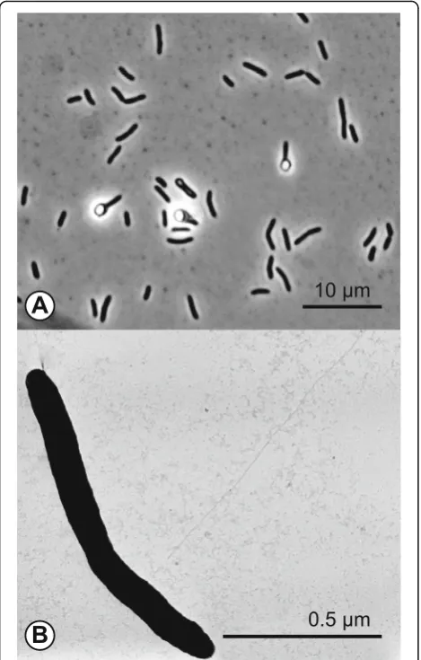
The haloalkaliphilic anaerobe D. alkaliphilus AHT1T was isolated from hypersaline soda lake sediments in northeastern Mongolia [10].D. alkaliphilusAHT1Tcells are Gram-positive and the motile rod-shaped cells form terminal ellipsoid endospores (Fig. 1). The strain toler-ates salt concentrations ranging from 0.2–0.8 M Na+ with an optimum at 0.4 M and is an obligate alkaliphile, growing within a pH range from 8.5–10.3 with an optimum at 9.5 [10]. Phylogenetic analysis showed that strain AHT1T is a member of the phylum Firmicutes
and the order Clostridiales(Fig. 2). Its closest relative is an acetate-oxidizing syntrophic alkaliphile, described as “Candidatus Contubernalis alkalaceticum” which was isolated from a soda lake [11] (Fig. 2). The 16S riboso-mal RNA of D. alkaliphilus AHT1T (EF422412) is 88% identical to the 16S rRNA of“CandidatusContubernalis alkalaceticum”(DQ124682) [12].
Extended feature descriptions
D. alkaliphilus AHT1T is an obligate anaerobe that can produce sulfide by using elemental sulfur and polysulfides
* Correspondence:[email protected]
1Department of Freshwater and Marine Ecology, Microbial Systems Ecology,
Institute for Biodiversity and Ecosystem Dynamics, University of Amsterdam, Amsterdam, The Netherlands
Full list of author information is available at the end of the article
as electron acceptor [10]. Additionally, it has been shown to incompletely reduce thiosulfate to sulfide and sulfite with hydrogen or formate as electron donor [10]. Strain AHT1Tis the first representative from theFirmicuteswith the metabolic capacity to grow by elemental sulfur dispro-portionation [13] and, therefore, is a very interesting organism to compare to the typical sulfur disproportiona-tors from theDeltaproteobacteria. This species may play an important role in the reductive sulfur cycle in soda lake environments [2] and possibly also in other alkaline anaer-obic habitats, such as serpentinization “cement springs”, where sequences closely related toDethiobacterhave been found [14, 15]. Also, its affiliation with the syntrophic
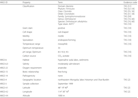
Clostridia “Candidatus Contubernalis alkalaceticum” (Fig. 2) implies that D. alkaliphilus AHT1T could be involved in syntrophic anaerobic metabolic activity. More classifications and features of this species are listed in Table 1.
Genome sequencing information
Genome project history
This organism was selected for sequencing at the JGI (http://jgi.doe.gov) based on its potential for bioremedi-ation and biotechnological applicbioremedi-ations. It is part of the Community Science Program: Haloalkaliphilic sulfate-, thiosulfate- and sulfur-reducing bacteria (CSP_788492). The project is registered in the Genomes OnLine Database (Ga0028528) [16] and the permanent draft genome sequence is deposited in GenBank (RefSeq: NZ_ACJM00000000.1). Draft sequencing and assem-bly were performed at the JGI using state of the art sequencing technology [17]. The project information is summarized in Table 2.
Growth conditions and genomic DNA preparation
Strain AHT1T was grown anaerobically at 30 °C in Na-carbonate buffered mineral medium (22 g/L Na2CO3, 8 g/L NaHCO3, 6 g/L NaCl, 1 g/L K2HPO4) with a pH of 10 and 0.6 M total Na+. Additionally, 4 mM NH4Cl, 1 mM MgCl2x 6H2O and 1 mlL−1 trace element solu-tion were added [18]. After sterilizasolu-tion, acetate serving as carbon source (2 mM) and thiosulfate (20 mM) the electron-acceptor, were also added to the medium. The culture (2 L) was grown in a 10 L bottle mounted on a magnetic stirrer whereby the headspace (8 L) was re-placed by 100% (v/v) H2, at 0.5 Bar overpressure, acting as the electron-donor. Half the culture volume (1 L) was centrifuged at 13,000 g for 30 min, the pellet was washed with 1 M NaCl and frozen at -80 °C until further down-stream processing. DNA was extracted from the pellet by the phenol-chloroform method after pre-treatment with SDS-proteinase K according to Marmur [19]. The concentration and molecular weight of the DNA were checked by UV spectroscopy and gel electrophoresis, respectively.
Genome sequencing and assembly
The size of the assembled D. alkaliphilus AHT1T gen-ome sequence was 3.12 Mbp. The draft gengen-ome was generated at the JGI using a combination of Sanger, Solexa/Illumina [20] and 454 DNA sequencing technolo-gies [21]. An 8 Kb Sanger library was constructed that provided 2.5 x coverage of the genome (15,321 reads generated) and a Solexa shotgun library and a 454 Titan-ium standard library, which provided 25× genome cover-age totalling 110.0 Mbp of 454 data. The 454 Titanium data were assembled with Newbler. The Newbler con-sensus sequences were computationally shredded into 2 Kb overlapping fake reads (shreds). Illumina sequencing data was assembled with VELVET, version 1.0.13 [22], and the consensus sequences were computationally shredded into 1.5 Kb overlapping fake reads (shreds). We then integrated Sanger reads, the 454 Newbler
Table 1Classification and general features ofD. alkaliphilusAHT1T
MIGS ID Property Term Evidence code Classification Domain:Bacteria
Phylum:Firmicutes
Class:Clostridia
Order:Clostridiales
Family:Syntrophomonadaceae
Genus:Dethiobacter
Species:Dethiobacter alkaliphilus
Type strain: AHT1T
TAS [51] TAS [52–54] TAS [55,56] TAS [57,58] TAS [59] TAS [10,60] TAS [10,60] TAS [10] Gram stain positive TAS [10] Cell shape rod-shaped TAS [10] Motility motile TAS [10] Sporulation endospore-forming TAS [10] Temperature range mesophile TAS [10] Optimum temperature 33
pH range; Optimum 8.5-10.3; 9.5 TAS [10] Carbon source CO2, acetate TAS [10]
MIGS-6 Habitat hypersaline soda lakes, sediments MIGS-6.3 Salinity moderately salt-tolerant MIGS-22 Oxygen requirement anaerobe
MIGS-15 Biotic relationship free-living MIGS-14 Pathogenicity none
MIGS-4 Geographic location northeastern Mongolia; lakes Hotontyn and Shar-Burdiin TAS [2] MIGS-5 Sample collection September 1999
MIGS-4.1 Latitude 48° 19′40″ TAS [2] MIGS-4.2 Longitude 114° 30′16″ TAS [2] MIGS-4.4 Altitude 1000 m
Evidence codes -IDAInferred from Direct Assay,TASTraceable Author Statement (i.e., a direct report exists in the literature);NASNon-traceable Author Statement (i.e., not directly observed for the living, isolated sample, but based on a generally accepted property for the species, or anecdotal evidence). These evidence codes are from the Gene Ontology project [Cite ontology project]
consensus shreds and the Illumina VELVET consensus shreds using the PGA assembler [23], to combine se-quence data from all three platforms for a most contigu-ous assembly. The software Consed [24] was used in the computational finishing process as described previously [25]. The final draft assembly contained 34 contigs in 5 scaffolds.
Genome annotation
The assembled sequence was automatically annotated with the JGI prokaryotic annotation pipeline [26] with additional manual review using the IMG-ER platform [27]. Genes were predicted using Prodigal [28], riboso-mal RNAs were detected using models built from SILVA [29] and tRNAs were predicted with tRNAScanSE [30]. The predicted CDs were translated and used to search the NCBI non-redundant database UniProt, TIGRFam, Pfam, KEGG, COG and InterPro databases. The final an-notated genome is available from the IMG system [31]. We performed a CheckM analysis [32] and assessed that the genome is 95.8% complete.
Genome properties
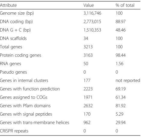
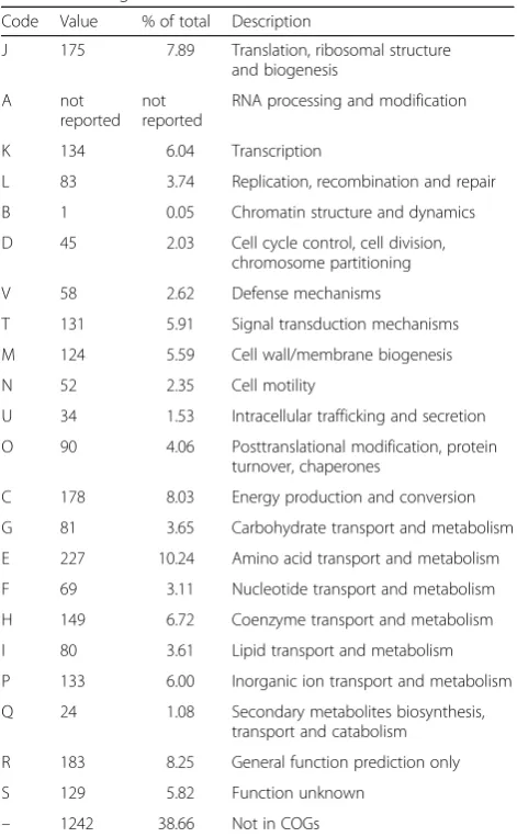
The genome is 3,116,746 bp long with a GC content of 48.46%. A total of 3213 genes were found, of which 3163 coded for proteins and 50 genes encoded only RNA. From the total genes, 69.19% was assigned a putative function. The IMG taxon ID is 643,886,183. The differ-ent functional gene groups are summarized in Table 3. Furthermore, the number of genes assigned to functional COG categories is displayed in Table 4.
Insights from the genome sequence
Extended insights: Metabolic potential
Hydrogen metabolism requires a number of hydrogenase operons, including thehyd operon, and a Ni-Fe metallo-center assembly (hyp) [33]. The first part of the hydro-genase hyd operon is the small hydrogenase subunit
hydA located at DealDRAFT_1217, the closest NCBI BLAST hit [12] of this protein is the hydA gene in
Desulfotomaculum gibsoniae (Desgi_1397) with 70.4% similarity in a pair-wise alignment [34]. Directly adjacent to hydA, is the large subunit hydB (DealDRAFT_1218) in the D. alkaliphilus AHT1T genome. This subunit is most similar (75.9%) to the hydBsubunit in Dehalobac-ter sp. UNSWDHB (UNSWDHB_1527) [12, 34]. Deal-DRAFT_1219 is a cytochrome B561 of 198 amino acids and could therefore be the interacting partner and gamma subunithydCin thehydoperon. The 6-genehyp
operonhypABCDEF is responsible for the assemblage of the Ni-Fe uptake hydrogenases [35]. The last 5 proteins of the hyp operon are annotated in the D. alkaliphilus
AHT1T genome (DealDRAFT_0838-DealDRAFT_0842) and follow the organizationhypBFCDE, as has been seen before in Rhizobium [36]. The first gene in the operon (DealDRAFT_0843) is a hypothetical protein of 88 nu-cleotides length and is assigned to pfam01155 hypA, which is 42.6% identical to the hypA gene in Moorella thermoaceticum. Therefore, this hypothetical protein is most likely hypA in D. alkaliphilus AHT1T. Using hydrogen as electron donor, D. alkaliphilus AHT1Tcan grow autotrophically by fixing inorganic carbon through the Wood Ljungdahl pathway, the key genes are all present in the genome (Fig. 3a), including the acs gene
Table 2Project information
MIGS ID Property Term
MIGS 31 Finishing quality permanent draft MIGS 28 Libraries used Solexa MIGS 29 Sequencing platforms 454 MIGS 31.2 Fold coverage 33.2
MIGS 30 Assemblers Newbler, (2.0.00.20-PostRelease-11-05-2008-gcc-3.4.6), PGA [23], VELVET [22]
MIGS 32 Gene calling method Prodigal [28] Locus Tag DealDRAFT Genbank ID ACJM00000000
Genbank Date of Release 12.12.2013 GOLD ID Gp0001962 BIOPROJECT PRJNA30985
Project relevance bioremediation, environmental biotechnology
Table 3Nucleotide content and gene count levels of the genome
Attribute Value % of total Genome size (bp) 3,116,746 100 DNA coding (bp) 2,773,015 88.97 DNA G + C (bp) 1,510,353 48.46 DNA scaffolds 34 100 Total genes 3213 100 Protein coding genes 3163 98.44 RNA genes 50 1.56 Pseudo genes 0 0
cluster (Fig. 3b). Heterotrophic growth byD. alkaliphilus
AHT1Tcan be achieved with glucose and fructose [10], the entire glycolysis pathway is present in the genome (Fig. 4). Carbohydrate metabolism in D. alkaliphilus
AHT1T also includes oxidation of short chain organic acids; the tetrameric pyruvate oxidoreductase is present in the conformation porBADC (DealDRAFT_1244 – DealDRAFT_1247). Lactate dehydrogenases could not be found, although there is an L-lactate permease DRAFT_0239), an L-lactate transport protein (Deal-DRAFT_1845) and a large and small subunit acetolactate synthase (DealDRAFT_2169 and 2170). For assimilation of acetate, strain AHT1Thas an acetyl coen-zyme A synthetase (DealDRAFT_1887).
D. alkaliphilusAHT1Tmight play a role in the reduc-tive sulfur cycle in alkaline habitats since it grows as a thiosulfate and sulfur/polysulfide reducer or by sulfur disproportionation in laboratory cultures [10]. The
genome sequence contains a thiosulfate sulfurtransferase (DealDRAFT_1917), which is located directly adjacent to another sulfur transferase (Rhodanese domain Deal-DRAFT_1918). Both alpha and beta subunits of the ade-nylylsulfate reductase apr operon were also found (DealDRAFT_1379, DealDRAFT_1380). The qmo elec-tron transfer complex, which usually accompanies the
Table 4Number of genes associated with general COG functional categories
Code Value % of total Description
J 175 7.89 Translation, ribosomal structure and biogenesis
A not reported
not reported
RNA processing and modification K 134 6.04 Transcription
L 83 3.74 Replication, recombination and repair B 1 0.05 Chromatin structure and dynamics D 45 2.03 Cell cycle control, cell division,
chromosome partitioning V 58 2.62 Defense mechanisms
T 131 5.91 Signal transduction mechanisms M 124 5.59 Cell wall/membrane biogenesis N 52 2.35 Cell motility
U 34 1.53 Intracellular trafficking and secretion O 90 4.06 Posttranslational modification, protein
turnover, chaperones
C 178 8.03 Energy production and conversion G 81 3.65 Carbohydrate transport and metabolism E 227 10.24 Amino acid transport and metabolism F 69 3.11 Nucleotide transport and metabolism H 149 6.72 Coenzyme transport and metabolism I 80 3.61 Lipid transport and metabolism P 133 6.00 Inorganic ion transport and metabolism Q 24 1.08 Secondary metabolites biosynthesis,
transport and catabolism R 183 8.25 General function prediction only S 129 5.82 Function unknown
– 1242 38.66 Not in COGs
The total is based on the number of protein coding genes in the genome
aproperon [37], is not found. Key sulfur reduction genes such assat (sulfate reduction),dsr(sulfite reduction) and
psr (sulfur reduction) were also not found in this draft genome. As D. alkaliphilus AHT1Tcan reduce and dis-proportionate elemental sulfur/polysulfide in laboratory
cultures [10, 13], the absence of these genes is surpris-ing. It is conceivable however, that the sequencing qual-ity of the permanent draft is insufficient to recover complete pathways. Indeed, CheckM analysis revealed that the genome was only 95.8% complete. Unfortu-nately, we can therefore not explain the key dissimila-tory disproportionation mechanism from this genomic data. The genome also contains some assimilatory sul-fate reduction genes, such ascysND (DealDRAFT_1193 and DealDRAFT_1192).
Extended insights: Haloalkaliphilic adaptations
In order to generate ATP,D. alkaliphilusAHT1Thas an
ntp gene operon encoding a vacuolar ATP synthase (V0V1-type) (DealDRAFT_1677 – DealDRAFT_1685) (Fig. 5a). This operon structure is conserved among the
Clostridia (Fig. 5b). The ntp operon encodes the ATP synthase for ATP generation and follows the GILEX-FABD organization in the Deinococcus-Thermusphylum [38]. In the Firmicutes, the gene organization is slightly different at GIKECFABD (Fig. 5a, b). In D. alkaliphilus
AHT1T these genes are located from DealDRAFT_1685
Fig. 4KEGG orthologs annotated in the Embden-Meyerhof pathway of organic carbon assimilation inD. alkaliphilusstrain AHT1T. The numbers of the locus tags of the genes catalyzing each reaction are indicated and must be preceded by DealDRAFT_
Fig. 5aThentpVacuole-type ATP synthase operon structure.b93
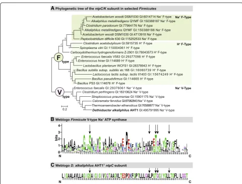
(ntpG) to DealDRAFT_1677 (ntpD). The ntpD subunit within the operon is annotated as being of the V-type. In order to confirm that the ATP synthase is indeed V-type [39], we constructed a phylogenetic tree of the trans-membranec/Ksubunits ofFirmicutesknown specifically to be V- or F-type [40] and NCBI annotation] and aligned theD. alkaliphilusAHT1TntpCsequence (Deal-DRAFT_1683) with these other sequences (Fig. 6a) [41]. As seen before, there was a clear separation between V-type and F-V-type ATP synthase, where the AHT1T se-quence clustered together with the V-type ATP synthase. In addition, the sequences are tentatively clustered into separate H+ or Na+ coupled ATPase branches. The AHT1T sequence was positioned within a Na+ coupled V-type ATP synthase group, indicating that this organ-ism’s ATP synthase is coupled specifically to Na+ trans-location across the membrane. In order to explore this further, we looked at specific Na+ binding residues and ligands on the transmembrane c/K subunit [40], and
created a Weblogo for the Na+ specific Firmicutes V-type ATP synthase (Fig. 6b) [42, 43]. When we aligned the ntpC sequence of D. alkaliphilus AHT1T we found that it contains all the conserved five amino acids (Ser26, Leu57, Thr60, Gln61 and Tyr64) specific for Na+ translocation [40] (Fig. 6c). Thus, the D. alkaliphilus
AHT1T genome contains a Na+ coupled V-type ATP synthase.
In order to import protons to retain the intracellular pH, the genome contains the multi-subunit electrogenic sodium/proton antiporter mrp (DealDRAFT_2487– 2497), that pumps protons into the cell and sodium out of the cell [44]. To retain osmotic balance,D. alkaliphi-lus AHT1Thas numerous substrate binding regions and transporters for glycine betaine (e.g. DealDRAFT_2378, _2380 and DealDRAFT2842, _2844), leading to the con-clusion that osmoprotectants are used to maintain cellu-lar turgor pressure, instead of the salt-in strategy. Another necessity for alkaliphilic bacteria is to prevent
Fig. 6aPhylogeny of the F- vs. V-type ATPase within theFirmicutes. Numbers on the tree nodes indicate bootstrap values (1000 replicates). Scale bar indicates 0.2% sequence difference.bWeblogo of conserved region within thentpC/K Firmicusubunit [42, 43].cWeblogo of aligned
proton leakage from cells, which they can achieve through structural membrane adaptations [1]. The genome con-tains the genes to synthesize the squalene precursors dimethylallyl diphosphate and isopentenylallyl diphos-phate through the non-mevalonate pathway [45]. The ac-companying locus tags within the KEGG non-mavalonate pathway (M00096) are dxs (DealDRAFT_0731), dxr/ispC
(DealDRAFT_2409),ispD(DealDRAFT_2331),ispE (Deal-DRAFT_2584), ispF (DealDRAFT_2332), ispG (Deal-DRAFT_2411) and ispH (DealDRAFT_0659). However, we did not find genes similar tohpnCDE, which function in the formation of squalene from its precursors [46]. Thus,D. alkaliphilus AHT1T does not seem to have this membrane adaptation to haloalkaline environments, al-though it could also be due to the incompleteness of the genome. Nevertheless, it has been shown thatBacillus len-tus C-125, also a Firmicute, survives in the haloalkaline environment by increased levels of acidic polymers in its cellular membrane resulting in a cell wall negative charge [47]. It is possible thatD. alkaliphilusAHT1Tsupports a similar mechanism to survive the alkaline pH values of its environment.
Conclusions
In this manuscript we globally characterize the genome of D. alkaliphilus AHT1T, which was isolated from hy-persaline soda lakes sediment in north-eastern Mongolia. Investigation of the genome of this anaerobic sulfidogen identified genes for the Wood Ljungdahl pathway (auto-trophic growth, Fig. 3) and the Embden-Meyerhof path-way (heterotrophic growth Fig. 4). Thus the carbon metabolism of this microbe is fairly versatile. D. alkali-philusAHT1Tis capable of disproportionation in labora-tory cultures, thus future genomic analyses with qPCR may provide insights into the disproportionation of sul-fur compounds. D. alkaliphilus AHT1T is well adapted to the haloalkaline environment, we found genes for active energy generation with a sodium V-type ATP syn-thase (Fig. 6). In addition, transporters for the osmopro-tectants glycine and betaine were found to maintain cellular homeostasis and protection from the saline ex-ternal environment. Further research will extend our knowledge on the ecophysiology of haloalkaliphiles, their role in nutrient cycling in extreme environments and their adaptations to this polyextreme environment. Moreover, insight in the genome sequence and subse-quent transcriptomic or proteomic analysis will be help-ful to infer the potential role of D. alkaliphilusAHT1T in the biotechnological removal of sulfur compounds from wastewater and gas streams.
Abbreviations
F-type:Phosphorylation factor-type; IMG: Integrated Microbial Genomes; IMG-ER: Integrated Microbial Genomes - Expert Review; JGI: Joint
Genome Institute; NCBI: National Center for Biotechnology Information; THF: tetrahydrofolate; V-type: Vacuole-type
Acknowledgements
Emily Denise Melton, Lex Overmars and Gerard Muyzer are supported by ERC Advanced Grant PARASOL (No. 322551); Dimitry Sorokin is supported by RFBR grant 16-04-00035 and by the Gravitation (SIAM) program (grant 24002002, Dutch Ministry of Education and Science). Alla L. Lapidus is supported by the St. Petersburg State University grant 15.61.951.2015. The work conducted by the U.S. Department of Energy Joint Genome Institute, a DOE Office of Science User Facility, was supported under Contract No. DE-AC02-05CH11231.
Authors’contributions
EDM drafted and wrote the manuscript. DYS, GM, LO, NCK and ALL contributed to the written manuscript. LO, DYS and GM stimulated critical discussions. DS cultured AHT1 and extracted the DNA. The sequencing and annotation of the genome were performed at the JGI by ALL, MP, NI, TGR, NCK and TW. All authors read and approved the final manuscript.
Competing interests
The authors declare that they have no competing interest.
Publisher’s Note
Springer Nature remains neutral with regard to jurisdictional claims in published maps and institutional affiliations.
Author details
1Department of Freshwater and Marine Ecology, Microbial Systems Ecology,
Institute for Biodiversity and Ecosystem Dynamics, University of Amsterdam, Amsterdam, The Netherlands.2Winogradsky Institute of Microbiology,
Research Centre of Biotechnology, RAS, Moscow, Russia.3Department of Biotechnology, Delft University of Technology, Delft, The Netherlands.
4Center for Algorithmic Biotechnology, Institute of Translational Biomedicine,
St. Petersburg State, University, St. Petersburg, Russia.5Joint Genome
Institute, Walnut Creek, CA, USA.6Biological Data Management and Technology Center, Lawrence Berkeley National Laboratory, Berkeley, CA, USA.7Department of Biological Sciences, Faculty of Science, King Abdulaziz
University, Jeddah, Saudi Arabia.
Received: 23 March 2017 Accepted: 8 September 2017
References
1. Sorokin DY, Berben T, Melton ED, Overmars L, Vavourakis CD, Muyzer G. Microbial diversity and biogeochemical cycling in soda lakes. Extremophiles. 2014;18:791–809.
2. Sorokin DY, Gorlenko VM, Namsaraev BB, Namsaraev ZB, Lysenko AM, Eshinimaev BT, Khmelenina VN, Trotsenko YA, Kuenen JG. Prokaryotic communities of the north-eastern Mongolian soda lakes. Hydrobiologia. 2004;522:235–48.
3. Sorokin DY, Rusanov I, Pimenov NV, Tourova TP, Abbas B, Muyzer G. Sulfidogenesis under extremely haloalkaline conditions in soda lakes of Kulunda steppe (Altai, Russia). FEMS Microbiol Ecol. 2010;73:278–90. 4. Sorokin DY, Kuenen JG, Muyzer G. The microbial sulfur cycle at extremely
haloalkaline conditions of soda lakes. Front Microbiol. 2011;2:44.
5. Zhao B, Mesbah NM, Dalin E, Goodwin L, Nolan M, Pitluck S, et al. Complete genome sequence of the anaerobic, halophilic alkalithermophileNatranaerobius thermphilusJW/NM-WN-LF. J Bacteriol. 2011;193:4023–4.
6. Junier P, Junier T, Podell S, Sims DR, Detter JC, Lykidis A, et al. The genome of the gram-positive metal- and sulfate-reducing bacteriumDesulfotomaculum reducensstrain MI-1. Environ Microbiol. 2010;12:2738–54.
7. Aüllo T, Ranchou-Peyruse A, Ollivier B, Magot M.Desulfotomaculumspp. and related gram-positive sulfate-reducing bacteria in deep subsurface environments. Front Microbiol. 2013;4:362.
8. Visser M, Parshina SN, Alves JI, Sousa DZ, Pereira IAC, Muyzer G, et al. Genome analyses of the carboxydotrophic sulfate-reducersDesulfotomaculum nigrificans
andDesulfotomaculum carboxydivoransand reclassification ofDesulfotomaculum nigrificans. Stand Genomic Sci. 2014;9:655–75.
Desulfosporosinus youngiaeDSM17734T,Desulfosporosinus meridiei
DSM13257T, andDesulfosporosinus acidiphilusDSM22704T. J Bacteriol. 2012;194:6300–1.
10. Sorokin DY, Tourova TP, Mussmann M, Muyzer G.Dethiobacter alkaliphilusgen. Nov. sp. nov., andDesulfurivibrio alkaliphilusgen. nov. sp. nov.: two novel representatives of reductive sulfur cycle from soda lakes. Extremophiles. 2008; 12:431–9.
11. Zhilina TN, Zavarzina DG, Kolganova TV, Tourova TP, Zavarzin GA.“Candidatus
Contubernalis alkalaceticum,”an obligately syntrophic alkaliphilic bacterium capable of anaerobic acetate oxidation in a coculture withDesulfonatronum cooperativum. Microbiology. 2005;74:800–9.
12. Altschul SF, Gish W, Miller W, Myers EW, Lipman DJ. Basic local alignment search tool. J Mol Biol. 1990;215:403–10.
13. Poser A, Lohmayer R, Vogt C, Knoeller K, Planer-Friedrich B, Sorokin D, et al. Disproportionation of elemental sulfur by haloalkaliphilic bacteria from soda lakes. Extremophiles. 2013;17:1003–12.
14. Tiago I, Verissimo A. Microbial and functional diversity of a subterrestrial high pH groundwater associated to serpentinization. Environ Microbiol. 2012;15:1687–706.
15. Suzuki S, Ishiia S, Wua A, Cheung A, Tenneya A, Wangera G, et al. Microbial diversity in the cedars, an ultrabasic, ultrareducing, and low salinity serpentinizing ecosystem. Proc Nat Acad Sci USA. 2013;110: 15336–41.
16. Reddy TBK, Thomas AD, Stamatis D, Bertsch J, Isbandi M, Jansson J,et al. The Genomes OnLine Database (GOLD) v.5: a metadata management system based on a four level (meta)genome project classification. Nucleic Acids Res. 2014;43:1099–106.
17. Mavromatis K, Land ML, Brettin TS, Quest DJ, Copeland A, Clum A,et al. The fast changing landscape of sequencing technologies and their impact on microbial assemblies and annotations. PLoS ONE 2012;doi: https://doi.org/ 10.1371/journal.pone.0048837.
18. Pfennig N, Lippert KD. Über das Vitamin B12-Bedürfnis phototropher Schwefelbakterien. Arch Mikrobiol. 1966;55:245–56.
19. Marmur J. A procedure for isolation of DNA from microorganisms. J Mol Biol. 1961;3:208–14.
20. Bennett S. Solexa Ltd. Pharmacogenomics. 2004;5:433–8.
21. Margulies M, Egholm M, Altman WE, Attiya S, Bader JS, Bemben LA, et al. Genome sequencing in microfabricated high-density picolitre reactors. Nature. 2005;437:326–7.
22. Zerbino DR, Birney E. Velvet: algorithms for de novo short read assembly using de Bruijn graphs. Genome Res. 2008;18:821–9.
23. Zhao F, Zhao F, Li T, Bryant DA. A new pheromone trail-based genetic algorithm for comparative genome assembly. Nucl Acids Res. 2008;36:3455–62. 24. Gordon D, Abajian C, Green P. Consed: a graphical tool for sequence
finishing. Genome Res. 1998;8:195–202.
25. Sims D, Brettin T, Detter JC, Han C, Lapidus A, Copeland A, et al. Complete genome sequence ofKytococcus sedentariustype strain (541T). Stand Genomic Sci. 2009;1:12–20.
26. Mavromatis K, Ivanova NN, Chen IM, Szeto E, Markowitz VM, Kyrpides NC. The DOE-JGI standard operating procedure for the annotations of microbial genomes. Stand Genomic Sci. 2009;1:63–7.
27. Markowitz VM, Ivanova NN, Chen IMA, Chu K, Kyrpides NC. IMG ER: a system for microbial genome annotation expert review and curation. Bioinformatics. 2009;25:2271–8.
28. Hyatt D, Chen G, LoCascio PF, Land ML, Larimer FW, Hauser LJ. Prodigal: prokaryotic gene recognition and translation initiation site identification. BMC Bioinf. 2010;11:119.
29. Quast C, Pruesse E, Yilmaz P, Gerken J, Schweer T, Yarza P,et al.The SILVA ribosomal RNA gene database project: improved data processing and web-based tools. Nucl Acids Res. 2013; doi: 10.1093/nar/gks1219.
30. Lowe TM, Eddy SR. tRNAscan-SE: a program for improved detection of transfer RNA genes in genomic sequence. Nucl Acids Res. 1997;25:955–64. 31. Markowitz VM, Chen I-M A, Palaniappan K, Chu K, Szeto E, Grechkin Y, et al.
IMG: the integrated microbial genomes database and comparative analysis system. Nucleic Acids Res. 2012;40:D115–22.
32. Parks DH, Imelfort M, Skennerton CT, Hugenholtz P, Tyson GW. CheckM: assessing the quality of microbial genomes recovered from isolates, single cells, and metagenomes. Genome Res. 2015;25:1043–55.
33. Schwartz E., Fritsch J., Friedrich B. H2-Metabolizing prokaryotes. In: Rosenberg E., DeLong E. F., Lory S., Stackebrandt E., Thompson F., editors. The Prokaryotes. Berlin: Springer; Verlag; 2013. p. 119–199.
34. Sievers F, Wilm A, Dineen D, Gibson TJ, Karplus K, Lopez R, et al. Fast, scalable generation of high-quality protein multiple sequence alignments using Clustal omega. Mol Systems Biol. 2011;7:539.
35. Agervald Å, Stensjö K, Holmqvist M, Lindblad P. Transcription of the extended
hyp-operon inNostocsp. strain PCC 7120. BMC Microbiol. 2008;8:69. 36. Hernando Y, Palacios JM, Imperial J, Ruiz-Argüeso T. ThehypBFCDEoperon
fromRhizobium leguminosarumbiovar viciae is expressed from an Fnr-type promotor that escapes mutagenesis of the fnrN gene. J Bacteriol. 1995;177: 5661–9.
37. Duarte AG. Santos AA and Pereira IAC electron transfer between the
QmoABC membrane complex and adenosine 5’phosphosulfate reductase. Biochim Biophys Acta. 1857;2016:380–6.
38. Yokoyama K, Ohkuma S, Taguchi H, Yasunaga T, Wakabayashi T, Yoshida M. V-type H+ ATPase/synthase from a thermophilic eubacterium,Thermus thermophilus. J Biol Chem. 2000;275:13955–61.
39. Hicks DB, Liu J, Fujisawa M, Krulwich TA. F1F0-ATP synthases of alkaliphilic bacteria: lessons from their adaptations. Biochim Biophys Acta. 2010;1797: 1362–77.
40. Mulkidjanian AY, Galperin MY, Makarova KS, Wolf YI, Koonin EV. Evolutionary primacy of sodium bioenergetics. Biol Direct. 2008;3:13.
41. Li W, Cowley A, Uludag M, Gur T, McWilliam H, Squizzato S, et al. The EMBL-EBI bioinformatics web and programmatic tools framework. Nucleic Acids Res. 2015;43:580–4.
42. Schneider TD, Stephens RM. Sequence logos: a new way to display consensus sequences. Nucleic Acids Res. 1990;18:6097–100.
43. Crooks GE, Hon G, Chandonia JM, Brenner SE. WebLogo: A sequence logo generator. Genome Res. 2004;14:1188–90.
44. Krulwich TA, Sachs G, Padan E. Molecular aspects of bacterial pH sensing and homeostasis. Nat Rev Microbiol. 2011;9:330–43.
45. Eisenreich W, Bacher A, Arigoni D, Rohdich F. Biosynthesis of isoprenoids 544 via the non-mevalonate pathway. Cell Mol Life Sci. 2004;61:1401–26. 46. Pan JJ, Solbiati JO, Ramamoorthy G, Hillerich BS, Seidel RD, Cronan JE, Almo
SC, Poulter CD. Biosynthesis of squalene from farnesyl diphosphate in bacteria: three steps catalysed by three enzymes. ACS Cent Sci. 2015;1:77–82. 47. Aono R, Ito M, Joblin KN, Horikoshi K. A high cell wall negative charge is
necessary for the growth of the alkaliphileBacillus lentusC-125 at elevated pH. Microbiology. 1995;141:2955–64.
48. Ludwig W, Strunk O, Westram R, Richter L, Meier H, Yadhukumar, et al. ARB: a software environment for sequence data. Nucl Acids Res. 2004;32:1363–71. 49. Tamura K, Stecher G, Peterson D, Filipski A, Kumar S. MEGA6: Molecular
evolutionary genetics analysis version 6.0. Mol Biol Evol. 2013;30:2725–9. 50. Overmars L, Kerkhoven R, Siezen RJ, Francke C. MGcV: the microbial
genomic context viewer for comparative genome analysis. BMC Genomics. 2013;14:209.
51. Woese CR, Kandler O, Wheelis ML. Towards a natural system of organisms: proposal for the domains Archaea, bacteria, and Eucarya. Proc Natl Acad Sci U S A. 1990;87:4576–9.
52. Gibbons NE, Murray RGE. Proposals concerning the higher taxa of bacteria. Int J Syst Bacteriol. 1978;28:1–6.
53. Garrity GM, Holt JG. The road map to the manual. In: Garrity GM, Boone DR, Castenholz RW, editors. Bergey's Manual of Systematic Bacteriology, 2nded. Volume 1, New York: Springer; 2001. p. 119–169.
54. Murray RGE. The higher taxa, or, a place for everything…? In: Holt JG editor. Bergey's Manual of Systematic Bacteriology, 1sted. Volume 1, The Williams and Wilkins Co., Baltimore; 1984. p. 31–34.
55. List of new names and new combinations previously effectively, but not validly, published. List no. 132. Int J Syst Evol Microbiol. 2010;60:469–472. 56. Rainey FA. Class II.Clostridiaclass nov. In: De Vos P, Garrity G, Jones D, Krieg
NR, Ludwig W, Rainey FA, Schleifer KH, Whitman WB, editors, Bergey's Manual of Systematic Bacteriology, 2nded. Volume 3, Springer-Verlag, New York; 2009. p. 736.
57. Skerman VBD, McGowan V, Sneath PHA. Approved lists of bacterial names. Int J Syst Bacteriol. 1980;30:225–420.
58. Prévot AR. In: Hauderoy P, Ehringer G, Guillot G, Magrou J, Prévot AR, Rosset D, Urbain A, editors. Dictionnaire des Bactéries Pathogènes. 2nd ed. Paris: Masson et Cie; 1953. p. 1–692.
59. Jumas-Bilak E, Roudière L, Marchandin H. Description of‘Synergistetes’phyl. nov. and emended description of the phylum‘Deferribacteres’and of the familySyntrophomonadaceae, phylum‘Firmicutes’. Int J Syst Bacteriol. 2009; 59:1028–35.
![Fig. 5 aaligned in Clustal Omega [34] and an unrooted neighbour-joiningtree was generated in MEGA-6 [49]](https://thumb-us.123doks.com/thumbv2/123dok_us/650311.2064704/6.595.300.538.371.624/aaligned-clustal-omega-unrooted-neighbour-joiningtree-generated-mega.webp)