R E S E A R C H
Open Access
Two structurally discrete GH7-cellobiohydrolases
compete for the same cellulosic substrate fiber
Fernando Segato
1,4, André R L Damasio
1,5, Thiago Augusto Gonçalves
4, Mario T Murakami
3, Fabio M Squina
4,
Maria de Lourdes T M Polizeli
6, Andrew J Mort
2and Rolf A Prade
1,4*Abstract
Background:Cellulose consisting of arrays of linear beta-1,4 linked glucans, is the most abundant carbon-containing polymer present in biomass. Recalcitrance of crystalline cellulose towards enzymatic degradation is widely reported and is the result of intra- and inter-molecular hydrogen bonds within and among the linear glucans. Cellobiohydrolases are enzymes that attack crystalline cellulose. Here we report on two forms of glycosyl hydrolase family 7 cellobiohydrolases common to allAspergilliithat attack Avicel, cotton cellulose and other forms of crystalline cellulose.
Results:Cellobiohydrolases Cbh1 and CelD have similar catalytic domains but only Cbh1 contains a
carbohydrate-binding domain (CBD) that binds to cellulose. Structural superpositioning of Cbh1 and CelD on the Talaromyces emersoniiCel7A 3-dimensional structure, identifies the typical tunnel-like catalytic active site while Cbh1 shows an additional loop that partially obstructs the substrate-fitting channel. CelD does not have a CBD and shows a four amino acid residue deletion on the tunnel-obstructing loop providing a continuous opening in the absence of a CBD. Cbh1 and CelD are catalytically functional and while specific activity against Avicel is 7.7 and 0.5 U.mg prot-1, respectively specific activity on pNPC is virtually identical. Cbh1 is slightly more stable to thermal inactivation compared to CelD and is much less sensitive to glucose inhibition suggesting that an open tunnel configuration, or absence of a CBD, alters the way the catalytic domain interacts with the substrate. Cbh1 and CelD enzyme mixtures on crystalline cellulosic substrates show a strong combinatorial effort response for mixtures where Cbh1 is present in 2:1 or 4:1 molar excess. When CelD was overrepresented the combinatorial effort could only be partially overcome. CelD appears to bind and hydrolyze only loose cellulosic chains while Cbh1 is capable of opening new cellulosic substrate molecules away from the cellulosic fiber.
Conclusion: Cellobiohydrolases both with and without a CBD occur in most fungal genomes where both
enzymes are secreted, and likely participate in cellulose degradation. The fact that only Cbh1 binds to the substrate and in combination with CelD exhibits strong synergy only when Cbh1 is present in excess, suggests that Cbh1 unties enough chains from cellulose fibers, thus enabling processive access of CelD.
Keywords: Cellobiohydrolase, Cellobiohydrolase I, Cellobiohydrolase D, Aspergillus niveus, Aspergillus fumigatus, Crystalline cellulose breakdown, Biofuels, Cellulases, Biomass decomposition
* Correspondence:[email protected]
1
Department of Microbiology & Molecular Genetics, Oklahoma State University, Stillwater, OK, USA
4
Laboratório Nacional de Ciência e Tecnologia do Bioetanol (CTBE), Centro Nacional de Pesquisas em Energia e Materiais, Campinas, Sao Paulo, Brazil Full list of author information is available at the end of the article
Background
Biofuel generation from sources such as cornstarch, sugar-cane or sweet sorghum syrups, produces large amounts of biomass waste products. For example, over 90% of the plant is unused in the case of ethanol production from cornstarch [1-3]. Current commercial enterprises produce ethanol from cornstarch, sugar cane or sweet sorghum syr-ups and large amounts of waste biomass accumulate alongside refineries and most of them are not recycled. Biofuels production would therefore be significantly more efficiently utilized if this biomass could be converted into fermentable sugars.
Biomass polysaccharides consist of cellulose, hemicel-lulose and pectin. Celhemicel-lulose is a linear, crystalline self-assembled nanofiber formed from the linear polymer containing exclusively glucose monomers linked through β-1,4-glycosydic bonds [1-4]. Recalcitrance of cellulose towards enzymatic degradation is a widely reported phenomenon [1,5,6] and is the result of the wide range of possible intra- and inter-molecular hydrogen bonds within and among linear cellulose molecules assuming crystalline or amorphous cellulosic nanofiber structures. Cell walls from different plants contain various amounts of crystalline/amorphous cellulose fibers [7], reflected by the relative crystalinity index (RCI).
Because cellulose is structurally complex and crystalline in nature, it is recalcitrant towards microbial or enzym-atic attack. Unfortunately, current pretreatment methods –e.g., acid hydrolysis [8,9] or pyrolysis [10,11], generate compounds that inhibit subsequent fermentation. Enzym-atic hydrolysis of cellulose results in glucose the universal carbon source for all organisms to drive oxidative metab-olism including the production of biofuels, chemicals and pharmaceuticals.
A complete enzymatic cellulose degrading system consists of at least three related partially redundant biochemical reactions. Endo-glucanases (EC 3.2.1.4) randomly hydrolyze internal glycosydic bonds to de-crease the length of the cellulose chain and multiply polymer ends [12-14]. Exo-glucanases (EC 3.2.1.91) split-off cellobiose from cellulose termini [15-17] and β-glucosidases (EC 3.2.2.21) hydrolyze cellobiose and oligomers to render glucose [18-20]. All three types of enzymes have similar catalytic domains all splitting the β-1,4-glycosidc bond between glucose molecules, however they differ in their binding and substrate interaction domains resulting in cooperation and syn-ergism in releasing glucose [21-23].
Exo-glucanases hydrolyze cellulose chains by removing cellobiose either from the non-reducing end (GH6, EC 3.2.1.91) or reducing end (GH7, EC 3.2.1.176), which in both cases results in the release of reducing sugars (cello-biose) but little polymer length reduction [24]. In fungi, exo-glucanases are commonly known as 1,4-β-D-glucan
cellobiohydrolases [25]. The main characteristic of a cellobiohydrolase is its processive action on individual cellulose chains by reiterated release of cellobiose [24,26-32].
The catalytic domain (CD) of a typical cellobiohydro-lase forms a channel-shaped cavity topped by several flexible loops resulting in a tunnel like structure [33-39], which is frequently connected through a linker to a carbohydrate binding domain (CBD) [40,41]. CBDs are thought to bind to the crystalline cellulosic fiber sliding down the cellulosic surface [6,42] and carrying the CD with it. Three tyrosine residues on the CBD hydrophobic surface align with the cellulose chain adjacent to the re-ducing end and a fourth tyrosine residue moves from its internal position to form van der Waals interactions with the cellulose surface resulting in an induced change near the surface allowing the CBD to progress [16,40].
From a comprehensive bioinformatics survey of seven Aspergilliicompletely sequenced genomes, we found be-tween three and five genes encoding cellobiohydrolases, typically two in each of the CAZy families GH6 and GH7, respectively. Remarkably, for each GH family 6 or 7 fungal cellobiohydrolase examined, one enzyme was linked to a CBD and a second was not.
Here we describe two GH7-cellobiohydrolases, Cbh1 and CelD from Aspergillus niveus. The structural differ-ences between these two enzymes were the lack of a CBD in CelD and a conserved loop that obstructed the catalytic tunnel in enzymes that were linked to a CBD, but missing in enzymes that did not link to a CBD, en-suring that the tunnel was always open. These structural differences reflected directly on kinetic properties, ther-mostability, inhibition by glucose and cellobiose and the way these proteins interacted with crystalline substrate molecules. We hypothesize that the cellobiohydrolase linked to a CBD is competent to release cellulosic chains from the crystalline nanofiber and the cellobiohydrolase without a CBD can only use loose ends, without the aid of a CBD.
Results
Aspergilliihold precisely two GH7-cellobiohydrolases, one with and another without a cellulose-binding domain
Fungi and bacteria produce enzymes that at least par-tially degrade plant cell wall polysaccharides [43,44]. More often than not, cellulase-encoding genes appear as multiple copies in the genome of sequenced microorgan-isms and enzymes occur as functionally redundant with overlapping biochemical functions.
and another without a CBD. The one exception is Aspergillus flavus/A. oryzae (both genomes contain an almost identical nucleotide sequence [45]), which en-code two GH7-cellobiohydrolase, both with no CBD. However, it is not clear if another gene with a CBD exists or the CBD domain has not yet been annotated. Other fungi such asNeurospora crassaOR74,Penicillium chrysogenumW54-1255 andPhanerochaete chrysosporium have a two-set of GH7-cellobiohydrolases, whileHypocrea jecorina, Hypocrea rufa, Hypocrea virens and other Trichodermales appear to contain only one GH7-cel-lobiohydrolase, all with a CBD.
A. niveusa filamentous fungus analogous toA. fumigatus (the genomic DNA sequence is up to 97% identical) grew at high temperatures, up to 50°C, on medium containing complex carbon sources such as cellulose and hemicellu-lose. As expected, the genome of A. niveus encoded two GH7-cellobiohydrolases: Cbh1-like 1,4-beta-D-glucan cel-lobiohydrolase with a linker and CBD and a CelD-like 1,4-beta-D-glucan cellobiohydrolase with no linker and
CBD. Genes, cbh1 andcelD were expressed and proteins secreted into the medium when the fungus was grown on cellulose (e.g., Avicel) as the carbon source (data not shown).
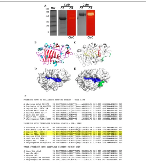
Both cbh1 and celD genes have been expressed as client proteins in A. nidulans, under the control of the maltose promoter, translated and secreted via the glucoamylase signal peptide [46] where they exhibited“in situ” activity towards carboxymethylcellulose (Figure 1A). Both proteins were purified by ion exchange followed by gel filtration chromatography and used for comprehensive biochemical analysis (See Methods and Additional file 1: Table S1).
The Cbh1 and CelD catalytic domains share 72% iden-tity and 82% similarity in their amino acid sequences. The main difference between these proteins is the pres-ence of a cellulose-binding domain in Cbh1 and the ab-sence of such a domain in CelD. The objective of this study was to investigate the mechanistic differences be-tween these two enzymes.
Minor structural differences in Cbh1 and CelD are catalytically relevant
Cbh1 and CelD catalytic domains (CD) were modeled based on the 3D-structural template derived from Talaromyces emersonii cellobiohydrolase 1 (PDBID: 1Q9H, [47]). Modeled structures were subjected to energy minimization and final models presented high-quality local and overall stereochemistry with approxi-mately 98% of amino acid residues lying in allowed regions of the Ramachandran plot (Figure 1B-E).
The Cbh1 and CelD catalytic core comprises a typical CaZy GH7 family β-sandwich surrounded by numerous surface loops, which outline the substrate-binding chan-nel (Figure 1B-E). The nativeT. emersoniistructure pre-sented a disordered loop encompassing the amino-acid segment 190–200. This sequence was fully conserved among all other Cbh1 proteins (Figure 1F), and likely be-came structured upon substrate binding similarly, to what has been reported for other glycosyl hydrolases [48]. Structural superposition resulted in a RMSD value of 0.15 Cα for Cα atoms indicating high conservation of Cbh1 and CelD three-dimensional structures. The cata-lytically relevant residues and the substrate channel are fully conserved including Trp38, Tyr168, Asp170, Glu209, Asp211, Glu214, Trp371 and Trp380 (Figure 1C). The only significant difference was at the entrance of the catalytic tunnel formed by loops 43–63, 94–103 and 190–200 that partially blocked the catalytic channel in Cbh1, due to a four-residue insertion in the 94–103 loop (Figure 1C and 1F). In all CelD proteins, this four amino acid insertion is missing (Figure 1F) and CelD proteins do not have a CBD thus suggesting that in the absence Table 1 Genome wide distribution of
gh7-cellobiohydrolases
Aspergillii gh7-cellobiohydrolases
no CBM CBM
Prot ID Locus tag Prot ID Locus tag
A. clavatusNRRL1 XP_001272622 ACLA_088870
XP_001272622 ACLA_085260
A. fumigatusAf293 XP_750600 AFUA 6G07070
XP_751044 AFUA 6G11610
A. nidulansFGSC A4 XP_662780 AN5176
XP_658098 AN0494
A. nigerCBS 513.88 XP_001392008 ANI_1_2134064
XP_001389576 ANI_1_1574014
A. terreusNIH 2624 XP_001212905 ATEG_03727
XP_001214180 ATEG_05002
A. oryzaeRIB40 XP_001818879 AOR_1_608164
none
XP_001727881 AOR_1_1654194
none
A. flavusNRRL-3357 XP_002380314 AFLA_067550
none
XP_002376207 AFLA_021870
none
P. chrysogenum W54-1255
CAP94773 Pc18g05490
CAP85526 Pc20g01970
Neurospora crassaOR 74A
XP_957090 NCU05104
XP_962498 NCU07340
Phanerochaete chrysosporium
CAA38274 Pccbh-1
CAA38275 Pccbh 1-2
Hypocrea jecorina none GUX_1TRIRE cbn1
Trichoderma viride (Hypocrea rufa)
none BAA36215 cbh1
of a CBD, the catalytic domain only functions if the sub-strate channel is open.
Cbh1 and CelD are catalytically functional
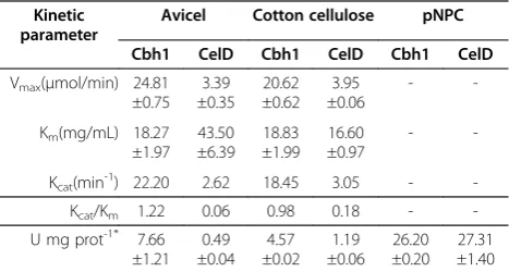
Purified Cbh1 and CelD were incubated in the presence of increasing amounts of two crystalline cellulosic substrates, Avicel and cotton cellulose and the specific velocity was determined after a 120 min reaction period (For the Michaelis Menten plot refer to Additional file 2: Figure S1). Table 2 shows both, Cbh1 and CelD responding to increas-ing amounts of crystalline cellulose substrates, the number of substrate molecules converted, kcat, over time (minutes)
significantly diverged between Cbh1 and CelD; while Cbh1 turned over 22.20 and 18.45 Avicel or cotton cellulose molecules, respectively, CelD turned over only 2.62 and 3.05 molecules respectively. Thus, Cbh1 was about 6 to 8 fold more efficient in hydrolyzing crystalline cellulosic sub-strates. This difference was also reflected in the determined catalytic efficiency Kcat/Km, which hovers from 1.22 to 0.98
and 0.06 to 0.18 depending on the substrate, respectively (see Table 2). Specific activity for both proteins reflected the catalytic differences as well as the substrate affinities for Avicel and cotton cellulose except when assayed with p-nitrophenyl-β-D-cellobioside (pNPC) where specific ac-tivity was identical, suggesting that Cbh1 and CelD catalytic domains function equally. However they differ in the way they interact with insoluble polymeric substrates.
Only Cbh1 binds to cellulose
Because CelD does not have a CBD and is biochemically active, we hypothesized that CelD could associate with Cbh1 in solution and use the Cbh1 CBD to slide along cel-lulose chains. However, our experiment (Figure 2) shows that CelD does not bind to cellulose alone or in combin-ation with Cbh1, thus ruling out an associcombin-ation between these two enzymes in natural substrate interactions.
Thermal inactivation leads to differential protein unfolding
To study thermal inactivation, Cbh1 and CelD were incu-bated at various temperatures in the absence of substrate in 50 mM ammonium acetate buffer for 40 min and residual activity was determined with pNPC. Figure 3 shows that Cbh1 was slightly more stable to thermal inactivation when compared to the inactivation of CelD, suggesting that the presence of a CBD, only present in Cbh1, had a thermo protection effect similar to that observed for other proteins with CBD’s [49,50].
Differential glucose inhibition implies two differentially adapted catalytic domains
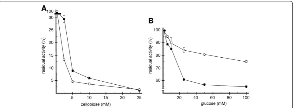
Catalytic inhibition studies of Cbh1 and CelD by cellobiose and glucose, were carried out with purified enzymes and pNPC as a substrate (Figure 4). In the presence of 5 mM cellobiose, 90% of the activity for both enzymes was inhib-ited. Addition of glucose however, had a differential effect: with 50 mM of glucose, Cbh1 retained 75% of its initial activity while CelD retained only 55%. In the presence of 100 mM of glucose, Cbh1 retained 80% of its initial activity and CelD had lost more than 50%. Thus, the rate of p-nitrophenyl formation from pNPC by Cbh1 is less se-verely affected by the presence of glucose than CelD. The fact that cellobiose does not differentially affect both enzymes, but glucose does, may be a consequence of the structural differences of the catalytic channel.
Cbh1 and CelD combine efforts to hydrolyze the same substrate molecules
Figure 5 shows that Cbh1/CelD combinations incubated with various cellulosic substrates develop strong synergy only under selective conditions. Cbh1 needs to be added Table 2 Cbh1 and CelD kinetic properties on crystalline
cellulosic substrates Kinetic
parameter
Avicel Cotton cellulose pNPC
Cbh1 CelD Cbh1 CelD Cbh1 CelD
Vmax(μmol/min) 24.81 ±0.75
3.39 ±0.35
20.62 ±0.62
3.95 ±0.06
-
-Km(mg/mL) 18.27 ±1.97
43.50 ±6.39
18.83 ±1.99
16.60 ±0.97
-
-Kcat(min-1) 22.20 2.62 18.45 3.05 -
-Kcat/Km 1.22 0.06 0.98 0.18 -
-U mg prot-1* 7.66 ±1.21
0.49 ±0.04
4.57 ±0.02
1.19 ±0.06
26.20 ±0.20
27.31 ±1.40
*-Specific activity (U.mg prot-1
) determined with 10 mg/ml avicel cotton cellulose or 18 mMpNPC at 40 C FOR 120 mins.
to molar excess of at least 2 fold and the substrate has to be a crystalline form of cellulose, while excess of CelD induces little or no gain in cellulose breakdown.
Cellobiohydrolase activity (U) was determined by mix-ing 20 nM (2:1) and 40 nM (4:1) of Cbh1 with 10 nM of CelD or 20 nM (1:2) and 40 nM (1:4) of CelD with 10 nM of Cbh1, combined with various forms of crystalline (Figure 5A) and partially hydrated (Figure 5B) cellulosic substrates and the degree of synergy (DS = ab/a + b) was determined [20].
Equimolar Cbh1/CelD mixtures had no combinatorial ef-fect (DS≤1) on cellulose breakdown. Excess of Cbh1 over
CelD resulted in a combinatorial effort (up to 345%) to at-tack crystalline (Figure 5A) forms of cellulose while less crystalline forms remained unchanged (Figure 5B). Excess of CelD over Cbh1 resulted in little or no gain in synergy (DS up to 1.91) for crystalline substrates (except 1:4 FP) and activity was inhibited with less crystalline substrates. For details on activity and degree of synergy, see Additional file 3: Figure S2 and Methods.
Thus, molar excess of Cbh1 improved the ability to hydrolyze cellulose chains while excess of CelD only par-tially improved the activity, which could be correlated to the crystalinity (or available loose cellulose chain ends) of the substrate. When the substrate was PASC, an Avi-cel artificially swollen with phosphoric acid, there was no apparent combinatorial effort because of the excess number of untied cellulose chains. When the substrate was CMC, a soluble but substituted form of cellulose, there was no consequence when Cbh1 was present in ex-cess or equimolar conditions, however activity was se-verely affected by the excess of CelD. Interestingly, when incubated alone, Cbh1 was far more active in CMC than was CelD (Figure 1A).
Discussion
It is a widespread feature of sequenced fungal genomes to contain multiple loci that encode similar plant cell wall degrading enzymes. InAspergillii, cellobiohydrolase genes are one such example. In A. fumigatus (or the closely related A. niveus), one GH6 cellobiohydrolase with a CBD and two GH7 cellobiohydrolases, one with and one without a CBD are present. Here we investi-gated the functionality of the two GH7 cellobiohydro-lases and focus on whether the CBD domain is an essential domain for typical cellobiohydrolase function.
Figure 3Cbh1 and CelD differential thermal inactivation. Cbh1 and CelD were incubated in 50 mM ammonium acetate buffer pH 5.0 in the absence of substrate for 40 min at the indicated temperature and residual activity (U) measured. Cbh1 with a CBD domain shows enhanced thermo protection in relation to CelD, which only has a catalytic domain. Time dependent inactivation curves are shown in Additional file 3: Figure S3.
Fungi growing in the presence of cellulose (Avicel or cot-ton cellulose) expressed and secreted both enzymes to the extracellular medium. Thus, we transferred both genescbh1 and celD to a controlled high-yield expression/secretion system to recover enriched and purified Cbh1 and CelD enzyme preparations useful to study their biochemical properties.
Initially we investigated enzyme thermal tolerance (Figure 3). Both enzymes were thermolabile at tempera-tures of 60°C and above. However Cbh1 appeared to be sig-nificantly more thermostable at lower temperatures, 40 and 50°C compared to CelD, thus indicating that the presence of a CBD provided some thermo-protection [49,50]. We then investigated the inhibition effect of cellobiose and glu-cose on both enzymes (Figure 4). Both enzymes were se-verely inhibited by cellobiose, however Cbh1 was much less sensitive to the presence of glucose (Figure 4B). It remains unclear how the presence of a CBM could affect the inhib-ition by glucose. However, the opened catalytic channel in the CelD catalytic channel, could explain the high sensitiv-ity towards the presence of glucose. Interestingly, Bu and cols [51] while probing absolute binding free energies for cellobiose and glucose onT. reeseiGH7-cellobiohydrolase show that glucose is less stable in the catalytic channel.
We compared specific activity and other enzyme kinetic parameters using crystalline cellulosic substrates and the artificial substrate,p-nitrophenyl cellobiose (Table 2 and Additional file 3: Figure S2). Cbh1 specific activity was between 16 and 4 fold more active on Avicel and cotton cellulose respectively, while CelD and Cbh1 exhibited identical specific activities when assayed with pNPC (Table 2). Nearly indistinguishable specific activities with
pNPC highlights the fact that both enzymes have almost identical catalytic domains and the differences in activity on crystalline substrates emphasizes involvement of other structural binding features such as the CBD in Cbh1 and a lid-type open-loop structure in CelD.
Cbh1 kcatsubstrate turnover rates (Table 2) were 8.47 fold
higher than CelD in Avicel but only 6.05 fold higher in cot-ton cellulose indicating that crystalinity of the substrate had a direct effect on catalytic efficiency (kcat/km), 1.22 versus
0.06 with Avicel and 0.98 versus 0.18 with cotton cellulose. The difference in substrate turnover rates was likely related to the presence of a CBD in Cbh1, which allowed the un-tying of cellulose chains from the original hydrogen bonded nanofiber while the lid-like open-loop feature on the CelD catalytic channel allowed binding and catalysis of already untied cellulose chains (Figure 1).
Thus, it seemed reasonable to assume that the amount of enzyme in the presence of a constant amount of crys-talline substrate should have a synergistic effect for the protein that was capable of binding to the substrate. In-deed that was precisely what Figure 5A showed, where increasing molar amounts of Cbh1 favors specific activity over CelD, which performs at a lesser level. Therefore, protein concentration as well as substrate crystalinity dif-ferentially affects Cbh1 and CelD activity. Moreover, this could suggest some sort of interaction at the one-on-one substrate molecule level. Kurasin and Väljamäe measured Cel7A processivity and found cellulose hydrolysis was more than an order of magnitude lower than the values of the ratio of catalytic and dissociation rate constants, suggesting that the length of the obstacle-free path avail-able for a processive run on a cellulose chain limits the
processivity of cellobiohydrolase [52]. Igarashi and cols found that the sliding velocity of Cel7A on crystalline cel-lulose was 3.5 nm/s, and interestingly, the catalytic domain without a cellulose-binding domain moved at similar rates to that of the intact enzyme [53]. Moreover, Cel7A mole-cules slide along the crystalline cellulose surface and at a given point undergo collective halting [54,55].
In our experiment, when both enzymes were mixed at equal molecular amounts, Cbh1 and CelD probably oc-cupied all untied cellulose chains and overall activity was reduced because of the poor performance of CelD on cellulose chains that were not loose. Excess of Cbh1 molecules rescued activity, because many Cbh1 molecules initiated fresh untying cellulosic fibers, halted and changed to a new strand allowing CelD to initiate a processive run. Thus, when both enzymes are mixed together they progress into a combinatorial effort whereas Cbh1 unties substrates chains and CelD hydrolyzes these cellulosic chains. When CelD was present in excess, the effect could only be partially overcome while CelD could not initiate new loose cellulosic chains only used the ones that were already available. Hence, the recovery of excessive CelD was dependent on the crystalinity of the substrate, less pronounced on CC than AV and FP showing little to no combinatorial effort ef-fect with PASC, an artificially swollen Avicel. Activity of cel-lobiohydrolases on soluble (CMC) and partially hydrated substrates has been reported [56].
Conclusions
The two cellobiohydrolases investigated in this study are similar in amino acid sequence differing mainly by the pres-ence of a cellulose-binding domain in Cbh1 which makes this enzyme a substrate bound and CelD a soluble substrate unbound enzyme. Both enzymes have similar catalytic properties however differ in thermostability, inhibition by glucose and protein concentration dependent specific activ-ity. The fact that Cbh1 binds to its substrate and specific ac-tivity is dependent on protein concentration suggests that both enzymes employ a combinatorial effort in attacking the crystalline forms of cellulose.
Methods
Materials
Cellulosic and hemicellulosic substrates were pur-chased from the best source possible, Sigma Aldrich, MO and Megazyme, UK. For synthesis of APTS-la-beled cellopentaose 1 mg of cellopentaose, β -D-Glc-[1!4])4-D-Glc, D(+)-cellopentaose (Sigma Aldrich,
MO) was mixed with 10 μl of 10 mg APTS (8-aminopyr-ene-1,3,6-trisulfonic acid trisodium salt) in 200 μl of 25% acetic acid and 10μl of 1 M sodium cyanoborohydride in DMSO, heated at 80°C for 60 min and purified as described in [57]. The cellulosic substrates used through-out were carboxymethylcellulose (CMC), cotton linters,
SigmaCel50 (CC) crystalinity index (CI) of 91.2, Avicel PH-101 91.7 CI as determined by the x-ray diffraction method [58], phosphoric acid swollen-Avicel (PASC) and filter paper, Whatman #3 (FP). Proteins were quantified by the Bradford method [59], validated for purity by SDS-PAGE [60] and used for biochemical studies.
StandardA. nidulansminimal medium (MM) and gen-eral cultivation techniques were used throughout this work and were based on [61,62].
Construction of pEXPYR-client protein plasmids The
pEXPYRAspergillus “shuttle” expression plasmid for ex-pression and secretion of client proteins was used [46]. PCR-amplified gene-fragments (for primers of gene models see Additional file 3: Figure S2) were digested with NotI and XbaI, isolated by gel excision of a thin-slice from a 0.8% agarose electrophoresis gel, purified with QIAquick Gel Extraction kit (Quiagen), ligated onto NotI/XbaI digested pEXPYR plasmid with T4-fast ligase (Promega, WI) and transformed into Ca+competentEscherichia coli TOP 10 F’ competent cells (Invitrogen, CA). Random ampicillin-resistant colonies were selected and grown in 5 ml LB-ampicillin broth, plasmids purified [63], restricted with NotI/XbaI and insert size verified by 1% agarose gel electrophoresis [63]. Plasmids with the correct insert size DNA were fully sequenced at the Oklahoma State Univer-sity Core Facility and clones with the correct DNA se-quence used for transformation. Recombinant pEXPYR-Cbh1 or pEXPYR-CelD plasmid was introduced through integrative transformation into the A. nidulans strain FGSC A773 (pyrG,pyroA) genome [64] and recombinants selected on MM supplemented with 1 mM pyridoxine and 100μg/ml of zeocin. FivepyrG+, zeocin resistant transfor-mants were grown on 10 ml MM, pyridoxine and 5% mal-tose containing plates for 48 h at 37°C. Accumulation of Cbh1 and CelD in the medium was analyzed by SDS-PAGE and one transformant for each enzyme was used for further investigation.
Production and secretion of client proteins 107-108
spores/ml were inoculated in liquid minimal medium supplemented with 0.5 to 15% of maltose, distributed onto dishes (20 ml in 150 mm Petri-dishes and 500 ml onto cafeteria trays) and incubated (stationary) at 37°C for 2–3 days. The mycelial mat was lifted with a spatula and discarded and the medium collected by filtration, centrifuged at 10,000xg for 10 min prior to concentration by ultra-filtration (10,000 kDa cutoff Amicon), quantified by the Bradford method [59], validated for purity by SDS-PAGE [60] and used for biochemical studies.
Cbh1/CelD purification For biochemical studies, the
by heavy inoculation of fresh conidia onto Petri dishes containing MM supplemented with pyridoxine and 5% maltose and incubated at 37°C for 48 h. Cbh1 and CelD were routinely recovered with this stationary in-cubation method and proteolysis avoided due to the 2 day incubation period. The medium was harvested by filtration, centrifuged at 10,000xg and concentrated by ultra filtration on Amicon 10 kDa cut-off micro columns. The majority of the protein content recov-ered from culture filtrates was Cbh1 or CelD.
Crude ultra filtrated protein extracts were resolved by SDS-PAGE, Comassie-blue stained (CB) and the SDS removed by successive washes with 25% isopropanol so-lution. After transferred to 50 mM ammonium acetate buffer pH 5.0 a 1% carboxymethylcellulose (CMC) solu-tion was infused into the SDS-free polyacrylamide gel, incubated at 37°C for 120 min, and stained with Congo red [65-68].
Cbh1 and CelD were purified by two steps. The con-centrated and dialyzed protein samples (500 μl aliquots) were applied to ion exchange Resource QWcolumn equi-librated with 20 mM sodium phosphate buffer, pH 7.4 and proteins eluted with a linear 0 to 1 M sodium chlor-ide gradient (Äkta Purifier, GE). Fractions active on pNPC were collected and loaded onto a Superdex G-75W (10x30 mm) gel filtration column, equilibrated with 50 mM ammonium acetate buffer, pH 5.0 and eluted fractions showing enzymatic activity were analyzed by SDS-PAGE. Single band containing fractions were com-bined, concentrated and used for further biochemical analysis. The flow rate used for both chromatographic steps was 0.5 ml min-1. Purified Cbh1 and CelD fractions were validated by SDS-PAGE (Additional file: 1 Table S1), and pNPC, pNPG activity measurement compari-sons (Additional file 3: Figure S2).
Optimal pH, temperature and thermostability
Opti-mal pH was measured at 50°C in the presence of 18 mM pNPC with the pH ranging from pH 3.0 to pH 8.0 using 50 mM phosphate/citrate buffer. Optimal enzyme oper-ating temperatures for Cbh1 and CelD were measured at their optimal pH, 5.0, with temperatures ranging from 30°C to 80°C. Thermal stability of Cbh1 and CelD was tested at optimal pH and exposure for various times (up to one hour). Purified enzyme in 50 mM ammonium acetate (without substrate) was incubated at tempera-tures ranging from 40 to 70°C. Samples were drawn from a master mix and residual activity assayed with 18 mM pNPC.
Cellulose specific CelD or Cbh1, CBD-dependent
bindingTo reveal the functionality and specificity of the
predicted CBM1 domain, the binding of Cbh1 or CelD was evaluated by a pull-down assay. 20 μg of Cbh1 or
CelD was incubated at 4 C for 30 min in a rotary shaker, with 200 μl of 30% cotton cellulose or Avicel slurry in 50 mM, pH 5.0, ammonium acetate buffer. The reac-tion was centrifuged at 14,000xg for 15 min, super-natant (free fraction) collected and concentrated in an Amicon, 10 kDa cutoff ultra filtration column mixed with 2X Laemmli buffer and subjected to SDS-PAGE. The bound fraction was released from the Avicel or cotton linter or Avicel slurry by addition of 40 μl of 2X Laemmli buffer, vigorous agitation and boiling for 10 min. The SDS protein-solubilized slurry was cen-trifuged and supernatant (bound fraction) subjected to SDS-PAGE.
Cellobiohydrolase activities, substrate- and
protein-dependent kineticsSubstrate specificity of
cellobiohydro-lases was determined by incubating 1μg of Cbh1 or CelD with a, 1% slurry of cotton cellulose (Sigmacell 50), Avicel PH-101, PASC or 1% solution of CMC or 18 mM pNPCel-lobiose, incubated for 120 min or as indicated at 50°C and the release of reducing sugars determined with the DNS method [69]. Specific activity was defined as U per mg pro-tein at 50°C whereas U was the amount of enzyme that produced 1μmole of reducing sugar (glucose or cellobiose) per minute. Activity towards starch, polygalacturonic acid, wheat arabinoxylan, arabinan from sugar beet, xylan birch-wood, xylan beechwood and xyloglucan from tamarind could not be detected and is not shown.
Michaelis-Menten kinetic constants were determined from Lineweaver–Burk plots. Reaction rates were mea-sured using Avicel and cotton cellulose ranging from 0 to 100 mg/ml of insoluble substrate suspended in 50 mM ammonium acetate buffer pH 5.0. Reactions were carried out over a 120-min period at 50°C, boiled, and the released reducing sugars determined by DNS assay [69]. The raw substrate activity plots are shown in Additional file 4: Figure S3.
The degree of synergy (D.S.) was calculated by div-iding the activity when Cbh1 and CelD were incu-bated together (ab) by the sum of the activity when Cbh1 and CelD were incubated separately (a+b) as proposed by [70]. Enzyme mixtures with dissimilar molar amounts (e.g., 4xCbh1:1xCelD), the individual activities were adjusted correspondingly (4a+ 1b). A value of >1 indicated synergy and a value below 1 indicated antagonist competition for the same sub-strate molecules.
Homology molecular modeling The atomic coordinates
sampling, an ensemble of 50 models was built, from which the best final model was selected based on evaluation of stereo chemical values from MOLPROBITY [72], the ob-jective function from MODELLER (DOPE score) and by visual inspection. Those models were then minimized using the steepest descent minimization algorithm as implemented in the UCSF chimera software [73]. Incom-plete side-chains were replaced using the Dunbrack rota-mer library [74].
Cellobiose and glucose Inhibition To analyze Cbh1 and
CelD inhibition, 1 μg enzyme was incubated in 18 mM pNPC dissolved in 50 mM ammonium acetate buffer pH 5.0 with glucose or cellobiose added in a range from 0 to 100 mM at 50°C. After 15 min, enzyme activity was stopped by adding 100 μl of a 2% Na2CO3. The pNPC
chromophore release was spectrophotometrically quanti-fied at 410 nm with a Multimode Infinte M200 Reader (Tecan, SC).
Additional files
Additional file 1:Table S1.Primers used in this study.Table S2.Cbh1 and CelD substrate binding competition.Table S3.Cbh1 and CelDpNPC andpNPG activity
Additional file 2:Figure S1.Time courseA. nidulansclient expression and secretion ofA. niveusCbh1 (A) and CelD (B) and purified enzymes (C). Note that after the second day native Cbh1 and CelD are subjected to proteolytic degradation in the medium.
Additional file 3:Figure S2.Cellobiohydrolase substrate dependent kinetics with crystalline cellulosic fibers. Michaelis-Menten substrate dependent, avicel (open symbols) and cotton linters (closed) cellobiohydrolase activity of Cbh1 (A) and CelD (B). Nearly equal amounts of enzyme (9.3 nM Cbh1 and 10.7 nM CelD) were incubated with increasing amounts of substrate, avicel or cotton linters and specific velocity (μmol/min) determined after a 120 min reaction period at 40 C.
Additional file 4:Figure S3.Cbh1 and CelD differential thermal inactivation
Abbreviations
CaZy: Carbohydrate-Active enZYmes Database (http://www.cazy.org/); Cbh1: CaZy GH7 (Cel7A) cellobiohydrolase 1; CelD: CaZy GH7 (Cel7B) cellobiohydrolase D; CI: Crystalinity index; CD: Catalytic domain; CBD: Cellulose-binding domain; CMC: Carboxymethylcellulose; AV: Avicel; CC: Cotton cellulose; DNS: Dintrosalicilic acid; DS: Degree of synergism; FP: Whatman #3 filter paper; MM: Minimal Aspergillus medium;pNPC: Para-nitrophenylcellobiose;pNPG:Para-nitophenylglucose; PASC: Phosphoric acid swollen (Avicel) cellulose; SDS-PAGE: Sodium dodecyl sulfate polyacrylamide gel electrophoresis.
Competing interests
The authors are not aware of any affiliations, memberships, funding, or financial holdings that might be perceived as affecting the objectivity of this article.
Acknowledgements
We are indebted for insightful discussions with Dr. Babu Fathepure and Dr. Richard J Ward, the expert enzyme kinetic analysis prepared by Junio Cota, at the Brazilian Bioethanol Science and Technology Laboratory and generous funding from the Oklahoma Bioenergy Center and Department of Energy, awards 06103-OKL and ZDJ-7-77608-01 and FAPESP award 2010/18198-3.
Author details 1
Department of Microbiology & Molecular Genetics, Oklahoma State University, Stillwater, OK, USA.2Department of Biochemistry and Molecular Biology, Oklahoma State University, Stillwater, OK, USA.3Laboratório Nacional de Biociências (LNBio), Campinas, Sao Paulo, Brazil.4Laboratório Nacional de Ciência e Tecnologia do Bioetanol (CTBE), Centro Nacional de Pesquisas em Energia e Materiais, Campinas, Sao Paulo, Brazil.5Department of Biochemistry, Ribeirão Preto School of Medicine, Ribeirão Preto, Sao Paulo, Brazil.6Biology Department, FFCLRP, Universidade de São Paulo, Ribeirão Preto, Sao Paulo, Brazil.
Authors’contributions
Conceived and designed the experiments: RAP, MLTMP and AM. Made the 3D-structural modeling: MTM and FMS. Performed the experiments: ARLD, FS and TAG and contributed equally to this work. Analyzed the data: FS, ARLD, TAG, RAP and MTM. Wrote the paper: RAP, ARLD, FS, FMS, MLTMP and MTM. All authors read and approved the final manuscript.
Received: 14 December 2011 Accepted: 30 March 2012 Published: 11 April 2012
References
1. Himmel ME, Ding SY, Johnson DK, Adney WS, Nimlos MR, Brady JW, Foust TD:Biomass recalcitrance: engineering plants and enzymes for biofuels production.Science2007,315(5813):804–807.
2. Chundawat SP, Bellesia G, Uppugundla N, da Costa Sousa L, Gao D, Cheh AM, Agarwal UP, Bianchetti CM, Phillips GN Jr, Langan P,et al:Restructuring the crystalline cellulose hydrogen bond network enhances its depolymerization rate.J Am Chem Soc2011,133(29):11163–11174. 3. Bayer EA, Lamed R, Himmel ME:The potential of cellulases and
cellulosomes for cellulosic waste management.Curr Opin Biotechnol2007, 18(3):237–245.
4. Chundawat SPS, Beckham GT, Himmel ME, Dale BE:Deconstruction of Lignocellulosic Biomass to Fuels and Chemicals.Annual Review of Chemical and Biomolecular Engineering2011,2(1):121–145.
5. Akin DE, Morrison WH 3rd, Rigsby LL, Barton FE 2nd, Himmelsbach DS, Hicks KB:Corn stover fractions and bioenergy: chemical composition, structure, and response to enzyme pretreatment.Appl Biochem Biotechnol2006, 129–132:104–116.
6. Ding SY, Xu Q, Ali MK, Baker JO, Bayer EA, Barak Y, Lamed R, Sugiyama J, Rumbles G, Himmel ME:Versatile derivatives of carbohydrate-binding modules for imaging of complex carbohydrates approaching the molecular level of resolution.Biotechniques2006,41(4):435–436. 438, 440 passim.
7. Harris D, DeBolt S:Relative crystallinity of plant biomass: studies on assembly, adaptation and acclimation.PLoS One2008,3(8):e2897. 8. Binder JB, Raines RT:Fermentable sugars by chemical hydrolysis of
biomass.Proc Natl Acad Sci U S A2010,107(10):4516–4521.
9. Miller EN, Jarboe LR, Turner PC, Pharkya P, Yomano LP, York SW, Nunn D, Shanmugam KT, Ingram LO:Furfural inhibits growth by limiting sulfur assimilation in ethanologenic Escherichia coli strain LY180.Appl Environ Microbiol2009,75(19):6132–6141.
10. Gaunt JL, Lehmann J:Energy balance and emissions associated with biochar sequestration and pyrolysis bioenergy production.Environ Sci Technol2008,42(11):4152–4158.
11. Khiyami MA, Pometto Iii AL, Brown RC:Detoxification of corn stover and corn starch pyrolysis liquors by Pseudomonas putida and Streptomyces setonii suspended cells and plastic compost support biofilms.J Agric Food Chem2005,53(8):2978–2987.
12. Macarron R, Acebal C, Castillon MP, Dominguez JM, de la Mata I, Pettersson G, Tomme P, Claeyssens M:Mode of action of endoglucanase III from Trichoderma reesei.Biochem J1993,289(Pt 3):867–873.
13. Macarron R, van Beeumen J, Henrissat B, de la Mata I, Claeyssens M: Identification of an essential glutamate residue in the active site of endoglucanase III from Trichoderma reesei.FEBS Lett1993,316(2):137–140. 14. McCarthy T, Hanniffy O, Savage AV, Tuohy MG:Catalytic properties and
15. Baker JO, Ehrman CI, Adney WS, Thomas SR, Himmel ME:Hydrolysis of cellulose using ternary mixtures of purified cellulases.Appl Biochem Biotechnol1998,70–72:395–403.
16. Nimlos MR, Matthews JF, Crowley MF, Walker RC, Chukkapalli G, Brady JW, Adney WS, Cleary JM, Zhong L, Himmel ME:Molecular modeling suggests induced fit of Family I carbohydrate-binding modules with a broken-chain cellulose surface.Protein Eng Des Sel2007,20(4):179–187. 17. Yaoi K, Kondo H, Hiyoshi A, Noro N, Sugimoto H, Tsuda S, Mitsuishi Y,
Miyazaki K:The structural basis for the exo-mode of action in GH74 oligoxyloglucan reducing end-specific cellobiohydrolase.J Mol Biol2007, 370(1):53–62.
18. Nunoura N, Ohdan K, Tanaka K, Tamaki H, Yano T, Inui M, Yukawa H, Yamamoto K, Kumagai H:Cloning and nucleotide sequence of the beta-D-glucosidase gene from Bifidobacterium breve clb, and expression of beta-D-glucosidase activity in Escherichia coli.Biosci Biotechnol Biochem
1996,60(12):2011–2018.
19. Nunoura N, Ohdan K, Yano T, Yamamoto K, Kumagai H:Purification and characterization of beta-D-glucosidase (beta-D-fucosidase) from Bifidobacterium breve clb acclimated to cellobiose.Biosci Biotechnol Biochem1996,60(2):188–193.
20. Sigurskjold BW, Duus B, Bock K:Hydrolysis of substrate analogues catalysed by beta-D-glucosidase from Aspergillus niger.Part II: Deoxy and deoxyhalo derivatives of cellobiose. Acta Chem Scand1991,45(10):1032–1041. 21. Boisset C, Fraschini C, Schulein M, Henrissat B, Chanzy H:Imaging the
enzymatic digestion of bacterial cellulose ribbons reveals the endo character of the cellobiohydrolase Cel6A from Humicola insolens and its mode of synergy with cellobiohydrolase Cel7A.Appl Environ Microbiol
2000,66(4):1444–1452.
22. Jeoh T, Wilson DB, Walker LP:Cooperative and competitive binding in synergistic mixtures of Thermobifida fusca cellulases Cel5A, Cel6B, and Cel9A.Biotechnol Prog2002,18(4):760–769.
23. Mishra C, Rao M:Mode of action and synergism of cellulases from Penicillium funiculosum.Appl Biochem Biotechnol1988,19(2):139–150. 24. Henrissat B, Callebaut I, Fabrega S, Lehn P, Mornon JP, Davies G:Conserved
catalytic machinery and the prediction of a common fold for several families of glycosyl hydrolases.Proc Natl Acad Sci U S A1995,92(15):7090–7094. 25. Zhong L, Matthews JF, Hansen PI, Crowley MF, Cleary JM, Walker RC, Nimlos
MR, Brooks CL 3rd, Adney WS, Himmel ME,et al:Computational simulations of the Trichoderma reesei cellobiohydrolase I acting on microcrystalline cellulose Ibeta: the enzyme-substrate complex.Carbohydr Res2009,344(15):1984–1992.
26. Boisset C, Petrequin C, Chanzy H, Henrissat B, Schulein M:Optimized mixtures of recombinant Humicola insolens cellulases for the biodegradation of crystalline cellulose.Biotechnol Bioeng2001,72(3):339– 345.
27. Davies G, Henrissat B:Structures and mechanisms of glycosyl hydrolases.
Structure1995,3(9):853–859.
28. Kipper K, Valjamae P, Johansson G:Processive action of cellobiohydrolase Cel7A from Trichoderma reesei is revealed as‘burst’kinetics on fluorescent polymeric model substrates.Biochem J2005,385(Pt 2):527– 535.
29. Murashima K, Kosugi A, Doi RH:Determination of subunit composition of Clostridium cellulovorans cellulosomes that degrade plant cell walls.Appl Environ Microbiol2002,68(4):1610–1615.
30. Palonen H, Tenkanen M, Linder M:Dynamic interaction of Trichoderma reesei cellobiohydrolases Cel6A and Cel7A and cellulose at equilibrium and during hydrolysis.Appl Environ Microbiol1999,65(12):5229–5233. 31. Rouvinen J, Bergfors T, Teeri T, Knowles JK, Jones TA:Three-dimensional
structure of cellobiohydrolase II from Trichoderma reesei.Science1990, 249(4967):380–386.
32. Zhang YH, Lynd LR:A functionally based model for hydrolysis of cellulose by fungal cellulase.Biotechnol Bioeng2006,94(5):888–898.
33. Divne C, Stahlberg J, Reinikainen T, Ruohonen L, Pettersson G, Knowles JK, Teeri TT, Jones TA:The three-dimensional crystal structure of the catalytic core of cellobiohydrolase I from Trichoderma reesei.Science1994,265 (5171):524–528.
34. Divne C, Stahlberg J, Teeri TT, Jones TA:High-resolution crystal structures reveal how a cellulose chain is bound in the 50 A long tunnel of cellobiohydrolase I from Trichoderma reesei.J Mol Biol1998, 275(2):309–325.
35. MacKenzie LF, Sulzenbacher G, Divne C, Jones TA, Woldike HF, Schulein M, Withers SG, Davies GJ:Crystal structure of the family 7 endoglucanase I (Cel7B) from Humicola insolens at 2.2 A resolution and identification of the catalytic nucleophile by trapping of the covalent glycosyl-enzyme intermediate.Biochem J1998,335(Pt 2):409–416.
36. Teeri TT, Koivula A, Linder M, Wohlfahrt G, Divne C, Jones TA:Trichoderma reesei cellobiohydrolases: why so efficient on crystalline cellulose?
Biochem Soc Trans1998,26(2):173–178.
37. Varrot A, Hastrup S, Schulein M, Davies GJ:Crystal structure of the catalytic core domain of the family 6 cellobiohydrolase II, Cel6A, from Humicola insolens, at 1.92 A resolution.Biochem J1999, 337(Pt 2):297–304.
38. Varrot A, Schulein M, Davies GJ:Structural changes of the active site tunnel of Humicola insolens cellobiohydrolase, Cel6A, upon oligosaccharide binding.Biochemistry1999,38(28):8884–8891. 39. Zou J, Kleywegt GJ, Stahlberg J, Driguez H, Nerinckx W, Claeyssens M,
Koivula A, Teeri TT, Jones TA:Crystallographic evidence for substrate ring distortion and protein conformational changes during catalysis in cellobiohydrolase Ce16A from trichoderma reesei.Structure1999,7 (9):1035–1045.
40. Beckham GT, Matthews JF, Bomble YJ, Bu L, Adney WS, Himmel ME, Nimlos MR, Crowley MF:Identification of amino acids responsible for processivity in a Family 1 carbohydrate-binding module from a fungal cellulase.J Phys Chem B2010,114(3):1447–1453.
41. Wang L, Zhang Y, Gao P:A novel function for the cellulose binding module of cellobiohydrolase I.Sci China C Life Sci2008,51(7):620–629. 42. Bu L, Beckham GT, Crowley MF, Chang CH, Matthews JF, Bomble YJ,
Adney WS, Himmel ME, Nimlos MR:The energy landscape for the interaction of the family 1 carbohydrate-binding module and the cellulose surface is altered by hydrolyzed glycosidic bonds.
J Phys Chem B2009,113(31):10994–11002.
43. Sato S, Liu F, Koc H, Tien M:Expression analysis of extracellular proteins from Phanerochaete chrysosporium grown on different liquid and solid substrates.Microbiology2007,153(Pt 9):3023–3033.
44. Tolonen AC, Haas W, Chilaka AC, Aach J, Gygi SP, Church GM: Proteome-wide systems analysis of a cellulosic biofuel-producing microbe.Mol Syst Biol2011,7:461.
45. Payne GA, Nierman WC, Wortman JR, Pritchard BL, Brown D, Dean RA, Bhatnagar D, Cleveland TE, Machida M, Yu J:Whole genome comparison of Aspergillus flavus and A. oryzae.Medical Mycology2006,44(s1):9–11. 46. Segato F, Damasio ARL, Gonçalvez TA, de Lucas RC, Squina FM, Decker SR,
Prade RA:High-yield secretion of multiple proteins in Aspergillus.Enzime Microbiol Technol2012,51(2):100–106.
47. Grassick A, Murray PG, Thompson R, Collins CM, Byrnes L, Birrane G, Higgins TM, Tuohy MG:Three-dimensional structure of a thermostable native cellobiohydrolase, CBH IB, and molecular characterization of the cel7 gene from the filamentous fungus, Talaromyces emersonii.Eur J Biochem
2004,271(22):4495–4506.
48. Santos CR, Tonoli CC, Trindade DM, Betzel C, Takata H, Kuriki T, Kanai T, Imanaka T, Arni RK, Murakami MT:Structural basis for branching-enzyme activity of glycoside hydrolase family 57: structure and stability studies of a novel branching enzyme from the hyperthermophilic archaeon Thermococcus kodakaraensis KOD1.Proteins2011,79(2):547–557. 49. Hall M, Rubin J, Behrens SH, Bommarius AS:The cellulose-binding domain
of cellobiohydrolase Cel7A from Trichoderma reesei is also a thermostabilizing domain.J Biotechnol2011,155(4):370–376.
50. Souza TA, Santos CR, Souza AR, Oldiges DP, Ruller R, Prade RA, Squina FM, Murakami MT:Structure of a novel thermostable GH51 alpha-L-arabinofuranosidase from Thermotoga petrophila RKU-1.Protein Sci2011, 20(9):1632–1637.
51. Bu L, Beckham GT, Shirts MR, Nimlos MR, Adney WS, Himmel ME, Crowley MF:Probing carbohydrate product expulsion from a processive cellulase with multiple absolute binding free energy methods.J Biol Chem2011, 286(20):18161–18169.
52. Kurasin M, Valjamae P:Processivity of cellobiohydrolases is limited by the substrate.J Biol Chem2011,286(1):169–177.
54. Igarashi K, Uchihashi T, Koivula A, Wada M, Kimura S, Okamoto T, Penttila M, Ando T, Samejima M:Traffic jams reduce hydrolytic efficiency of cellulase on cellulose surface.Science2011,333(6047):1279–1282.
55. Fox JM, Levine SE, Clark DS, Blanch HW:Initial- and processive-cut products reveal cellobiohydrolase rate limitations and the role of companion enzymes.Biochemistry2012,51(1):442–452.
56. Gilkes NR, Kwan E, Kilburn DG, Miller RC, Warren RAJ:Attack of
carboxymethylcellulose at opposite ends by two cellobiohydrolases from Cellulomonas fimi.Journal of Biotechnology1997,57(1–3):83–90. 57. Naran R, Pierce ML, Mort AJ:Detection and identification of
rhamnogalacturonan lyase activity in intercellular spaces of expanding cotton cotyledons.Plant J2007,50(1):95–107.
58. Park S, Baker JO, Himmel ME, Parilla PA, Johnson DK:Cellulose crystallinity index: measurement techniques and their impact on interpreting cellulase performance.Biotechnology for biofuels2010,3(1):10. 59. Marshall T, Williams KM:Coomassie blue protein dye-binding assays
measure formation of an insoluble protein-dye complex.Anal Biochem
1992,204(1):107–109.
60. Shapiro AL, Vinuela E, Maizel JV Jr:Molecular weight estimation of polypeptide chains by electrophoresis in SDS-polyacrylamide gels.
Biochem Biophys Res Commun1967,28(5):815–820.
61. Pontecorvo G, Roper JA, Hemmons LM, McDonald KD, Bufton AWJ:The genetics ofAspergillus nidulans.Adv Genet1953,5:141–238.
62. Clutterbuck AJ:Sexual and parasexual genetics of Aspergillus species.
Biotechnology1992,23:3–18.
63. JSambrookEFFritschTManiatis1987Molecular cloning: A laboratory manual, 2nd ednCold Spring HarberSambrook J, Fritsch EF, Maniatis T:Molecular cloning: A laboratory manual, 2nd edn.: Cold Spring Harber; 1987. 64. Yelton MM, Hamer JE, Timberlake WE:Transformation ofAspergillus
nidulansby using atrpCplasmid.Proc Natl Acad Sci U S A1984,81(5):1470– 1474.
65. Mathew R, Rao KK:Activity staining of endoglucanases in polyacrylamide gels.Anal Biochem1992,206(1):50–52.
66. Schwarz WH, Bronnenmeier K, Grabnitz F, Staudenbauer WL:Activity staining of cellulases in polyacrylamide gels containing mixed linkage beta-glucans.Anal Biochem1987,164(1):72–77.
67. Bartley TD, Murphy-Holland K, Eveleigh DE:A method for the detection and differentiation of cellulase components in polyacrylamide gels.Anal Biochem1984,140(1):157–161.
68. Beguin P:Detection of cellulase activity in polyacrylamide gels using Congo red-stained agar replicas.Anal Biochem1983,131(2):333–336. 69. Miller GL:Use of dintirosalicilic acid reagent for determination of
reducing sugar.Analytical Chemistry1959,31:426–428.
70. Srisodsuk M, Kleman-Leyer K, Keranen S, Kirk TK, Teeri TT:Modes of action on cotton and bacterial cellulose of a homologous endoglucanase-exoglucanase pair from Trichoderma reesei.Eur J Biochem1998,251 (3):885–892.
71. Fiser A, Sali A:Modeller: generation and refinement of homology-based protein structure models.Methods Enzymol2003,374:461–491. 72. Chen VB, Arendall WB 3rd, Headd JJ, Keedy DA, Immormino RM, Kapral GJ,
Murray LW, Richardson JS, Richardson DC:MolProbity: all-atom structure validation for macromolecular crystallography.Acta Crystallogr D Biol Crystallogr2010,66(Pt 1):12–21.
73. Pettersen EF, Goddard TD, Huang CC, Couch GS, Greenblatt DM, Meng EC, Ferrin TE:UCSF Chimera–a visualization system for exploratory research and analysis.J Comput Chem2004,25(13):1605–1612.
74. Dunbrack RL Jr, Cohen FE:Bayesian statistical analysis of protein side-chain rotamer preferences.Protein Sci1997,6(8):1661–1681.
doi:10.1186/1754-6834-5-21
Cite this article as:Segatoet al.:Two structurally discrete GH7-cellobiohydrolases compete for the same cellulosic substrate fiber.
Biotechnology for Biofuels20125:21.
Submit your next manuscript to BioMed Central and take full advantage of:
• Convenient online submission
• Thorough peer review
• No space constraints or color figure charges
• Immediate publication on acceptance
• Inclusion in PubMed, CAS, Scopus and Google Scholar
• Research which is freely available for redistribution