©1998 Biological Procedures Online. All rights reserved. Paper-based copying permitted for internal use for educational or non-profit purposes only. Otherwise, this article may be copied to paper provided that $US15 per copy is paid directly to Biological Procedures Online. Electronic copying, storage or redistribution prohibited. ISSN: 1480-9222
Methods for direct determination of mitomycin C in aqueous solutions and in urine
Dolores Marín *, Pedro Pérez , Carmen Teijeiro , Emil Pale
1 1 1ek
2Departamento de Química Física, Universidad de Alcalá, 28871 Alcalá de Henares, Madrid, Spain and 1
Institute of Biophysics, Academy of Sciences of the Czech Republic, Královopolská 135, 61265 Brno, 2
Czech Republic. *To whom correspondence should be addressed. E-mail: [email protected]
ABSTRACT
Stripping voltammetry (SV) is used to quantitatively determine concentrations of the anti-neoplastic drug mitomycin C (MMC) alone and in mixtures with 5-fluorouracil and cisplatin, both of which are used in combined chemotherapy with MMC. If the accumulation is performed at the potentials of MMC reduction (-0.35 V vs. SCE), reduced MMC is strongly adsorbed at the electrode. It is possible to prepare a MMC-modified electrode, which, after a washing step, is transferred to the background electrolyte to determine MMC by voltammetry. This procedure, which is termed transfer stripping voltammetry (TSV), helps to eliminate interferences and can be applied for a direct determination of MMC alone or in mixtures with other drugs in urine.
INTRODUCTION
The determination of the antibiotic mitomycin C (MMC) in aqueous solutions and in biological fluids has been a subject of special interest in several papers (reviewed in (1)) because MMC is an anti-neoplastic drug widely used in clinical chemotherapy. The anti-cancer activity of MMC is based on its covalent binding to DNA (2) after chemical or enzymatic reductive activation. This reductive activation is a complex process involving reduction of the quinone group and opening of the aziridine ring. These processes occur at the hanging mercury drop electrode (HMDE) (3). In a previous paper (4), we reported that stripping voltammetry (SV) of MMC at the accumulation potentials corresponding to MMC reduction (between -0.3 and -0.4 V vs SCE) produces a useful analytical signal in aqueous solutions. Also, at these accumulation potentials, MMC is so strongly adsorbed at the mercury electrode that the MMC layer can be washed and transferred to the electrolyte (not containing any dissolved MMC), where it produces a response similar to that obtained with the electrode immersed in the MMC solution. This method, termed transfer stripping voltammetry (TSV), is useful for MMC determination in biological fluids containing interfering substances that are either not adsorbed at the electrode or that are separated in the washing step.
MATERIALS AND METHODS
Apparatus
an Amel 863 XY recorder with time-base. As a working electrode, a Metrohm (6.0335.000) hanging mercury drop electrode (HMDE) was used with a drop area of 1.39 mm . A saturated calomel electrode2 (SCE) and a platinum electrode, both from Ingold, were used as reference and auxiliary electrodes respectively. All measurements were carried out in a nitrogen atmosphere at room temperature. The pH values were measured with a Radiometer pH meter model 62.
Reagents
MMC was received from Merck. 5-fluorouracil (5-FU) was from Sigma. Cisplatin (cis-Pt) was from Farmitalia (Barcelona, Spain). Other chemicals were purchased from Merck and were of analytical grade.
Urine samples containing MMC and other anti-neoplastic drugs were prepared by dissolving the appropriate quantity of the drug in urine specimens obtained from healthy volunteers. No sample treatment steps were performed, except for a urine dilution (1:3) with the background electrolyte. Voltammograms of urine obtained from different healthy individuals did not show substantial differences.
A solution consisting of 0.3 M ammonium formate, 50 mM sodium phosphate, pH 6.9 (AFP) was used as the background electrolyte, because it is suitable for measurements of the cathodic response of MMC (4), and also for measurements of the DNA signals (5,6), which allows for future study of the interaction of MMC with DNA. Wang et al. (7) used 0.025 M boric acid solution, adjusted to pH 10.2 with sodium hydroxide as supporting electrolyte in the MMC determination by means of stripping voltammetry. Considering the poor stability of MMC outside the pH range 5-8 (8), using the above neutral AFP solution allowed us to obtain reproducible results even after storing the MMC solution at room temperature for periods as long as one day. On the other hand, when we used the previously applied alkaline medium (pH 10.2), the MMC peak used for analytical purposes decreased with time. In 2.5 h, this peak current decreased by 60%.
Stripping Voltammetry (SV)
SV is an extremely sensitive electrochemical technique in which the substance is preconcentrated by electrodeposition at a controlled time and potential into a small-volume electrode. The substance of interest can be measured at concentration levels down to 10 M. Its remarkable sensitivity is attributed to the-10 combination of an effective preconcentration step with advanced measurement procedures that generate an extremely favorable signal-to-background ratio.
Accumulation measurementCV AFP
double washing MMC
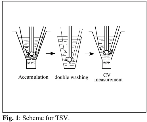
Fig. 1: Scheme for TSV. Transfer Stripping Voltammetry (TSV)
This technique was developed in the late 1980's (9,10) to determine sub-nanogram quantities of nucleic acids (NA) exploiting the strong affinity of NA for the mercury and carbon electrodes (11,12).
Attachment of low molecular mass substances such as MMC to the mercury electrode has rarely been observed. However, we observed a strong immobilization of MMC at the HMDE when applied potentials corresponded to the MMC reduction (between -0.3 and -0.4 V vs. SCE), making possible the easy preparation of a MMC-modified electrode. Such a modified electrode is used in MMC analysis in mixtures containing other substances which are not strongly attached to the electrode under the given conditions and which would otherwise interfere with the determination of MMC in conventional voltammetric analysis, as occurs with the MMC determination in urine samples.
In TSV of urine samples containing MMC and its mixtures with 5-FU or cis-Pt, the HMDE was immersed in the solution at an accumulation potential of -0.35 V for an accumulation time of 5 min without stirring (urine samples produce foam with stirring, and the electrode surface is partially blocked, causing the MMC signals to be weaker and not as well defined as those obtained without stirring). The electrode was then removed from the solution and washed (nitrogen was passed through the washing medium) with distilled water for 15 s (to eliminate interfering substances) and with the background electrolyte for 15s (to keep the electrolyte concentration in the voltammetric cell constant.) The electrode was finally placed in t h e voltammetric cell with a previously
deoxygenated background electrolyte (not containing any dissolved drug) and nitrogen was bubbled again through the solution for 120 s to remove the oxygen introduced in the process of transferring the HMDE. The circuit was closed and following the application of the initial potential for 15 s, the voltammetric measurement was taken at the same settings as in SV. The whole procedure is shown in Fig. 1.
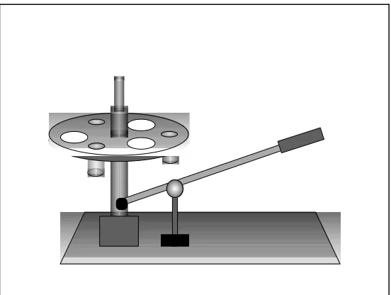
Fig. 2: Carousel used to perform the TSV experiments. carousel is “down.” Revolving the carousel
transfers the electrodes through the solutions in a chosen order.
Determining MMC by TSV made it possible: (a) to adsorb MMC at the HMDE from media not suitable for conventional voltammetric analysis and to perform the electrochemical measurements in the medium of choice (i.e. AFP); (b) to exploit the differences in the adsorbability of MMC and other substances to separate them at the electrode in the washing step; (c) to study the influence of the electrode potential on MMC signals; (d) to study the interaction of immobilized MMC with other substances as DNA, since it is known that MMC interacts with guanine residues in DNA.
For substances accumulated at the electrode surface by adsorption that do not require any accumulation potential (i.e. DNA), the modified electrode can be prepared by immersing the electrode in a drop of the substance at open circuit. This method is called Adsorption Transfer Stripping Voltammetry (AdTSV). Using 5-10 µ L drops makes it possible to reduce the volume of the analyzed sample by two orders of magnitude as compared to the conventional voltammetric experiments.
RESULTS AND DISCUSSION
Determination of MMC in aqueous solutions by using SV
Conventional voltammetry of MMC solutions in AFP yields two reversible cathodic peaks at -0.31 and -0.43 V (peaks Ic and IIc). SV responses are highly influenced by the accumulation potential (E ) (4). At E lessA A negative than -0.3 V (corresponding to reduction of the MMC quinone moiety which caused the peak I ),c both peaks were obtained. However, at E between -0.3 and -0.4 V, only the peak II appeared. This peakA c was chosen for analytical purposes, thus E -0.35 V was selected for the determination of MMC.A
Using concentrations of MMC of 9x10 M, increasing the accumulation time (t ), resulted in peak II-7
A c
showing a linear dependence up to 3 min (stirring at 500 rpm); at longer t , linear dependence leveled off.A Stirring resulted in a 3-fold enhancement of the response, hence indicating the fast rate of adsorption on the electrode surface. The proposed idealized conditions for determining MMC in AFP solutions are then EA of -0.35 V, and t at 3 min (stirring at 500 rpm).A
Mixtures of MMC with other anti-neoplasic drugs used in combined chemotherapy (13) were tested by using SV. Doxorubicin, vinblastine, bleomycin and vincristine produced their own signals at submicromolar level. 5-Fluorouracil (5-FU) produced no peak in the studied potential range and cisplatin (cis-Pt) showed only a little peak at -0.52 V. The MMC calibration curves were done in mixtures MMC and 5-FU, and MMC and cis-Pt, and the same results as in MMC alone were obtained. Similar experiments were performed with 5 µM MMC, 5-FU and cis-Pt concentrations varying between 10 and 75 µM, and no differences in the MMC peak current were observed.
These results show that SV (at E -0.35 V, t 3 min, stirring at 500 rpm) is suitable to determine MMC inA A an excess of 5-FU and cis-Pt.
Determination of MMC in urine samples by using TSV
Attempts to determine MMC in urine (diluted 4-fold by AFP) by conventional SV were not successful because urine samples alone produced peaks in the potential region close to the MMC peak. This interference of urine was efficiently eliminated by TSV in which MMC was adsorbed at the electrode by applying an accumulation potential of -0.35 V followed by washing, medium exchange and determining MMC immobilized at the electrode in a blank background electrolyte.
A comparison of the measurements with and without stirring of the analyte during the accumulation time showed that the MMC signal is better and of higher amplitude without stirring. With 1 µM MMC, the electrode surface was fully covered at the accumulation time (t ) of 5 min, without stirring. ReproducibilityA suffers when the electrode is not covered fully. Optimal conditions for MMC analysis in urine were t 5 min,A E -0.35 V, without stirring. A linear relationship of the peak was observed in the range 3-30x10 M,A -7 corresponding to 100-1000 ng ml . Additional data are shown in Table 1. The voltammograms did not show-1 any difference by comparing the urine samples spiked with MMC alone, MMC and 5-FU or MMC and cis-Pt. The MMC calibration curves were performed in these mixtures and the same results as in MMC alone were obtained.
Table 1: Mitomycin C calibration curves in aqueous solutions (using SV) and in urine (using TSV) in both cases as much alone as in mixtures with 5-fluorouracil or cisplatin
Aqueous solutions Urine
linearity over range (ng ml ) 3-70 100-1000-1
slope. 10 (µA/ng ml ) 13.6 1.204 -1
standard deviation of the slope.10 0.2 0.024
intercept (µA) 0.002 0.000
standard deviation of the intercept 0.001 0.001
correlation coefficient 0.9988 0.9992
Quantitatively, the MMC peak was lower in urine than in pure aqueous solutions. This might be partly due to adsorption of surface-active urine components (resistant to washing) at the electrode surface. This effect produces a 10-fold decrease in sensitivity (slope) upon changing the adsorption medium from water to urine, but does not affect the accuracy of the measurement, since urine specimens of different healthy volunteers yield the same calibration curve, indicating that the signal is independent of the urine matrix.
Although the detection limit (100 ng ml ) is higher than the previously obtained by Tjaden et al. (1 ng ml )-1 -1 by HPLC with on-line sample pre-treatment (14), the TSV determination of MMC alone and in the presence of other drugs in urine proposed in this paper is sufficiently sensitive to be of potential use in the clinical analysis, since the concentrations of MMC in urine of clinical patients range between 500-1000 ng ml (14)-1 while those of 5-FU (15,16) and cis-Pt (17) usually exceed 2000 and 750 ng ml , respectively. The main-1 advantage of the technique proposed here is that in variance to HPLC and other methods, where it is mandatory to isolate the drug from the biological matrix, usually by solvent or solid phase extraction, the MMC determination in urine described here is very simple and does not require urine pre-treatment. Most interfering substances present in urine samples are either not adsorbed at the electrode or are separated in the washing step, if their adsorption at the electrode is weaker than the attachment of MMC to the surface. The TSV determination of MMC is rapid and inexpensive and may become the method of choice for MMC determination in patients.
ACKNOWLEDGEMENTS
P. Pérez acknowledges the receipt of a grant from Comunidad Autónoma de Castilla-La Mancha (Spain). Fig. 2 was generously designed by Dr. Javier Pozuelo.
REFERENCES
1. Tjaden, U.R., and de Bruijn, E.A. 1990. Chromatographic analysis of anticancer drugs. J. Chromatogr.
Biomed. Appl. 531, 253-255.
2. Franck, R.W., and Tomasz, M. 1990. The Chemistry of Antitumor Agents. Ch. 15. D.E.V. Wilman, Blackie: Glasgow.
3. Rao, G.M., Begleiter, A., Lown, J.W., and Plambeck, J.A. 1977. Electrochemical studies of antitumor antibiotics. II. Polarographic and cyclic voltammetry studies of mitomycin C. J. Electrochem. Soc. 124, 199-202.
4. Teijeiro, C., Pérez, P., Marín, D., and Pale ek, E. 1995. Cyclic voltammetry of mitomycin C and DNA.
Bioelectrochem. Bioenerg. 38, 77-83.
5. Pale ek, E. 1996. From polarography of DNA to microanalysis with nucleic acid-modified electrodes.
Electroanal. 8(1), 7-14.
6. Pale ek, E. 1983. Topics in Bioelectrochemistry and Bioenergetics. Vol. 5. J. Wiley: London.
7. Wang, J., Lin, M.S., and Villa, V. 1986. Trace measurements of mitomycin C based on adsorptive stripping voltammetry. Anal. Lett. 19, 2293-2305
8. van Bennekom, W.P., Tjaden, U.R., de Bruijn, E.A., and van Oosterom, A.T. 1984. Determination of mitomycin C in human blood plasma and urine by high-performance differential pulse polarography.
9. Pale ek, E. 1988. Adsorptive transfer stripping voltammetry: Determination of nanogram quantities of DNA immobilized at the electrode surface. Anal. Biochem. 170, 421-431.
10. Pale ek, E., Jelen, F., and Postbieglová, I. 1989. Adsorptive transfer stripping voltammetry offers new possibilities in DNA research. Stud. Biophys. 130, 51-54.
11. Pale ek, E., Jelen, F., Teijeiro, C., Fu ik, V., and Jovin, T.M. 1993. Biopolymer-modified electrodes. Voltammetric analysis of nucleic acids and proteins at the submicrogram level. Anal. Chim. Acta. 273, 175-186.
12. Teijeiro, C., Nejedly, K., and Pale ek, E. 1993. Cyclic voltammetry of submicrograms quantities of supercoiled, linear and denatured DNAs with DNA-modified electrode. J. Biomol. Struct.& Dyn. 11, 313-331.
13. Fischer, D.S., Knobf, M.T., and Durivage, H.J. 1993. The Cancer Chemotherapy Handbook (4th Ed). Ch.6. Mosby-Year Book, Inc.: St.Louis.
14. Tjaden, U.R., de Bruijn, E.A., van der Hoeven, R.A.M., Jol, C., van der Greef, J., and Lingeman, H. 1987. Automated analysis of mitomycin C in body fluids by high-performance liquid chromatography with on-line sample pre-treatment. J. Chromatogr. 420, 53-62.
15. Schaaf, L.J., Ferry, D.G., Hung, C.T., Perrier, D.G., and Edwards, I.R. 1985. Analysis of 5'-deoxy-5-fluorouridine and 5-fluorouracil in human plasma and urine by high-performance liquid chromatography.
J. Chromatogr. 342, 303-313.
16. Stetson, P.L., Shukla, U.A., and Ensminger, W.D. 1985. Sensitive high-performance liquid chromatographic method for the determination of 5-fluorouracil in plasma. J. Chromatogr. 344, 391-396.