R E S E A R C H
Open Access
Contribution of nonconsensus base pairs
within ArsR binding sequences toward
ArsR-DNA binding and arsenic-mediated
transcriptional induction
Xingjuan Chen
1,2†, Xin Jiang
4†, Cuijuan Tie
4, Jinnon Yoo
4, Yan Wang
3, Meiying Xu
1,2*, Guoping Sun
1,2,
Jun Guo
1,2and Xianqiang Li
1,2,4*Abstract
Background:A transcriptional reporter is the key component in bacterial biosensors which are employed to monitor the induction or repression of a reporter gene corresponding to environmental change. Interaction of a transcription factor with its consensus sequence generated by using a position weight matrix (PWM) model is crucial for its sensitivity of the reporter. However, recent studies suggest that PWM model based on independent contribution of individual consensus base pairs to protein interaction is often insufficient to explain complex regulation, such as the effect of nonconsensus sequences on the protein-DNA binding affinity. In the present study, we employed a simpler prokaryotic arsenic repressor (ArsR) regulation system to access the protein-DNA
recognition. Contribution of nonconsensus base pairs within ArsR binding sequences toward ArsR-DNA binding and arsenic-mediated transcriptional induction was studied.
Results:We constructed a series of arsenic responsive reporters, each comprising two copies of the ArsR binding sequences from different resources. We found that high arsenic-mediated induction specifically requires the
binding sequence fromEscherichia colito be placed at the first binding sequence; however, no such preference
was observed for the second binding sequence, which could be fromAcidithiobacillus ferrooxidans, plasmid R773,
Synechococcus, or a core binding sequence ofarsR. By creating a series of reporters differed at the nonconsensus base pairs of the second binding sequence, we observed that some constructs bound weakly while others strongly to ArsR. Most interestingly, although a number of these reporters showed similar binding affinity to ArsR, their arsenic-dependent induction differed significantly.
Conclusions:The results indicated that nonconsensus base pairs could have profound influence on protein binding and may also modulate post-binding function. These findings provide new insights into the complex regulation of gene expression and facilitate the development of transcriptional reporter-based biosensors.
Keywords:Bacterial biosensor, Arsenic bioreporter, Nonconsensus base pair, Arsenic repressor, Arsenic binding sequence, Protein-DNA recognition
© The Author(s). 2019Open AccessThis article is distributed under the terms of the Creative Commons Attribution 4.0 International License (http://creativecommons.org/licenses/by/4.0/), which permits unrestricted use, distribution, and reproduction in any medium, provided you give appropriate credit to the original author(s) and the source, provide a link to the Creative Commons license, and indicate if changes were made. The Creative Commons Public Domain Dedication waiver (http://creativecommons.org/publicdomain/zero/1.0/) applies to the data made available in this article, unless otherwise stated.
* Correspondence:[email protected];[email protected]
†Xingjuan Chen and Xin Jiang contributed equally to this work.
1Guangdong Provincial Key Laboratory of Microbial Culture Collection and
Background
The interaction between a transcription factor (TF) and its corresponding DNA binding sequence is cru-cial in gene regulation [1, 2]. The base composition determines the binding affinity of the sequence to the TF. The contribution of individual base pairs in the interaction with a TF can be assessed by their conser-vation. Algorithms typically use the statistically simple position weight matrix (PWM) model for a binding consensus sequence [3, 4]. Moreover, the binding consensus sequence can be determined by sequencing a group of DNA fragments or oligonucleotides se-lected by a TF using in vitro methods such as ChIP-seq or SELEX [5]. Nevertheless, more than 40% of TFs still remain unknown for their binding sequences [5]. In Escherichia coli (E. coli), most TFs bind to a single binding site in chromosomal DNA, such as ar-senic repressor (ArsR), a metalloregulatory transcrip-tional repressor to its operator/promoter (O/P) sequence [6]. Due to the abundant presence of ArsR binding sequences in microbial chromosomes, the alignment of these binding sequences via comparison and analysis with PWM can lead to the identification of its binding consensus sequence or motif [7]. However, recent studies suggest that PWM model based on independent contribution of individual con-sensus base pairs to protein interaction is often insuf-ficient to explain various complex regulations [8], such as the effect of nonconsensus sequences on the protein-DNA binding affinity. In the present study, we employed a simpler prokaryotic ArsR regulation system to access the protein-DNA recognition.
ArsR, belonging to the Smt/ArsR family, is a regula-tory protein that controls the expression of the genes in-volved in arsenical resistance via interaction with the arsenic-responsive operon. ArsR binding prevents the RNA polymerase from interacting with the O/P se-quence of its targeted genes in the absence of arsenicals [7,9]. Upon arsenic binding, the protein dissociates from the promoter, subsequently activating the gene expres-sion [9–11]. ArsR protein is well characterized in plas-mid R773 and E. colichromosome. Both of these ArsR proteins are able to form homodimer, each with a Cys32-Val-Cys-Asp-Leu-Cys arsenic-binding sequence located at the start of their DNA binding domain [11]. ArsR from Acidithiobacillus ferrooxidans (A. ferrooxi-dans) does not have the binding sequence at this loca-tion, instead, their cysteine residues are located at amino acid residues of 95, 96, and 102 [12]. Both binding and the consensus sequences of Smt/ArsR family proteins, including those inA. ferrooxidans, have been character-ized [6,7,12–14].
In a previous study, we created two arsenic reporters, pLHPars9 and pLLPars9, in order to rapidly and
cost-effectively monitor arsenic on site and measure arsenic bioavailability. The bioreporters pLHPars9 and pLLPars9 comprised either a high or low copy-number plasmid, along with common elements of ArsR-luciferase fusion and addition of two binding sequences, one each fromE. coli (ECBS) and A. ferrooxidans (AFBS) chromosome, before the R773 arsR operon (arsRBC) [15]. Both of these reporters were highly sensitive to arsenite, with a low detection limit of 0.04μM arsenite (~ 5μg/L) and differed in their metal specificity, with pLLPars9 being more specific to arsenite and pLHPars9 to both arsenite and antimonite. The only difference between pLHPars9 and pLLPars9 is their copy numbers.
In the present study, we constructed a set of arsenic bioreporters comprising two copies of different binding sequences. We found that high arsenic-mediated induc-tion specifically requires ECBS to be placed at the first binding sequence; however, no such preference was ob-served for the second binding sequence. By creating a series of reporters differed at the nonconsensus base pairs of the second binding sequence, we tested the interaction of these probes with the protein. Interest-ingly, while some of the nonconsensus base pairs resem-bling the consensus are needed for the interaction with the ArsR protein, some of the nonconsensus base pairs appear to also affect the post-binding function of the TF.
Results
Arsenic transcriptional induction with a promoter
containing ECBS binding sequence in arsenic bioreporters In a previous study, we found that a luciferase reporter construct pLLPars9 (or pECBS-AFBS in this study) con-taining ECBS-AFBS comprising two copies of ArsR binding sequences (BS), one from E. coli chromosome (EC) and another from A. ferrooxidans (AF) chromo-somal DNA, responded better more robustly to arsenic treatment than the reporters comprising either one or two identical copies of EC or AF [15]. In this study, we swapped the position of ECBS and AFBS to create pAFBS-ECBS. After transformed into DH5a, luciferase activates of pECBS-AFBS and pAFBS-ECBS were mea-sured and compared (Fig.1a). Relative to untreated con-trol cells, pAFBS-ECBS showed only 2-fold induction in arsenic-treated cells, compared to the 9-fold induction with pECBS-AFBS (Fig.1b). This dramatic induction dif-ference suggested that the order of these two binding se-quences is crucial in the arsenic-mediated induction of the reporter.
Furthermore, we replaced the AFBS moiety within ECBS-AFBS with the binding sequence ofSynechococcus
treatment. As presented in Fig.1b, the induction of ECBS-smt2/1BS and ECBS-arsRBCBS moderately declined, los-ing approximately 15–25% induction folds of ECBS-AFBS. This suggested that AFBS at this position is not crucial for induction and can be substituted by other ArsR binding sequences. When we replaced ECBS moiety within ECBS-AFBS with the binding sequence of smt2/1 or arsRBC to create reporters psmt2/1BS-ECBS and parsRBCBS-ECBS, we found that the ratio of luciferase activities significantly declined, losing approximately 70% compared to ECBS-AFBS, as shown in Fig. 1b. The aforementioned results demonstrated that ECBS needs to be the first binding se-quence in order to robustly respond to arsenic.
The consensus sequence of a DNA-binding protein can be determined by comparison of a group of binding sequences. Those consensus base pairs are believed to be crucial for the protein to bind DNA and the noncon-sensus base pairs are not important to the binding. Ar-senic binding proteins from different microbes are DNA-binding proteins. The DNA sequences that they bind to display a consensus sequence [7]. Our above data indicated that the second binding sequence within the biosensors was relative flexible, which could be more tolerant to bioengineering manipulation, such as a con-sensus sequence. According to the concon-sensus sequence of arsRBC and cadCA, we designed a binding sequence
CS (Fig. 1c) and swapped it with the AFBS moiety to construct pECBS-CS, with 3 Ts in between. Luciferase assay revealed that pECBS-CS showed no significant dif-ference in the response to arsenic treatment when com-pared to pECBS-AFBS, suggesting that the CS can be used to replace AFBS within the biosensors. However, when we swapped ECBS with the CS to make pCS-AFBS, it demonstrated a significant change (Fig. 1d). Moreover, when we replaced ECBS of ECBS-CS with arsRBCBS or AFBS to construct parsRBCBS-CS and pAFBS-CS, they lost induction significantly like any other constructs without ECBS being at the first position as shown as above. These results with CS indicated that ECBS must be the first binding sequence.
Arsenic cannot remove the repressor protein from AFBS-ECBS and CS-AFBS-ECBS binding sequences
To examine whether there exists any difference between ECBS-AFBS and AFBS-ECBS in ArsR binding, we per-formed EMSA. Biotin-labeled probes of ECBS-AFBS and AFBS-ECBS were mixed with lysates prepared from cells with and without arsenic treatment, respectively. As pre-vious reported, two shifted bands were observed with the ECBS-AFBS probe in mock-treated cell lysate [15] and the intensity of both shifted bands dramatically de-clined in arsenic-treated cells (Fig. 2a), indicating that
A
C
D
B
the arsenic treatment disrupted the interaction between the probe and the bound repressor protein. In contrast, when we utilized the AFBS-ECBS probe to execute EMSA, we found no difference in both the number and intensity of the shifted bands between mock-treated and arsenic-treated cells. This indicated that the arsenic treatment was unable to remove the repressor protein from the AFBS-ECBS (Fig.2a).
Next, we compared ECBS-CS and CS-ECBS probes with EMSA. The ECBS-CS probe revealed two shifted bands in control cell lysate and much weaker inten-sity of the shifted bands in arsenic-treated cell lysate (Fig. 2b). Again, we observed no significant difference in the intensity of the shifted bands between control and treated cells with CS-ECBS probe. This result is in accordance with AFBS-ECBS, suggesting no re-moval of the repressor protein from CS-ECBS probe under arsenic treatment.
Arsenic removal of the repressor protein from ECBS-CS binding sequence required a linker of 3Ts
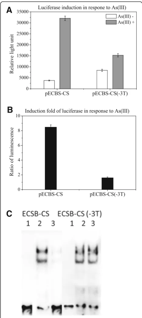
As a linker of 3Ts was inserted between ECBS and CS, we examined whether it was necessary for the induction by removing the linker from pECBS-CS to create pECBS-CS(−3 T). After transformation, the luciferase activity of pECBS-CS(−3 T) transformed cells were treated with and without arsenite. A significant reduc-tion of luciferase activity in pECBS-CS transformed cells with arsenic treatment was not observed when com-pared to untreated control cells; however, it was ob-served in the pECBS-CS(−3 T) cells treated with arsenic (Fig.3a; b). This result suggested that the linker of 3Ts is needed for arsenic-mediated induction.
Moreover, to examine whether the absence of 3Ts caused a steric hindrance for binding of the dimers to the binding sequences or prevented the removal of the bound repressor protein from the bound sequence, we
performed EMSA with biotin-labeled probes of ECBS-CS and ECBS-EC(−3Ts). As shown in Fig. 3c, like ECBS-AFBS probe, the result with ECBS-CS probe dis-played two shift bands in arsenic-untreated cells but sig-nificant decline in arsenic-treated cells. Without the linker, ECBS-CS(−3 T) displayed two shifted bands in both control and treated cells, indicating the absence of any interference with protein binding, thus ruling out the possibility of steric hindrance. Therefore, this result suggested that the absence of the linker hampered arsenic-mediated removal of the repressor protein from the bound sequence.
Fast analysis of the DNA binding sequences of ArsR with DNA filter assay
The above results indicated that two ArsR binding el-ements within the biosensors were needed in order to have a sensitive response to arsenic treatment. The first element must be from E. coli and the second one was more flexible, such as arsRBCBS or CS. Al-though arsRBC of pECBS-arsRBCBS and CS of pECBS-CS contained the same consensus sequence, their responses to arsenic treatment were distinct with moderate difference. We assumed that the differ-ence could arise only from the contribution of non-consensus base pairs of the second binding sequence. To investigate the contribution of the nonconsensus base pairs in both binding and induction, we con-structed a series of probes and reporters exclusively with alternative nonconsensus base pairs in the sec-ond binding site within ECBS-arsRBCBS. According to the consensus sequence of arsRBC, only 4 base pairs are not conserved. Investigation of different combinations of these 4 base pairs required testing of a large series of probes.
EMSA is usually used for monitoring protein/DNA in-teractions. Due to the low throughput nature of the
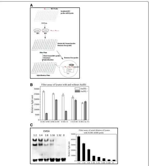
assay, the analysis becomes time consuming when hand-ling a large sample size. We therefore developed a fast filter binding assay that enabled us to efficiently monitor the interaction of several probes with their binding pro-teins simultaneously (Fig. 4a). In this assay, probes were
first mixed with lysates prepared from E. coli treated with or without arsenite, respectively. After incubation, the mixtures were loaded onto a nitrocellulose mem-brane (NC)-coated 96-well plate. Only protein-bound probes could stay on the plate and free probes passed through the membrane upon centrifugation. After wash-ing, the plate was then treated with sodium dodecyl sul-fate (SDS) to denature proteins, thereby releasing the probes. The released biotin-labeled probes were sub-jected to hybridization using a plate pre-coated with complementary sequences, and further monitored with streptavidin-HRP for luminescent detection.
To examine the feasibility of the filter assay, we employed probes ECBS-AFBS, AFBS-ECBS, ECBS-CS, CS-ECBS and ECBS-CS(−3 T) and mixed them with ly-sates prepared from arsenic-treated and mock-treated cells. The probe mixtures with lysates were first vali-dated with EMSA before using for the filter assay. The filter assay indicated that the binding of ECBS-AFBS probe with the lysate without arsenic treatment was much stronger than the lysate with arsenic treat-ment, and the ratio of the binding intensities of con-trol to treated cells was about 5-fold (Fig. 4b). As expected, binding of the AFBS-ECBS probe with con-trol and arsenic treated cell lysates was both strong and no obvious difference in binding was observed. The result with ECBS-CS probe was similar to that with the ECBS-AFBS probe and the binding ratio of control to treated cells was about 3-fold. In addition, both ECBS-CS(−3 T) and CS-ECBS probes displayed no difference in the binding ratio of the two different lysates (Fig. 4b). These results demonstrated that the filter assay in general was in accordance with EMSA. The only difference of the filter assay was that it could not present two distinct shifted bands like EMSA.
Next, we employed both assays to perform two-fold dilutions of lysates from both arsenic treated and control cells with the ECBS-AFBS probe. As shown in Fig. 4c, EMSA could detect the complex in 1:16 diluted lysates and filter assay in 1:64, indicating that the filter assay is 4 times more sensitive than EMSA. Therefore, the filter binding assay was able effectively to analyze the binding of several probes with target protein in a quick mode.
Identification of alternations at nonconsensus base pairs crucial in protein binding using DNA filter assay
The original E. coli ArsR binding sequence was identi-fied as acacattcg TT AA GT CA TA TA (TG) TT TT TG AC TT A [6]. Based on the comparison with other ArsR binding sequences, we noted an extra tail of 9 base pairs at the 5′ end that are unlikely to contribute to arsenic-mediated induction. To reduce the cost in
A
C
B
C
B
A
Fig. 4Filter assay.a: Schematic diagram of the filter assay comprising 3 steps; the biotin-labeled (* indicates biotin labeled) probe BS1 (Binding Sequence) was first mixed with lysates; BS1 probe bound to the protein in the cell lysate to form protein/DNA complexes; second, the mixture loaded onto nitrocellulose (NC) membrane-based filter plate, the protein/DNA complexes retained on membrane, the free probe washed away, and the protein-bound probe eluted, denatured and hybridized on the hybridization plate; third, the hybridization and detection within one of 96 wells was illustrated in the Cycle. The capture oligos were pre-coated on the bottom of the well, the denatured biotin-labeled probe BS1 hybridized to the capture oligo, and further detected with StreptavidinHorseradish Peroxidase (STV-HRP) and measured with a luminescence plate reader. Theluminescencesignal directly corresponds to the binding activity.b: Filter assay of probes ECBS-AFBS, AFBS-ECBS, ECBS-CS, CS-ECBS and ECBS-CS(−3 T) using lysates prepared from cells treated with (grey) and without (open) 10μM arsenite for 1 h. C: Serial 2-fold dilutions ofE. coli
oligonucleotide synthesis, we removed 5 base pairs at the 5′ end to make sECBS as ttcg TT AA GT CA TA TA (TG) TT TT TG AC TT A. Functional analysis using reporters with the shorter version, sECBS to re-place ECBS revealed no difference (data not shown).
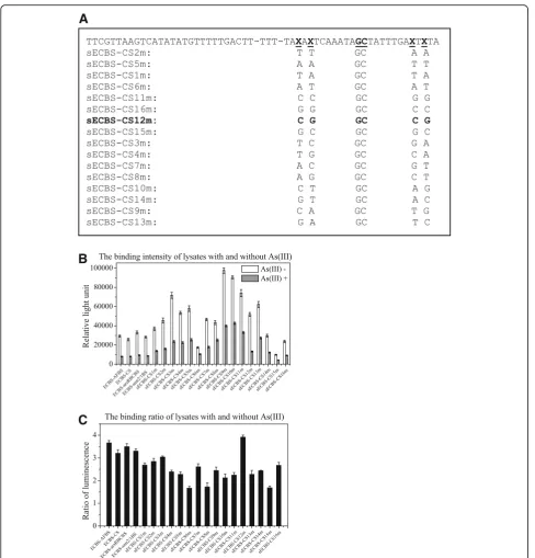
Among the 4 base pairs that are not conserved, two base pairs locate on each side of the inverted repeat re-gion, TAxAxTCAAATA xx TATTTGAxTxTA, the core binding sequence was obtained by the alignment of ArsR binding sequences among O/P sequences of arsRBC, cadCA, smtS2/S1, smtS4/S3, ziaA, czrAB, and nmtA [7]. To investigate the contribution of these nonconsensus base pairs to ArsR binding, we systematically designated different nucleotides at the position on the left and cated complementary nucleotides on the right of the re-peat. We constructed a series of probes with AA, TT, TA, AT CC, GG CG, GC, TC, TG, AC, AG, CT, GT, CA, and GA on the left side of the repeat, as presented in Fig. 5a. Biotin-labeled probes were employed to exe-cute the filter binding assay using cell lysate of E. coli,
with or without arsenic treatment. As shown in Fig. 5b, certain probes such as sECBS-CS9m and sECBS-CS10m bound to the protein from both cell lysates, with or without arsenic treatment, were stronger than ECBS-AFBS, sECBS-arsRBCBS, or sECBS-CS, whereas another probe sECBS-CS15m bound to the protein from both cell lysates, with or without arsenic treatment, was weaker than ECBS-AFBS, arsRBCBS, or sECBS-CS. Therefore, the nonconsensus base pairs could change the binding affinity to both directions. In addition to the alternations at nonconsensus base pairs, we included an alternation in the consensus base pairs to construct the probes ECBS-CS-SM as a control. With the filer binding assay, we observed that a change of 2 base pairs in the consensus sequence destroyed the dif-ference in binding between cell lysates with and without arsenic treatment (data not shown). Moreover, we found one probe sECBS-CS12m bound the protein strongly in control cells, but weakly in arsenic-treated cells. The binding difference of this probe in control versus treated cells appeared to be the biggest among all other probes (Fig.5c).
Contribution of nonconsensus base pairs to protein binding and luciferase induction
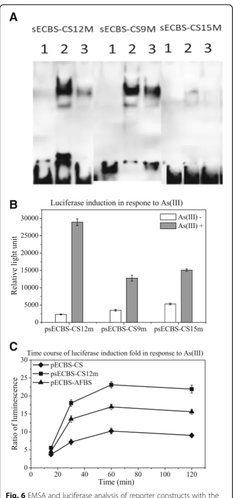
From the filter binding assay, we chose three probes, the strongest sECBS-CS9m in binding, the weakest CS15m in binding, and the highest sECBS-CS12m in induction, to validate the results with EMSA. As shown in Fig. 6a, the intensities of the shifted bands in EMSA were equivalent to the bind-ing strengths in the filter bindbind-ing assay, sECBS-CS9m being the strongest and sECBS-CS15m being the weakest. The difference of sECBS-CS12m shifted
bands in binding intensity is the highest between con-trol and induced cells. These results again demon-strated that changes in nonconsensus base pairs could lead to critical differences in protein binding. Import-antly, our data also revealed that changes in noncon-sensus base pairs could enhance arsenic-mediated removal of the bound protein, an additional func-tional impact after the binding is established.
To investigate the induction of few binding sequences, we used CS12m, CS9m, and sECBS-CS15m to replace the ECBS-AFBS of pECBS-AFBS, in order to construct psECBS-CS12m, psECBS-CS9m, and psECBS-CS15m. As expected, psECBS-CS12m resulted in a better induction than the other two reporters psECBS-CS9m and psECBS-CS15m (Fig. 6b). Further-more, we compared psECBS-CS12m with pECBS-AFBS and pECBS-CS. The transformants were treated with 10μM arsenite for 15, 30, 60 and 120 min. As shown in Fig. 6c, arsenic-mediated induction of psECBS-CS12m was significantly better than that with either pECBS-AFBS or pECBS-CS.
Discussion
Arsenic, as a naturally occurring element, is widely distributed throughout the environment. Long-term exposure to arsenic from drinking water and food can cause human diseases [16]. Prevention of further ex-posure to arsenic needs rapid and cost-effective on-site analytical techniques to monitor arsenic in water supplies. Bacteria-based assays are an emerging tech-nology, in the case of arsenical contamination, to monitor arsenic-induced gene expression. Compared to the traditional capital equipment-based methods that are inappropriate for on-site detection, bacteria-based assays are robust and inexpensive for detecting arsenic in the field [17]. More significantly, they could measure arsenic bioavailability that accounts for the difference between exposure and dose [18]. The cru-cial component of bacteria-based assays is the re-porter, comprising a promoter/operator (or an operon) and a reporter gene [19]. Ideally, a good re-porter should display high sensitivity and specificity, low endogenous background, and a wide dynamic range of response [20]. In our previous study of mak-ing sensitive arsenic reporter, we constructed pLLPars9 (the same construct as pECBS-AFBS in this study) reporter and demonstrated that it is equivalent to some of the best reporters constructed to date in response to arsenic [15]. In this study, we demon-strated that the reporter psECBS-CS12m is signifi-cantly better than pLLPars9.
occupied by an ArsR homodimer. In the previous study, we designed, such as smt operon, two binding sequences ECBS-AFBS and found that the induction of the lucifer-ase reporter in response to the treatment of arsenic is better than either of single binding sequences [15]. We also found that the induction with these two different
binding sequences is better than the two identical se-quences either from EC or AF. In the present study, we uncovered that ECBS must be at the front position and the induction dramatically declined if it was replaced by other binding sequences such as smt2/1 or arsRBC. In contrast, AFBS at the second position can be replaced
A
C
B
by other binding sequences without affecting the induc-tion to a significant degree. This indicated that an ap-propriate order of these two binding sequences is important for achieving maximal induction. As the two binding sequences bind to two dimers, the complex could be stabilized by dimer-dimer interaction [7]. Change in the order of these two binding sequences, that is AFBS-ECBS, could allow the binding of two di-mers, but the order might affect arsenic interaction with the repressor protein or removal of the repressor protein from the binding sequences. Therefore, unlike ECBS-AFBS, arsenic binding sites within AFBS-ECBS might be hidden due to steric structure, which prevents arsenic binding or dissociation the repressor from the binding sequence.
Protein-DNA recognition has been increasingly ap-preciated to be more complex than previously thought. Although the simple model of PWM has been widely used to define the DNA binding motifs of individual TFs, recent studies suggest that this model based on independent contribution of individ-ual consensus base pairs to protein interaction is often insufficient to explain various complex regula-tion [8], such as the relevant dinucleotides or trinu-cleotides crucial to protein-DNA recognition [21–24], significant difference of low-affinity binding sites from the consensus sequence [25, 26], novel DNA-binding specificities of multi-protein complexes formed with a TF [27–29], and the effect of flanking sequences on the binding affinity [30]. In the present study, we employed a simpler prokaryotic ArsR regulation sys-tem to access the protein-DNA recognition. We found base pairs at nonconsensus positions within the second binding sequences such as ECBS-CS15m could result in lower binding with target protein, whereas others such as ECBS-CS9m resulted in binding with higher affinities, although both still maintained the consensus sequence. PWM was unable to explain these results. More interestingly, our study demon-strated that one of the base pairs at the nonconsensus position could also affect induction, the function be-yond DNA binding. We found sECBS-CS12m, which could bind to target protein as well as ECBS-CS; however, its response to arsenic was much stronger than ECBS-CS. Their similar basal binding levels but differential induction rates suggest that arsenic-mediated removal of the binding protein from the DNA binding sequence of CS12m is faster than CS. Therefore, like AFBS-ECBS, the interaction of these nonconsensus base pairs with the repressor protein could influence arsenic binding or arsenic-induced conformational change of the repressor protein, lead-ing to differential turnover of the bound protein from the binding sequence, as exemplified by the
A
B
C
observations that AFBS-ECBS was no longer sensitive to arsenic while sECBS-CS12m became more sensitive to the arsenic.
Conclusions
In the present study, we found that nonconsensus base pairs played important roles in protein-DNA binding and gene transcriptional regulation. More sensitive and accurate biosensors for arsenic detection can be devel-oped through the design of nonconsensus base pairs. Our current findings illustrate an innovative strategy to construct better reporters, which will facilitate the devel-opment of more sensitive biosensors to monitor envir-onmental arsenic via the induction of reporter gene expression.
Methods
Plasmid construction
Reporter constructs with different orders and sources of binding sequences were made by modifying the binding sequence of pLLPars9 [15], which was renamed as pECBS-AFBS in this study. The sense and antisense strand sequences were synthesized and annealed to gen-erate double strand fragments with the sticky end of XbaI and HindIII, which were subsequently cloned into the XbaI and HindIII site of pLLPars9 [15] to replace the ECBS-AFBS to make constructs, pAFBS-ECBS, pECBS-smt2/1BS, pECBS-arsRBCBS, psmt2/1BS-ECBS, parsRBCBS-ECBS, pCS-AFBS, parsRBCBS-CS, pAFBS-CS, pECBS-pAFBS-CS, and pECBS-CS(−3 T). Five base pairs at the 5′end of ECBS were removed to make sECBS. Dif-ferent nucleotides at the nonconsensus position of CS were designated to make CS1-16 m. Then sECBS and CS1-16 m were subsequently cloned into the XbaI and HindIII site of pLLPars9 [15] to replace the ECBS-AFBS to make constructs, sECBS-CS9m, sECBS-CS12m and sECBS-CS15m.
Luciferase assay
E. coliDH5αcompetent cells were transformed with the recombinant plasmids constructed in this study. Single colonies were picked and inoculated in 2 mL Luria-Bertani (LB) media supplied with 25μg/mL chloram-phenicol for 12–16 h at 37 °C with vigorous shaking. The overnight culture was 1:50 diluted in a 1.5 mL microcen-trifuge tube with pre-warm and fresh-prepared 2 mL LB media supplied with chloramphenicol. The diluted cells were cultured for additional 3 h at 37 °C until the optical density (OD) reached 0.5. Cells were treated with or without 10μM sodium arsenite [As (III)] for 60 min at 37 °C. The cell samples were sonicated to lyse the cells, and the protein concentration was measured with Brad-ford Protein Assay (Bio-Rad, Cat#5000201) to confirm the equal protein concentration among the treated and
untreated cell samples. Twenty μL of induced sample was taken and mixed 50μL luciferase substrate, and the luciferase activities were measured on the luminescence plate reader (Veritas).
Preparation of cell lysates
One mL of cell culture with or without sodium arsenite was centrifuged at 10,000 g for 1 min and the pellet was resuspended in 300μL of lysis buffer (10 mM Tris-HCl, pH 8.0, 0.1 M NaCl, 1 mM ethylene diamine tetraacetic acid (EDTA), and 0.1% [w/v] polyethylene glycol octyl-phenol ether (TRITON X-100)). Senve point fiveμL of a freshly prepared lysozyme solution (10 mg/mL in 10 mM Tris-HCl, pH 8.0, final concentration is 0.25 mg/mL) was added and mixed well by tapping the tube gently, and the lysis mixture was incubated for 10–20 min at room temperature. After centrifugation, the supernatant was used for electrophoretic mobility shift assay (EMSA) or Filter assay.
EMSA
One to 3μg cell lysate was mixed with 2μL of 5× bind-ing buffer and 1μL polyd(I-C) and incubated on ice for 5 min. One μL of biotin-labeled probe was added to the mixture and incubated at 22 °C for 30 min. Each reaction mixture was separated using a 6.5% non-denaturing polyacrylamide gel at 100 V at 4 °C in 0.5 × Tris-borate-EDTA (TBE) for about 50 to 60 min. After the gel was transferred onto an NC membrane and blocked by add-ing 15 mL of blockadd-ing buffer for 20 min at room temperature, the biotin-labeled probe on the blot was then detected with streptavidin–HRP and chemilumines-cent substrates (enhanced chemiluminescence by lumi-nol, Pierce). The image was acquired using an imager.
Filter assay method
In this assay, 2μL cell lysate (2–10μg) was mixed with 10μL 2× Binding Buffer Mix (40 mM 4-(2-hydro-xyethyl)-1-piperazineethanesulfonic acid (HEPES), pH 7.6, 20 mM ammonia sulfate, 2 mM dithiothreitol (DTT), 20 mM KCl, and 0.4% Tween-20), 1μL biotin-labeled probe, and 7μL ddH2O in a 96-well PCR plate.
probe was eluted from the filter and collected for quan-titative analysis through DNA plate hybridization. The captured DNA probe was further detected with streptavidin-HRP and the signals were read by a lumi-nescence plate reader (Beckman Coulter, LD-400), and reported as relative light units (RLUs). Induction fold was the ratio of luminescence of arsenic-treated cells to that of arsenic-untreated cells.
Abbreviations
AFBS:binding sequences fromA. ferrooxidans; ArsR: arsenic repressor; arsRBC: R773arsR operon; arsRBCBS: binding sequence from arsRBC; CS: consensus sequence of arsRBC and cadCA; DTT: dithiothreitol; ECBS: binding sequences fromE. coli; EDTA: ethylene diamine tetraacetic acid; EMSA: electrophoretic mobility shift assay; HEPES: 4-(2-hydroxyethyl)-1-piperazineethanesulfonic acid; LB: Luria-Bertani media; NC: nitrocellulose membrane; O/P: operator/promoter; OD: optical density; PWM: position weight matrix; RLUs: relative light units; SDS: sodium dodecyl sulfate; smt2/ 1BS: binding sequence fromSynechococcussmt2/1; TBE: Tris-borate-EDTA; TF: transcription factor; TRITON X-100: polyethylene glycol octylphenol ether
Acknowledgements
Thank Professors Yinghua Cen and Xiangdong Fu for reading the manuscript.
Authors’contributions
YW, MX, GS, JG, and XL developed the protocol. XC, XJ, CT, and JY performed the experiments. XC, XJ, MX, and XL analyzed the data and wrote the manuscript. XC and XJ contributed equally to this work. All authors read and approved the final manuscript.
Funding
This work was supported by the High-level Leading Talent Introduction Pro-gram of GDAS (2016GDASRC-0208) and the Science and Technology Plan-ning Project of Guangzhou City (201707020021) to XL, the National Natural Science Foundation of China (21677042) and the Natural Science Foundation of Guangdong Province (2018B0303110010) to XC, and the National Natural Science Foundation of China (91851202, 51678163) to MX.
Availability of data and materials
All data generated or analysed during this study are included in this published article.
Ethics approval and consent to participate
Not applicable.
Consent for publication
Not applicable.
Competing interests
The authors declare that they have no competing interests.
Author details
1Guangdong Provincial Key Laboratory of Microbial Culture Collection and
Application, Guangdong Institute of Microbiology, Guangzhou, China.2State Key Laboratory of Applied Microbiology Southern China, Guangzhou, China.
3Science and Technology Library of Guangdong Province, Guangdong
Institute of Science and Technology Information and Development Strategy, Guangzhou, China.4Signosis Inc., 1700 Wyatt Drive, suite10-12, Santa Clara, CA, USA.
Received: 12 March 2019 Accepted: 27 May 2019
References
1. Vaquerizas JM, Kummerfeld SK, Teichmann SA, Luscombe NM. A census of human transcription factors: function, expression and evolution. Nat Rev Genet. 2009;10(4):252–63.
2. Albert FW, Kruglyak L. The role of regulatory variation in complex traits and disease. Nat Rev Genet. 2015;16(4):197–212.
3. Stormo GD, Schneider TD, Gold L, Ehrenfeucht A. Use of the‘perceptron’ algorithm to distinguish translational initiation sites inE. coli. Nucleic Acids Res. 1982;10(9):2997–3011.
4. Stormo GD. Modeling the specificity of protein-DNA interactions. Quant Biol. 2013;1(2):115–30.
5. Inukai S, Kock KH, Bulyk ML. Transcription factor-DNA binding: beyond binding site motifs. Curr Opin Genet Dev. 2017;43:110–9.
6. Xu C, Shi W, Rosen BP. The chromosomearsRgene ofEscherichia coli
encodes a trans-acting metalloregulatory protein. J Biol Chem. 1996;271(5): 2427–32.
7. Busenlehner LS, Pennella MA, Giedroc DP. The SmtB/ArsR family of metalloregulatory transcriptional repressors: structural insights into prokaryotic metal resistance. FEMS Microbiol Rev. 2003;27(2–3):131–43. 8. Siggers T, Gordan R. Protein-DNA binding: complexities and multi-protein
codes. Nucleic Acids Res. 2014;42(4):2099–111.
9. San Francisco MJ, Hope CL, Owolabi JB, Tisa LS, Rosen BP. Identification of the metalloregulatory element of the plasmid-encoded arsenical resistance operon. Nucleic Acids Res. 1990;18(3):619–24.
10. Wu J, Rosen BP. Metalloregulated expression of the ars operon. J Biol Chem. 1993;268(1):52–8.
11. Shi W, Wu J, Rosen BP. Identification of a putative metal binding site in a new family of metalloregulatory proteins. J Biol Chem. 1994;269(31): 19826–9.
12. Qin J, Fu HL, Ye J, Bencze KZ, Stemmler TL, Rawlings DE, Rosen BP. Convergent evolution of a new arsenic binding site in the ArsR/SmtB family of metalloregulators. J Biol Chem. 2007;282(47):34346–55.
13. Butcher BG, Rawlings DE. The divergent chromosomal ars operon of
Acidithiobacillus ferrooxidansis regulated by an atypical ArsR protein. Microbiology. 2002;148(Pt 12:3983–92.
14. Hödar C, Moreno P, Genova A, Latorre M, Reyes-Jara A, Maass A, González M, Cambiazo V. Genome wide identification ofAcidithiobacillus ferrooxidans
(ATCC 23270) transcription factors and comparative analysis of ArsR and MerR metal regulators. Biometals. 2012;25(1):75–93.
15. Fang Y, Zhu C, Chen X, Wang Y, Xu M, Sun G, Guo J, Yoo J, Tie C, Jiang X, Li X. Copy number of ArsR reporter plasmid determines its arsenite response and metal specificity. Appl Microbiol Biotechnol. 2018;102(13):5753–61. 16. Jomova K, Jenisova Z, Feszterova M, Baros S, Liska J, Hudecova D, Rhodes
CJ, Valko M. Arsenic: toxicity, oxidative stress and human disease. J Appl Toxicol. 2011;31(2):95–107.
17. Chen J, Rosen BP. Biosensors for inorganic and organic arsenicals. Biosensors (Basel). 2014;4(4):494–512.
18. Soangra R, Majumder B, Roy P. Whole cell arsenic biosensor - a cheap technology for bioavailable arsenic (as) determination. Eur J Adv Eng Technol. 2015;2:52–61.
19. Close DM, Ripp S, Sayler GS. Reporter proteins in whole-cell optical bioreporter detection systems, biosensor integrations, and biosensing applications. Sensors. 2009;9(11):9147–74.
20. Kaur H, Kumar R, Babu JN, Mittal S. Advances in arsenic biosensor development - a comprehensive review. Biosens Bioelectron. 2015; 63(15):533–45.
21. Gershenzon NI, Stormo GD, Ioshikhes IP. Computational technique for improvement of the position-weight matrices for the DNA/protein binding sites. Nucleic Acids Res. 2005;33(7):2290–301.
22. Zhao Y, Ruan S, Pandey M, Stormo GD. Improved models for transcription factor binding site identification using nonindependent interactions. Genetics. 2012;191(3):781–90.
23. Weirauch MT, Cote A, Norel R, Annala M, Zhao Y, Riley TR, Saez-Rodriguez J, Cokelaer T, Vedenko A, Talukder S. Evaluation of methods for modeling transcription factor sequence specificity. Nat Biotechnol. 2013;31(2):126–34. 24. Mathelier A, Wasserman WW. The next generation of transcription factor
binding site prediction. PLoS Comput Biol. 2013;9(9):e1003214. 25. Jaeger SA, Chan ET, Berger MF, Stottmann R, Hughes TR, Bulyk ML.
Conservation and regulatory associations of a wide affinity range of mouse transcription factor binding sites. Genomics. 2010;95(4):185–95.
26. Tanay A. Extensive low-affinity transcriptional interactions in the yeast genome. Genome Res. 2006;16(8):962–72.
28. Delwel R, Funabiki T, Kreider BL, Morishita K, Ihle JN. Four of the seven zinc fingers of the Evi-1 myeloid-transforming gene are required for sequence-specific binding to GA(C/T)AAGA(T/C)AAGATAA. Mol Cell Biol. 1993;13(7):4291–300.
29. Funabiki T, Kreider BL, Ihle JN. The carboxyl domain of zinc fingers of the Evi-1 myeloid transforming gene binds a consensus sequence of GAAG ATGAG. Oncogene. 1994;9(6):1575–81.
30. Gordan R, Shen N, Dror I, Zhou T, Horton J, Rohs R, Bulyk ML. Genomic regions flanking E-box binding sites influence DNA binding specificity of bHLH transcription factors through DNA shape. Cell Rep. 2013;3(4): 1093–104.
Publisher’s Note