A Simple Precipitation Method for Synthesis CoFe
2O
4Nanoparticles
G. Nabiyouni a, S. Sharifi a, D. Ghanbari b, M. Salavati-Niasari c, *a
Derpartment of Physics, Faculty of Science, Arak University, Arak 38156-88349, Iran
b
Young Researchers and Elite Club, Arak Branch, Islamic Azad University, Arak, Iran
c
Institute of Nano Science and Nano Technology, University of Kashan, Kashan, P.O. Box 87317-51167, Iran
Abstract
Magnetic CoFe2O4 nanoparticles were synthesized via a simple
chemical reaction using precipitation method. The obtained materials consist of ferrite particles with average diameter of 25 nm. The effect of different surfactants such as cationic, anionic and neutral on the morphology of the products was investigated. Scanning electron microscopy was used to study the structure and particle size of CoFe2O4 nanoparticles.
Magnetic properties of the product were also examined by vibrating sample magnetometer at room temperature. By using ammonia and sodium hydroxide cobalt ferrite nanoparticles exhibit different super-paramagnetic and ferrimagnetic behaviors respectively.
2014 JNS All rights reserved
Article history:
Received 4/10/2014 Accepted 7/11/2014 Published online 1/12/2014
Keywords:
Precipitation CoFe2O4
Nanoparticles
*Corresponding author:
E-mail address:
[email protected] Phone: + 98 31 55555 333 Fax: + 98 31 5555 29 30
1. Introduction
Magnetic nanoparticles have been the subject of intense research because of their potential applications in high-density magnetic recording, magnetic fluids, high coercive force, mechanical hardness, chemical stability and temperate saturation magnetization[1-2].
There is an increasing interest in magnetic ferrite nanoparticles because of their broad applications in several technological fields including permanent magnets, magnetic fluids, drug delivery, microwave devices, and high density information storage [3–5]. Among the various ferrite materials for magnetic
recording applications, cobalt ferrite (CoFe2O4) has been widely studied because it
possesses excellent chemical stability and suitable mechanical hardness. In addition to the precise control on the composition and structure of CoFe2O4, the success of its
chemical synthesis methods, such as precipitation, sol-gel, hydrothermal are used to produce cobalt ferrite. Among the reported methods, the precipitation method is an efficient and economical way to mass production of ultrafine cobalt ferrite powder
[9-16]. In the present work, cobalt ferrite (CoFe2O4) nano-particles were synthesized by
the precipitation method at reaction temperature of 60 °C. The obtained samples were characterized by scanning electron microscopy (SEM) and X-ray diffraction pattern (XRD). The magnetic properties were investigated using a vibrating sample magnetometer (VSM).
2 Experimental
2.1. Materials and Instruments
Co(CH3COO)2 4H2O, Fe(NO3)3 9H2O, cetyl
trimethyl ammonium bromide (CTAB), sodium dodecyl sulphate (SDS), polyvinyl alcohol (PVA), NaOH and NH3 were purchased from
Merck Company. All of the chemicals were used as received without further purifications. XRD patterns were recorded by a Philips, X-ray diffracttometer using Ni-filtered Cu Kα
radiation. For SEM images the samples were coated by a very thin layer of Au to make the sample surface conductor and prevent charge accumulation, and obtaining a better contrast. Room temperature magnetic properties were investigated using a vibrating sample magnetometer (VSM, made by Meghnatis Daghigh Kavir Company) in an applied magnetic field sweeping between ±10000 Oe.
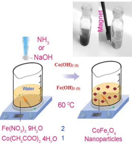
2.2. Synthesis of CoFe2O4 nanoparticles
1.62g of Fe(NO3)3 9H2O and 0.5g of
Co(CH3COO)2 4H2O are dissolved in 75 ml of
distilled water. 0.25g CTAB (or SDS, PVA) is
then added to the solution. 4 ml of NH3 12M (or
14 ml of NaOH 1M) solution is slowly added to the solution and is heated at 60° C for 60 min. A
black-brown precipitate is obtained confirming
the synthesis of CoFe2O4. The precipitate of
CoFe2O4 is then centrifuged and rinsed with
distilled water, followed by being left in an atmosphere environment to dry. Fig.1 shows the
schematic diagram for experimental setup used in this precipitation method.
Fig. 1. Schematic of precipitation reaction
3. Results and discussion
The XRD pattern of CoFe2O4 nanoparticles is
shown in Fig. 2. The pattern of as-prepared
CoFe2O4 nanoparticles is indexed as a pure cubic
phase which is very close to the literature values
(JCPDS No. 01-1121). Space group of cobalt iron oxide is Fd3m with cell constant of 8.39
angstrom. The narrow sharp peaks indicate that
the CoFe2O4 nanoparticles are well crystallized.
The crystallite size measurements were also carried out using the Scherrer equation,
Dc=Kλ/βCosθ
Where β is the width of the observed diffraction
and λ is the X-ray wavelength (CuKα radiation,
equals to 0.154 nm). The estimated crystallite size
was about 16 nm.
Fig. 2. XRD pattern of the CoFe2O4 nanoparticles
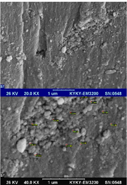
Fig. 3 illustrates SEM images of nanoparticles synthesized by ammonia and confirms average diameter size product is less than 45 nm. As the images show some agglomeration are observed in the product. Surfactant effect on the particle size and morphology of the products was investigated. Three different water-soluble additives were used as capping agent and surfactant.
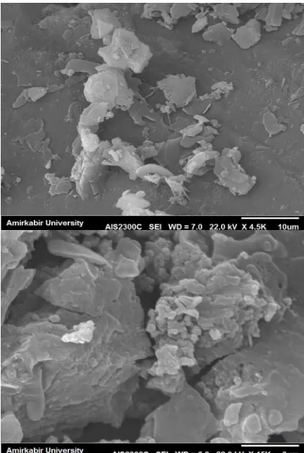
The influence of cetyl trimethyl ammonium bromide (CTAB: cationic surfactant) on the morphology of nanoparticles (synthesized by NH3) is shown in Fig. 4. Using CTAB leads to
synthesize of nanoparticles with average diameter less than 100 nm albeit lots of agglomerations were observed and the sample consists of larger particles compare to nanoparticles achieved without surfactant.
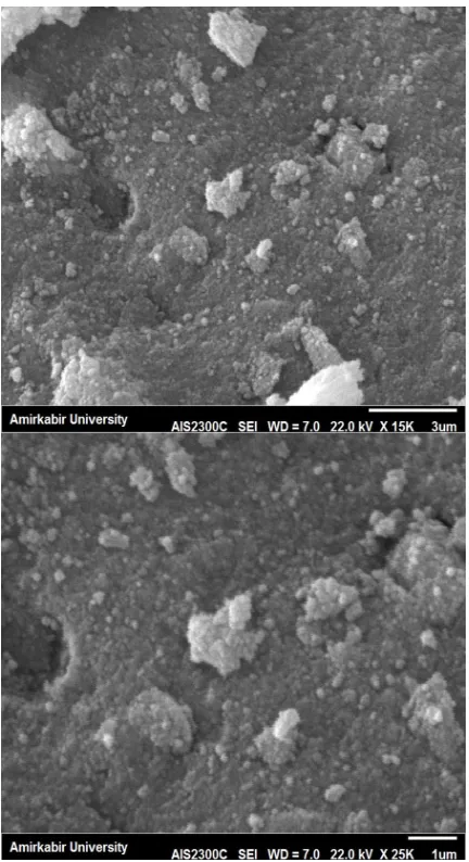
The effect of polyvinyl alcohol (PVA: neutral surfactant) on the morphology of nanoparticles (obtained by NH3) is studied and is illustrated in
Fig.5. It seems polyvinyl alcohol remains on the
surface of the particles and nanocomposite is formed. For better investigation transmission electron microscopy is needed that shows polymeric matrix remains on the surface of particles.
The effect of sodium dodecyl sulphate (SDS: anionic surfactant) on the morphology of
nanoparticles (synthesized by NH3) is depicted in
Fig.6. The image shows that the sample consists
of larger particles compare to surfactant-free nanoparticles and bulk particles were synthesized. Outcomes approve in these conditions using various surfactants like cationic, anionic and neutral have negative effect on the size of samples.
It seems by applying surfactants growth stage overcome to nucleation stage and leads to magnetic nucleuses grow together and bigger particles are obtained.
Consequently in these conditions and procedure surfactant-free sample shows smaller particle size.
Fig. 3. SEM images of nanoparticles achieved by NH3
According to SEM images in these conditions ammonia is not appropriate precipitation-agent in comparison with sodium hydroxide and products with bigger particle size was obtained.
This method proposes easy manipulation in particle size and magnetic properties by a simple change in precursors.
Fig. 4. SEM images of the nanoparticles obtained by CTAB
Fig. 5. SEM image of the CoFe2O4 achieved by PVA
Fig. 7 illustrates SEM image of sample obtained by NaOH and confirms average diameter size of surfactant-free product is about 25 nm.
Fig. 6. SEM images of the CoFe2O4 obtained by SDS
Fig. 7. SEM image of CoFe2O4 nanoparticles
The annealing effect on the morphology of the products is shown in Fig. 8. Using annealing at 200 °C leads to synthesize nanoparticles with average diameter less than 50 nm and show some agglomeration compare to blank sample.
At 800 °C some bulk products simultaneously were formed besides nanoparticles with average diameter less than 80 nm. Relatively sample consists of bigger particles compare to surfactant-free product (Fig. 9).
Fig. 8. Nanoparticles that are annealed at 200° C
Room temperature magnetic properties of our samples are studied using a VSM device.
Coercivity, remanence and saturation
magnetization of the cobalt ferrite nanoparticles
which synthesized with sodium hydroxide and
ammonia are mentioned in magnetic curves.
Hysteresis loops for CoFe2O4 nanoparticles
obtained with sodium hydroxide is depicted in
Fig. 10.
CoFe2O4 synthesized nanoparticles show
ferrimagnetic behavior and have a saturation
magnetization of 21.05 emu/g, remanence of
3.8 emu/g and a coercivity of 200.9 Oersted.
Fig .10. Hysteresis curve of nanoparticles synthesized by NaOH
Fig. 11 shows magnetization curve of CoFe2O4 nanoparticles (obtained with ammonia)
that exhibits super-paramagnetic behavior with a very low coercivity and a saturation magnetization of 5.4 emu/g. It is an interesting outcome because by a simple change in precursor, property of product easily is altered. In this procedure ammonia leads to lower magnetization and coercivity. By using NaOH totally new magnetic product with higher amounts of magnetization and coercivity is synthesized. Applying annealing at higher temperatures also goes to higher coercivity.
Fig. 11. Magnetization curve of nanoparticles synthesized by NH3
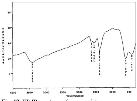
Fig. 12. FT-IR spectrum of nanoparticles
Fourier transform infra-red (FT-IR) spectrum of synthesized nanoparticles was recorded in the range of 400–4000 cm -1 at room temperature and results is shown in Fig. 12. The spectrum exhibits absorption peaks between 3400-3500 cm−1, corresponding to the stretching mode of O-H group adsorbed on the surface of nanoparticles. Absorption peaks around 410 and 590 cm-1 are related to metal-oxygen (Fe-O, Co-O) bonds.
4.
Conclusion
CoFe2O4 nanoparticles were synthesized via a
facile chemical precipitation method. The particle size of products synthesized by sodium hydroxide was smaller than ammonia. By applying surfactants growth stage overcome to nucleation stage and leads to magnetic nucleuses grow together and bigger particles be obtained. The magnetic properties of the product were also investigated using a vibrating sample magnetometer. Interestingly cobalt ferrite nanoparticles exhibit different super-paramagnetic and ferrimagnetic behaviors by changing precipitation-agent. Annealing leads to nanoparticles with average diameter less than 80 nm and also sample consists of bigger particles compare to blank sample.
Acknowledgments
References
[1] F. Zhang, S. Kantake, Y. Kitamoto, M. Abe, IEEE Trans. Magn. 35 (1999) 2751–2753.
[2] R. Arulmurugan, G. Vaidyanathan, S. Sendhilnathan, B. Jeyadevan, Mn–Zn J. Magn. Magn. Mater. 298 (2006) 83–94. [3] A. Goldman, Modern Ferrite Technology, Van Nostrand–Reinhold, New York. (1990).
[4] B.M. Berkovsky, V.F. Medvedev, M.S. Krakov, Magnetic Fluids: Engineering Applications, Oxford Univ. Press, Oxford, (1993).
[5] Y. Kitamoto, S. Kantake, S. Shirasaki, F. Abe, M. Naoe, J. Appl. Phys. 85 (1999) 4708-4710.
[6] A.E. Berkowitz, W. Schuele, J. Appl. Phys. 30 (1959) 134–135.
[7] C.N. Chinnasamy, B. Jeyadevan, O. Perales-Perez, K. Shinoda, K. Tohji, A. Kasuya, IEEE Trans. Magn. 38 (2002) 2640–2642.
[8] O. Perales-Perez, H. Sasaki, A. Kasuya, B. Jeyadevan, K. Tohji, T.
Hihara, K. Sumiyama, J. Appl. Phys. 91 (2002) 6958–6960.
[9] K. Maaz, A. Mumtaz, S.K. Hasanain, A. Ceylan, J. Magn. Magn. Mater 308 (2007) 289-295.
[10] X. Chu, D. Jiang, Y. Guo, C. Zheng, Sens. Actuator B. 120 (2006) 177.-181 [11] C.C. Wang, I.H. Chen, C.R. Lin, J. Magn.
[12] Y.I. Kim, D. Kim, C.S. Lee, Phys. B 337 (2003) 42-51.
[13] Y. Shi, J. Ding, H. Yin, J. Alloys Compd. 308 (2000) 290-295.
[14] D. Ghanbari, M. Salavati-Niasari, J
Indus Eng Chem. DOI:
10.1016/j.jiec.2014.09.043 (2014). [15] D. Ghanbari, M. Salavati-Niasari, M.Ghasemi-Koch, J Indus Eng Chem. 20 (2014) 3970-3974.