
www.fm.viamedica.pl
Address for correspondence: F. Burdan, MD, PhD, Experimental Teratology Unit of the Human Anatomy Department, Jaczewskiego 4, 20–090 Lublin, Poland, tel: +48 603 767 649, fax: +48 81 532 89 03; e-mail: [email protected]
Association of maternal pancreatic function
and foetal growth in rats treated with DFU,
a selective cyclooxygenase-2 inhibitor
F. Burdan
1, J. Szumiło
2, J. Dudka
2, M. Szumiło
3, A. Korobowicz
4,
S. Chatterjee
1, R. Klepacz
21Experimental Teratology Unit of the Human Anatomy Department, Medical University of Lublin, Poland 2Clinical Pathomorphology Department, Medical University of Lublin, Poland
3Applayed Pharmacy Department, Medical University of Lublin, Poland
4Paediatrics Pulmonology and Rheumatology Department, Medical University of Lublin, Poland
[Received 1 March 2007; Accepted 23 May 2007]
Constitutive (COX-1) and inducible (COX-2) cyclooxygenase isoforms have been detected in various mammalian tissues. Their activity is blocked by non-steroidal anti-inflammatory drugs that may induce various side reactions. The aim of the study was to evaluate the effects of DFU, a selective COX-2 inhibitor, on exo-crine and endoexo-crine pancreatic function and the immunoexpression of both COX isoforms in maternal and foetal rat pancreases. The compound was admin-istered to pregnant Wistar rats once daily from the 8th to the 21st day of
gesta-tion. Glucose level and amylase activity were determined in the maternal sera. Maternal and foetal pancreases were examined histologically. Immunoexpression of COX-1 and COX-2 was also evaluated. Both biochemical parameters, as well as the histological structure of the pancreas were undisturbed in the dams and their foetuses. The maternal glucose level was found to be an important factor for foetal growth. Strong cytoplasmic COX-1 immunostaining was observed in acinar secretory cells, whereas in islets the immune reaction was weak. Endocrine cells also revealed strong cytoplasmic COX-2 staining in the maternal and foetal pan-creases. Acinar cells exhibited nuclear reaction, which was strong in the foetal but weak in the maternal pancreases. No differences in COX immunoexpression were found between the DFU-exposed and the control groups in either mothers or foetuses. It should be stressed that DFU administered throughout mid and late pregnancy in rats did not change maternal or foetal pancreatic morphology or immunoexpression of either of the main COX isoforms in the organ.
Key words: coxibs, cyclooxygenase, diabetes, foetus, glucose, NSAID, macrosomia, pregnancy
INTRODUCTION
The two main isoforms of cyclooxygenase (COX), namely constitutive (COX-1) and inducible (COX-2), have been detected in various mammalian tissues and organs. The third one (COX-3), also known as
inhibited by the specific reversible or irreversible COX inhibitors. The non-selective COX inhibitors (e.g., ibu-profen and naproxen) block both constitutive and inducible isoforms, while the selective ones (e.g., DFU and rofecoxib) inhibit inducible isoform only. How-ever, in higher than therapeutic doses COX-1 inhibi-tion could appear as well [3, 12, 24].
A number of animal and human studies have indicated that a decrease in COX-1 activity induces various side reactions, including gastrointestinal and renal toxicity and others which depend on the block-ade of the housekeeping functions of the inducible isoform [3, 12]. However, the constitutive expres-sion of COX-2 has also been found in some organs. It should be noted that high expression of this iso-form is typical of foetal organs and decreases rapid-ly after delivery. In mature tissues the highest ex-pression of COX-2 is characteristic of the macula densa, pancreatic islets and some structures of the central nervous system [12, 28, 31].
In previous studies COX-1 immunoexpression in the pancreas has been limited to acinar secretory cells, while COX-2 was found in secretory cells of the endo-crine pancreatic islets and in epithelial cells of the pancreatic ducts and smooth muscle cells of the blood vessels of the pancreatic stroma [6, 21, 31]. On the other hand, Zabel-Langhennig et al. [32] revealed that both COX-1 and COX-2 isoenzymes are synthesised in rat pancreatic acinar cells. The differences are prob-ably secondary to methodology, since the authors examined both isoenzymes on the mRNA level [32]. It was also reported that indomethacin, a non-selec-tive COX-inhibitor, decreased basal, glucose- and glu-cagon-stimulated acute insulin response [31]. Accord-ing to Koliopanos et al. [21] pancreatic islets displayed a variable COX-2 staining pattern, which was associ-ated with the distribution of insulin-positive cells and with the clinical diabetes mellitus status. In normal insulin production or latent diabetes COX-2 immu-noreactivity was found, whereas in diabetic patients the COX-2 expression was decreased or absent from the pancreatic islets.
The present study was undertaken to evaluate the effects of DFU (i.e., 5,5-dimethyl-3-(fluorophe-nyl)-4-(4-methylsulphonyl)phenyl-2(5H)-furanone), a selective COX-2 inhibitor, on exocrine and endo-crine pancreatic functions and immunoexpression of the constitutive and inducible cyclooxygenase iso-forms in the rat pancreas during pregnancy. The data were collected during a large scientific project un-dertaken to evaluate the prenatal toxicity of the ex-amined compound, and the article partially covers
foetal findings previously published in detail [8]. Since COX-2 is expressed almost exclusively in islet paren-chyma [3, 6, 21, 31], the influence of the maternal glucose level on foetal and placental development was also studied.
MATERIAL AND METHODS
The study was conducted on sexually mature al-bino rats of the Wistar CRL:(WI)WUBR strain and approved by the Local Bioethical Committee (guide-line No.372/2002).All the animals were obtained from an accredited breeder (Warsaw-Rembertow, Poland), and housed and maintained in an animal care facility. On mating days females (weight 200–250 g) were placed in cag-es with malcag-es (5:2) for approximately 14 hours. The following morning a vaginal smear test was performed to determine if copulation had occurred. The day when sperm was found was designated gestation day 1. Sperm-positive females were randomly selected to the drug-treated and control groups.
DFU (Merck Research Laboratory, a gift from Merck & Co. Inc., Rahway NJ, USA; purity > 99%) was intragastrically administered to pregnant Wist-ar rats (n = 20/group) once daily from the 8th
to the 21st day of gestation in the following doses: 0.2, 2.0
and 20.0 mg/kg/dose. Animals in the control group (n = 20) received the Tween 80 water suspension in a volume that corresponded to the drug-exposed groups (10 ml/kg).
On gestation day 21 the animals were anaesthe-tised and blood was taken from the beating heart. The dams were sacrificed by decapitation. During the autopsy the internal organs were removed and ex-amined. The foetuses were removed and separated from the placenta. The weights of the foetuses and placentae, as well as the foetal crown-rump lengths, were individually checked. The body mass indices (BMI) were calculated: BMI [kg/m2] = body
weight/(crown-rump length)2. Comprehensive teratological
measure-ments and observations were performed [8]. The maternal blood was placed in standard plas-tic test tubes, and centrifuged. The amylase and glu-cose levels were determined using colorimetric en-zymatic methods and commercial laboratory kits by Cormay (Poland).
The maternal and foetal pancreatic samples were fixed in 10% buffered formalin, embedded in paraf-fin blocks, sectioned at 5 mm and then stained rou-tinely with haematoxylin and eosin.
from the paraffin blocks used previously for histo-logical examination. After dewaxing and rehydration the slides were placed for three cycles of heating in a microwave oven (750 W) for 5 min in citrate buff-er (0.01 M, pH 6.0) for antigen retrieval. Endoge-nous peroxidase activity was then blocked with 3% hydrogen peroxide for 5 min and the slides were in-cubated for 60 min with primary mouse monoclonal anti-human antibodies (Novocastra; Newcastle, UK) against COX-1 (clone 12E12, dilution 1:20) and COX-2 (clone 4H12, dilution 1:200). The next step was in-cubation with DakoEnvisionTM+/HRP, Mouse kit
(Da-koCytomation; Glostrup, Denmark) according to the manufacturer’s directions. The specific immune reac-tion was visualised by 3’,3-diaminobenzidine tetrahy-drochloride (DAB) (DakoCytomation; Glostrup, Den-mark), and finally the sections were counterstained with Mayer’s haematoxylin. TBS buffer rinsing was used after each step. The whole procedure was per-formed at room temperature. In all the cases the ap-propriate positive and negative controls were per-formed. Sections treated in the same way but with mouse pre-immune serum without the examined pri-mary antibodies were used as negative controls. For the positive COX-1 and COX-2 controls human colon-ic mucosa and osteochondroma respectively were applied. Before starting the immunohistochemical study proper, the cross-reactivity with rat tissues was verified. All the slides were evaluated, without knowl-edge of the group treated, under a light microscope (Olympus BX45; Tokyo, Japan).
Quantitative continuous data were compared between the experimental groups using the Kolmo-gorov-Smirnov test. Differences in continuous vari-ables were evaluated by the ANOVA, ANOVA Kruskal-Wallis and Student’s t tests. If the data were not normally distributed, the Mann-Whitney U test was employed. The nominal scale measures were analy-sed by the c² test with Yates’ correction for indepen-dence for differences among treatment groups. On the basis of the normal glucose level, calculated as a 95% coefficient interval (CI) of the glucose level in the control group, the drug-exposed litters were di-vided into three groups, those below, those over and those at the 95% CI level. Correlations between all the examined parameters were analysed by the Spearman rank R test. A 0.05 level (p < 0.05) of proba-bility was used as the criterion of significance.
RESULTS
No maternal mortality was noted in either the drug-treated or the control groups. Maternal body
weight gain, as well as food and water intake dif-fered insignificantly from the control group. Except for sporadic enlargement of the intestinal loops and a small amount of peritoneal fluid, no other signs of gastrointestinal toxicity were found during necropsy. However, in a few cases gastric and intestinal injuries of varying severity, includ-ing one deeply penetratinclud-ing ulcer, were revealed microscopically. Mild hepatic and renal abnormal-ities were also noted during histological examina-tion. A statistically significant decrease in foetal body length observed in the groups exposed to the highest dose of DFU was the only sign of de-velopmental toxicity of the tested substance (Fig. 1). A detailed report on the gastrointestinal, hepatic and renal effects and foetal observations are presented in detail elsewhere [7, 8].
In none of the drug-exposed groups was the glu-cose level significantly altered when compared with the controls (Table 1). However, a slight increase in glucose level was found in the group treated with the tested compound (Fig. 2). No significant chang-es in amylase activity were noted.
A statistically significant negative correlation [Spearman rank (Rs) = –0.4581] between maternal glucose level and foetal weight was found in the control group. A positive correlation (Rs = 0.5088) was also revealed between foetal weight and length. A similar but less distinct (Rs = 0.3907)
correlation between both foetal parameters was ob-served in the common group that contained all the DFU-treated litters. However, a statistically signifi-cant correlation (Rs = 0.2772) was only found be-tween maternal glucose level and foetal/maternal weight ratio. When the whole group was divided into three, corresponding to the normal glucose level (191.44–244.16 mmol/l) calculated as 95% of the coefficient interval of values in the control group, insignificant correlations between maternal and foe-tal parameters were revealed in litters characterised by glucose levels below or over the control values. The only positive correlation was revealed for foetal length and glucose level (Rs = 0.4932) in litters with the control glucose level. In all the groups examined a significant correlation was seen between BMI and foetal length and/or weight. No significant correla-tion was consistently noted between placental weight and glucose level and amylase activity. The only statistically significant correlation (Rs = –0.3054) between maternal amylase activity was noted for
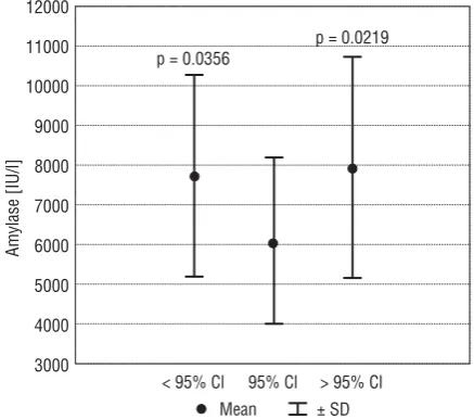
dam’s body weight gain in the common DFU-ex-posed group. However, it should be stressed that an increase in enzyme activity was found in the drug-treated mothers with lower and higher glucose activity (Fig. 3).
It was also shown that in the control group a greater placental weight was found in litters ob-tained from mothers with high body weight gain and high glucose levels (Fig. 4A). In the DFU-treated groups a greater placental weight was observed in litters with low glucose levels (Fig. 4B). No sim-ilar data were revealed for foetal weight and length in the control groups (Fig. 4C, E). Howev-er, a high glucose level and low maternal weight gain predicted high foetal weight, while high foe-tal length was observed in litters with a high
Figure 2. Relative (lg% — logarithm%; Mean ± SD) amylase ac-tivity and glucose level in group exposed to DFU [mg/kg] when compared with the corresponding control value.
Figure 3. Absolute amylase activity in mothers treated with DFU with low (< 95% CI), normal (95% CI) and higher (> 95% CI) glu-cose levels (p value vs. group with normal glucose level). Table 1. Absolute amylase activity and glucose level in maternal sera in control and DFU-treated groups
DFU [mg/kg] Min. Max. Mean SD Median p1 p2
Amylase [IU/l] – 3249.00 11714.9 7017.11 2537.47 6948.60 – –
0.2 3493.00 11093.4 7256.22 2337.12 7313.90 0.796 0.992
2.0 3078.90 12697.2 7152.47 2835.52 7390.60 0.870 0.992
20.0 3441.70 11861.9 7214.35 2676.87 7418.10 0.824 0.992
Glucose [mmol/l] – 112.00 308.0 217.80 56.33 212.50 – –
0.2 165.00 465.0 234.05 78.34 204.50 0.499 0.899
2.0 143.00 457.0 226.60 71.54 217.00 0.684 0.899
20.0 145.00 398.0 228.45 63.97 211.00 0.645 0.899
Min. — minimum, Max. — maximum, SD — standard deviation, 1p value when compared with the control value, 2p value in the whole group exposed to DFU when
glucose level and a high maternal weight gain (Fig. 4D, F).
The histological structure of the pancreas was found to be undisturbed in all the slides examined from the DFU-exposed and control groups, in both mothers and foetuses (Fig. 5A, B). Strong
cytoplas-mic COX-1 immunostaining was seen in pancreatic acinar secretory cells, whereas in the endocrine cells of islets the immune reaction was weak (Fig. 5C, D). Endocrine cells were strongly COX-2-positive in the maternal and foetal organs (Fig. 5E, F). In all the cases the staining pattern was cytoplasmic.
Furthermore, acinar cells exhibited a nuclear reac-tion, which was strong in foetal, while weak and only focal in maternal pancreases. A positive reac-tion was also observed in the epithelial cells of the
pancreatic ducts and the smooth muscle cells of blood vessels. No differences in COX immunoexpres-sion were found between DFU-exposed and control groups in either mothers or foetuses.
Figure 5. Normal pancreatic morphology in maternal (A) and 21-day old foetal (B) rat exposed to DFU at a dose of 20.0 mg/kg (haema-toxylin and eosin, lens mag. 20¥). Immunostaining for COX-1 (C, D) and COX-2 (E, F) in maternal (C, E) and 21-day old foetal (D, F) pan-creas (DakoEnvisionTM+/HRP; C-F: lens magn. 20¥).
A
C
E
F
DISCUSSION
The study demonstrated that organ morphology and function were undisturbed after administration of a selective COX-2 inhibitor, in spite of the consti-tutive expression of the inducible isoform in the pan-creas. However, since endocrine pancreatic function was monitored only by the glucose level and the morphology of the islets of Langerhans, other more sophisticated methods are desirable to confirm our results. On the other hand, the data obtained veri-fied previous experimental findings on the good tol-erability of the tested compound. In the above-cited paper by Burdan et al. [7] a good gastrointestinal, hepatic and renal toxicological profile was demon-strated for both pregnant and non-pregnant rats. Since DFU is still in the pre-clinical study, no similar human data were available in the literature. It should also be noted that, unlike the non-selective COX in-hibitor, DFU did not induce severe gastrointestinal changes such as deeply penetrating ulcers, which may result in secondary peritoneal inflammation [7]. Thus the direct effect of local inflammation as a mechanism that may explain an insignificant in-crease in glucose level cannot be apply [26]. Similar results were also reported in our previous study with another selective COX-2 inhibitor, namely DuP-697, which did not change the morphology and immu-noexpression of the constitutive and inducible COX isoforms in the rat pancreas during pregnancy [5]. Biochemical tests were not performed, however. On the other hand, an analgesic and antipyretic mix-ture containing a non-selective COX inhibitor, pre-pared with paracetamol, propyphenazone and caf-feine in a constant ratio of 5:3:1, increases the amy-lase activity in non-pregnant female rats exposed to the highest doses of the examined drugs (0.35, 0.21 and 0.07 g/kg respectively) [4]. An insignificant decrease in enzyme activity was noted in other groups of non-pregnant, as well as pregnant animals that were treat-ed with the same or lower doses of the mixture.
As was stressed in the introduction, different pancreatic COX immunoreactivity has already been reported in healthy and diabetic patients, as well as after administration of COX-inhibitors [31]. The roles of prostanoid, especially prostaglandin E2
(PGE2), and the COX-related mechanism in the
aeti-ology of diabetes and pancreatitis have already been reported [1, 9, 10, 11, 15, 17, 23]. Histologically, an inflammatory infiltration of the islets of Langer-hans is typical of type I diabetes, which is classified as an autoimmune disease [2]. Additionally, inflam-matory stimulators such as cytokines,
liposaccha-ride and mitogens increase an otherwise high phys-iological expression of COX-2 in insulin-secreting cells and secondarily initiate their cytotoxicity [3, 31]. At the same time a low physiological level of COX-1 is not significantly changed [15]. However, chronic hyperglycaemia impairs insulin-secreting cell func-tion and decreases the number and/or size of islets of Langerhans in type II diabetes. This may be the consequence of oversynthesis of interleukin 1 (IL-1) followed by apoptosis of insulin-secreting cells [23]. The pioneer study co-ordinated by Hughes et al. [17] indicated that recombinant human IL-1α was able to induce PGE2 accumulation in isolated rat islets
of Langerhans at concentrations similar to those at which the cytokine inhibits glucose-induced insulin secretion and islet glucose oxidation. Prior studies also showed that a high glucose concentration in-creased COX-2 expression [26]. The data were par-tially confirmed in mice treated with NS-398, a selective COX-2 inhibitor, which prevented the on-set of diabetes in mice brought on by multiple low doses of streptozotocin [29]. Moreover, COX-2 se-lective inhibitors such as NS-398 and SC-236 pre-vent the destruction of insulin-secreting cells initi-ated by IL-1 [30].
It has also been reported that COX-2 inhibition reduces the severity of pancreatitis and pancreatitis-associated lung injury [1, 10, 11]. With results simi-lar to our data, Alhan et al. [1] demonstrated that celecoxib did not change amylase activity, glucose level or other biochemical parameters and pancre-atic morphology in the rat. It was also found that treatment improved lung and renal function, de-creased the severity of pancreatic damage and nor-malised serum levels of IL-6 in cerulean-induced pan-creatitis in rats. Similar results were obtained in rats with taurocholate-induced acute pancreatitis treat-ed with parecoxib [10]. However, celecoxib-inductreat-ed acute pancreatitis and hepatitis have also been found in humans [9].
The data presented above for the influence of the glucose level on foetal and placental development partially confirmed the previous animal and human observations. It was shown that prolonged eleva-tion of the glucose level, a typical sign of diabetes and glucose intolerance, may result in foetal over-growth, as well as higher perinatal mortality, inci-dence of respiratory distress and congenital devel-opmental abnormalities such as anencephaly, en-cephalomeningocoele, caudal regression syndrome, transposition of the great vessels and coarctation of the aorta [16, 20, 22]. During uncomplicated preg-nancy the influence of hypoglycaemic factors is much stronger than hyperglycaemic ones. In consequence, higher maternal insulin secretion prevents hypergly-caemia [19]. However, unlike the normal levels of adrenal, noradrenaline, growth hormone and glu-cagons, which remain similar throughout pregnan-cy, the higher cortisol level correlates with gestational age. During pregnancy extra placental-relted hyper-glycaemic factors, such as lactogen (HPL), prolactin, insulinases (transhydrogenase glutathione insulinase and sulfhydrylprotease, oestrogens and progester-one are also synthesised. HPL and prolactin are char-acterised by the strongest anti-insulin activity and are blamed for the glucose intolerance [13, 14]. It has also been suggested that during pregnancy the content of insulin receptors decreases, while the insulin-binding protein increases significantly [18, 25]. In spite of the constitutive secretion of foetal insulin at an early developmental stage, the foetus does not regulate hyperglycaemia by increasing in-sulin secretion owing to the insufficiency of the ade-nyl cyclase-dependent pathway. Even later, when an extra amount of endogenous insulin is secreted, its total activity cannot normalise the increase in glucose level, since the main role of foetal insulin is to regulate growth [19, 27]. Such activity may ex-plain foetal overgrowth in offspring born to dia-betic mothers. Furthermore, both clinical observa-tions in type 1 diabetic pregnancies and prelimi-nary animal experimental studies suggest that even short periods of metabolic perturbation early in pregnancy may affect placental growth and trans-port functions for the remaining period of preg-nancy, thereby contributing to foetal overgrowth. An in vitro simultaneous study has demonstrated an up-regulation of placental transport systems for glucose and certain amino acids, as well as placen-tal lipoprotein lipase. In such a situation an up-reg-ulation of inflammatory mediators and Leptin genes is also revealed [19]. However, since none of the
above-mentioned hormones and enzymes was eval-uated in our study, it is not possible to fully explain the phenomena observed.
CONCLUSION
In conclusion, it was found that DFU administered throughout mid and late pregnancy in rats did not change maternal or foetal pancreatic morphology and function, or the immunoexpression of constitutive and inducible cyclooxygenase isoforms in the organ. How-ever, administration of a COX-2 selective inhibitor may affect foetal development in a mechanism that is re-lated to abnormal glucose levels.
ACKNOWLEDGEMENTS
We are very grateful to Merck Research Labora-tories (Rahway NJ, USA) for providing DFU. The study was not performed at this company’s request, al-though it had full access to the final version of the manuscript. The work was supported by the Polish Committee of Scientific Research, Grant KBN# 3 P05A 048 25.
REFERENCES
1. Alhan E, Kalyoncu NI, Ercin C, Kural BV (2004) Effects of the celecoxib on the acute necrotizing pancreatitis in rats. Inflammation, 28: 303–309.
2. Alm P, Ekstrom P, Henningsson R, Lundquist I (1999) Morphological evidence for the existence of nitric ox-ide and carbon monoxox-ide pathways in the rat islets of Langerhans: an immunocytochemical and confocal microscopical study. Diabetologia, 42: 978–986. 3. Burdan F, Chalas A, Szumilo J (2006) Cyclooxygenase
and prostanoids — biological implications. Post Hig Med Dosw, 60: 129–141.
4. Burdan F, Radzikowska E, Wyskiel M, Urbanowicz Z (1999) Drug-induced pancreatic injury during pregnan-cy. In: Mackiewicz Z (ed) Selected surgical problems. Vol. 4. Fundacja Polski Przegląd Chirurgiczny, Warsza-wa, pp. 168–171.
5. Burdan F, Szumilo J, Dudka J, Korobowicz A, Fronczek A, Klepacz R, Wojtowicz Z (2007) Pancreatic morphology in pregnant rats exposed to DuP-697 — the irrevers-ible, highly selective cyclooxygenase-2 inhibitor. Ann Univ Mariae Curie Sklodowska Med, 62 (in press). 6. Burdan F, Szumilo J, Dudka J, Radzikowska E, Klepacz R
(2006) Constitutive and inducible cyclooxygenase iso-forms expressed in rat pancreas. Ann Univ Mariae Cu-rie Sklodowska Med, 61: 572–575.
7. Burdan F, Szumilo J, Klepacz R, Dudka J, Korobowicz A, Tokarska E, Cendrowska-Pinkosz M, Madej B, Klepacz L (2004) Gastrointestinal and hepatic toxicity of selec-tive and non-selecselec-tive cyclooxygenase-2 inhibitors in pregnant and non-pregnant rats. Pharmacol Res, 50: 533–543.
inhibitors in CRL:(WI)WUBR Wistar rats — DFU and piroxicam study. Toxicology, 211: 12–25.
9. Carrillo-Jimenez R, Nurnberger M (2000) Celecoxib--induced acute pancreatitis and hepatitis: a case re-port. Arch Intern Med, 160: 553–554.
10. de Almeida JL, Jukemura J, Coelho AM, Patzina RA, Machado MC, da Cunha JE (2006) Inhibition of cyclo-oxygenase-2 in experimental severe acute pancreatitis. Clinics, 61: 301–306.
11. Ethridge RT, Chung DH, Slogoff M, Ehlers RA, Hellmich MR, Rajaraman S, Saito H, Uchida T, Evers BM (2002) Cy-clooxygenase-2 gene disruption attenuates the sever-ity of acute pancreatitis and pancreatitis-associated lung injury. Gastroenterology, 123: 1311–1322. 12. Fitzpatrick FA (2004) Cyclooxygenase enzymes:
regu-lation and function. Curr Pharm Des, 10: 577–588. 13. Fuglsang J, Lauszus FF, Fisker S, Flyvbjerg A, Ovesen P
(2005) Growth hormone binding protein and maternal body mass index in relation to placental growth hor-mone and insulin requirements during pregnancy in ty-pe 1 diabetic women. Growth Horm IGF Res, 15: 223– –230.
14. Gabbe SG, Quilligan EJ (1977) Fetal carbohydrate me-tabolism: its clinical importance. Am J Obstet Gynecol, 127: 92–103.
15. Han X, Chen S, Sun Y, Nadler JL, Bleich D (2002) In-duction of cyclooxygenase-2 gene in pancreatic beta-cells by 12-lipoxygenase pathway product 12-hydro-xyeicosatetraenoic acid. Mol Endocrinol, 16: 2145–2154. 16. Hedderson MM, Weiss NS, Sacks DA, Pettitt DJ, Selby JV, Quesenberry CP, Ferrara A (2006) Pregnancy weight gain and risk of neonatal complications: macrosomia, hypoglycemia, and hyperbilirubinemia. Obstet Gy-necol, 108: 1153–161.
17. Hughes JH, Easom RA, Wolf BA, Turk J, McDaniel ML (1989) Interleukin 1-induced prostaglandin E2 accu-mulation by isolated pancreatic islets. Diabetes, 38: 1251–1257.
18. Ishizuka T, Klepcyk P, Liu S, Panko L, Liu S, Gibbs EM, Friedman JE (1999) Effects of overexpression of hu-man GLUT4 gene on maternal diabetes and fetal growth in spontaneous gestational diabetic C57BLKS/J Lepr(db/+) mice. Diabetes, 48: 1061–1069.
19. Jansson T, Cetin I, Powell TL, Desoye G, Radaelli T, Ericsson A, Sibley CP (2006) Placental transport and metabolism in fetal overgrowth — a workshop report. Placenta, 27 (Suppl. A): S109–S113.
20. Kautzky-Willer A, Bancher-Todesca D (2003) Gestation-al diabetes. Wien Med Wochenschr, 153: 478–484.
21. Koliopanos A, Friess H, Kleeff J, Roggo A, Zimmermann A, Buchler MW (2001) Cyclooxygenase 2 expression in chronic pancreatitis: correlation with stage of the disease and diabetes mellitus. Digestion, 64: 240– –247.
22. Lucas MJ (2001) Diabetes complicating pregnancy. Obstet Gynecol Clin North Am, 28: 513–536. 23. Maedler K, Sergeev P, Ris F, Oberholzer J,
Joller--Jemelka HI, Spinas GA, Kaiser N, Halban PA, Donath MY (2002) Glucose-induced beta cell production of IL-1beta contributes to glucotoxicity in human pancreatic is-lets. J Clin Invest, 110: 851–860.
24. Miedzybrodzki R (2004) Trends in nonsteroidal anti-inflammatory drug development and application. Post Hig Med Dosw, 58: 438–448.
25. Neufeld ND, Corbo L (1982) Increased fetal insulin re-ceptors and changes in membrane fluidity and lipid composition. Am J Physiol, 243: E246–E250.
26. Shanmugam N, Todorov IT, Nair I, Omori K, Reddy MA, Natarajan R (2006) Increased expression of cyclooxy-genase-2 in human pancreatic islets treated with high glucose or ligands of the advanced glycation endprod-uct-specific receptor (AGER), and in islets from diabet-ic mdiabet-ice. Diabetologia, 49: 100–107.
27. Sjoholm A (1991) Inhibition of fetal rat pancreatic beta-cell replication by interleukin-1 beta in vitro is not mediated through pertussis toxin-sensitive G-proteins, a decrease in cyclic AMP, or protease activation. FEBS Lett, 289: 249–252.
28. Streck RD, Kumpf SW, Ozolins TR, Stedman DB (2003) Rat embryos express transcripts for cyclooxygenase-1 and carbonic anhydrase-4, but not for cyclooxygena-se-2 during organogenesis. Birth Defects Res B Dev Reprod Toxicol, 68: 57–69.
29. Tabatabaie T, Waldon AM, Jacob JM, Floyd RA, Kotake Y (2000) COX-2 inhibition prevents insulin-de-pendent diabetes in low-dose streptozotocin-treated mice. Biochem Biophys Res Commun, 273: 699–704. 30. Tran PO, Gleason CE, Poitout V, Robertson RP (1999) Pros-taglandin E(2) mediates inhibition of insulin secretion by interleukin-1 beta. J Biol Chem, 274: 31245–3128. 31. Warner TD, Mitchell JA (2004) Cyclooxygenases: new
forms, new inhibitors, and lessons from the clinic. FASEB J, 18: 790–804.
![Figure 1. Relative (%; mean ± SD) body weight and length,body mass index (BMI), as well as placental weight in groupexposed to DFU [mg/kg] when compared with the correspondingcontrol value (*p < 0.05 vs](https://thumb-us.123doks.com/thumbv2/123dok_us/9941882.1981705/3.581.306.513.76.306/figure-relative-weight-length-placental-groupexposed-compared-correspondingcontrol.webp)
![Figure 4. Placental (gain (%) and absolute glucose level [mmol/l] in control (A, C, E) and DFU-exposed (B, D, F) groups; PW — placental weight; FW — foet-A, B) and foetal (C, D) weights [g], as well as foetal length [mm] (E, F) in relation to relative maternal body weightal weight; FL — foetal length.](https://thumb-us.123doks.com/thumbv2/123dok_us/9941882.1981705/5.581.75.509.69.607/placental-absolute-exposed-placental-relation-relative-maternal-weightal.webp)

