Xerocomus
s. l. in the light of the present state of knowledge
JOSEFŠUTARA
Prosetická 239, 415 01 Teplice, Czech Republic [email protected]
Šutara J. (2008):Xerocomuss. l. in the light of the present state of knowledge. – Czech Mycol. 60(1): 29–62.
The definition of the generic limits ofXerocomuss. l. and particularly the delimitation of this genus fromBoletusis very unclear and controversial. During his study of European species of theBoletaceae, the author has come to the conclusion thatXerocomusin a wide concept is a heterogeneous mixture of several groups of species. These groups are separated from each other by different anatomical and some other characters. Also recent molecular studies show thatXerocomuss. l. is not a monophyletic group. In agreement with these facts, the European species ofXerocomuss. l. whose anatomy was studied by the present author are here classified into the following, more distinctly delimited genera: Xerocomuss. str.,Phylloporus,Xerocomellusgen. nov.,Hemileccinumgen. nov. andPseudoboletus. Boletus badiusandBoletus moravicus, also often treated as species ofXerocomus, are retained for the present in the genusBoletus. The differences betweenXerocomuss. str.,Phylloporus,Xerocomellus, Hemileccinum, PseudoboletusandBoletus(which is related to this group of genera) are discussed in detail. Two new genera,XerocomellusandHemileccinum,and necessary new combinations of species names are proposed.
Key words:Boletaceae,Xerocomus, Xerocomellus, Hemileccinum,generic taxonomy, anatomy, histology.
Šutara J. (2008): RodXerocomuss. l. ve světle současného stavu znalostí. – Czech Mycol. 60(1): 29–62.
Definování hranic roduXerocomuss. l. a zejména vymezení tohoto rodu od roduBoletusje velice nejasné a kontroverzní. Během studia evropských druhů čelediBoletaceaeautor dospěl k závěru, že Xerocomusv širokém pojetí je nesourodou směsicí několika skupin druhů. Tyto skupiny jsou od sebe odděleny rozdílnými anatomickými a některými dalšími znaky. Také novodobé molekulární studie uka-zují, žeXerocomuss. l. není monofyletickou skupinou. V souladu s těmito skutečnostmi jsou zde evrop-ské druhy roduXerocomuss. l., jejichž anatomie byla autorem studována, roztříděny do následujících, zřetelněji vymezených rodů:Xerocomuss. str.,Phylloporus,Xerocomellusgen. nov.,Hemileccinum gen. nov. aPseudoboletus.Boletus badiusaBoletus moravicus, také často považované za druhy rodu Xerocomus, jsou prozatím ponechány v roděBoletus. Rozdíly mezi rodyXerocomuss. str., Phyllopo-rus,Xerocomellus,Hemileccinum, PseudoboletusaBoletus(který je této skupině rodů příbuzný) jsou detailně diskutovány. Jsou navrženy dva nové rody,XerocomellusaHemileccinum,a nezbytné nové kombinace druhových jmen.
INTRODUCTION
In the course of his study of European boletes of theBoletaceaethe present au-thor has come to the conclusion that the genusXerocomusQuél. in a wider concept as adopted by many authors (e. g. Singer 1986, Ladurner and Simonini 2003, and oth-ers) is a heterogeneous mixture of several groups of species. These groups are sepa-rated from each other by different anatomical and some other characters. The groups whose anatomy has been studied by the present author are briefly described in the following paragraphs (for the meaning of some less common terms concerning the anatomical and histological characters used in this paper, see Šutara 1989, 2005) .
A significant part ofXerocomuss. l. is a group containing type species of this genus, Xerocomus subtomentosus (L.: Fr.) Quél., and Xerocomus ferrugineus
(Schaeff.) Bon. These species are characterised mainly by an unusual and very typical surface ornamentation of their spores (see Figs.1, 2), a phylloporoid, non-gelatinous hymenophoral trama with hyphae of the lateral strata touching or al-most touching each other, a pileipellis consisting of a non-gelatinous trichoderm (Figs. 8, 10a), and a divergent, loosely arranged, non-gelatinous lateral stratum in the peripheral layers of the stipe of young fruit bodies.
Another European species close to X. subtomentosus, which has been re-cently placed inXerocomuss. l. by some authors, isPhylloporus pelletieri(Lév.) Quél. (see Bresinsky and Besl 2003, Ladurner and Simonini 2003).P. pelletierihas a spore ornamentation similar to that in the Xerocomus subtomentosus group (see Fig. 5) and its hymenophoral trama is phylloporoid but its hymenophore is lamellate and its stipe lacks a distinct lateral stratum.
Another part ofXerocomuss. l. is formed by a group of species close to Bo-letus chrysenteronBull. [≡Xerocomus chrysenteron(Bull.) Quél.], including Bo-letus armeniacus Quél. [≡ Xerocomus armeniacus (Quél.) Quél.], Boletus porosporusImler ex Moreno et Bon [≡Xerocomus porosporus(Imler ex Moreno et Bon) Contu],Boletus pruinatusFr. [≡Xerocomus pruinatus(Fr.) Quél.], etc. This group is distant from theXerocomus subtomentosusgroup by its anatomical characters more than it seems at first sight. The species of the Boletus chrysenterongroup have a smooth or longitudinally striate spore surface which is different from that in theX. subtomentosusgroup, their hymenophoral trama is of a structure intermediate between the boletoid and phylloporoid types, with hyphae of the lateral strata not touching each other (Šutara 2005), their pileipellis is composed of a characteristic palisadoderm (Figs. 9, 10b), and in the peripheral layers of their stipe there is no or very reduced lateral stratum.
Representatives of another group of species recently included inXerocomuss. l. areBoletus impolitusFr. andBoletus depilatusRedeuilh (see e. g. Binder and Besl 2000, Ladurner and Simonini 2003). These two species, which are different from typical xerocomoid boletes in several macro– and micromorphological
char-acters, have smooth spores (Figs. 6, 7), a partly boletoid and partly leccinoid gen-eral appearance, their hymenophoral trama is true boletoid with distinctly gelati-nous lateral strata, and their arrangement of the peripheral layers of the stipe is in essence of the same type as inLeccinumspecies.
Pseudoboletus parasiticus (Bull.) Šutara, another species which has been sometimes considered a member ofXerocomuss. l., is clearly separated from the other European xerocomoid boletes by several exceptional characters. Its stipe is covered with a sterile filamentous trichoderm (without a caulohymenium and fer-tile caulobasidia), its mode of obtaining nutrition – at least in the phase of forming fruit bodies – is parasitic, its spores are characteristically pitted and its develop-ment of fruit bodies is hypo-(para)-velangiocarpous and pileostipiticarpous (see Reijnders 1963, Singer 1986). Unlike typical xerocomoid boletes,P. parasiticushas moreover a structure of its hymenophoral trama intermediate between the boletoid and phylloporoid types, sometimes even nearly boletoid (Šutara 1991, 2005).
Boletus badius (Fr.): Fr. and Boletus moravicus Vacek, often treated as
Xerocomusspecies,have a boletoid or almost boletoid hymenophoral trama. The pileus surface of these two species is sticky when moist and the pileipellis of
B. badiuseven often changes into a gelatinous ixotrichoderm. Such a pileipellis does not occur in any typical xerocomoid species but is a characteristic feature of some representatives of the genusBoletusFr.
If we summarise the characters of species which are now placed inXerocomus
s. l., we can see that the range of these characters is extraordinarily wide. Very dif-ferent characters of some species included in this widely circumscribed genus even seem to be almost contradictory. For example,Xerocomuss. l. includes side by side boletes with arrangements of the hymenophoral trama both phylloporoid (in theXerocomus subtomentosusgroup andPhylloporus pelletieri) and interme-diate between the phylloporoid and boletoid types (in theBoletus chrysenteron
group) or true boletoid (inBoletus impolitus, B. depilatus, B. badius,etc.). The presence of these three types (or subtypes) of the trama together in one genus is very unusual. In each of the other European boletaceous genera there is always only one type of the structure of the hymenophoral trama, viz. boletoid inBoletus,
Leccinum,Aureoboletus,Buchwaldoboletus,Tylopilus,SuillusandBoletinus, in-termediate between the phylloporoid and boletoid types in Pseudoboletus,
Chalciporus,RubinoboletusandPorphyrellus, and phylloporoid inPhylloporus
[for further information on types (or subtypes) of the hymenophoral trama in boletes, see Šutara 2005: 3–7, 31–33].
The pileus surface of species ofXerocomuss. l. is mostly dry and non-gelati-nous, but sometimes it is viscid, composed of a gelatinous ixotrichoderm (e. g. in
Boletus badius). In most xerocomoid boletes (e. g.Xerocomus subtomentosus,
X. ferrugineus,theBoletus chrysenterongroup, etc.) the pores are at maturity an-gular and relatively large (ca 1–3 mm), but in several species (e. g. Boletus
impolitusandB. depilatus) they are roundish and minute, smaller than 1 mm and similar to those inBoletusand Leccinum. The stipe surface of almost all Euro-pean xerocomoid species is covered by a caulohymenium with fertile caulobasidia, but in one exceptional case it is composed of a sterile filamentous trichoderm (inPseudoboletus parasiticus).
The various groups ofXerocomuss. l. have also different types of lateral stipe stratum. In Boletus impolitusandB. depilatus, the lateral stipe stratum is of the leccinoid type, up to 400(–640) μm thick, at the very beginning densely arranged but soon breaking up into characteristic, predominantly anticlinally arranged fascicles of hyphae (for further information on the leccinoid type of lateral stipe stratum, see Šutara 1989). The lateral stipe stratum which occurs in young fruit bodies of
Xerocomus subtomentosusandX. ferrugineusreaches a thickness of 80(–200) μm, is loosely arranged and does not break up into hyphal fascicles as in Boletus impolitusandB. depilatus. In theBoletus chrysenterongroup the lateral stipe stra-tum is absent or present only in a reduced form, at most 30(–40) μm thick. In
Pseudoboletus parasiticus the lateral stipe stratum does not exist at all and in
Phylloporus pelletieriit is probably absent too (for further information on the periph-eral layers of the stipe in boletes, including the latperiph-eral stipe stratum, see Šutara 2005: 8–15). Certain differences also exist in mechanisms for obtaining nutrition: almost all species ofXerocomuss. l. are permanently ectomycorrhizal, whereasPseudoboletus parasiticus,at least for a certain period of its life, is parasitic (see Kavina 1935).
The very wide range of characters ofXerocomuss. l. overlaps characteristics of some related genera. A precise definition of the limits between the widely cir-cumscribed Xerocomus and the other boletaceous genera, particularly Boletus
Fr., is therefore practically impossible.
The above discussed opinion thatXerocomus s. l. is a heterogeneous taxo-nomic group corresponds with results of recent molecular analyses (e. g. Binder and Hibbett 2004, 2007; Eberhardt and Taylor 2005; Bakker and Noordeloos 2005). For example, Eberhardt and Taylor remarked on this matter (see p. 39): ‘As mo-lecular data accumulate, it would appear thatXerocomusmay be paraphyletic – the taxa have multiple origin. Some indication of this situation can be seen in Fig-ure 1, whereXerocomus species are split into three groups. Binder (1999) sug-gested thatXerocomuscould be split intoXerocomuss. str.,Paraxerocomus, and
Pseudoboletusand, potentially, other groups.’
The multiple origin ofXerocomuss. l. is most obvious from the phylogram pub-lished by Binder and Hibbett (2007) in one of their latest work concerning molecu-lar systematics ofBoletales. These authors commented on this phylogram as fol-lows (see p. 976): ‘The analyses on the nuc-lsu dataset (Supplementary Fig. 1) with increased taxon sampling show that relationships among genera are poorly resolved and moreover that most of the larger genera (e. g.Boletus,Tylopilus,
The recent opinions on the taxonomic status ofXerocomusare very controver-sial. Some mycologists considerXerocomusto be a good separate genus (e. g. Singer 1986, Engel et al. 1996, Lannoy and Estades 2001, Peintner et al. 2003, Ladurner and Simonini 2003), whereas some other authors do not accept it at the generic level (e. g. Watling 1968, 1970; Smith and Thiers 1971; Kirk et al. 2001; Watling and Hills 2005; Legon and Henrici 2005). It is obvious that the question of the status and delimitation ofXerocomusis a taxonomic problem requiring a solution.
MATERIALS AND METHODS
The results of observations presented in this paper are based on the author’s macroscopic and mi-croscopic study of boletes in the years 1974–2008. Mimi-croscopic characters of boletes were examined predominantly on dried material. Microscopic sections were made by hand with a razor blade. Sections from dried material were revived and examined in a 3–10 % solution of ammonium hydroxide (NH4OH)
with Congo Red and, in many cases, moreover in Melzer’s reagent or a pure solution of ammonium hy-droxide (without Congo Red). Sections from fresh fruit bodies were mounted in Melzer’s reagent, pure water or an aqueous solution of Congo Red.
In the course of microscopic examination of anatomical characters, attention was paid not only to the aspects which are already relatively well-known but also to some structures which have been little studied so far, e. g. histological structures of the peripheral layers of the stipe. Particular attention was also focused on the study of the changes which some tissue layers (e. g. hymenophoral trama, lateral stipe stratum, and pileipellis) undergo in the course of development of fruit bodies. For this reason, the microscopic structures were examined on a sufficiently large number of specimens in various develop-mental stages (from very young to quite mature fruit bodies) whenever possible. A larger number of studied fruit bodies can also show which of the changes of the anatomical structures are typical of a certain species or a certain group of species and which of them are atypical or even abnormal.
The results of anatomical examinations are very dependent on the quality of the herbarium mate-rial examined. For this reason, observations obtained from matemate-rial of poor quality, with various abnor-malities or defects, were not taken into consideration. The quality of herbarium material can also be negatively influenced by the use of an inappropriate drying process of collected fruit bodies. Herbar-ium specimens which have not been dried in the right way can give skewed results because their tis-sues do not revive properly in microscopic preparations. Such herbarium specimens were excluded from the anatomical examinations.
Surface ornamentation of the spores was examined with the following scanning electron micro-scopes: FE SEM JEOL 7401 F (with cryo-attachment Alto 2500) and SEM JEOL 6300 in the Laboratory of Electron Microscopy, Biological Centre, Institute of Parasitology, Academy of Sciences of the Czech Republic, České Budějovice, and with SEM FEI Quanta 200 at the Institute of Botany, Academy of Sci-ences of the Czech Republic, Průhonice near Prague.
The material which was collected by the author and his colleagues is deposited in J. Šutara’s pri-vate herbarium (abbreviation: JŠ). The other material was loaned from herbaria of the following insti-tutions: National Museum, Prague (PRM), Moravian Museum, Brno (BRNM), South Bohemian Mu-seum, České Budějovice (CB), Slovak National MuMu-seum, Bratislava (BRA), Regional Museum of Litoměřice (LIT), Regional Museum of Hradec Králové (HK), and Forestry and Forest Products Re-search Institute, Tsukuba, Japan (TFM).
Species and herbarium specimens examined microscopically by the author in connection with tax-onomic problems discussed in this contribution are included in the following list.
Material examined
E u r o p e a n s p e c i e s:Boletus aereusBull.: Fr., BRNM 236110, 236125, 265699, 265727, 265773, 266067; CB 1348; JŠ 4128, 4269. –Boletus appendiculatusSchaeff., PRM 775391; JŠ 2136, 5095, 5099, 5101. – Boletus armeniacusQuél. [≡Xerocomus armeniacus(Quél.) Quél.], JŠ 2629, 4123, 4129, 4284–85, 4451, 4455, 4459. –Boletus badius(Fr.): Fr. [≡Xerocomus badius(Fr.: Fr.) Kühner], JŠ 373, 395, 1814, 1856, 1858, 1875, 1931–32, 1980–81, 2102–03, 3089, 3116, 3120, 3223–25, 4299. –Boletus calopusPers.: Fr., BRA 13.9.1988; LIT 3719/171; JŠ 285, 2139, 2329, 5241. –Boletus chrysenteronBull. [≡ Xerocomus chrysenteron(Bull.) Quél.], JŠ 256, 331, 1692, 2315, 2319–20, 2401, 2416, 2518, 2674, 2701, 3085, 3242, 4280, 4304, 4312–16, 4373, 4388, 5003–04, 5006. –Boletus depilatusRedeuilh [≡Xerocomus depilatus(Redeuilh) Binder et Besl], PRM 647825, 717049; BRNM 265678–79, 265688, 265692, 265711, 265823, 265858, 265867 (all asBoletus impolitus); JŠ 3155, 5103–04, 5159, 5168, 5200, 5202–04. –Boletus edulisBull.: Fr., JŠ 048, 163, 241, 319, 320, 638, 1724, 2344, 3088, 3146, 3149–51, 4032–33, 5088. –Boletus engeliiHlaváček [=Boletus declivitatum(C. Martin) Watling andXerocomus communis(Bull.) Bon s. auct.], JŠ 4133, 4206–10, 4452, 4454–61, 5229, 5232. –Boletus erythropusPers.: Fr., LIT 3719/173; JŠ 058, 060, 282, 316, 404, 2001–07, 3125–26, 4212. –Boletus erythropusvar.junquilleus(Quél.) Bon [≡Boletus junquilleus(Quél.) Boud.], BRNM 236072, 236073, 236075, 265822, 267708; JŠ 3128, 4044. –Boletus fechtneriVelen., PRM 518242, 617615, 647783. –Boletus fennicus(Harmaja) Šutara [≡Xerocomus fennicus(Harmaja) Ladurner et Simonini], BRNM 693777. –Boletus fragransVittad., herb. Drescher. – Boletus fuscoroseusSmotl. [=Boletus pseudoregius(Huber) Estades], CB 1352, 1782, 1784 (all as Bo-letus speciosus); JŠ 4244. –Boletus impolitusFr. [≡Xerocomus impolitus(Fr.) Quél.], PRM 647825, 682521, 717049; BRNM 236061–62, 265671, 265681, 265685–86, 265689–91, 265704–05, 265717, 265728, 265747, 265779, 265789, 265816–17, 265825, 265828, 265831, 265851, 265864, 265877, 265885; BRA 99, 118, 132, 155, 1/10/1967, 4/8/1969, 18/8/1970; LIT 3719/1148; JŠ 1704, 2079–80, 2535–36, 3005–06, 4254–55, 5151, 5205. –Boletus kluzakiiŠutara et Špinar, PRM 857093; JŠ 3889, 4215; herb. P. Špinar 234. – Bo-letus legaliaePilát, CB 3408; JŠ 3541, 3546, 5093, 5121. –Boletus luridusSchaeff.: Fr., PRM 583462; JŠ 059, 317, 1622, 2015, 2769, 4041. –Boletus marekiiŠutara et Skála [≡Xerocomus marekii(Šutara et Skála) Klofac], PRM 857255; JŠ 4311, 4433–35, 5045, 5074, 5133–37, 5141–44. –Boletus moravicus Vacek [≡Xerocomus moravicus(Vacek) Herink], PRM 203551; BRNM 265479, 265530–31, 265534, 265540; CB 3435–38; JŠ 4155. –Boletus porosporusImler ex Moreno et Bon [≡Xerocomus porosporus (Imler ex Moreno et Bon) Contu], JŠ 016, 248–50, 1626–31, 1659, 1703, 2021, 2189, 2802, 2808, 3096, 3253–55, 4131, 4463–64. –Boletus pinophilusPilát et Dermek, JŠ 2387, 2800, 3139–40, 5233. –Boletus pruinatusFr. [≡Xerocomus pruinatus(Fr.) Quél.], JŠ 078, 302, 336–37, 341, 345, 1836, 1963, 1965, 1986, 2112, 4053, 4397–99. –Boletus pulverulentusOpat., BRNM 265815, 265862; CB 922, 1127, 1647; JŠ 041–42, 3463. –Boletus queletiiSchulzer, PRM 647965, 824842, 830105, 830114, 836121; BRNM 236086, 265835, 266120; JŠ 4113, 5224–26. –Boletus radicansPers.: Fr., JŠ 2120, 2534, 2785, 3143–45, 3661–62, 3876, 3880–86, 4035–37, 4216–33, 4254–56, 4261. – Boletus regius Krombh., PRM 614841, 615709, 866632; JŠ 044, 3525, 5227. –Boletus reticulatusSchaeff., JŠ 2395, 3129–30, 4034, 5109, 4478, 5236. – Bo-letus rhodopurpureusSmotl., BRNM 265731, 265746; CB 1145–47, 1779, 2608, JŠ 3552, 5105, 5117. – Bo-letus rhodoxanthus (Krombh.) Kallenb., BRNM 265697, 265732, 265764, 265767, 265791, 265794, 265799, 265806, 265821, 265863; BRA 17.9.1988; CB 1143–44, 1781, 3386; JŠ 5237, 5240. –Boletus ripariellus(Redeuilh) Watling et A. E. Hills (≡Xerocomus ripariellusRedeuilh), JŠ 008, 011, 052, 205, 721, 1624, 1698, 1806, 2043–62, 4440–49, 4466. –Boletus rubellusKrombh. [≡Xerocomus rubellus (Krombh.) Quél.], PRM 894395; JŠ 2135, 2321–25, 2327, 2353–54 2371–77, 4405, 4465, 4467–71. –Boletus satanasLenz, JŠ 3091, 3142, 4039, 4473, 4487. –Boletus subappendiculatusDermek, Lazebníček et Veselský, JŠ 318. –Phylloporus pelletieri(Lév.) Quél. [≡Xerocomus pelletieri(Lév.) Bresinsky and M. Binder], CB 448; HK 292/81; JŠ 281, 738, 2121, 2722, 3251, 3412–13, 3415–16. – Pseudoboletus parasiticus(Bull.: Fr.) Šutara [≡Xerocomus parasiticus(Bull.) Quél.], CB 1302, 2310, 2329, 2360; JŠ 2088–92, 2106, 2108–09, 2307, 2342, 3115, 3243–44. –Xerocomus chrysonemusA. E. Hills et A. F. S. Tay-lor, JŠ 5145. –Xerocomus ferrugineus(Schaeff.) Bon, JŠ 034, 039, 251, 326–27, 543, 1548, 1707, 1848–49,
2039–40, 2042, 2133, 2144–45, 2147–49, 2173, 2402, 2560–61, 2586, 2660, 3136, 5010. – Xerocomus silwoodensisA. E. Hills, U. Eberhart et A. F. S. Taylor, JŠ 5146. –Xerocomus subtomentosus(L.: Fr.) Quél. (≡Boletus subtomentosusL.: Fr.), JŠ 266, 410, 1168, 1632, 1643–45, 1647–55, 3231–33, 4119, 4121, 4134, 4198–99, 5120, 5182, 5184, 5186, 5188–89, 5191, 5194, 5196–97. –Boletus subtomentosusvar. luteolusVelen. (=Xerocomus subtomentosusvar.xanthusGilbert), JŠ 5178–80, 5198–99.
E x t r a - E u r o p e a n s p e c i e s: –Boletus auripesPeck, PRM 487595. –Boletus bicolorPeck, PRM 704838. –Boletus frostiiRussell in Frost, PRM 647800, 833580. –Boletus inflexusPeck, PRM 647837. – Boletus variipes Peck, PRM 704846. –Boletus vermiculosusPeck, PRM 487648. –Pseudoboletus astraeicola(Imazeki) Šutara (≡Xerocomus astraeicolaImazeki), TFM 972.
RESULTS AND DISCUSSION
CLASSIFICATION OFEUROPEAN SPECIES OFXEROCOMUSS. L.
On the basis of anatomical differences and some other distinctive characters and in view of the results of present molecular analyses (for references, see Intro-duction), the European species whose anatomy has been studied by the present author and which have been mostly placed inXerocomuss. l. (except for two ex-ceptions mentioned below) are here classified into the following genera:
Xerocomuss. str.,Phylloporus,Xerocomellus,PseudoboletusandHemileccinum. The two above-mentioned exceptions whose generic placement is still rather unclear are the speciesBoletus badiusand Boletus moravicus.In the author’s opinion, these species belong neither toXerocomuss. str. nor to the remaining four above-mentioned genera. In this contribution, Boletus badius and Boletus moravicusare placed inBoletus, but neither this solution seems to be fully satis-factory. The generic position of these two species remains a difficult taxonomic problem whose definitive solution will necessitate further research.
XerocomusQuél. s. str.
in Mougeot et Ferry, Fl. Vosges, Champ.: 477, 1887, nom. conserv.
S y n o n y m s:VersipellisQuél., Enchir. Fung.: 157, 1886. –XerocomopsisReichert, Palest. Journ. Bot. Rehov. Ser. 3: 229, 1940.
Ty p u s:Xerocomus subtomentosus(L.: Fr.) Quél. (≡Boletus subtomentosusL.: Fr.).
C h a r a c t e r s. Pileus surface dry, matt, subtomentose, neither viscid nor sticky when moist. Pileipellis from an early stage composed of a trichoderm whose hyphae are more or less intertwined, unequal in length, at first suberect but later gradually collapsing with age (see Figs. 8, 10a). The trichodermal elements are more or less cylindrical (although in some cases a smaller number of some-what broadened terminal or subterminal cells may also occur), non-gelatinous
and mostly without a distinct incrustation. But hyphae in the tissue layer under the trichoderm are sometimes incrusted.
Tubes at most 15 mm long, nearly adnate or somewhat depressed around the stipe apex and shortly decurrent with a tooth, when young yellow or yellow– ochreous, later ochreous, olive-yellow or olive-brownish, when cut bluing or un-changing. Pores concolorous or, in some cases, more ochreous or more brownish than the tube-sides, at maturity angular and relatively large (ca 1–3 mm), in bruised places bluing or unchanging. Hymenophoral trama phylloporoid, without a distinct gelification of lateral strata. Lateral strata slightly divergent, densely ar-ranged in all developmental stages, with hyphae touching or almost touching each other (Šutara 1987: Fig. 1). In microscopic sections stained with Congo Red there is not a conspicuous colour difference between layers of the trama. The lateral strata are red in the same or almost the same intensity as the mediostratum.
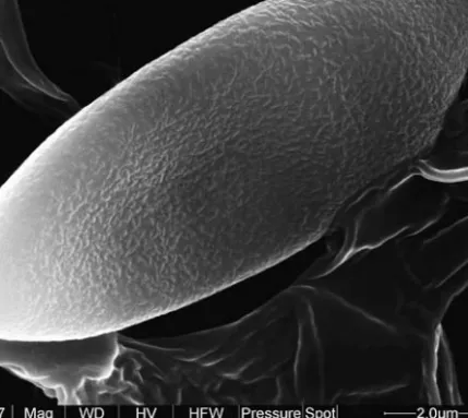
Spores of boletoid shape, subfusoid or fusoid-ellipsoid in face view, inequilat-eral with a shallow suprahilar depression in profile. Spore surface bacillate. [Note: The term ‘bacillate’ (‘bacillées’ in French) was used by Heinemann and his collab-orators for a fine but very characteristic ornamentation appearing in SEM as if the spore surface is covered with rod-like bacteria (bacilli), see Heinemann and Rammeloo 1987, Heinemann et al. 1988]. The bacillate ornamentation is detect-able with ca x 10 000 or x 15 000 magnification inXerocomus subtomentosus(see Fig. 1, and Heinemann et al. 1988: Figs. 15, 16) and with x 30 000 or higher magnifi-cation inXerocomus ferrugineus(Fig. 2). Spore print olive-brown.
Stipe relatively slender, non-reticulate or, particularly in the upper part, with a more or less distinct reticulation with elongate meshes, sometimes looking like longitudinal ribs. A substantial part of the stipe is covered by a caulohymenium with scattered fertile caulobasidia. In an early stage the caulohymenium is formed by a continuous, unbroken hymenial layer but during further development it grad-ually fragments into small islands of caulohymenial elements, which macroscopi-cally looks like minute floccose granules on the stipe surface. Under the caulohymenium in the upper half of the stipe of young fruit bodies it is sometimes developed a non-gelatinous lateral stipe stratum which is differentiated from the longitudinal stipe trama by its divergent and looser arrangement of hyphae (Šutara 1991: Fig. 3). In places between the ridges of the stipe reticulation the lat-eral stratum is up to 80(–120) μm thick inX. subtomentosusand up to 200 μm inX. ferrugineus. Within the ridges of the reticulation the lateral stratum is sometimes even thicker. During further developmental stages this lateral stratum soon loses its loose and divergent arrangement, becomes gradually less distinct and finally disappears. At times remnants of this layer persist in the ridges of the stipe reticu-lation up to maturity. (Note: As the lateral stipe stratum of species ofXerocomus
s. str. is developed best in an early stage, this layer should be examined mainly in young fruit bodies. It is also important to mention that the development of lateral
Fig. 1.Spore ofXerocomus subtomentosus(JŠ 4134) with bacillate surface ornamentation (x 24 000).
Fig. 3.Spores ofXerocomus chrysonemus(JŠ 5145) with bacillate surface ornamentation (x 11 000).
stipe stratum is rather often negatively influenced by unfavourable weather con-ditions. In dry weather, this layer is not sometimes developed at all.). Stipe trama composed of densely longitudinally arranged hyphae. Surface of the stipe base sterile, covered with a tangled filamentous tomentum which is whitish, pale yel-lowish or yellow, the mycelium threads on the base sometimes up to bright yellow. Partial veil and annulus absent.
Context yellow, pale yellowish, whitish or pale cream-coloured, in the stipe base sometimes brownish and usually rather firm, when cut or bruised partly blu-ing, particularly above the tubes, or unchanging. Clamp connections not found in the fruit body.
Ontogenetic development of fruit bodies gymnocarpous (according to Reijnders 1963 and the author’s own observations).
Ectomycorrhizal with both conifers and deciduous trees.
D e l i m i t a t i o n. Characters distinguishing Xerocomus s. str. from Phyllo-porus,Xerocomellus, PseudoboletusandHemileccinumare given under each of the four last mentioned genera.
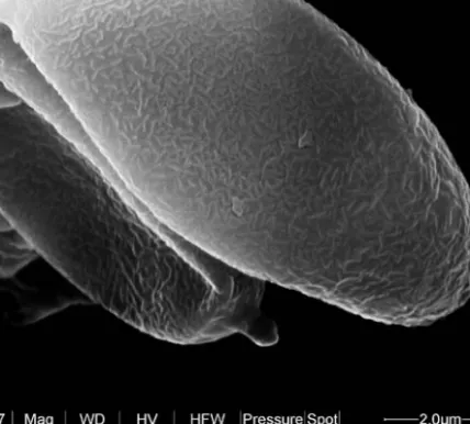
Fig. 6.Spores ofHemileccinum impolitum(JŠ 4289) with a smooth surface (x 19 000).
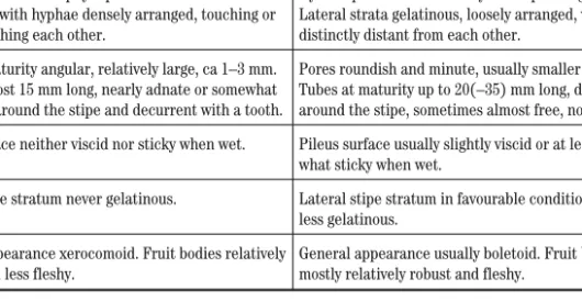
Tab. 1.Differences betweenXerocomuss. str. andBoletus.
Xerocomuss. str. Boletus
Spore surface bacillate. Spore surface smooth. Hymenophoral trama phylloporoid. Lateral strata
non-gelatinous, with hyphae densely arranged, touching or almost touching each other.
Hymenophoral trama in fully developed stage boletoid. Lateral strata gelatinous, loosely arranged, with hyphae distinctly distant from each other.
Pores at maturity angular, relatively large, ca 1–3 mm. Tubes at most 15 mm long, nearly adnate or somewhat depressed around the stipe and decurrent with a tooth.
Pores roundish and minute, usually smaller than 1 mm. Tubes at maturity up to 20(–35) mm long, depressed around the stipe, sometimes almost free, not decurrent. Pileus surface neither viscid nor sticky when wet. Pileus surface usually slightly viscid or at least
some-what sticky when wet.
Lateral stipe stratum never gelatinous. Lateral stipe stratum in favourable conditions more or less gelatinous.
General appearance xerocomoid. Fruit bodies relatively slender and less fleshy.
General appearance usually boletoid. Fruit bodies mostly relatively robust and fleshy.
N e w c o m b i n a t i o n
Xerocomus subtomentosusvar.luteolus(Velen.) Šutaracomb. nov.
(MycoBank MB511903)
B a s i o n y m:Boletus subtomentosusvar.luteolusVelenovský, České houby 2: 717, 1922. S y n o n y m s:Xerocomus subtomentosusvar.xanthusGilbert, Les Bolets: 142, 1931. –Xerocomus flavusSinger et Kuthan, Česká Mykol. 30(3–4): 153, 1976.
S p e c i e s:Xerocomus ferrugineus(Schaeff.) Bon,Xerocomus subtomentosus
(L.: Fr.) Quél., includingX. subtomentosusvar.luteolus(Velen.) Šutara, and prob-ably Xerocomus chrysonemus A. E. Hills et A. F. S. Taylor and Xerocomus silwoodensisA. E. Hills, U. Eberhart et A. F. S. Taylor, which were described only recently (see Taylor et al. 2006, 2007).
N o t e. The present author had the opportunity to examine dried material of a middle-aged fruit body ofX. chrysonemusand a mature one ofX. silwoodensis
obtained from A. E. Hills during the preparation of the manuscript of this paper. The examination of these specimens showed that X. chrysonemus and X. silwoodensishave a bacillate spore ornamentation (Figs. 3, 4) which is one of the typical characters of members ofXerocomuss. str. The present author, however, has not yet sufficiently studied some other anatomical characters of these two species (e. g. their peripheral stipe layers, early developmental stages of their pileipellis, etc.), which usually require examination of a larger number of younger fruit bodies.
Fig. 8.Pileipellis of a very young fruit body ofXerocomus subtomentosus. The pileus of this dried fruit body (JŠ 4198) is 14 mm wide. At this early stage the pileipellis of species ofXerocomuss. str. is com-posed of a trichoderm whose hyphae are unequal in length, loosely arranged and distinctly intertwined.
PhylloporusQuél. Fl. Mycol. Fr.: 409, 1888.
Ty p u s:Phylloporus pelletieri(Lév.) Quélet (≡Agaricus pelletieriLév.).
C h a r a c t e r s. Pileus surface dry, matt, subtomentose. Pileipellis consisting of a trichoderm whose hyphae sometimes partly collapse with age.
Hymenophore lamellate. Lamellae with numerous anastomoses, subdecurrent or decurrent. Hymenophoral trama phylloporoid, with lateral strata non-gelati-nous. Hyphae of the lateral strata touching each other (Šutara 2005: Figs. 2c, 2d).
Spores of boletoid shape, fusoid-ellipsoid in face view, inequilateral in profile. In P. pelletieri, the only European species of this genus, the spore surface is bacillate (see Fig. 5). Spore print brown with an olivaceous shade.
Stipe solid, composed of a longitudinally arranged trama. A large part of the stipe is covered by a gradually fragmenting caulohymenium with very sparsely scattered fertile caulobasidia. The fragments of the caulohymenium on the stipe surface macroscopically look like very minute granules. Surface of the stipe base sterile, covered with a tangled filamentous tomentum. InP. pelletieriprobably no distinct lateral stipe stratum occurs (Šutara 2005: Fig. 2f). Partial veil and annulus absent. Clamp connections not found in the carpophore.
Ectomycorrhizal with various trees.
D e l i m i t a t i o n.P. pelletieri(type species of this genus) is very similar in ana-tomical characters to species of Xerocomus s. str., from which it differs in its decurrent lamellate hymenophore and in the absence of a distinctly developed lat-eral stipe stratum. At least in material ofP. pelletieriexamined by the present au-thor (which contained specimes in all developmental stages, including young fruit bodies), no lateral stipe stratum was found. On the other hand, in young fruit bod-ies ofXerocomuss. str. the lateral stipe stratum is sometimes up to 80(–200) μm thick.
Also results of molecular studies show thatP. pelletieriis rather closely re-lated toXerocomus subtomentosus, type species ofXerocomus(see Binder 1999, Binder and Bresinsky 2002, Bakker and Noordeloos 2005, Binder and Hibbett 2007). For this reason,P. pelletieriwas transferred by Binder and Bresinsky to
Xerocomusin 2003. In this paper,Phylloporusis retained as a genus separate from
Xerocomusbecause of the somewhat different arrangement of the surface layers of the stipe. The present author, however, is aware that the taxonomic question concerning the whole genusPhylloporusmay be much more complicated than it appears from the European viewpoint, becausePhylloporusis a large genus with a world-wide distribution, occurring predominantly in tropical Africa, Indonesia, East Asia, and South and Central America.
XerocomellusŠutaragen. nov.
(MycoBank MB511890)
Carposomata pileata, stipitata, parva vel mediocriter magna. Superficies pilei sicca, nec viscida nec adhearens, initio glabra, velutina vel pruinosa, postea interdum subtomentosa, saepe rimoso-areolata. Pileipellis e palisadodermide formata. Hyphae palisadodermidis saepe incrustatae. Tubuli subadnati vel aliquantum depressi juxta stipitem et frequenter dente decurrentes, iuventute flavi, deinde olivaceo-flavi vel viridi-olivacei. Pori concolores, in vetustate angulati et satis magni (circa 1–2,5 mm). Tubuli et pori tactu plus minusve caerulescentes vel virescentes, raro fere immutabili. Structura tramae hymenophori inter typum boletoideum et phylloporoideum intermedia; strata lateralia tenuiter gelatinosa cum hyphis mutuo non contiguis. Sporae boletoideae, subfusoideae vel fusoideo-ellipsoideae, interdum truncatae, laeves vel longitudinaliter striatae. Pulvis sporarum olivaceo-brunneus. Stipes gracilior et minus carnosus quam in genereBoletus, superficies stipitis subtiliter granulosa. Pars magna superficiei stipitis caulohymenio cum basidiis fertilibus obducta. Stratum laterale stipitis nullum vel deminutum, maxime 30(–40) μm crassum. Tomentum basalis et mycelium albidum, flavo-albidum, pallide griseollo-flavum vel sordide flavum. Carne flava, pallide flavida vel albida, interdum intra stipitem partim rubeolla vel rubro-vinosa et intra basem brunneolla, aurantiaca vel carotino-rubra, fracta plus minusve cyanescens, raro fere immutabilis. Fibulae in carposomate nullae. GenusXerocomellusdiffert: – a genereXerocomuss. str. sporis laevibus vel striatis, structura tramae hymenophori non phylloporoidea, sed inter typum boletoideum et phylloporoideum intermedia cum hyphis stratis lateralibus mutuo non contiguis, pileipelle e palisadodermide formata atque strato laterali stipitis nullo vel valde tenui, – a genereBoletushabito dissimili, pileipelle e palisadodermide formata, nec viscida nec adhaerens, poris maioribus, trama hymenophori minus gelatinosa atque strato laterali stipitis nullo vel alio, – a generePseudoboletussuperficie stipitis caulohymenio cum basidiis fertilibus obducta, modo nutrimenti non-parasitico, pileipelle e palisadodermide formata atque ontogenese carposomatum gymnocarpa.
Ty p u s:Boletus chrysenteronBulliard
C h a r a c t e r s. Fruit bodies stipitate-pileate, small or medium-sized.
Pileus surface dry, matt, neither viscid nor sticky when moist, at first glabrous, velutinous or pruinose, usually without a distinct fibrillose aspect when young, only in later stages sometimes becoming subtomentose, in some species gradually cracking with age in a typical manner and becoming areolate-rimose along the margin or overall, exposing the pallid context in the cracks. Pileipellis composed of a palisadoderm consisting of anticlinally arranged, parallel or subparallel hyphae whose terminal elements reach the same or almost the same level. The hyphae consist of chains of cells which are short to moderately long, usually more or less broadened and often incrusted in a typical way. Thickness of the palisadodermal layer varies according to species, age of fruit bodies and location on the pileus in the range of (80–)120–350(–500) μm (for the meaning of the term ‘palisadoderm’, see Clémençon 1997: 685). The palisadoderm, which is one of the most typical characters of this genus and which does not occur in this form in the other European boletes, is usually most typically developed and arranged in an early stage (see Fig. 9, and Šutara 2007: Fig. 5). In further stages the palisadoderm sometimes changes secondarily into a more or less disarranged layer. In most
characteris-tic appearance and arrangement for a longer time (see Fig. 10b), sometimes up to maturity.
Tubes at most 10(–14) mm long, nearly adnate or somewhat depressed around the stipe apex and shortly decurrent with a small tooth, light yellow to deep yel-low when young, later olive-yelyel-low to greenish olivaceous. Pores concolorous, at maturity angular and – particularly in comparison with the smaller size of fruit bodies – relatively large (ca 1–2.5 mm). Tubes and pores more or less bluing or greening when bruised, rarely almost unchanging. The structure of the hymenophoral trama in a fully developed state is not phylloporoid but intermedi-ate between the phylloporoid and boletoid types, with lintermedi-ateral strata weakly but distinctly gelatinous. (Note: the hymenophoral trama of Xerocomellus species, like tramas of almost all other boletes, is developed best in younger developmen-tal stages; therefore the arrangement of this trama should be studied above all on younger fruit bodies). In a fully developed stage the hyphae of the lateral strata are 2–4(–8) μm distant from each other. In microscopic sections stained with Congo Red there is a distinct difference in coloration of layers of the trama. The mediostratum is stained much redder than the lateral strata. This colour contrast is substantially greater than in species ofXerocomuss. str. (for further informa-tion on the tramal structure intermediate between the boletoid and phylloporoid types, see Šutara 2005). Pleurocystidia scattered, mostly fusoid to lageniform, with walls smooth and thin, rarely slightly thickened (up to 0.6 μm).
Spores of boletoid shape, subfusoid or fusoid-ellipsoid, sometimes truncate at the top, with a more or less distinct suprahilar depression in profile. Spore surface smooth or longitudinally striate, never bacillate as inXerocomuss. str. (for SEM-microphotographs of the spores of someXerocomellusspecies, see Heinemann et al. 1988, Oolbekkink 1991, Klofac and Krisai-Greilhuber 1992, Engel et al. 1996, and others). Spore print brownish or brown, usually with a more or less discern-ible olivaceous shade when fresh.
Stipe less fleshy and more slender than inBoletus.With the exception of the basal part, the stipe is covered by a caulohymenium with scattered fertile caulobasidia. The caulohymenium, which is at first formed by a continuous, un-broken hymenial layer, soon fragments into small islands of caulohymenial ele-ments as the stipe gradually lengthens and expands. Macroscopically the islands of the caulohymenium look like very minute floccose granules on the stipe sur-face. Stipe surface sometimes longitudinally striate, usually non-reticulate, rarely with reticulation (e. g. inXerocomus armeniacusvar.venosipesRedeuilh). Lat-eral stipe stratum mostly absent, rarely present in a very reduced form, not thicker than 30(–40) μm. Stipe trama composed of densely longitudinally ar-ranged hyphae. Surface of the stipe base sterile, covered with a tangled tomentum of filamentous hyphae. Basal tomentum and mycelium whitish, yellow-whitish, grey-yellowish or dirty yellow. Partial veil and annulus absent.
Fig. 9.Pileipellis of a very young fruit body ofXerocomellus chrysenteron. The pileus of this dried fruit body (JŠ 5004) measures 14 mm in diameter. At this initial stage the pileipellis ofXerocomellusspecies is composed of a characteristic palisadoderm whose hyphae are densely arranged, parallel or subparallel, with terminal elements reaching approximatelly the same level.
Context yellow, light yellowish or whitish, sometimes partly reddish or wine-red in the middle or lower part of the stipe and brownish, dirty brown, orange-coloured or carrot-red in the stipe base, when cut or bruised more or less bluing, rarely almost unchanging. Hyphal system monomitic. Clamp connections not found in the fruit body.
Ontogenetic development of fruit bodies gymnocarpous (according to Watling 1985 and the author’s own observations).
Ectomycorrhizal with a range of trees, both frondose and coniferous.
D e l i m i t a t i o n. The macroscopic appearance of Xerocomellus species is mainly characterised by the following features: small or at most medium-sized and often vividly coloured fruit bodies, a dry, at first velvety and later often rimose-areolate pileus surface, and a minutely granulose, sometimes longitudi-nally striate but mostly non-reticulate stipe which is usually slender and not very firm. The general appearance of Xerocomellus species, which is called ‘xerocomelloid’ in this paper, is so typical that the members of this genus are usu-ally distinguishable from the other boletes (including the macroscopicusu-ally some-what similar species ofXerocomuss. str.) without great difficulties.
In 1989 the present author came to the conclusion that the Boletus chrysenteron group is well characterised by its anatomical characters, which moreover correlate with macromorphological features and therefore this group should be separated from the other xerocomoid boletes as a new genus. The au-thor hesitated for a very long time and finally decided to realize this intention in this contribution.
The separation of theB. chrysenterongroup as an independent genus, Xeroco-mellus, is supported by results of recent molecular analyses, which show that in the phylogenetic tree this group forms a separate monophyletic branch which is sufficiently distant both from theXerocomus subtomentosusgroup (Xerocomus
s. str.) and from the other xerocomoid species (see Binder and Hibbett 2004, Eberhardt and Taylor 2005, Bakker and Noordeloos 2005, Binder and Hibbett 2007, etc.). Binder (1999), in his dissertation concerning molecular systematics of theBoletales, used the term „Paraxerocomus” for the group in question. This pro-visional name was, however, never validly published (as far as the present author knows).
Tab. 2.Differences betweenXerocomellusandXerocomuss. str.
Xerocomellus Xerocomuss. str. Spore surface longitudinally striate or smooth, never
bacillate.
Spore surface bacillate.
Hymenophoral trama in fully developed stage of a structure intermediate between the boletoid and phylloporoid types. Lateral strata weakly but distinctly gelatinous, loosely arranged, with hyphae distinctly dis-tant from each other. In preparations stained with Congo Red the mediostratum is much redder than the lateral strata.
Hymenophoral trama phylloporoid. Lateral strata non-gelatinous, with hyphae densely arranged, touching or almost touching each other. In preparations with Congo Red the mediostratum is stained red in the same or almost the same intensity as the lateral strata.
Pileipellis at the initial stage composed of a character-istic palisadoderm (see Figs. 9, 10b).
Pileipellis from an early stage composed of a trichoderm (see Figs. 8, 10a).
Lateral stipe stratum none or very reduced, at most 30(–40) µm thick.
Lateral stipe stratum in some young fruit bodies up to 80(–200 ) µm thick.
Context in stipe base not so firm as inXerocomuss. str. Context in stipe base relatively firm.
Tab. 3.Differences betweenXerocomellusandBoletus.
Xerocomellus Boletuss. str. Fruit bodies mostly smaller and more slender, of a
char-acteristic xerocomelloid appearance.
Fruit bodies mostly fleshy and more robust, usually with a boletoid appearance.
Pileipellis at the initial stage composed of a palisadoderm.
Pileipellis composed of a trichoderm, sometimes col-lapsing, rarely passing to an ixotrichoderm or otherwise modified.
Pileus surface neither viscid nor sticky when moist. Pileus surface usually slightly viscid or at least sticky when moist.
Hymenophoral trama of a structure intermediate be-tween the boletoid and phylloporoid types, with lateral strata weakly gelatinous.
Hymenophoral trama true boletoid, lateral strata in fully developed stage more gelatinous than in
Xerocomellus. Pores at maturity angular and relatively large,
ca 1–2.5 mm. Tubes not longer than 10(–14) mm,
nearly adnate or somewhat depressed around the stipe and shortly decurrent with a tooth.
Pores roundish and minute, usually not larger than 1 mm. Tubes at maturity up to 20(–35) mm long, usu-ally depressed around the stipe, sometimes almost free, not decurrent.
Lateral stipe stratum none or very reduced, not thicker than 30(–40) µm and non-gelatinous.
Lateral stipe stratum in favourable conditions usually well-developed, up to 60(–90) µm thick, often gelati-nous.
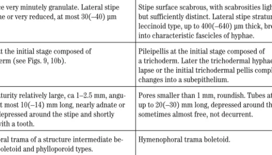
Tab. 4.Differences betweenXerocomellusandHemileccinum.
Xerocomellus Hemileccinum
Fruit bodies smaller and more slender than in
Hemileccinum. General appearance xerocomelloid.
Fruit bodies larger and more fleshy, with general ap-pearance intermediate betweenBoletusandLeccinum. Stipe surface very minutely granulate. Lateral stipe
stratum none or very reduced, at most 30(–40) µm thick.
Stipe surface scabrous, with scabrosities light-coloured but sufficiently distinct. Lateral stipe stratum of the leccinoid type, up to 400(–640) µm thick, breaking up into characteristic fascicles of hyphae.
Pileipellis at the initial stage composed of a palisadoderm (see Figs. 9, 10b).
Pileipellis at the initial stage composed of
a trichoderm. Later the trichodermal hyphae partly col-lapse or the initial trichodermal pellis completely changes into a subepithelium.
Pores at maturity relatively large, ca 1–2.5 mm, angu-lar. Tubes at most 10(–14) mm long,nearly adnate or somewhat depressed around the stipe and shortly decurrent with a tooth.
Pores smaller than 1 mm, roundish. Tubes at maturity up to 20(–30) mm long, depressed around the stipe, sometimes almost free, not decurrent.
Hymenophoral trama of a structure intermediate be-tween the boletoid and phylloporoid types.
Hymenophoral trama boletoid.
N e w c o m b i n a t i o n s
Xerocomellus armeniacus(Quél.) Šutaracomb. nov.
(MycoBank MB511892)
B a s i o n y m:Boletus armeniacusQuélet, in Guillaud, Forq. et Merlet, Ann. Sci. Nat. Bord. Sud-ouest Mém. 2: 42, 1884.
S y n o n y m:Xerocomus armeniacus(Quél.) Quél., Fl. mycol. France: 419, 1888.
Xerocomellus chrysenteron(Bull.) Šutaracomb. nov.
(MycoBank MB511893)
B a s i o n y m:Boletus chrysenteronBulliard, Histoire des champignons: 328, 1791. S y n o n y m:Xerocomus chrysenteron(Bull.) Quélet, Fl. mycol. France: 418, 1888.
Xerocomellus engelii(Hlaváček) Šutaracomb. nov.
(MycoBank MB511894)
B a s i o n y m:Boletus engeliiHlaváček, Mykologický Sborník 78(2): 66, 2001.
S y n o n y m s:Xerocomus quercinusEngel et Brückner nom. prov., Pilzfl. Nordwestoberfrankens 13/A: 75–78, 1990 (“1989”). –Boletus declivitatum(C. Martin) Watling, Edinb. J. Bot. 61(1): 43, 2005 (“2004”). –Xerocomus communis(Bull.) Bon, Doc. Mycol. 14(no. 56): 16, 1985 (“1984”) s. auct. non orig.
Xerocomellus fennicus(Harmaja) Šutaracomb. nov.
(MycoBank MB511895)
B a s i o n y m:Boletellus fennicusHarmaja, Karstenia 39: 37, 1999.
S y n o n y m:Xerocomus fennicus(Harmaja) Ladurner et Simonini, Mycol. Research 107: 672, 2003.
Xerocomellus marekii(Šutara et Skála) Šutaracomb. nov.
(MycoBank MB511896)
B a s i o n y m:Boletus marekiiŠutara et Skála, Czech Mycol. 59(1): 12, 2007.
S y n o n y m:Xerocomus marekii(Šutara et Skála) Klofac, Österr. Z. Pilzk. 16: 258, 2007.
Xerocomellus porosporus(Imler ex Moreno et Bon) Šutaracomb. nov.
(MycoBank MB511897)
B a s i o n y m:Boletus porosporusImler ex Moreno et Bon, Doc. Mycol. 7(nos. 27–28): 6, 1977. S y n o n y m:Xerocomus porosporus (Imler ex Moreno et Bon) Contu, Boletim da Sociedade Broteriana 63: 385, 1990.
In the mycological literature the basionym of this species is often cited in the following form: Boletus porosporus Imler ex Watling, Notes Roy. Bot. Gard. Edinb. 28(3): 305, 1968. In the author’s opinion, however, this basionym, particu-larly the designation of the type, was not published fully in agreement with no-menclatural rules. For this reason, the present author has here cited the later but validly published basionym of Moreno and Bon.
Xerocomellus pruinatus(Fr.) Šutaracomb. nov.
(MycoBank MB511898)
B a s i o n y m:Boletus pruinatusFries in Fr. et Hök, Boleti, fungorum generis, illustratio: 9, 1835. S y n o n y m:Xerocomus pruinatus(Fr.) Quél., Fl. mycol. France: 420, 1888.
Xerocomellus ripariellus(Redeuilh) Šutaracomb. nov.
(MycoBank MB511899)
B a s i o n y m:Xerocomus ripariellusRedeuilh, Doc. Mycol. 26(no. 104): 30, 1997.
S y n o n y m:Boletus ripariellus(Redeuilh) Watling et A. E. Hills, in Watling, Edinb. J. Bot. 61(1): 45, 2005 (“2004”).
Xerocomellus rubellus(Krombh.) Šutaracomb. nov.
(MycoBank MB511900)
Basionym:Boletus rubellusKrombholz, Naturgetr. Abbild. Beschr. Schwämme 5: 12, 1836. Synonym:Xerocomus rubellus(Krombh.) Quél., C. R. Ass. franc. Av. Sci. 24: 620, 1896 (“1895”).
In some phylograms published in recent phylogenetic analyses,Xerocomellus rubellusappeared (asBoletus rubellusorXerocomus rubellus) in several various places of the phylogenetic tree. In the author’s opinion, this was apparently
Fig. 10.–a:Pileipellis of a middle-aged fruit body ofXerocomus subtomentosus(JŠ 4121) composed of a typical, partly collapsed trichoderm. The pileus of this dried fruit body is 43 mm wide. –b: Pileipellis of a middle-aged fruit body ofXerocomellus porosporus(JŠ 4464) composed of a character-istic palisadoderm. The pileus of this fruit body in dried state measures 42 mm in diameter. The differ-ence in the structure of the pileipellis of these species is evident at first sight.
caused by misdetermination of some sampled specimens.Xerocomellus rubellus
is a bolete which has been sometimes confused with some other species.
S p e c i e s:Xerocomellus armeniacus(Quél.) Šutara, Xerocomellus chrysen-teron (Bull.) Šutara, Xerocomellus engelii (Hlaváček) Šutara, Xerocomellus fennicus (Harmaja) Šutara, Xerocomellus marekii (Šutara et Skála) Šutara,
Xerocomellus porosporus(Imler ex Moreno et Bon) Šutara,Xerocomellus prui-natus (Fr.) Šutara, Xerocomellus ripariellus (Redeuilh) Šutara, Xerocomellus rubellus(Krombh.) Šutara, and apparently some other, both European and extra-European xerocomoid species whose anatomy has the present author not yet studied, e. g. Boletus dryophilus Thiers, Boletus intermediusSmith et Thiers, Xerocomus truncatus Singer, Snell et Dick, Boletus zelleri(Murrill) Murrill and others.
HemileccinumŠutaragen. nov.
(MycoBank MB511891)
Carposomata pileata, stipitata, mediocriter magna vel magna. Superficies pilei initio subtomentosa, postea subtomentosa vel glabra. Pileipellis e trichodermide vel subepithelio formata. Tubuli depressi juxta stipitem, iuventute flavi, deinde olivaceo-flavi. Pori concolores, orbiculati et minuti (circa 0,5–1 mm). Tubuli et pori tactu immutabili. Structura tramae hymenophori boletoidea, cum stratis lateralibus gelatinosis. Sporae boletoideae, subfusoideae vel fusoideo-ellipsoideae, laeves. Pulvis sporarum olivaceo-brunneus. Stipes satis carnosus, superficies stipitis scabra. Structura stratorum periphericorum stipitis (strato laterali incluso) similis sicut in genereLeccinum. Tomentum basalis albidum vel luteolum, interdum partim pallide ochraceum vel brunneolum. Carne flava, pallide flavida vel albida, interdum intra basem stipitis brunneo vel rubro-brunneo maculata, fracta immutabilis. Fibulae in carposomate nullae.
Genus Hemileccinumdiffert: – a genere Xerocomus s. str. sporis laevibus, structura tramae hymenophori boletoidea cum stratis lateralibus gelatinosis, superficie stipitis scabra cum structura stratorum periphericorum stipitis simili sicut in genereLeccinumatque poris minutis, – a genere Leccinumhymenophoro flavo in combinatione cum carne immutabili, – a genereBoletussuperficie stipitis scabra cum structura stratorum periphericorum stipitis alia.
Ty p u s:Boletus impolitusFries.
C h a r a c t e r s. Fruit bodies stipitate-pileate, medium-sized to relatively large, with a general appearance partly resembling some species of Leccinum sect.
Luteoscabraand partly some representatives ofBoletuss. str.
Pileipellis composed of two rather different modifications of a trichoderm. In
Hemileccinum impolitum(≡Boletus impolitus) the trichoderm consists of cylin-drical, filamentous hyphae and only a small number of terminal cells is somewhat broadened at the very top. In later stages the trichodermal hyphae more or less collapse. InHemileccinum depilatum(≡Boletus depilatus) the pileipellis under-goes a conspicuous change during development. At the initial stage the pileipellis of this species consists of two layers: a suprapellis, which is composed of a trichodermal layer with loosely intertwined filamentous hyphae, and a subpellis whose hyphae are arranged more anticlinally and much densely. In this stage the
pileus surface looks finely tomentose. The pileus surface, however, soon becomes glabrous as the filamentous hyphae of the trichodermal suprapellis are washed away and completely disappear. In further stages, the pileus surface is thus com-posed of only the layer which was earlier the subpellis. The hyphae of this layer gradually broaden, become more or less inflate so that finally the pileipellis often looks like a subepithelium with predominantly ellipsoid to subglobose cells. The macrochemical reaction of the pileus surface with NH3vapours is violet.
Tubes depressed around the stipe apex, sometimes almost free, up to 20(–30) mm long when mature, when young light yellow to deep yellow, later olive-yellow. Pores concolorous, at maturity roundish and minute (ca 0.5–1 mm). Tubes and pores unchanging when bruised. Hymenophoral trama in a fully developed stage (i. e. in younger fruit bodies) boletoid. Lateral strata divergent and gelatinous, with hyphae distinctly distant from each other. In microscopic sections stained with Congo Red there is a conspicuous colour contrast between layers of the trama. Mediostratum is stained deep red and the lateral strata remain very light-coloured, with a hyaline gelatinous substance in the space between the hyphae. Pleurocystidia scattered, usually fusoid to lageniform, smooth and thin-walled.
Spores of boletoid shape, subfusoid or fusoid-ellipsoid in face view, with a more or less distinct suprahilar depression in profile. Spore surface smooth (Figs. 6, 7). Spore print olive-brown.
Stipe relatively fleshy, nearly cylindrical, elongate clavate or elongate subfusoid, covered with a scabrous ornamentation similar to that inLeccinum
with the only difference that the stipe scabrosities ofHemileccinumspecies are less conspicuous because they persistently remain light-coloured or darken only slightly. With age the stipe scabrosities at times collapse so that in some old fruit bodies the stipe surface seems to be almost glabrous. Except for the basal part, the surface of the stipe is composed of the following peripheral layers: a caulohymenium with scattered fertile caulobasidia, a caulosubhymenium (which is, however, often indistinct), and a lateral stipe stratum of the leccinoid type. The lateral stipe stratum is non-gelatinous, predominantly anticlinally ar-ranged, during growth of the stipe soon breaking up into characteristic fascicles of hyphae ending in elements of the caulohymenium, in well-developed scabrosities up to 400(–640) μm thick [the arrangement of the peripheral layers of the stipe of Hemileccinum impolitum (mistakenly named Leccinum fragrans) is depicted in Fig. 2, Šutara 1989]. Stipe trama composed of densely longitudinally arranged hyphae. Surface of the stipe base sterile, covered with a tangled tomentum of filamentous hyphae. The basal tomentum whitish or yel-lowish, in places sometimes pale ochraceous or brownish. Partial veil and annu-lus absent.
Context yellow, pale yellowish or whitish, sometimes with brownish or brown-red spots in the stipe base, when cut unchanging. Taste mild, odour usually
un-pleasant, reminding of iodoform, particularly in the lower part of the stipe. Hyphal system monomitic. Clamp connections not found in the fruit body.
Ectomycorhizal with frondose trees.
D e l i m i t a t i o n. In 1980 Bertault transferredBoletus impolitustoLeccinum.
Later, the present author studied the microscopic structure of the peripheral lay-ers of the stipes ofBoletus impolitusandBoletus depilatusand came to the con-clusion that the anatomy of the stipe of both these species is in essence of the same type as inLeccinum(Šutara 1989). For this reason, he proposed to transfer alsoB. depilatustoLeccinum. In recent years, however, it turned out that this so-lution was not very fortunate. Recent molecular analyses have shown that from the phylogenetic viewpoint these two species are very distant from Leccinum
(see Binder and Besl 2000, Eberhardt and Taylor 2005, Bakker and Noordeloos 2005, Binder and Hibbett 2007, etc.). The molecular analyses have moreover shown thatB. impolitusandB. depilatusare distant as well as fromBoletus, in which these species have been placed most frequently.
On the basis of molecular analyses, Binder and Besl (2000) placed the above-mentioned species in Xerocomus. In the phylogram published in 2000, B. impolitus and B. depilatus appeared close to Xerocomus subtomentosus, but new data published in the most comprehensive work concerning the phylogeny of
Boletales(Binder and Hibbett 2007, Supplementary: Fig. 1) show that the distance betweenX. subtomentosusand the first two species (which are here transferred to the new genusHemileccinum) is much greater than it seemed in 2000.
As suggested above, the anatomical structure of the peripheral stipe layers having a lateral stipe stratum of the leccinoid type is a very typical character of
Hemileccinum,which distinguishes this genus not only from all the other species which have been placed inXerocomuss. l. but also from the genusBoletus. Spe-cies ofXerocomuss. str. moreover differ fromHemileccinumin some other ana-tomical features, particularly in the spore ornamentation and the arrangement of the hymenophoral trama. Characters distinguishing Hemileccinum from
Xerocomellusare given in Tab. 4, and those distinguishing it fromXerocomuss. str. in Tab. 5.
As far asLeccinumis concerned,Hemileccinumis distinguished from this ge-nus by its yellow hymenophore in combination with the unchanging context. Fur-ther characters distinguishing Hemileccinum from Leccinumare the odour of iodoform and the violet macrochemical reaction of the pileus surface with NH3 va-pours.
Tab. 5.Differences betweenHemileccinumandXerocomus.
Hemileccinum Xerocomuss. str. Spore surface smooth. Spore surface bacillate. Hymenophoral trama in fully developed stage boletoid.
Lateral strata gelatinous, loosely arranged, with hyphae distinctly distant from each other. In preparations stained with Congo Red the mediostratum is deep red and the lateral strata are very light-coloured.
Hymenophoral trama phylloporoid. Lateral strata non-gelatinous, densely arranged, with hyphae touching or almost touching each other. In preparations with Congo Red the mediostratum is stained red in the same or almost the same intensity as the lateral strata. Ornamentation of the stipe scabrous. Lateral stipe
stra-tum of the leccinoid type, up to 400(–640) µm thick, breaking up into characteristic, predominantly anticlinally arranged hyphal fascicles.
Ornamentation of the stipe not scabrous. Lateral stipe stratum not leccinoid, at most 80(–200) µm thick, not breaking up into hyphal fascicles.
General appearance intermediate betweenBoletusand
Leccinum.
General appearance xerocomoid.
Pores roundish and minute, not larger than 1 mm. Tubes at maturity up to 20(–30) mm long, depressed around the stipe apex, often nearly free, not decurrent.
Pores when mature angular and relatively large, ca 1–3 mm. Tubes at most 15 mm long, nearly adnate or some-what depressed around the stipe and decurrent with a tooth.
N e w c o m b i n a t i o n s
Hemileccinum depilatum(Redeuilh) Šutaracomb. nov.
(MycoBank MB511901)
B a s i o n y m:Boletus depilatusRedeuilh, Bull. Soc. Mycol. France 101(4): 389, 1986 (“1985”). S y n o n y m s:Leccinum depilatum(Redeuilh) Šutara, Česká Mykol. 43(1): 4, 1989. –Xerocomus depilatus(Redeuilh) Binder et Besl, Micologia 2000: 85, 2000.
Hemileccinum impolitum(Fr.) Šutaracomb. nov.
(MycoBank MB511902)
B a s i o n y m:Boletus impolitusFries, Epicrisis systematis mycologici: 421, 1838.
S y n o n y m s: Xerocomus impolitus (Fr.) Quél., Fl. mycol. France: 417, 1888. – Leccinum impolitum(Fr.) Bertault, Bull. Soc. Mycol. France 96(3): 287, 1980.
S p e c i e s: Hemileccinum depilatum (Redeuilh) Šutara and Hemileccinum impolitum(Fr.) Šutara.
PseudoboletusŠutara Česká Mykol. 45: 2, 1991.
Ty p u s:Pseudoboletus parasiticus(Bull.: Fr.) Šutara (≡Boletus parasiticusBull.: Fr.). C h a r a c t e r s. Fruit bodies usually small, with a xerocomoid appearance.
Pileipellis composed of a trichoderm whose hyphae more or less collapse with age. Gelification of the trichodermal hyphae not observed.
Tubes adnate or subdecurrent. Pores medium large. Structure of the hymenophoral trama in a well-developed stage intermediate between the boletoid and phylloporoid types, sometimes nearly boletoid. Spores of boletoid shape, elongate fusoid-cylindric in face view, with a shallow suprahilar depression in pro-file. In the only European species of this genus (P. parasiticus) the spore surface has an unusual pitted ornamentation which is detectable only with electron mi-croscopy (see Oolbekkink 1991, Holec 1994). Spore print olive-brown.
Stipe solid, attached with its basal part to the fruit body of the host. Stipe sur-face sterile, composed of a filamentous trichoderm (see Šutara 1991: Figs. 1, 2; 2005: Fig. 4b). Caulobasidia absent. Stipe trama consisting of longitudinally ar-ranged hyphae. Lateral stipe stratum absent. No clamp connections found in the fruit body.
Ontogenetic development of fruit bodies hypo-(para)-velangiocarpous and pileostipiticarpous (Reijnders 1963, Singer 1986).
In the period of formation of fruit bodies, Pseudoboletus species live as mycoparasites on gasteromycetes (SclerodermaandAstraeus).
D e l i m i t a t i o n. Some molecular analyses (e. g. Bakker and Noordeloos 2005) indicate that from the phylogenetic viewpoint,Pseudoboletus parasiticus
(type species of the genus) is sufficiently distant from the other xerocomoid spe-cies.
Pseudoboletusdiffers from the other European species of theBoletaceaein the following characters: (1) its stipe surface is sterile, without caulobasidia, covered with a filamentous trichoderm, (2) its mode of obtaining nutrition, at least in a cer-tain period of its life, is parasitic, and (3) its ontogenetic development of fruit bod-ies is hypo-(para)-velangiocarpous. Another character which distinguishes P. parasiticusfrom other EuropeanBoletaceaeis the pitted surface ornamentation of the spores.
Pseudoboletusis moreover distinguished from genera which are macroscopi-cally most similar (Xerocomuss. str. andXerocomellus) by the following charac-ters: fromXerocomuss. str. by the structure of its hymenophoral trama which is intermediate between the boletoid and phylloporoid types, sometimes almost boletoid, and from Xerocomellus by its pileipellis composed of a filamentous trichoderm.
S p e c i e s:Pseudoboletus parasiticus (Bull.: Fr.) Šutara and the extra-Euro-peanPseudoboletus astraeicola(Imazeki) Šutara.
BoletusL.
Sp. Pl.: 1176, 1753 (nom. conserv.)
S y n o n y m s:TubiporusPaulet ex P. Karst., Rev. Mycol. 3: 16, 1881. –DictyopusQuél., Enchir. fung.: 159, 1886. –OedipusBataille, Les Bolets: 13, 1908. –SuillellusMurrill, Mycologia 1: 16, 1909. – CeriomycesMurrill, Mycologia 1: 140, 1909.
Ty p u s:Boletus edulisBull.: Fr. (typ. conserv.)
C h a r a c t e r s. Fruit bodies medium-sized or large, fleshy.
Pileipellis in an early stage usually composed of a trichoderm, which occurs in this genus in several modifications. The trichoderm moreover often changes its appearance according to weather conditions and age of fruit bodies. In cases that the trichodermal hyphae strongly collapse with age, the initial trichoderm may even change into a layer resembling a cutis with a predominantly periclinal hyphal orientation. The trichodermal hyphae of most Boletus species have also a ten-dency to gelatinise so that in favourable conditions their surface is covered with a thin layer of a gelatinous substance. For this reason, the pileus surface in this ge-nus is slightly viscid or at least sticky in wet weather conditions. In a few species the gelification is even so strong that the pileipellis forms a completely gelatinised layer – an ixotrichoderm.
Tubes when mature up to 20(–35) mm long, depressed around the stipe apex, sometimes almost free, when young white or yellow, later olive-yellow, yellow-brown, yellow-green or olive-brown. Pores usually roundish and minute, mostly concolorous with tube-sides, but sometimes coloured differently, e. g. red-orange, deep red or wine-red (in sectionLuridi). Hymenophoral trama in a fully devel-oped stage (i. e. in younger fruit bodies) boletoid, with a distinct gelification of the lateral strata (see Lohwag and Peringer 1937: Figs. 3, 5, 9). In microscopic sec-tions stained with Congo Red there is a conspicuous colour contrast between lay-ers of the trama. The mediostratum is stained deep red and the lateral strata are very light-coloured, hyaline in the space between the hyphae.
Spores smooth, of boletoid shape, elongate subfusoid or fusoid-ellipsoid, with suprahilar depression in profile. Spore print olivaceous or olive-brown when fresh.
Stipe usually fleshy, ornamented with a reticulation and/or floccose granules. Although reticulate and non-reticulate stipe surfaces have a somewhat different macroscopic appearance, from the anatomical viewpoint they are only two modi-fications of one type of arrangement of peripheral stipe layers. Stipe surface fer-tile, composed of a gradually fragmenting caulohymenium with scattered spore-bearing caulobasidia. Under the caulohymenium there is often a lateral stipe stra-tum which is usually developed best in young or middle-aged fruit bodies. With age this layer usually gradually disappears. The lateral stipe stratum is of the boletoid type, up to 60(–90) μm thick, loosely arranged, often gelatinous, consist-ing of hyphae runnconsist-ing divergently or irregularly from the longitudinal stipe trama