PII S0736-4679(01)00477-2
Clinical
Communications
TRAUMATIC RETROBULBAR HEMORRHAGE: EMERGENT DECOMPRESSION BY
LATERAL CANTHOTOMY AND CANTHOLYSIS
Susi Vassallo, MD,* Morris Hartstein, MD,† David Howard, MD,‡ and Jessica Stetz, MD* *Department of Surgery/Division of Emergency Medicine, New York University School of Medicine, Bellevue Hospital Center, New York, New York, †Department of Ophthalmology, New York University School of Medicine, Bellevue Hospital Center, New York, New
York, ‡Department of Ophthalmology and Cell Biology, New York University School of Medicine, Bellevue Hospital Center, New York, New York,
Reprint Address:Susi Vassallo,MD, FACEP, FACMT, 545 First Avenue 10C, New York, NY 10016
e Abstract—Traumatic retrobulbar hemorrhage may re-sult in acute loss of vision that is reversible when recognized and treated promptly. A case of traumatic retrobulbar hemorrhage is presented. The technique of emergent or-bital decompression by lateral canthotomy and cantholysis is described. The anatomy of the lateral canthus and the surgical procedure are illustrated by gross dissection. © 2002 Elsevier Science Inc.
e Keywords—retrobulbar hematoma; retrobulbar hemor-rhage; eye trauma; orbital trauma; lateral canthotomy; lateral cantholysis; orbital decompression; orbital hemor-rhage
INTRODUCTION
Traumatic hemorrhage into the retrobulbar space may result in acute visual loss. Although this ophthalmologic emergency presents most frequently to the Emergency Department (ED), discussion of this entity is notably absent from the Emergency Medicine literature. Prompt recognition and early decompression of retrobulbar he-matoma may prevent loss of vision or lead to return of vision. Although consultation with an ophthalmologist is ideal, specialty support is not always available in a timely fashion. The physician encountering eye emer-gencies must be comfortable evaluating, diagnosing, and
treating acute visual compromise secondary to retrobul-bar hematoma.
CASE REPORT
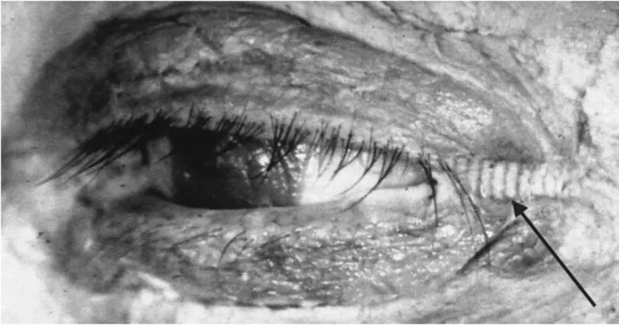
A 74-year-old man was struck with a blunt object over his left eye. He presented to the Emergency Department 3 h after the injury with complaints of pain and loss of vision. Initial visual acuity in the injured eye showed the ability to count fingers at two feet. Proptosis, severe periorbital edema and ecchymosis were noted (Figure 1). No sclera was visible secondary to diffuse subconjunc-tival hemorrhage and chemosis. The pupil was fixed at 3 mm. Extraocular muscle movements in the left eye were restricted in all directions. There was moderate resistance to retropulsion but formal intraocular pressure testing was not performed. The cornea was clear and the anterior chamber unremarkable. The optic nerve and retina also appeared grossly normal.
Orbital decompression was performed in the ED via lateral canthotomy and cantholysis. Almost immediately, extraocular muscle movements improved. Within 20 to 30 min, the patient’s vision improved to 20/70 in the affected eye. The following day the patient’s vision remained 20/70, with decreased proptosis and full ocular motility. Several intraretinal hemorrhages were noted; the optic nerve appeared normal.
RECEIVED: 25 October 2001; FINAL SUBMISSION RECEIVED: 2 July 2001;
ACCEPTED: 17 July 2001
The Journal of Emergency Medicine, Vol. 22, No. 3, pp. 251–256, 2002 Copyright © 2002 Elsevier Science Inc. Printed in the USA. All rights reserved 0736-4679/02 $–see front matter
Pathophysiology
The orbit is an enclosed space bound laterally and pos-teriorly by bony walls and anpos-teriorly by the globe and the superior and inferior orbital septa. When bleeding occurs within this cavity, there is little room to accommodate the increase in volume. The globe and septum are dis-placed anteriorly to some extent (proptosis); however, the capacity for forward movement is limited by the eyelids and the length of the optic nerve (1–3). Posteri-orly, the orbit is effectively closed as well, demonstrated by the fact that hemorrhages do not decompress sponta-neously by posterior dissection (1).
The orbital space can be likened to the intracranial
space (1). Because of the limited capacity for expansion, increased volume because of hemorrhage results in in-creased pressure and compression of contained struc-tures. The precise mechanism of the resultant neuropathy in the eye is not completely understood. The optic nerve may be compressed directly, or the vascular supply to the nerve may be compromised. Central retinal artery (CRA) ischemia is another proposed mechanism. Animal mod-els suggest that visual loss secondary to CRA ischemia may be reversible for up to 100 min (25).
Traumatic optic neuropathy may result from causes other than hemorrhage. The optic nerve can be transected at the moment of impact, or stretched and contused, leading to edema and visual loss (2,6–9). Emergent
Figure 2. The inferior and superior crura (thin arrows) of the lateral canthal tendon form a common tendon at the point of attachment to the inner aspect of the lateral orbital wall (thick arrow). This point of attachment is called Whitnall’s tubercle. The inferior crus of the lateral canthal tendon is cut in lateral cantholysis.
canthotomy is not indicated in instances when hemor-rhage into the enclosed orbital space is not present.
Emergent orbital decompression is reserved for the patient with a history of trauma who has visual loss, severe proptosis, diffuse subconjunctival hemorrhage, and marked periorbital edema. Partial or complete oph-thalmoplegia, an afferent pupillary defect, and resistance to retropulsion may be present as well. Usually visual acuity is decreased, though not necessarily initially. Vi-sion may be intact and then deteriorate, suggesting re-versible compression rather than nerve transection at the moment of impact (1,2,10). Less specific signs include periorbital crepitus and infraorbital hypesthesia.
The differential diagnosis of orbital hemorrhage in-cludes orbital cellulitis, isolated orbital fracture, and globe rupture. Orbital fractures may be accompanied by
enophthalmos because of depression of the supporting orbitalfloor. Proptosis is not present with fracture unless accompanied by orbital hemorrhage. Globe ruptures usu-ally cause low intraocular pressure and enophthalmos, without proptosis (11).
Retrobulbar bleeding, though most often because of trauma, may occur spontaneously. Many various causes have been reported, including venous anomalies, athero-sclerosis, intraorbital aneurysm of the ophthalmic artery, hemophilia, leukemia, von Willebrand’s disease, hyper-tension, and straining (12–19).
Anatomy
Understanding the anatomy of the lateral canthus is crit-ical for proper performance of lateral canthotomy and
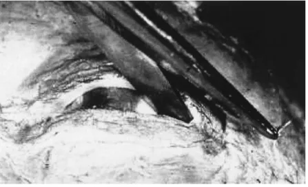
Figure 3. A clamp is placed horizontally across the lateral canthus to compress tissue and reduce bleeding.
Figure 4. The clamp is removed leaving an impression in the soft tissue.
cantholysis. The lateral canthal tendon is a combined tendon-ligament that provides structural fixation of the lids (tarsal plates) and orbicularis oculi muscle to the inner aspect of the bony lateral orbital wall (zygoma), just posterior to the orbital rim (Figure 2). The tendon has an inferior and superior portion (Figure 2). The osseous point at which the tendon attaches is called Whitnall’s tubercle (Figure 2). Dissection of the medial ends of the lateral canthal tendon demonstrates the liga-mentous attachments to the tarsal plates. The orbicularis oculi muscle lies anterior to the tarsal plates and also has tendinous attachments to Whitnall’s tubercle. Anterior to the lateral canthal tendon is a small pocket of orbital fat known as Eisler’s pocket (22). When the inferior portion of the lateral canthal tendon is cut, the lower lid loses its structuralfixation to the lateral orbital wall. It becomes
lax and is easily pulled away from the lid margin (2,20–22).
Procedure
The lateral canthal area is prepared and draped in sterile fashion. Because of the emergent nature of the clinical situation, some authors recommend only simple irriga-tion with normal saline (26). Lidocaine with epinephrine is administered to obtain anesthesia and hemostasis. A straight clamp is placed horizontally across the lateral canthus for about one to two minutes to compress the tissues and reduce bleeding (11) (Figure 3). The clamp is removed, leaving an impression in the edematous soft tissues (Figure 4). Sterile scissors are then used to make
Figure 5. Sterile scissors are used to make a one centimeter horizontal incision into the tissue at the clamp site.
Figure 6. It is helpful to grab the lower lid and pull it down and away from the lateral orbital margin. This serves to separate the skin and conjunctiva. The inferior crus is more easily palpated than visualized at this point. A 1 to 2 cm cut is made to lyse the inferior crus.
an approximately 1 cm horizontal incision in the tissue at the clamp site (11) (Figure 5). This initial canthotomy opens skin, orbicularis muscle, orbital septum, palpebral conjunctiva, and exposes Eisler’s fat pocket. It is helpful to grab the lower lid margin with a toothed forceps. Pulling the lid down and away from the lateral orbital margin separates the skin and conjunctiva (Figure 6). The inferior portion of the lateral canthal tendon can be easily palpated using a finger or the tip of the scissors (11). The tendon is more easily palpated than visualized (23). With the scissors pointed inferoposteriorly toward the lateral orbital rim, the inferior arm of the lateral canthal tendon is cut (Figure 7). This critical incision is approximately 1 to 2 cm in depth and length. The lower lid should easily pull away from the lid margin. Progres-sively deeper cuts are to be made until this effect is achieved (2).
Despite the decompression of high intraorbital pres-sure, only a small amount of blood is usually expressed with the release of the hematoma; there is no need to empty the orbit of its contents. Simply releasing the pressure of the hematoma is enough to restore vision or prevent further visual loss.
Regarding the closure of the canthotomy, most au-thors agree that it is to be performed several days after the procedure once the swelling has decreased (27).
Complications of Cantholysis
Extensive cantholysis may result in loss of adequate lower lid suspension and ectropion. However, this is easily repaired at a later date (24). Improper direction of the scissors superiorly may cause injury to the levator aponeurosis, resulting in ptosis. The lacrimal gland and lacrimal artery lie superiorly as well, and care must be taken to avoid these structures. After the acute process has resolved, the patient must be watched for infection and abscess formation. Fibrosis may develop, limiting extraocular motility (11).
CONCLUSION
Bleeding into the intraorbital space may cause acute visual loss by compressing the optic nerve and its vas-cular supply. Decompression of the tense orbit can re-verse a mechanical optic neuropathy or retinal ischemia. Emergent decompression of the orbital space is the treat-ment of choice for retrobulbar hematoma and may pre-serve or restore vision.
Acknowledgments—Special thanks to Robert S. Hoffman, MD, NYU School of Medicine, for his expertise in computer graph-ics. This work is supported in part by Research to Prevent Blindness.
REFERENCES
1. Katz B, Herschler J, Brick DC. Orbital haemorrhage and prolonged blindness: a treatable posterior optic neuropathy. Br J Ophthalmol 1983;67:549–53.
2. Linberg JV. Orbital compartment syndromes following trauma. Adv Ophthal Plastic and Reconstruct Surgery 1987;6:51–62. 3. Duke-Elder S. Text-book of ophthalmology. St. Louis: C. V.
Mosby Co.; 1954:5935.
4. Rosen P, Barkin R. Emergency medicine: concepts and clinical practice. St Louis, MO: Mosby; 1998:2703.
5. McCartney DL, Char DH. Return of vision following orbital de-compression after 36 h of postoperative blindness. Am J Ophthal-mol 1985;100:602–4.
6. Fry HJH. Reversible visual loss after proptosis from retrobulbar hemorrhage. Reproduction of the syndrome in the Cynomolgus Monkey. Plastic and Reconstructive Surgery 1969;44:5:480–3. 7. Park JH, Frenkel M, Dobbie JG, Choromoko E. Evulsion of the
optic nerve. Am J Ophthalmol 1971;72:969–91.
8. Hughes B. Indirect injury of the optic nerve and chiasma. Bull Johns Hopkins Hosp 1962;111:98–126.
9. Loewenstein A. Marginal haemorrhage of the disc. Partial Cross tearing of the optic nerve. Br J Ophthalmol 1943;27:208–15. 10. Anderson RL, Panje WR, Gross CE. Optic nerve blindness
fol-lowing blunt forehead trauma. Ophthalmology 1982;89:445–55. 11. Krausen AS, Ogura JH, Burde RM, Ostrow DE. Emergency orbital
decompression: a reprieve from blindness. Otolaryngol Head Neck Surg 1981;89:252–6.
12. Friedberg MA, Rapuano JB. Wills Eye Hospital Office and
Emer-Figure 7. With the scissors pointed inferoposteriorly toward the lateral orbital rim, the inferior arm of the lateral canthal tendon is cut. Reprinted with permission from Soll, D. B. (ed.), Management of complications in ophthalmic plastic surgery. Birmingham, AL: Aesculapius Publishing Co.; 1976: 261.
gency Room diagnosis and treatment of eye disease. New York: JB Lippincott Co.; 1990:36–9.
13. Petrelli RL, Petrelli A, Allen W. Orbital hemorrhage with loss of vision. Am J Ophthalmol 1980;89:593–7.
14. Krohel GB, Wright J. Orbital Hemorrhage. Am J Ophthalmol 1979;88:254–8.
15. Myerson L, Lazar SJ. Intraorbital aneurysm of the ophthalmic artery. Br J Ophthalmol 1971;55:199–204.
16. Rubenstein RA, Albert DM Scheie HG. Ocular Complications of hemophilia. Arch Ophthalmol 1966;76:230–2.
17. Zimmerman A, Merigan TC. Retrobulbar hemorrhage in a hemo-philiac with irreversible loss of vision. Arch Ophthalmol 1960;64: 949.
18. Reese AB, Guy L. Exopthalmos in leukemia. Am J Ophthalmol 1933;16:718.
19. Kubik J. Spontaneous hemorrhage into the orbit in hypertension. Cesk Oftalmol 1963;19:267–9.
20. Law FW. Spontaneous orbital hemorrhage. Br J Ophthamol 1971; 55:556–8.
21. Whitnall SE. On a tubercle on the malar bone, and on the lateral attachments of tarsal plates. J Anat Physiol 1911;45:426–32. 22. Gioia VM, Linberg JV, McCormick SA. Anatomy of the lateral
canthal tendon. Arch Ophthalmol 1987;105:529–32.
23. Kestenbaum A. Applied anatomy of the eye. New York: Grune and Stratton; 1976:18–19.
24. Roberts J, Hedges J. Clinical procedures in emergency medicine. Portland, OR: WB Saunders Company; 2000:1116.
25. Soll DB. Management of complications in ophthalmic plastic surgery. Birmingham, AL: Aesculapius Publishing Co.; 1976:261. 26. Albert D, Jakobiec F. Principles and practice of ophthalmology.
Portland, OR: WB Saunders Company; 2000:5195.
27. Anderson RL, Gordy DD. The tarsal procedure. Arch Ophthalmol 1979;97:2192–6.