PDF hosted at the Radboud Repository of the Radboud University
Nijmegen
The following full text is a publisher's version.
For additional information about this publication click this link.
http://hdl.handle.net/2066/150248
Please be advised that this information was generated on 2017-12-05 and may be subject to
change.
Bio-inspired cross-linking methods for hydrogel formation
Proefschrift
ter verkrijging van de graad van doctor aan de Radboud Universiteit Nijmegen op gezag van de rector magnificus, volgens besluit van het college van decanen
in het openbaar te verdedigen op woensdag 20 januari 2016 om 14.30 uur precies
door
Anika Maartje Jonker
geboren op 30 maart 1987 te Almere
Promotor
Prof. dr. ir. J.C.M. van Hest Co-promotor
Dr. D.W.P.M. Löwik Manuscriptcommissie Prof. dr. A.E. Rowan
Prof. dr. R.P. Sijbesma (TU Eindhoven) Dr. K.M. Bonger
Paranimfen Saskia Bode Annika Borrmann
Cover design Angela van de Weg
Kristel Schumacher, Studio Lakmoes, Arnhem Barbara Wagensveld, Studio Lakmoes, Arnhem Layout and printed by Gildeprint - Enschede ISBN: 978-94-6233-183-9
The research presented in this thesis was financially supported by Agentschap NL, Ministry of Education, Culture and Science
Table of Contents
List of abbreviations 5
List of amino acids 7
Chapter 1 Peptide- and protein based hydrogels 9
Chapter 2 Physical cross-linking of star-PEG polymers 51
using calcium-binding peptides
Chapter 3 Soft PEG-hydrogels with independently tunable 89
stiffness and RGDS-content for cell adhesion studies
Chapter 4 A fast and activatable cross-linking strategy 119
for hydrogel formation
Chapter 5 SPAAC and SPOCQ cross-linked hydrogels as matrices 143
for encapsulation of aligned peptide amphiphile fibres
Chapter 6 Dual cross-linked hydrogels by combining chemical and 169
physical cross-linking
Chapter 7 Future perspectives 195
Summary 205
Samenvatting 211
Dankwoord 217
Publications 221
List of abbreviations
α-cyano α-cyano-4-hydroxycinnamic acid BCN (1R,8S,9S)-bicyclo[6.1.0]non-4-yn-9-yl Boc tert-Butyloxycarbonyl
BOP Benzotriazol-1-yl-oxy-tris dimethylaminophosphonium hexafluorophosphate
CD Circular dichroism
CuAAC Cu(I)-catalyzed azide-alkyne cycloaddition DCM Dichloromethane
DIPCDI N,N’-diisopropylcarbodiimide DiPEA N,N-diisopropylethylamine
DLS Dynamic light scattering
DMEM Dulbecco’s Modified Eagle Medium DMF Dimethylformamide
DMSO Dimethylsulfoxide
DOPA 3,4-dihydroxyphenylalanine DHPA 3,4-dihydroxyphenylacetic acid
DMPC 1,2-dimyristoyl-sn-glycero-3-phosphocholine DPPC 1,2-dipalmitoyl-sn-glycero-3-phosphocholine
ECM Extracellular matrix
EDTA Ethylenediaminetetraacetic acid
EthD-1 Ethidium homodimer-1
Et3N Triethylamine Et2O Diethylether EtOAc Ethylacetate Fmoc Fluorenylmethyloxycarbonyl G’ Storage modulus G’’ Loss modulus
HBTU N,N,N’,N’-Tetramethyl-O-(1H-benzotriazol-1-yl)uronium hexafluorophosphate
HeLa Henrietta Lacks
HMPA Hexamethylphosphoramide
HOAc Acetic acid
HOBt N-hydroxybenzotriazole
HOS Human osteosarcoma
HPLC High Performance Liquid Chromatography IR Infrared
LC-MS Liquid chromatography-Mass Spectrometry
MALDI-TOF Matrix-assisted laser desorption/ionization – time of flight
MAS Magic-angle spinning
MeCN Acetonitrile MeOH Methanol MMP Matrix metalloproteinase MS Mass Spectrometry MT Mushroom Tyrosinase MW Molecular weight
NaIO4 Sodium periodate
NHS N-hydroxy succinimide
NMR Nuclear Magnetic Resonance
OSu N-hydroxysuccinimide
PBS Phosphate buffered saline
PEG Poly(ethylene)glycol
PyBOP Benzotriazol-1-yl-oxy-tripyrrolidinophosphonium hexafluorophosphate PyBrOP Bromotripyrrolidinophosphonium hexafluorophosphate
r.t. Room temperature
Rt Retention time
SEC Size-exclusion chromatography
SPAAC Strain-promoted azide-alkyne cycloaddition
SPOCQ Strain-promoted oxidation-controlled cyclooctyne–1,2-quinone cycloaddition SPPS Solid phase peptide synthesis
TEM Transmission electron microscopy
TFA Trifluoroacetic acid
TFE Trifluoroethanol
TIS Triisopropylsilane
TLC Thin-layer chromatography
List of amino acids
Ala A Alanine Arg R Arginine
Asp D Aspartic acid
Asn N Asparagine Cys C Cysteine Gln Q Glutamine
Glu E Glutamic acid
Gly G Glycine His H Histidine Ile I Isoleucine Leu L Leucine Lys K Lysine Met M Methionine Phe F Phenylalanine Pro P Proline Ser S Serine Thr T Threonine Trp W Tryptophan Tyr Y Tyrosine Val V Valine
Chapter 1
Peptide- and protein based hydrogels
Temperature, pH changes in the body
Solution Hydrogel formation
Part of this chapter has been published as:
R1 R2 R3 R4 R5 R6 R7 R8 R9 R10 R11 R12 R13 R14 R15 R16 R17 R18 R19 R20 R21 R22 R23 R24 R25 R26 R27 R28 R29 R30 R31 R32 R33 R34 R35 R36 R37 R38 R39
R1 R2 R3 R4 R5 R6 R7 R8 R9 R10 R11 R12 R13 R14 R15 R16 R17 R18 R19 R20 R21 R22 R23 R24 R25 R26 R27 R28 R29 R30 R31 R32 R33 R34 R35 R36 R37 R38 R39
Peptide- and protein based hydrogels 11
1
1.1. Introduction
Hydrogels are three-dimensional, hydrophilic polymer networks, capable of absorbing large amounts of water or biological fluids, up to thousand times their dry weight. They are insoluble due to the presence of cross-links between the constituents that form the polymeric network. They are often made of homopolymers or copolymers, but can also consist of small molecules that self-assemble into high molecular weight structures.1,2 The first hydrogel report dates back to the study by Wichterle and Lim (1960), who polymerized and cross-linked 2-hydroxyethyl methacrylate into a transparent gel, which showed promising results in the manufacturing of contact lenses.3 Since this pioneering work, the number of publications on hydrogels has increased every year.4,5 Hydrogels can be classified in a number of ways. They can for example be categorized based on the nature of their building blocks, being either neutral or ionic, or natural or synthetic in origin. Furthermore, hydrogels are often distinguished based on their mechanical and structural characteristics, such as the degree of cross-linking and the integrity of the gels. Finally, an important classification is the way in which cross-linking occurs, which can be either chemical or physical.2 Hydrogels formed by chemical cross-links undergo significant volume changes during the transition from solution (sol) to gel state. These chemical cross-links can be formed in many different ways, such as disulfide formation, (photo)polymerisation or the reaction between thiols and acrylates or sulfones. The density of cross-linking can easily be varied and with this also the mechanical properties of the final material. Physically cross-linked hydrogels, on the other hand, do not undergo significant volume changes during the sol-gel transition. They can be prepared via the self-assembly of polymers, which occurs in response to external stimuli such as pH and temperature. The polymers generally provide relatively low viscous solutions that can rapidly form a gel when an external trigger is applied. Often physical cross-linking leads to weaker gels than chemical cross-linking; therefore these gels are more susceptible to mechanical forces such as shear. This can also be used to its advantage, as it makes physical hydrogels suitable as injectable materials (Figure1.1). Another advantage of physical linking is that it mostly does not depend on the addition of organic solvents or cross-linking reagents, offering possibilities for these hydrogels in biomedical applications such as the controlled delivery of drugs and cells and in tissue engineering.6
Tissue engineering is an important research field, since every year millions of people suffer from the loss or failure of organs or tissues. These patients are treated by organ or tissue transplantations. Since there is a huge shortage of donors, new treatments such as tissue engineering gain increasing attention.7-10 In this research field, man-made organs or tissues are developed to restore, improve or maintain tissue function.9 These materials are usually made by incorporation of the patient’s own cells into polymer scaffolds, such as poly(glycolic acid) (PGA) and poly(lactic acid) (PLA). These scaffolds are capable of delivering the cells into the body,
R1 R2 R3 R4 R5 R6 R7 R8 R9 R10 R11 R12 R13 R14 R15 R16 R17 R18 R19 R20 R21 R22 R23 R24 R25 R26 R27 R28 R29 R30 R31 R32 R33 R34 R35 R36 R37 R38 R39
thereby providing a site for new tissue formation. Administration of these polymer scaffolds into the body occurs via cuts or incisions. In addition, polymer scaffolds can also be used as a substrate for the attachment and migration of cell populations.7,8 As an alternative, hydrogels are thought to constitute an excellent starting point as a matrix to grow new tissue in. The gels can be designed to be biodegradable and their mechanical and structural properties to resemble those of many tissues. Importantly, hydrogels can be designed to be able to transplant the cells into the body by simple injection, thereby minimizing the invasive manner (Figure 1.1).11
Temperature, pH changes in the body
Solution Hydrogel formation
Figure 1.1. A schematic representation of an injectable hydrogel. At low temperatures, these hydrogels are soluble. Cells or drugs (represented by the black dots) are mixed with the solvent. After injection, temperature or pH changes in the body are responsible for hydrogel formation, resulting in the delivery of the cells or drugs to the body.
In the design of these hydrogels, several criteria have to be taken into account. A highly important factor is the biocompatibility. The injectable materials should be biocompatible, in the sense that they do not damage adjacent cells or in some other way induce undesired responses. Another issue that needs to be addressed is cell adhesion; hydrogels should interact favourably with the type of cells they want to deliver. Inappropriate interaction might lead to undesirable tissue formation. Furthermore, the degradation behaviour of the hydrogels is important for tissue engineering applications. The rate of degradation should be in line with the tissue development, since the degradation of the hydrogels may influence the mechanical properties. Importantly, the degradation products should be biocompatible as well.7
An important guide in the design of hydrogels for tissue engineering is the extracellular matrix (ECM). The ECM is the biological material that surrounds cells in tissues. It is furthermore responsible for mechanical support and provides an environment for new tissue formation.12 The ECM itself consists mainly of proteoglycan filaments, collagen fibres and elastin. The collagen
R1 R2 R3 R4 R5 R6 R7 R8 R9 R10 R11 R12 R13 R14 R15 R16 R17 R18 R19 R20 R21 R22 R23 R24 R25 R26 R27 R28 R29 R30 R31 R32 R33 R34 R35 R36 R37 R38 R39
Peptide- and protein based hydrogels 13
1
fibres provide tensile strength and durability to the surrounding tissue. Elastin enables thestretch and recoil of the ECM network. The proteoglycan filaments are built up from proteins and hyaluronic acid to form coiled structures. Proteoglycan binds water, sequesters growth factors and supplies compression resistance. At low protein concentrations, the ECM structure can be considered gel-like.13,14 Hence, mimicking the ECM is an important goal in the design of hydrogels for tissue engineering and is considered as a good way to develop biomaterials with control over cell adhesion, proliferation, migration and differentiation.15
Peptides and proteins are often used in the development of hydrogels, since they exhibit some advantageous characteristics, such as biocompatibility and biodegradability. The incorporation of peptides into biomaterials can be performed using several chemical reactions including click chemistry, Michael addition of cysteine residues to vinyl sulfones or maleimides, UV-initiated cross-linking and native chemical ligation. These reactions all take place with a high level of chemical specificity. This is often not possible for proteins, since the exact composition of the interaction region is not precisely known. Incorporation of proteins into biomaterials is therefore often performed via non-specific amine-carboxylic acid couplings. Peptides can be incorporated into biomaterials to infer a number of functions. This includes cell-binding, growth factor binding, surface binding and/or proteolytic susceptibility.16 The most frequently used peptide sequence in this respect is the arginine-glycine-aspartic acid (RGD) sequence.17 The incorporation of this cell adhesion motif into biomaterials has already been reviewed extensively in literature. 8,18-21 Efforts are being made to introduce combinations of peptides during the synthesis of biomaterials, in order to integrate multiple functions.16
On the other hand, peptides and proteins can also be employed as moieties that provide structural integrity to a gel, e.g. via self-assembly or aggregation. In this chapter an overview is given of hydrogels in which peptides and proteins are structural elements of the polymer network. Starting from hydrogels which are derived from naturally occurring structural proteins, we will gradually move from all-protein and peptide-based synthetic systems to hybrid hydrogels which are composed of synthetic polymeric and peptide structural elements. We will illustrate the potential of these hydrogels with applications which are mainly derived from the field of tissue engineering. Finally, an overview of chemical cross-linking methods is described. For articles which give a broader overview of biomaterials used in hydrogel formation or which discuss in more detail the mechanical properties of peptide-based hydrogels one is referred to excellent reviews by Schacht et al. and Pochan et al.4,22
In summary, hydrogels are promising materials for use as biomaterials, since they have high water content, have a tuneable viscoelasticity, are biocompatible and can be made injectable. Both peptides and proteins have advantageous features for use as building blocks in the synthesis of
R1 R2 R3 R4 R5 R6 R7 R8 R9 R10 R11 R12 R13 R14 R15 R16 R17 R18 R19 R20 R21 R22 R23 R24 R25 R26 R27 R28 R29 R30 R31 R32 R33 R34 R35 R36 R37 R38 R39
biomaterials and hydrogels in particular. As mentioned above, an important requirement for the design of hydrogels is the biocompatibility. Hydrogels based on peptides or proteins often fulfil this prerequisite and are easily degraded by the body.
1.2. Natural protein-based hydrogels
As described above, proteins display interesting properties for use in the synthesis of hydrogels. Especially their excellent biocompatibility and biodegradability make them promising candidates in biomedical applications. A concise description of proteins derived from natural sources which are used for the preparation of biomaterials is provided. Since usually biosynthetic procedures are used to mimic the sequences of natural proteins, these are more extensively described in Paragraph 1.3. One is referred to excellent reviews for more information about elastin23,24, collagen and gelatin.25,26
1.2.1. Elastin
Elastin is a major structural protein of the extracellular matrix (ECM) that regulates the cell/cell interactions and the cell/ECM interactions. Elastin provides the ECM with tensile strength and elasticity, required for proper functioning.27 Typically, natural elastin hardly has been used as a biomaterial in tissue engineering. An important requirement when using proteins as biomaterial is the purity. In the synthesis of elastin, elastic fibres are formed on microfibrillar components acting as scaffold. Elastin then has to be purified from the elastic fibres. Contaminations from the preparation, like calcium-binding microfibrillar components, can lead to immunological responses in the human body. Moreover, elastin is insoluble and has a strong tendency to calcify, making purification even more difficult.28
1.2.2. Collagen and gelatin
Most of the collagens are fibrous proteins, responsible for important mechanical functions throughout the body. Collagens are particularly present in articular and bone tissues, where they provide most of the biochemical properties essential for proper functioning. Furthermore, collagens can bind and release cellular mediators such as cytokines and growth factors.29 The structural element of collagen is the triple helix, consisting of a -Gly-Xaa-Yaa- repeat. The amino acids Xaa and Yaa are usually proline and hydroxyproline (Hyp).30 Collagen is widely used as biomaterial in tissue engineering. It is used in its native fibrillar form, as well as after denaturation. Denaturation offers the possibility to fabricate several collagen forms, including sheets, tablets, pellets and sponges.31 Collagen has for example been used to repair tendon defects in rabbits32, for curing of chronic wounds caused by diabetes33 and as delivery system for proteins, drugs and genes.34 When the triple-helix structure of collagen is broken into single-strand molecules,
R1 R2 R3 R4 R5 R6 R7 R8 R9 R10 R11 R12 R13 R14 R15 R16 R17 R18 R19 R20 R21 R22 R23 R24 R25 R26 R27 R28 R29 R30 R31 R32 R33 R34 R35 R36 R37 R38 R39
Peptide- and protein based hydrogels 15
1
gelatin is obtained. Gelation of a gelatin solution occurs easily by a change in temperature.Gelatin gels have shown to be non-immunogenic, biodegradable and biocompatible, making them suitable candidates for biomedical applications. However, the thermal and mechanical stability of the gelatin gels is rather low. For long term biomedical applications, the stability of gelatin gels is improved by chemical modifications such as cross-linking.7,35 Gelatin-based materials have already been used in several tissue engineering applications, for example in adipose tissue repair36 and as bone tissue engineering material.37
1.2.3. Silk fibroin
Silks are natural structural proteins produced by insects such as spiders and worms. Silk produced by the silkworm Bombyx mori is called silk fibroin. This fibrous protein contains two protein components: fibroin and sericin. Fibroin consists of light (~26 kDa) and heavy chain (~390 kDa) polypeptides, which are present in a 1:1 ratio and are connected by a disulfide bridge. Silk fibroin forms β-sheets, due to the presence of the highly repetitive amino acid motif GAGAGS (Figure 1.2).38,39 H N O N H O H N O N H O H N O N H O OH n
Figure 1.2. The ubiquitous GAGAGS motif present in silk fibroin induces the formation of β-sheets.
These β-sheets are responsible for the formation of silk fibroin hydrogels. Due to the formation of the β-sheets, the silk fibroin becomes insoluble in water. Hydrophobic interactions among the protein chains in fibroin lead to the assembly of the material into hydrogels. Gelation can be enhanced by an increase in temperature or fibroin concentration. Furthermore, gelation can be promoted by a decrease in pH or by addition of a hydrophilic polymer. Both factors stimulate desorption of water and decrease the repulsion between the protein chains. Addition of Ca2+ ions also accelerates the formation of hydrophobic interactions, presumably because the pH is lowered by ionic interactions with the COO- ions of the amino acid side chains.40 Silk fibroin hydrogels are biocompatible, have unique mechanical properties, are biodegradable and display cellular interactions.41 For example, in vitro and in vivo tests on rabbits with cancellous bone defects showed bone healing and stimulated cell proliferation.42
1.2.4. Globular proteins
Many studies have been performed on the gelation processes of globular proteins such as bovine serum albumin, β-lactoglobulin and ovalbumin. From these proteins, β-lactoglobulin is the one most intensively studied. This protein, with a molecular mass of 18.6 kDa, is widely used in the food industry. This globular protein denatures when heated, leading to either large linear
R1 R2 R3 R4 R5 R6 R7 R8 R9 R10 R11 R12 R13 R14 R15 R16 R17 R18 R19 R20 R21 R22 R23 R24 R25 R26 R27 R28 R29 R30 R31 R32 R33 R34 R35 R36 R37 R38 R39
aggregates or small primary aggregates, both consisting of peptide fragments.43,44 The small aggregates can further grow into larger aggregates and at a certain concentration self-assemble into gels. Alternatively, the large linear aggregates can phase separate, leading to protein-rich domains. These can also further aggregate and form gels.43
1.3. Biosynthetic polypeptide-based hydrogels
Natural protein-based hydrogels also display some disadvantages for use as biomaterials. As outlined above, their purification and low stability might be problematic. Furthermore, when using samples from natural sources, it is difficult to obtain identical compositions. As an alternative, synthetic polypeptide-based hydrogels have been investigated. These materials are prepared using methods such as gene expression in bacteria, via recombinant technology and solid phase peptide synthesis. Synthetic hydrogels can contain sequences mimicking those of natural proteins. Functionalities to increase the biological or mechanical properties of the hydrogels can easily be incorporated. Moreover, modifications in the peptide sequences can simply be made, in order to improve the features of the hydrogels. The following part of this chapter gives an overview of biosynthetic polypeptide-based hydrogels.
1.3.1. Collagen-based synthetic hydrogels
Hydrogels based on collagen have been designed for the purpose of tissue engineering. As an example of the solid phase peptide synthesis approach, a small library of Nap-Gly-Phe-Phe-Tyr-Gly-Gly-Xaa-Hyp was synthesized (Figure 1.3). The repeating collagen sequence Gly-Xaa-Hyp was used, in which amino acid Xaa was Lys, Glu, Ser, Ala or Pro. This sequence was connected to Nap-Gly-Phe-Phe-Tyr, a peptide with an N-terminal naphthyl (Nap) moiety that is able to form gels. O H N O N H O H N O N H H N O NH O H N O N O OH R OH COOH
Figure 1.3. Nap-Gly-Phe-Phe-Tyr-Gly-Gly-Xaa-Hyp. Residue Xaa can be Lys, Glu, Ser, Ala or Pro. Due to the naphthyl moiety (Nap), all 5 members are capable of forming hydrogels.
All five members were capable of forming hydrogels in phosphate buffered saline. The construct with lysine proved to give the strongest hydrogel. A construct without the collagen domain Gly-Xaa-Hyp was also prepared. This compound also formed a hydrogel, but more extensive
R1 R2 R3 R4 R5 R6 R7 R8 R9 R10 R11 R12 R13 R14 R15 R16 R17 R18 R19 R20 R21 R22 R23 R24 R25 R26 R27 R28 R29 R30 R31 R32 R33 R34 R35 R36 R37 R38 R39
Peptide- and protein based hydrogels 17
1
hydrogen bonds between the peptides were found in the other, collagen-based constructs. Inorder to test the biocompatibility of the gels, in vitro cell culture experiments were performed. Mouse embryonic fibroblast cells (3T3) were placed on top of the hydrogels. After 72 h, most cells on the surface of the Lys-, Glu- and Ser-containing constructs were still alive. Fewer 3T3 cells were observed on the surface of Ala- and Pro-containing hydrogels and, if present, many of them were dead. These two hydrogels are thus not suitable for 3T3 cell culture, possibly due to the less polar side chains of Ala and Pro. The gel with lysine proved to be the most suitable in these tests, probably because the positively charged lysine increases the binding to the negative cell surface.45
E. coli cannot be used for the synthesis of hydroxyproline containing collagens, since this bacterial host lacks the ability to perform the post-translational modification of proline to hydroxyproline. In order to broaden the applications of collagen as injectable biomaterial, hydroxyproline-free collagen mimetic peptides (CMP) were synthesized. An example is the (GPP)3 GPR GEK GER GPR (GPP)3 GPCCG peptide, containing a (GPP)3 triplet, that stabilizes the formation of a triple-helix. Analysis of the CMP indeed revealed the formation of stable triple helices, the important structural feature of collagen. The observed assembly behaviour suggests that these hydroxyproline-free peptides can be used as collagen mimetics, with the advantageous possibility to use E. coli for their production.46 An application of collagen-based hydrogels as delivery system was recently reported. CMPs were used to create DNA polyplex-modified collagens. The release of the DNA polyplexes was demonstrated and could also be tuned by varying the CMP content.47 In another example, the release of green fluorescent protein and human epidermal growth factor was shown from a highly cross-linked CMP-based 3-D matrix. After release of the growth factor, it remained active, which was shown by its ability to stimulate cell growth. When cell binding motif RGDS was incorporated in the CMP constructs, encapsulated epithelial cells formed spheroid structures. This result shows the potential of collagen-based hydrogels for mimicking the ECM.48
Collagen was also taken as a basis for the preparation of triblock thermo-reversible gels as shown by Skrzeszewska and co-workers via a protein engineering approach.49 The end blocks consisted of the (Pro-Gly-Pro)9 sequence, which forms collagen-like triple helices. The middle block was a 399 amino acid long sequence containing the (Gly-Xaa-Yaa)n structure and, besides proline, only hydrophilic residues such as asparagine and glutamine.49,50 This highly hydrophilic sequence is not able to form secondary structures and hence maintains a random coil conformation. Stable gels were only obtained when three of the collagen-like sequences form a triple helix, since double helices are not stable. The triblock construct was present as a viscous solution at temperatures above 50°C, but formed a gel at room temperature. This thermo-responsive behaviour was shown to be reversible, hence an increase in temperature
R1 R2 R3 R4 R5 R6 R7 R8 R9 R10 R11 R12 R13 R14 R15 R16 R17 R18 R19 R20 R21 R22 R23 R24 R25 R26 R27 R28 R29 R30 R31 R32 R33 R34 R35 R36 R37 R38 R39
led to the soluble form. The gelation process of the triblock construct was used to develop a theoretical model that is able to predict the viscoelastic behaviour of polymer gels.49 In a later study rheology, light scattering and atomic force microscopy were conducted to investigate the pH-dependent fibre- and gel formation of the triblock constructs. Monomers were found at low pH, but with a pH value above 6, these self-assembled into fibres and then hydrogels. At physiological pH, gels appeared to have the highest moduli, although gel strength was found to be dependent on the purification and preparation method of the gels.51 As a first test, rat bone marrow cells were cultured in the presence of the triblock hydrogels. These cells remained viable, but comparison with collagen controls showed lower cell proliferation and mineralization.52 Possibly these results could be employed in the design of novel collagen-based gels with predictable and controllable properties.49
1.3.2. Elastin-like polypeptides
As indicated above, elastin has important biological functions but is very difficult to be used in biomedical applications. Therefore, elastin-like polypeptides (ELPs) have been designed, inspired by natural elastin. These ELPs contain a pentapeptide repeat VPGXG, in which X can be any natural amino acid, except proline (Figure 1.4).53
N H O N O H N O N H O H N O n R
Figure 1.4. The ELP pentapeptide repeat VPGXG. Residue X can be every natural amino acid, except proline.
The gelation behaviour of ELPs is temperature-dependent. At low temperatures, the hydrophobic residues are surrounded by ordered water from hydrophobic hydration, resulting in a soluble aqueous ELP. Above a certain transition temperature, the surrounding water becomes less-ordered and bulky, leading to the collapse of the polymer. This gives rise to folding and self-assembly of the ELP, hence gelation occurs.54 The stimulus responsive behaviour of ELPs has extensively been described by the Chilkoti and the Urry groups.55-58
Since the immune system does not differentiate between ELPs and natural elastin, these materials hold great promise as drug deliverers or in tissue engineering. Additionally, the degradation products of ELPs only contain natural amino acids, making them perfectly biocompatible. Poly(VPAVG) is a member of the elastin-like polymers, in which the central glycine has been replaced by an alanine. Poly(VPAVG) shows a similar self-assembly process. It has been studied
R1 R2 R3 R4 R5 R6 R7 R8 R9 R10 R11 R12 R13 R14 R15 R16 R17 R18 R19 R20 R21 R22 R23 R24 R25 R26 R27 R28 R29 R30 R31 R32 R33 R34 R35 R36 R37 R38 R39
Peptide- and protein based hydrogels 19
1
whether poly(VPAVG) is suitable as carrier for the delivery of bone morphogenetic proteins(BMPs). These BMPs are responsible for triggering the differentiation of stem cells during bone formation. BMP-2 and BMP-14 were successfully released from the elastin-like polymers and proved to increase the mineralization of C2C12 cells. Poly(VPAVG) thus has potential to be used in tissue engineering applications in bone.53
Apart from changes in temperature, the ordering of ELPs into hydrogels can also be initiated by performing chemical cross-linking. A variety of cross-linking strategies is known, but in order to use the ELPs for tissue engineering, the choice is limited. Many methods cannot be used because the cross-linking reagents are toxic to cells. Cross-linking reactions that require organic solvents are also not suitable, since this eliminates the possibility of encapsulating cells before cross-linking.56 Lim et al. therefore developed a new cross-linking strategy for ELPs.59 Lysine-containing ELPs in aqueous solution were reacted with the amine reactive cross-linker β[tris(hydroxymethyl) phosphino] propionic acid (THPP), resulting in the rapid formation of hydrogels (Figure 1.5). The mechanical properties of the cross-linked ELP gels can be controlled by the number of lysine residues and by the pH at which the cross-linking reaction takes place. Cell viability studies after encapsulation of mouse fibroblasts in these ELP hydrogels revealed that cross-linking with THPP is not cytotoxic.59 NH H N NH2 O HN O H N NH2 P COO HO HO OH P COO HN N H NH H2O ELPs THPP THPP crosslinked ELP hydrogel
Figure 1.5. Hydrogel formation by the cross-linking between the ELP Lys-residues and THPP.
The THPP cross-linking strategy has been used by Nettles et al. to develop an ELP hydrogel for repair of the cartilage matrix in goats.60 For this purpose, the knees of goats with osteochondral defects were filled by injecting the THPP-cross-linked ELP gel. After 3 months, a significant increase in integration was observed for the ELP filled knees, compared to the unfilled knees. However, after 6 months this increase was not maintained, due to degradation of the ELP-hydrogel. Optimizations of these hydrogels should make them useful for long-term benefits in cartilage repair applications.60 Lim et al. further optimized ELP gels for biological applications.61 ELP triblock copolymers were synthesized with an ABA structure. The central B-block was hydrophilic and surrounded by hydrophobic cross-linkable A domains. Gel formation occurred
R1 R2 R3 R4 R5 R6 R7 R8 R9 R10 R11 R12 R13 R14 R15 R16 R17 R18 R19 R20 R21 R22 R23 R24 R25 R26 R27 R28 R29 R30 R31 R32 R33 R34 R35 R36 R37 R38 R39
rapidly with THPP as cross-linking agent. It was shown that the presence of the non-cross-linkable B block and the variation of its length influenced the swelling, structure and mechanical properties of the gels. Cell viability of fibroblasts was also proven; it was actually found that the ELP triblock copolymers led to a greater cell proliferation than a monoblock ELP hydrogel (containing only the A block). Since the mechanical properties of ELP hydrogels can be tuned, gels can be designed in such a way that they support cell viability and that they are suitable for tissue repair applications.61
Another chemical cross-linking method used in combination with ELPs is the formation of disulfide linkages. Asai et al. used this method to form hydrogels via intramolecular disulfide cross-links between cysteine containing ELPs. Hydrogen peroxide (0.3%) was used to accelerate the gelation process, leading to gels formed within 2.5 min.62 Cysteine containing ELPs were also used by the Chaikof group for functionalization with an RGD peptide utilizing maleimide-thiol chemistry.63 The properties of ELPs can furthermore be modified with chemical cross-linkers. Genipin, glutaraldehyde and disulfide formation can all be used to cross-link ELP polymer films. Varying the cross-linking method and strategy enables tailoring of mechanical and physical properties as well as drug release profiles of ELP hydrogels.64 The porous ELP materials offer space for transplanted cells to grow in, to generate their own extracellular matrix. Rodríguez-Cabello and co-workers developed a method to synthesize macroporous ELP hydrogels as polymeric scaffolds, because they anticipated that materials containing (VPGIG)n would display elastin-like properties such as elasticity and biocompatibility.65 Some lysine residues were incorporated in the elastin blocks for cross-linking purposes. The second block contained the cell adhesive motive REDV, specific for endothelial cells. The final block consisted of a hexapeptide sequence for elastase-proteolytic action. Cross-linking of this ELP triblock material was performed using a so-called salt-leaching/gas-foaming technique, in which the amount of salt and the particle size control porosity and pore size of the material. This technique allowed the authors to prepare macroporous ELP hydrogels with tuneable pore size. It was found that HUVEC cells were present both on the surface and in the inner layers of the ELP hydrogel, showing that this type of ELP hydrogels indeed has potential for use as an artificial extracellular matrix.65
1.3.3. Silk-like polypeptides
Schacht et al. examined hydrogels made from β-sheet forming silk proteins.66 To this purpose, a spider silk protein called eADF4 (C16) was produced, consisting of 16 repeats of molecule C. Its sequence is GSSAAAAAAAASGPGGYGPENQGPSGPGGYGPGGP and mimics the silk protein ADF4 of the garden cross spider. Hydrogelation of the eADF4 (C16) protein took place at increasing concentration, invoked by dialysis. Gelation occurred via the transition of random coil conformations and α-helices into β-sheets. In order to increase the stability, the hydrogels
R1 R2 R3 R4 R5 R6 R7 R8 R9 R10 R11 R12 R13 R14 R15 R16 R17 R18 R19 R20 R21 R22 R23 R24 R25 R26 R27 R28 R29 R30 R31 R32 R33 R34 R35 R36 R37 R38 R39
Peptide- and protein based hydrogels 21
1
that formed were chemically cross-linked. The tyrosine residue side chains of the eADF4 (C16)protein were connected using ammonium peroxodisulfate and tris(2,2’-bipyridyl)dichloro-ruthenium(II). The cross-linking resulted in a higher mechanical strength for the hydrogels. The mechanical properties of the eADF4 (C16) hydrogels were furthermore influenced by addition of fluorescein. This interfered with the assembly into β-sheets resulting in less stable gels.66
1.3.4. Silk-elastin-like polypeptides
Silk-elastin-like polypeptides (SELPs) are composed of elastin-like and silk-like peptide components. The silk-like blocks provide chemical and thermal stability, since they have the ability to form hydrogen-bonded β-sheet crystals. The degree of crystallization is reduced by the elastin-like blocks, thereby introducing flexibility and aqueous solubility to the SELPs. These silk-elastin-like materials are liquid at room temperature, but form gels at elevated temperature. It appeared that the gel formation was caused by the crystallization of the silk-like components. This hypothesis was tested by the addition of urea, which prevents the formation of hydrogen bonding. Gelation was indeed not observed, since the silk-like blocks were unable to form the hydrogen-bonded β-sheet crystals. The addition of seed crystals enhanced the rate of gelation, further proving that the crystal formation of the silk-like blocks is responsible for the gelation. In a further study, the gelation of SELPs was found to be a two-step process. In the first step, it indeed is the hydrogen bonding between the silk blocks which causes spontaneous self-assembly. In this first step, micellar-like particles are formed. In the second step, the SELP molecules are ordered by hydrophobic interactions between the elastin blocks, leading to hydrogel or nanoparticle formation. 67-69 The ratio of elastin-like and silk-like components can be varied, as well as the block lengths and the sequence. This influences the biological, physicochemical and material properties of the formed hydrogels.67 Successful diffusion out of the SELP hydrogels was demonstrated for small molecules like vitamin B12, cytochrome c and theophylline70, for DNA71 and for adenoviruses.72 This shows that SELP hydrogels are already applied as biomaterials. Since their gelation is dependent on both elastin-like and silk-like proteins, a high level of control is possible. This makes SELP hydrogels very suitable for more applications in the controlled release of therapeutics.
By introducing stimulus sensitivity, more applications of SELPs are foreseen, including use as sensors and drug delivery devices. Nagarsekar et al. synthesized protein polymers, aiming for pH and temperature sensitivity.73,74 These SELPs were composed of repeating silk-like (GAGAGS) and elastin-like (VPGVG) sequences. The general structure of the synthesized SELPs is [(Gly-Val-Gly-Val-Pro)4-Gly-X-Gly-Val-Pro-(Gly-Val-Gly-Val-Pro)4-Gly-Ala-Gly-Ala-Gly-Ser]11, in which residue X can be either glutamic acid or valine. A transition from a solution state to a turbid state was observed for these protein polymers upon increasing temperature. Increasing ionic strength leads to less hydration of the polymer by water molecules, meaning that precipitation
R1 R2 R3 R4 R5 R6 R7 R8 R9 R10 R11 R12 R13 R14 R15 R16 R17 R18 R19 R20 R21 R22 R23 R24 R25 R26 R27 R28 R29 R30 R31 R32 R33 R34 R35 R36 R37 R38 R39
of the polymer occurs at lower temperatures. Furthermore, a decrease in transition values was also shown with increasing protein polymer concentrations. Altogether, these results show that mutations of neutral into ionic residues and changes in polymer length, gives the possibility to control the sensitivity of SELPs towards pH, temperature and ionic strength.73,74
1.3.5. Hydrogels based on a coiled-coil motif
A natural way of self-assembly of biological systems is the formation of an α-helical coiled-coil. Most of these coiled-coil sequences contain the characteristic heptad repeat abcdefg, with hydrophobic residues at positions a and d and with charged amino acids usually located at positions e and g (Figure 1.6). These peptides form a helical conformation, with the hydrophobic amino acids located at one side of the helix. The hydrophobic interhelical interfaces thus obtained, promote the aggregation into coiled-coil dimers. These aggregates are stabilized by the pH-dependent interactions between the charged residues.75 In the Woolfson group, two 28-residue peptides were designed to yield hydrogel materials. To ensure the formation of coiled-coil dimers, each peptide contained heptad repeats with isoleucines at position a and leucines at position d. To form a dimer with sticky ends, oppositely charged glutamate and lysine residues were placed on positions e and g. The complementary sticky ends promoted the assembly of the two peptides in α-helical coiled-coil fibrils.76
a
g
e
d
a
c
f
b
a
e
g
a
d
b
f
c
Figure 1.6. The coiled-coil dimer based on the heptad repeat (abcdefg). Positions a and d are usually occupied by hydrophobic amino acids, whereas charged residues are often found at positions e and g. The residues located at positions b, c and f are exposed on the surface of the coiled coil.
By choosing the right amino acids for position b, c and f, the interactions between the fibres might increase, resulting in a further assembly into hydrogels. It was chosen to make combinations of alanine and glutamine, since alanine can enhance the weak hydrophobic interactions between the fibrils and glutamine has the tendency to make hydrogen bonds. The constructs containing three alanines and the one with three glutamines on positions b,c and f were shown to form a gel. The possibility to use the three alanine- containing hydrogels as a substrate for cell growth was tested using rat adrenal pheochromocytoma cells. It was shown that the two peptide component hydrogel supports cell differentiation and cell growth.77
In another study, the coiled-coil domain of the cartilage oligomeric matrix protein was used to design hydrogels. This polypeptide is able to self-assemble into pentamers. After functionalization
R1 R2 R3 R4 R5 R6 R7 R8 R9 R10 R11 R12 R13 R14 R15 R16 R17 R18 R19 R20 R21 R22 R23 R24 R25 R26 R27 R28 R29 R30 R31 R32 R33 R34 R35 R36 R37 R38 R39
Peptide- and protein based hydrogels 23
1
with a cysteine residue, linkage to polymer PEGDA was performed using a Michael-typeaddition reaction. These polypeptide-polymers formed hydrogels upon exposure with UV-light. Fibroblasts encapsulated in the hydrogels showed spreading and migration, making these materials suitable for tissue engineering applications.78
The Tirrell group studied a protein containing six heptad repeats to make up a leucine zipper helix.75 The design of the peptide was based on the sequence of the Jun oncogene product and on a database containing naturally occurring coiled-coil proteins. A triblock copolymer was designed with the leucine zipper domains flanking the central polyelectrolyte domain. This central region was based on the Ala-Gly-rich sequence [(AG)3PEG]10 and was water soluble. The triblock copolymer formed a switchable hydrogel. Gelation was lost at pH values above 9.5 and at temperatures above 55°C, due to dissociation of the coiled-coil domains. The biosynthetic approach allowed controlling the characteristics of both the leucine zipper domains and the polyelectrolyte region. This gives the possibility to design hydrogels with specific features, aiming at the controlled release of therapeutics.75
Another series of triblock polypeptides were designed by Xu and co-workers based on the coiled-coil domain.79 Polypeptides ABA, CBA, ABC and CBC all contained two terminal coiled-coil motifs, A and C. The sequence of block A was (VSSLESK)6 and the C block sequence was (VSSLESK)2-VSKLESK-KSKLESK-VSKLESK-VSSLESK. Block C was obtained by changing one valine and three serine residues from block A in lysine residues. The central B block was the water-soluble random coil polyelectrolyte segment [(AG)3PEG]10. The balance between oligomerisation of blocks A and C and the swelling of block B should result in self-assembly of the triblocks. The thermal stability of the four triblocks was measured. The difference between the constructs containing block C, is the presence of four more lysine residues compared to block A. The introduced lysine amino acids disrupted hydrophobic interactions and introduced electrostatic repulsive forces. As a consequence, the thermal stability decreased, resulting in a lower temperature needed for coiled coil association. Triblock CBC indeed gave the lowest temperature (45°C) for unfolding of the polymer, whereas ABA was thermally stable even at temperatures as high as 95°C. All four constructs formed viscoelastic hydrogels at sufficiently high concentrations. The concentration to obtain elastic gels was the lowest for triblock CBC. When the same concentration range was taken for all constructs, it was found that the solution of ABA only contained monomers and dimers. In contrast, dimer to tetramer association was shown for triblock CBC. This explains why lower concentrations of CBC already form gels and furthermore indicates that gelation is related to the association of the coiled coil blocks. The triblocks were also shown to be pH sensitive. Finally, the reversibility of secondary structure formation was tested for polymer CBC. Addition of denaturant GdnHCl resulted in unfolding of the α-helical structure. After removal of the denaturant, the polymer folded back to its
R1 R2 R3 R4 R5 R6 R7 R8 R9 R10 R11 R12 R13 R14 R15 R16 R17 R18 R19 R20 R21 R22 R23 R24 R25 R26 R27 R28 R29 R30 R31 R32 R33 R34 R35 R36 R37 R38 R39
original structure and hydrogels were formed again. Since these triblocks are stimuli responsive and hydrogelation is reversible, applications in the biomedical field are foreseen.79
In contrast to these examples where the coiled-coil motif was essential for hydrogel formation, Mehl et al. designed a protein hydrogel not forming a coiled-coil. This gel was based on the four-helix bundle of the E. coli protein GrpE. This protein contained an amphipathic four-helix at each end and a random coil as the middle domain. The domain in between these two was an α-helix, not able to form a coiled coil structure. The amphipathic helices of the protein dimer were able to form a four-helix bundle. Instead of coiled-coil driven gelation, hydrogels were formed by simple hydrophilic and hydrophobic interactions forming the four-helix bundle.80
1.3.6. Two-component hydrogels
Two-component protein-engineered hydrogels have been designed by the Heilshorn group.81 For the synthesis of the two components, two protein association domains were chosen. The first was domain WW, consisting of conserved tryptophan residues. This WW domain is known to fold into antiparallel β-sheets. Secondly, a proline-rich protein domain was chosen. The first component consisted of multiple WW domains, linked together by hydrophilic spacers containing the cell adhesion motif RGDS. These spacers were designed to introduce flexibility and biofunctionality. The second component contained the proline-rich proteins, also linked by hydrophilic spacers. Upon mixing of the two components, physical cross-linking occurred, leading to the formation of a hydrogel. This type of material is therefore called a mixing-induced two-component hydrogel (MITCH). The viscoelasticity of the hydrogels can be altered, by adjusting the binding strength between the two domains and by changing the frequency of the repeated domains per component. This type of gels could be advantageous for use in the encapsulation of cells and proteins. Since gelation is not induced upon a change in temperature, pH or ionic strength, these cells and proteins can be kept under constant physiological conditions. The three cell types PC-12, HUVEC and NSC were encapsulated in these hydrogels. In all cases, the cells were distributed throughout the gels and were highly viable. Moreover, the two-component hydrogels showed complete self-healing after shear-thinning and can thus be useful as injectable materials for clinical use.81 In a follow-up study, the proline-rich protein was linked to 8-armed PEG. Upon mixing with the WW domain, a MITCH-PEG construct was obtained. In this way, multiple weak interactions were combined to get a stronger binding. This system allows altering the cross-linking density of hydrogels both by changing the PEG content as well as by peptide avidity. When endothelial cells were cultured in the MITCH-PEG hydrogels, a well-spread morphology was found.82
R1 R2 R3 R4 R5 R6 R7 R8 R9 R10 R11 R12 R13 R14 R15 R16 R17 R18 R19 R20 R21 R22 R23 R24 R25 R26 R27 R28 R29 R30 R31 R32 R33 R34 R35 R36 R37 R38 R39
Peptide- and protein based hydrogels 25
1
Two-component hydrogels were also investigated by Kiick et al.83 In their research, a PEG starpolymer was modified with heparin (PEG-LMWH), since this is known to sequester and stabilize growth factors. The second component consisted of the heparin-binding growth factor VEGF. Hydrogels were formed by mixing the PEG-LMWH and the VEGF component in phosphate buffered saline. Gelation was induced by the effective cross-linking between VEGF and LMWH. VEGF-receptors were shown to selectively remove VEGF, which thereby causes gel erosion (Figure 1.7). These results demonstrate that therapeutically relevant growth factors can be used as cross-linkers in hydrogel formation and can afterwards selectively be released in the body upon action of receptors.83 Another two-component hydrogel with heparin was studied by Wieduwild and co-workers. Star-PEG was functionalized with several heparin-binding peptide motifs. These peptides form α-helices upon interaction with heparin, leading to assembly and thus hydrogel formation. The peptide-star-PEG / heparin hydrogels could encapsulate cells which remained metabolically active.84
Figure 1.7. The two-component hydrogel PEG-LMWH/VEGF and the receptor-mediated gel erosion. Reproduced with permission from ref 83.83
1.4. Oligopeptides
1.4.1. Hydrogels based on β-sheet forming peptides
Another type of hydrogels is based on the β-sheet motif of peptides. The peptides used to make this type of hydrogels are designed to be soluble in aqueous solutions, displaying a random coil conformation. Only after addition of an external stimulus, the peptides form a β-hairpin conformation. After rapid self-assembly, a cross-linked, β-sheet rich hydrogel is obtained. External stimuli used to induce gel formation include heat, light and changes in pH or ionic strength.85 In the Messersmith group, the 16 residues containing peptide (FEFEFKFK)2 (FEK16) was studied.86 This peptide consists of alternating hydrophilic and hydrophobic amino acids and is able to self-assemble into β-sheet structures. The self-assembly was induced by the addition of salts. This resulted in shielding of the repulsive electrostatic interactions, thereby favouring attractive hydrophobic and Van der Waals forces. Peptide FEK16 is fully soluble in water and was shown to form β-sheets when the salts NaCl, CaCl2 and KCl were added in mM concentrations. It was investigated whether this assembly could be triggered by the addition of stimuli-responsive liposomes. Both temperature- and light-sensitive liposomes containing CaCl2 were prepared
R1 R2 R3 R4 R5 R6 R7 R8 R9 R10 R11 R12 R13 R14 R15 R16 R17 R18 R19 R20 R21 R22 R23 R24 R25 R26 R27 R28 R29 R30 R31 R32 R33 R34 R35 R36 R37 R38 R39
and added to a FEK16 solution. CaCl2 was released after applying the thermal or photochemical trigger, resulting in the formation of highly cross-linked β-sheet hydrogels. This peptide hydrogel was suggested to have potential in drug delivery, tissue engineering and wound healing.86 In the Schneider and Pochan groups, the 20 amino acid containing peptide MAX1 was synthesized.87 This peptide (VKVKVKVK-VDPPT-KVKVKVKV-NH
2) contains a tetra-peptide surrounded by hydrophobic valine and hydrophilic lysine residues. MAX1 formed an amphiphilic
β-hairpin at high pH (9.0) or after addition of salt at physiological pH. Addition of the cell culture growth media DMEM (Dulbecco’s Modified Eagle Medium) to the aqueous peptide solution resulted in β-hairpin folding, followed by self-assembly into a gel (Figure 1.8). Studies performed on model fibroblasts revealed that MAX1 hydrogels are cytocompatible and maintain their rigidity during cell proliferation.87
+ DMEM (cell culture
media) Self-Assembly
Unfolded peptide Foldedβ-hairpin nano-structureHydrogel
Figure 1.8. Hydrogelation of the MAX1 peptide. Addition of cell culture medium results in folding of the peptide in a β-hairpin, followed by self-assembly into a gel. Reproduced with permission from ref 87.87
The same researchers also synthesized peptide MAX8, in which one lysine was replaced by a glutamic acid (VKVKVKVK-VDPPT-KVEVKVKV-NH
2). This lowered the overall positive charge of the peptide, which should make folding and self-assembly faster in the same cell culture conditions. It was indeed shown that the MAX8 hydrogel was formed faster than MAX1, thereby maintaining the way of folding and self-assembly. The MAX8 hydrogels were used for the encapsulation of C3H10t1/2 mesenchymal stem cells, resulting in a homogeneous distribution. To be able to deliver the gel-cell constructs by syringe, the gels must show shear-thinning behaviour. The MAX8 hydrogel became a low-viscosity gel after shear stress was applied. When the stress was no longer applied, the gel quickly recovered its initial rigidity. These properties make MAX8 hydrogels useful for the delivery of cells in tissue-regeneration applications.85 MAX8 hydrogels are more rigid and contain more cross-links than MAX1 gels, leading to smaller mesh sizes. Dextran macromolecules and protein probes encapsulated in the MAX gels were released in rates varying from days till a month. Fastest release rates were recorded for MAX1, due to the larger mesh size.88 An additional study further demonstrated that electrostatic interactions have an influence on the release rate. Positively charged proteins encapsulated in the MAX8 hydrogel experience repulsive electrostatic interactions, which enhanced the protein release
R1 R2 R3 R4 R5 R6 R7 R8 R9 R10 R11 R12 R13 R14 R15 R16 R17 R18 R19 R20 R21 R22 R23 R24 R25 R26 R27 R28 R29 R30 R31 R32 R33 R34 R35 R36 R37 R38 R39
Peptide- and protein based hydrogels 27
1
from the gel. On the other hand, less than 10% of the initially loaded amount of negativelycharged proteins is released from these gels. Since the peptide hydrogel is positively charged, the strong attraction with the negatively charged proteins reduces the release.89 Furthermore, electrostatic interactions are also known to play a role in cell adhesion, cell viability and cell proliferation. Therefore, the MAX8 alternative HLT2 was designed, with a lower overall charge state (+5 compared to +7) and thus a less electropositive character. These constructs showed comparable gel formation and could both be used for cell encapsulation and syringe delivery of cells. However, when primary bovine chondrocytes were encapsulated, the less electropositive HLT2 showed higher cell viability and better supported maintenance of morphology.90 Further research on MAX1 and the more hydrophobic analogue MAX35 focussed on the lytic behaviour. Both constructs bound to negatively charged liposomes and induced release of their content by pore formation.91
1.4.2. Short aromatic peptides
Short aromatic peptide derivatives also have the ability to form hydrogels.92,93 Dipeptides protected with the fluorenylmethoxycarbonyl (Fmoc) group can self-assemble through the formation of hydrogen bonds and π-π interactions. The latter involves the attractions between the π-electrons in the aromatic fluorenyl rings of Fmoc. The gel formation of dipeptide Fmoc-Phe2 was tested, by dissolving the peptide in a basic solution (pH > 8.0). The drop wise addition of hydrochloric acid, to lower the pH, resulted in gel formation (Figure 1.9).94 Gelation of the dipeptide was also possible in cell culture medium DMEM. Fmoc-Phe2 dipeptides were also mixed in a 1:1 ratio with Fmoc-Ser, Fmoc-Lys and Fmoc-Asp. All four resulting hydrogels were shown to support the viability of chondrocytes. Cell viability of human dermal fibroblasts was increased by the Fmoc-Phe2/Ser and Fmoc-Phe2/Asp gels. The Fmoc-Phe2/Ser gels furthermore also supported viability of 3T3 fibroblasts and retention of cell morphology of chondrocytes.95 In another study, the effect of Fmoc-Phe2 and Fmoc-RGD gels on dermal fibroblasts was further investigated. The fibroblasts were encapsulated in a gel containing both Fmoc-Phe2 and Fmoc-RGD. Adhesion of the fibroblasts was promoted and cell spreading was observed, through specific binding of the RGD-motif to integrins. These results show that short aromatic peptide derivatives might be used as scaffolds for in vitro tissue regeneration.96 Inspired by the results obtained for the natural amino acid phenylalanine, a short peptide sequence containing the non-proteinogenic amino acid taurine was developed. This motif also proved to be a hydrogelator, resulting in gels containing up to 98% water.97
R1 R2 R3 R4 R5 R6 R7 R8 R9 R10 R11 R12 R13 R14 R15 R16 R17 R18 R19 R20 R21 R22 R23 R24 R25 R26 R27 R28 R29 R30 R31 R32 R33 R34 R35 R36 R37 R38 R39
Figure 1.9. Hydrogelation of short aromatic peptides. Upon applying an external trigger, the peptides associate with each other, leading to self-assembly into hydrogels. Adapted from ref. 92.92
The gelation of short peptide sequences was further investigated by utilizing different aromatic capping groups. Peptide sequence Gly-Phe-Phe-Tyr was capped with naphthalene, phenothiazine, benzyloxycarbonyl (Cbz) and with Fmoc. All constructs formed nanofibres, which entangled with each other to form the hydrogel network. The peptide with capping group phenothiazine was the most efficient gelator, as it already could form hydrogels at a very low concentration (0.01 wt%).98 Analogous to this, pyrene and Fmoc were used as capping group for tyrosine-leucine hydrogelators. Both constructs were assembled and co-assembled with serine surfactants, also capped with both pyrene and Fmoc. This allows variation in peptide and aromatic content. In all cases self-assembly behaviour was found, caused by aromatic interactions as wells as by
β-sheet-type arrangements.99 In another research, the Phe-Phe sequence was capped with indole, resulting in gels with a high storage modulus.100 Constructs with only one amino acid were also able to form hydrogels, for example Fmoc-Phe-OH101,102 and Fmoc-Tyr-OH.102 Even without the Fmoc group, but with the aromatic pyrene moiety as capping group, gelation of the single amino acid Phe was possible.103
1.4.3. Peptide amphiphiles
An important driving force for self-assembly is amphiphilicity. There are three types of amphiphilic peptides distinguished in literature.104 One type of peptide-amphiphiles is composed of a hydrophobic aliphatic tail and a hydrophilic peptide sequence, linked to each other via an amide bond. Amphiphiles have the ability to self-assemble in aqueous solution, in such a way that the hydrophilic domains are exposed to the water, while the hydrophobic regions are hidden from the aqueous environment. Amphiphiles can self-assemble in several organizations, including spheres, rods, disks, channels and sheets. With the introduction of peptide elements
R1 R2 R3 R4 R5 R6 R7 R8 R9 R10 R11 R12 R13 R14 R15 R16 R17 R18 R19 R20 R21 R22 R23 R24 R25 R26 R27 R28 R29 R30 R31 R32 R33 R34 R35 R36 R37 R38 R39
Peptide- and protein based hydrogels 29
1
in amphiphilic molecules, chemical functionalities can be introduced. Peptide amphiphiles oftenself-assemble in cylindrical micelles, called nanofibres. The core of these nanofibres is formed by the aliphatic tail, while the peptide sequence is displayed on the outer surface. This self-assembly can be controlled by adjustment of the pH or by addition of divalent cations such as Ca2+ and Mg2+. When the peptide-amphiphile concentration is sufficient, the nanofibres further assemble into a gel.15,105 Since the pioneering work of Stupp et al., peptide amphiphile nanofibre gels have been applied in the field of tissue engineering.106 One example is the use of peptide-amphiphiles containing the cell adhesion motif RGDS. These constructs were tested as scaffolds for the delivery of bone marrow derived stem and progenitor cells. In vitro tests showed that the cells are viable and proliferative. In vivo tests revealed an increase in the relative bioluminescent signal intensity coming from viable cells, indicating cell proliferation. Furthermore, the nanofibre gel showed only a mild tissue reaction, suggesting biocompatibility and biodegradability of the peptide amphiphiles. These results demonstrate the use of peptide-amphiphiles as injectable materials for bone marrow cell therapies.107
Another application of peptide-amphiphiles has been demonstrated by the Kokkoli group.108 They developed a fibronectin-mimetic peptide amphiphile containing the peptide sequences GRGDSP and PHSRN. The sequence GRGDSP contains the tripeptide RGD, which is naturally present in the cell binding domain of natural extracellular matrix (ECM) proteins, like fibronectin. Peptide sequence PHSRN was incorporated to enhance the activity and specificity of the fibronectin-mimetic. These two peptide domains were connected by a linker to mimic the natural distance between the respective domains and the overall hydrophobicity/ hydrophilicity. The thus designed PR_b peptide [KSSPHSRN(SG)5RGDSP] was linked to a C16 double hydrocarbon tail, to form the peptide-amphiphile.108 This construct showed excellent cell adhesion properties compared to other fibronectin-mimetic peptides, but was unable to form hydrogels.108,109 For this purpose, peptide PR_g [GGGSSPHSRN(SG)
5RGDSP] was synthesized, containing the spacer GGGSS. In contrast to KSS, this spacer was able to promote nanofibre formation. Peptide-amphiphiles were created by linkage of peptide PR_g to a C16 hydrocarbon tail. The PR_g construct self-assembled into nanofibres, followed by a further aggregation into hydrogels. In order to test their biological properties, HUVECs (human umbilical vein endothelial cells) were adhered on the surface on the gels. A comparison with PEG gels, fibronectin-functionalized PEG and commercially available peptide hydrogels (PuraMatrix) showed that PR_g hydrogels support cell adhesion and proliferation and thereby outperforms the comparison gels. Peptide-amphiphile nanofibre hydrogels are therefore promising as ECM mimetic scaffolds in tissue engineering.109
Several small peptides have been synthesized by the Zhang group as another kind of amphiphilic biomaterials.110 These peptides contain alternating hydrophobic and hydrophilic amino acids. The charged residues used have an alternating positive or negative charge. Solutions of these
R1 R2 R3 R4 R5 R6 R7 R8 R9 R10 R11 R12 R13 R14 R15 R16 R17 R18 R19 R20 R21 R22 R23 R24 R25 R26 R27 R28 R29 R30 R31 R32 R33 R34 R35 R36 R37 R38 R39
peptides in deionized water formed stable β-sheets. After exposure to electrolytes, self-assembly into nanofibres took place. These fibres further self-assembled into stable hydrogels. Peptide concentrations of 0.1-1% were sufficient for gelation to occur. Oligopeptide KLD-12 was synthesized, aiming for cartilage tissue repair (Figure 1.10).
Figure 1.10. Oligopeptide KLD-12. The peptide contains alternating positive and negative charges, leading to the formation of ß-sheets and assembly into nanofibres and gels. Reproduced with permission from ref 110.110
An efficient encapsulation of chondrocytes into the hydrogels was achieved. It was shown that the chondrocytes were evenly distributed over the hydrogel and maintained a high cell viability (>80%). Furthermore, the chondrocytes retained their morphology. The peptide hydrogels formed a cartilage-like ECM, showing their potential as scaffold for cartilage tissue repair.110 Apart from the chondrocytes, other cell interactions with these hydrogels have also been investigated, including osteoblasts,111 neural cells112 and endothelial cells.113
Another example of a small amphiphilic peptide for hydrogel formation was recently given by the Arosio group. Model peptide RADA 16-1 (RADARADARADARADA) was chosen as it is known to self-assemble into fibrils and higher ordered structures. The peptide first formed short rigid fibrils with a length of approximately 100 nm. Strong hydrophobic interactions led to the formation of longer fibrils and aggregates. At higher peptide concentrations hydrogels were formed, which were broken down upon dilution.114
Also peptide-based amphiphilic block copolymers have been designed for hydrogel formation. This category contains polymers prepared entirely from amino acids, as well as hybrid systems in which polypeptides are combined with synthetic polymers.104 The hydrogelation of diblock copolypeptide amphiphiles was investigated by Deming and coworkers. One part of these amphiphiles was built up from the polyelectrolytes (poly-L-lysine) or poly(L-glutamate). Both of these polyelectrolytes are soluble in water and are highly charged at pH 7. The other part of the diblock amphiphiles contained the hydrophobic domains poly(L-leucine) or poly(L-valine), which adopt an α-helical or a β-sheet conformation, respectively. Rigid hydrogels were formed
R1 R2 R3 R4 R5 R6 R7 R8 R9 R10 R11 R12 R13 R14 R15 R16 R17 R18 R19 R20 R21 R22 R23 R24 R25 R26 R27 R28 R29 R30 R31 R32 R33 R34 R35 R36 R37 R38 R39
Peptide- and protein based hydrogels 31
1
at low concentrations of the diblock amphiphiles in water. These hydrogels were thermallystable, even at temperatures as high as 90°C. When the hydrophobic part was short in length, hydrogels could not be formed. This is caused by the fact that the peptides are unable to adopt their desired conformation. A slightly better gelation was found for amphiphiles with the α-helical conformation. Gelation was furthermore influenced by the balance between the hydrophobic and the hydrophilic blocks. The hydrophobic interactions need to be strong enough, to stop the swelling caused by the electrostatic repulsion in the polyelectrolyte part of the gel. The hydrogels were broken down by applying large-amplitude oscillations, to test their shear thinning behaviour. An 80-90% recovery of the mechanical strength was observed within the short time period of 10 s. A slower reorganization process then led to the complete recovery of hydrogel strength.115 Several variations in the compositions of the diblock amphiphiles have been made, to optimize the hydrogel strength in ionic media. This type of hydrogels has shown to be stable in cell growth media, salt concentrations up to 0.5 M and in aqueous media of varying pH. These features, together with the ability to rapidly recover after shear thinning, make these hydrogels promising candidates in biomedical applications such as drug delivery and tissue regeneration.116
1.4.4. Multidomain peptides
Besides relatively simple peptide amphiphiles, coiled-coil and β-sheet forming peptides, also multidomain peptides have been designed to form gels. In these peptides, factors favouring assembly and factors disfavouring assembly are both present. Mostly, these multidomain peptides have an ABA structure. The central B domain contains both hydrophilic and hydrophobic amino acids, creating an amphiphile that has the ability to self-assemble. The A domains surrounding the B domain consist of charged residues, causing electrostatic repulsion at neutral pH, thereby working against the assembly. When these forces are properly balanced, multidomain peptides self-assemble into nanofibres and further into hydrogels. Several multidomain peptides have been synthesized and compared with respect to their ability to control gelation. Peptides containing the positively charged lysine in domain A, self-assemble in the presence of negatively charged ions such as phosphate. On the other hand, positively charged ions are required for gelation of peptides with glutamic acid in domain A. When hydrophilic glutamines in domain B were replaced for serines, a large increase in mechanical strength of the gels was obtained. More importantly, these serine-containing peptides were able to undergo shear-thinning and recovery of their mechanical strength after shear. Peptides containing both serine and cysteine residues in domain B showed a further increase in strength, due to the possibility to make cysteine disulfide bonds.117 In a follow-up study, the aromatic residues phenylalanine, tryptophan and tyrosine were incorporated in domain B. All multidomain peptides retained their ability to form nanofibres, but the presence of aromatic residues influenced the ability to form hydrogels. Constructs containing the motif QFQL or QF in the B-block still formed good hydrogels, while
R1 R2 R3 R4 R5 R6 R7 R8 R9 R10 R11 R12 R13 R14 R15 R16 R17 R18 R19 R20 R21 R22 R23 R24 R25 R26 R27 R28 R29 R30 R31 R32 R33 R34 R35 R36 R37 R38 R39
constructs with QW or QY only formed short fibres and therefore also poor hydrogels.118 These results show that gelation of multidomain peptides can be controlled through changes in chemical functionalities.117,118 As mentioned, an important goal in tissue engineering is to generate materials providing an ECM-like environment for cells. Matrix metalloproteinases (MMPs) are used by cells to degrade the matrix, so that it can be replaced by newly synthesized ECM proteins. With this in mind, a peptide was synthesized containing the MMP specific cleavage motif LRG in domain B and the adhesion motif RGD in one of the A domains. Compared to a multidomain peptide without these motifs, increased cell viability, cell spreading and an improved cell migration into the hydrogel was observed.14
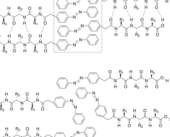
Peptide-based photo-responsive hydrogels have been synthesized by Huang and co-workers.119 Their materials contained an azobenzene moiety, which was used as a conformational switch. Upon UV-irradiation, a conformational change from (E) to (Z) took place, which changed the position of the phenylalanine residues and thereby led to less effective π-π stacking (Figure 1.11). The azobenzene moiety was coupled to the N-terminus of dipeptides using (E)-2-(4-(phenyldiazenyl)phenyl) acetic acid. Self-assembly of these azo-dipeptides was thought to occur via intermolecular π-π stacking of the phenyl rings from azobenzene and via hydrogen bonding of the peptides. A series of azo-dipeptides was synthesized. Constructs containing aromatic residues such as phenylalanine and tyrosine easily formed hydrogels. Poor gelation behaviour was observed for dipeptides with the cationic residues Arg and Lys. Insertion of phenylalanine or tyrosine into these dipeptides (azo-Arg-Ala, azo-Lys-Ala) facilitated gelation. Salt concentration and pH also had an effect on the hydrogelation behaviour. Azo-dipeptide hydrogels collapsed upon UV-irradiation, due to the expected less effective π-π stacking. The gel reformed after an average of two days under ambient visible light. To test the biological applications of these gels, vitamin B12 was trapped into azo-Gln-Phe-Ala. Release of B12 took more than two days, but was greatly enhanced upon UV-irradiation (4h).119 Velema et al. incorporated the azobenzene moiety into the structure of dichromyl molecules. Upon triggering with light, these compounds self-assembled into fibres, resulting in gel formation. Another trigger for assembly was NaCl, addition of this salt enhanced aggregate formation.120 Recently, constructs were synthesized containing an azobenzene motif, as well as alkyl chains and a polyether moiety. These constructs readily formed nanofibres in water and displayed multistimuli responsive behaviour. Apart from light, changes in temperature, pH and mechanical stress also initiated hydrogel formation.121
R1 R2 R3 R4 R5 R6 R7 R8 R9 R10 R11 R12 R13 R14 R15 R16 R17 R18 R19 R20 R21 R22 R23 R24 R25 R26 R27 R28 R29 R30 R31 R32 R33 R34 R35 R36 R37 R38 R39
Peptide- and protein based hydrogels 33
1
O O N R3 O N R2 O N R1 O N N N N H N N O O N R3 O N R2 O N R1 O N N H H H H H H H A O O N R3 O N R2 O N R1 O H H H N N N N O O N R3 O N R2 O N R1 O N N H H H O N H R1 O N H R2 O N H R3 O O H O N H R1 O N H R2 O N H R3 O O H O N H R1 O N H R2 O N H R3 O O H O N H R1 O N H R2 O N H R3 O O H N N H H BFigure 1.11. Balanced (A) and disturbed (B) interactions between the azo-dipeptides. Conformation A leads to the formation of hydrog
![Figure 2.11. CD spectra of peptide G[LEELLEE] 2 G (8) with several equivalents of calcium, showing an α-helical conformation in all cases](https://thumb-us.123doks.com/thumbv2/123dok_us/439783.2550848/66.850.154.537.306.593/figure-spectra-peptide-leellee-equivalents-calcium-showing-conformation.webp)
![Figure 2.13. DLS spectra of peptide G[LEELLEE] 2 G (8) and CaCl 2 , showing the formation of aggregates.](https://thumb-us.123doks.com/thumbv2/123dok_us/439783.2550848/68.850.158.551.173.457/figure-spectra-peptide-leellee-cacl-showing-formation-aggregates.webp)
![Figure 2.15. DLS spectra of peptide C 15 H 31 C(O)-[LEELLEE] 2 G (10) after addition of calcium and EDTA](https://thumb-us.123doks.com/thumbv2/123dok_us/439783.2550848/69.850.159.544.344.632/figure-dls-spectra-peptide-leellee-addition-calcium-edta.webp)