*Corresponding author: Isabella Princess
PHARMACEUTICAL RESEARCH
ISSN: 2395-6429, Impact Factor: 4.656
Available Online at www.journalcmpr.com
Volume 5; Issue 01(A); January 2019; Page No. 3994-3999
DOI: http://dx.doi.org/10.24327/23956429.ijcmpr201901607
Research Article
LABORATORY DIAGNOSIS OF PULMONARY TUBERCULOSIS – HOW RAPID CAN IT GET?
Isabella Princess
1and Rohit Vadala
21
Microbiology Apollo Speciality Hospitals Vanagaram, Chennai - 600095
2
Department of Pulmonary and Critical Care Medicine Primus Super Speciality Hospital
Chanakyapuri, New Delhi – 110021, India
ARTICLE INFO ABSTRACT
The aim of any diagnostic procedure is rapidity and reduced turnaround time. This becomes an urgent necessity especially for common infectious diseases. In India, the commonest infectious disease being tuberculosis, diagnosis plays a major role more so the rapidity in diagnosis. Many patients can be lost to follow up if the time taken for diagnosis is delayed. With available tests and advancements in diagnosis, tuberculosis treatment can be started at the earliest with judicious use of diagnostic tests.
Copyright © 2019 Isabella Princess and RohitVadala. This is an open access article distributed under the Creative Commons Attribution License, which permits unrestricted use, distribution, and reproduction in any medium, provided the original work is properly cited.
INTRODUCTION
Laboratory diagnosis remains the gold standard for diagnosis of tuberculosis (TB), although this disease has a characteristic presentation. Over past years, techniques have evolved to reach the present diagnostic era where diagnosis of tuberculosis can be made within few minutes.[1]The luxuries of using rapid diagnostics is reserved to high income countries where the disease burden is lower than low income countries. With increase in number of patients with tuberculosis and Multi Drug Resistant tuberculosis, rapid diagnosis is the need of the hour to effectively manage patients and prevent the spread of infection.[2] World Health Organization also recommends the use of rapid molecular tests for initial diagnosis of tuberculosis. The evolution of different diagnostic modalities for TB diagnosis is briefly summarized in this review.
Conventional methods versus newer methods of TB diagnosis
Methods used for diagnosis of tuberculosis include microscopy, culture, serological methods and tuberculin skin test. Although newer diagnostic tools exist, microscopy has still not been replaced in many primary health care and resource poor health facilities.
Microscopy
Conventional microscopy
Microscopic examination by acid fast staining is the age old method for diagnosis of tuberculosis. The procedure was discovered by two German doctors Franz Ziehl (Bacteriologist) and Friedrich Neelsen (Pathologist) in the 1890s and derived its name as Ziehl-Neelsen stain.[3] Microscopic screening of Mycobacterium tuberculosis cannot be used as stand-alone test for detection of tuberculosis. Rather than calling it a diagnostic test, it should be used as screening test since all Mycobacteria species take up the acid fast stain. Another drawback is the inability in distinguishing dead and live bacilli on stain, hence leaving the viability and bacillary load questionable.[4] Taking into consideration the low sensitivity of acid fast stain (20-80%), microscopy should be coupled with culture or molecular methods to make a diagnosis subject to their availability.
Two consecutive sputum samples are necessary to identify 95-98% cases of pulmonary tuberculosis.[1] Probability of detecting tuberculosis with one sputum sample is around 85.8%.[5] Reliable detection of Mycobacterium tuberculosis is achieved only if the bacillary load is 1000-10,000 CFU/ml of sputum.[6] Sample collection is crucial and specific guidelines should be followed in order to contain the spread of tuberculosis. Therefore, sputum collection booths are provided
Article History:
Received 4th October, 2018 Received in revised form 25th October, 2018
Accepted 18th December, 2018 Published online 28th January, 2019
Key words:
Microscopy, culture, drug
at sample collection areas. Designated place away from crowded areas are chosen for this purpose and specialized staff can be designated to instruct patients on sample collection.[7] Two spot samples or one spot and one early morning sample can be collected for smear and culture. A mucopurulent sample devoid of salivary contamination is mandatory to arrive at the appropriate diagnosis.[8]
Newer microscopy
Original microscopic procedure (Ziehl-Neelsen staining) has been replaced by fluorescent LED (Light Emitting Diode) microscopy in recent days. First endorsed by the WHO in 2009 and recommended thereafter by the Revised National Tuberculosis Control Program (RNTCP), LED microscopy has replaced ZN staining in many centers of India especially the Designated Microscopy Centers (DMCs). Fluorescent microscopes are of two major types, ones using mercury vapor lamps and ones using LED lamps. Various advantages of using fluorescent LED microscopes include rapidity, higher sensitivity and specificity by 10% over ZN staining.[9] Technical benefits include long life spans, minimal requirements of power, no emission of UV light. Time taken to screen smears using fluorescent LED microscope is also very less (mean time: 2 minutes) when compared to light microscope (mean time: 5 minutes).[10] Major benefits using LED microscopes are achieved in high throughput labs where rapid examination of multiple slides is enabled.
Culture
Mycobacterial culture is the gold standard of TB diagnosis. Mycobacterial species identification is necessary since specific drugs are targeted against each species of Mycobacteria. Detection limit of M.tuberculosis in culture is 100 bacilli/ml.[8] Mandatory requirement for laboratories to perform culture are those with Biosafety level 2 for sample processing and inoculation, however manipulation of positive cultures requires a biosafety level 3 facility.[11] A decontamination procedure should be done prior to sample inoculation into the media in order to eliminate the growth of other bacterial contaminants. Two commonly used decontamination agents are N-acetyl-L-cysteine (NALC) and sodium hydroxide (NaOH).
Conventional TB culture
Mycobacterium tuberculosis was successfully cultured for the first time by the German Microbiologist Robert Koch in 1882. Lowenstein Jensen medium used for culture of M.tuberculosisis an egg based medium containing malachite green, potato starch, salts and glycerol. Turnaround time for isolation of M.tuberculosis using this media is 4-8 weeks.[12]If growth occurs, further processing and drug susceptibility testing takes 4 weeks. Other types of solid media used are: Agar based media, Middlebrook 7H10 and Middlebrook 7H11 with antibiotics, potato based and serum based media. Rate of growth of Mycobacteria is quicker in agar based media compared to egg based media. Media is clear with a large surface area but there is requirement of CO2 for culture in agar
based media.[13] Drug susceptibility testing using agar and egg based media is cumbersome and reserved for performing in reference laboratories and other well equipped laboratories. This process takes up to six weeks or more. Methods by which drug susceptibility of M.tuberculosis can be done are proportion method, absolute concentration method and resistance ratio method.[14]
Newer culture methods for TB diagnosis
Automated culture methods were designed in 1955 with the introduction of Middlebrook 7H9 broth. Middlebrook 7H12 is another broth used for culture which is a modification of
7H9.[13] Since then, various other methods used for automated
TB culture are Mycobacterial Growth Indicator Tube (MGIT), BacT/Alert 3D,VersaTREK etc.
MGIT is the automated Mycobacterial culture system which uses modified Middlebrook 7H9 broth. Supplemented with casein peptone, growth supplements like oleic acid, albumin, dextrose, antibiotics PANTA (Polymyxin B, Amphotericin B, Nalidixic acid, Trimethoprim, and Azlocillin).[15] Average turnaround time for isolation of M.tuberculosis using MGIT is ≤14 days, with some studies reporting a mean detection time as 12.18 days.[15]Drug Susceptibility Testing (DST) can also be done using MGIT. The average time taken for DST after growth of the organism in culture is 7-14 days.[16]Working principle involves non radiometric detection of fluorescence emitted by O2consumption during bacterial growth in the
media.[16]However, MGIT is not approved for Mycobacterial culture from blood and urine samples.
BacT/Alert 3D is suitable for Mycobacterial culture; however blood samples should be inoculated into a specific BacT/Alert bottle. Another difference is in the BacT/Alert media with incorporation of Vancomycin in addition to PANTA used in MGIT. Average turnaround time using BacT/Alert is 13.7 days.[17] It works on the principle of colorimetric sensor for detection of CO2 produced during metabolism and growth of
M.tuberculosis. A major drawback in BacT/Alert is the unavailability of performance of DST.
Versa TREK is another method which detects M.tuberculosis using the principle of measurement of pressure released during growth of the organism by oxygen consumption. Turnaround time using this method is an average of 15.7 days.[18]Antibiotic supplement used is Polymyxin B, Amphotericin B, Nalidixic acid and Vancomycin. Drug susceptibility testing can also be performed on VersaTREK.
Microscopic observation broth-drug susceptibility assay (MODS) is useful in developing countries and laboratories which cannot afford automated instruments. It does not require radioactive isotopes or fluorescent indicators.[19]
Immunochromatography for TB diagnosis
Immunochromatography / lateral flow assay is available in order to differentiate Mycobacterium tuberculosis complex from other atypical Mycobacteria species. Once growth occurs in liquid or solid culture media, an immunochromatographic method for detection of a specific M.tuberculosis antigen (MPT 64) can be performed on the culture filtrate. It is necessary to perform this lateral flow immunoassay after growth occurs since the limit of detection is 105 CFU/ml.[2] This makes it mandatory to be performed from culture and therefore cannot be used directly on patient samples. Another major advantage of its use in resource poor settings is that this rapid test replaces a battery of biochemical tests (Nitrate reductase, catalase, niacin, pyrazinamidase, arylsulfatase, tween 80 hydrolysis) which are usually cumbersome to perform.
rapid immunochromatography. A small volume of the culture filtrate is placed on the card. Anti MPT64 antibodies immobilized on nitrocellulose membrane acts as capture antibody for antigen present in the filtrate. Test reading can be taken within 15 minutes. Overall sensitivity of this test is 98.6% and specificity is 97.9%. Studies from India have shown
a sensitivity of 99.19% and specificity of 100%.[21]Mutation of
MPT64 gene renders the test false negative since the antigen is not expressed due to mutation of the gene and cannot be detected by the kit.[22]
Lateral Flow Lipoarabinomannan assay (LF-LAM) is a rapid test used for detection of M.tuberculosis from samples. It is very sensitive for detection of M.tuberculosis from urine samples. It works on the principle of detecting Lipoarabinomannan, a polysaccharide antigen released by the cell wall of bacilli during metabolism and replication. Turnaround time is 25 minutes.[23] This assay has gained its importance mainly for use in HIV positive individuals who are suspected to have coinfection with pulmonary or extrapulmonary tuberculosis.
Matrix Assisted Laser Desorption Ionization Time of Flight – Mass Spectrometry (MALDI TOF – MS)
Mass spectrometry which is recently being used to detect microorganisms within few hours is also promising option for confirmation of Mycobacterial species. MALDI-TOF MS works on the principle of mass spectrometry to identify microorganisms based ontheir mass to charge ratio. It has an excellent turnaround time for identifying multiple organisms within 3 hours. In MALDI-TOF MS, the sample to be analyzed (bacterial colony) is mixed with another compound, called a matrix. An on-plate extraction should be performed using ethanol / heat / 1µl of 70% formic acid. The matrix is then added to this mixture and subjected to analysis .The results are displayed as a series of lines (spectrum) which correspond to different fragments that have broken away from the original molecule. A database is then used to compare this pattern with the known data of yeasts and bacteria in the test
system.[24] It is necessary to perform this test on culture isolates
and therefore in order to process TB cultures, the mandatory requirement is a BSL-3 laboratory. Apparently, currently available MALDI TOF systems are still not FDA approved for
diagnosis of M.tuberculosis and thus proper standardization is
required before use.
Molecular diagnosis of tuberculosis
Molecular methods have gained importance is the recent past due to tremendous reduction in turnaround time compared to conventional culture based diagnosis. Apart from that, patient follow up is easier and drug resistance detection is rapidly detected using molecular techniques.
Conventional PCR assays: They gained importance for TB
diagnosis in early 1990s with development of in-house PCR kits. Commonly used gene targets were against insertion element IC 6110.[25]Tedious nature of the procedure which involves DNA extraction and amplification made it almost obsolete. Another drawback was the difficulty in standardization and long processing time. Slowly these conventional PCR methods are now being replaced by automated real time and newer PCR assays.
Xpert MTB/RIF: Introduced and recommended by the WHO
in 2010, Xpert MTB/RIF is a cartridge based nucleic acid amplification assay.[26]It is a semi quantitative real time nested
PCR where the nucleic acid extraction and amplification occurs in a single cartridge device. Most Government hospitals and Reference laboratories in India are provided with Xpert MTB under RNTCP for diagnosis of TB. Since Rifampicin resistance can be detected early, treatment can be tailored to initiating second line drugs. However certain drawbacks of Xpert MTB/RIF have been recorded in literature as follows:
1. Xpert MTB/RIF detects both live and dead bacilli which makes it obsolete to be used for monitoring treatment prognosis.
2. Isoniazid monoresistance which results is 7-11% of treatment failure is missed on performing Xpert MTB/RIF.[27]
3. False positive Rifampicin resistance is seen with Xpert MTB, which makes it mandatory to confirm it with a repeat test or by using line probe assay.[28] 4. Trained personnel are required along with stable
electricity supply as well as air-conditioned processing rooms.
Loop Mediated isothermal Amplification (LAMP)
Isothermal amplification signifies the detection of DNA under isothermal conditions (65°C) rather than the usual PCR procedure of gradient thermo cycling process. Denaturation of nucleic acid is not necessary as the whole process takes place in a single centrifuge tube. Turnaround time is less than two hours with requirement of minimal instrumentation.[29]A DNA chelating fluorescent dye is used as the reporter molecule. If DNA amplification occurs in the sample due to presence of Mycobacteria, the fluorescence produced is detected using UV illuminator. Appearance of fluorescence denotes positive reaction and absence of the same denotes no amplification. This method is highly specific with a detection limit of 5-50 copies of bacilli. However, detection of drug resistance is not possible by using LAMP. WHO has also endorsed its use in diagnosis of tuberculosis due to the many advantages of this assay.[30]
Line Probe Assay (LPA)
Every year, new cases of Multi Drug Resistant tuberculosis is an emerging burden on the already existing load. The markers of MDR TB being resistance to Isoniazid and Rifampic in, the Line Probe Assay was first designed and recommended by the WHO in 2008 to detect resistance genes encoding both these anti TB drugs (rpoB, katG, inhA). There are two methods of testing: Direct method (performed on sputum samples) and indirect method (performed on culture isolates), both with a turnaround time of 5-6 hours. The specificity of indirect method was slightly higher than that of direct method. Second line drug susceptibility testing can also be done using LPA for detecting genes encoding resistance to fluoroquinolones and injectable second line drugs (gyrA, embB, rrs).[31]With ability to detect resistance to second line anti TB drugs, it makes LPA the WHO recommended rapid test for detection of MDR as well as Extensively Drug Resistant (XDR) TB.[31]Therefore, at present both Xpert MTB/RIF as well as LPA are WHO recommended diagnostic methods adopted by RNTCP in India.
assays enable remarkable reduction in turnaround time for detection of M.tuberculosis, MDR TB, XDR TB surpassing conventional solid based and liquid based drug susceptibility testing methods.
Serological methods
Antibodies such as anti-α-crystallin (ACR), lipoarabinomannan, trehalose 6,6’-dimycolate, and anti-tubercular-glycolipid antibodies were found to be elevated among active TB patients in studies from developing countries.[33]These can be used as surrogate / complementary tests but cannot be used as confirmatory diagnostic tests for tuberculosis. World Health Organization does not recommend the use of serological tests for diagnosing clinical pulmonary and extra pulmonary tuberculosis. The commercial kits have inconsistent and imprecise estimates of sensitivity and specificity. Risk of erroneous results due to various reasons is the major drawback making serological diagnosis of TB obsolete in India and other high prevalence countries.
Interferon Gamma Release Assay (IGRA)
Principle of IGRAs is the measurement of Interferon γ released as the result of cell mediate immune response to tubercle bacilli. Commercially available IGRA formats are: QuantiFERON-TB Gold (QFT-G) and Quanti FERON-TB Gold In- Tube (QFT-IT), (Cellestis, Australia); T.SPOT.TB (Immunotec, UK).[34] The procedure is technically complex and cumbersome. The results of IGRAs get affected by BCG vaccine as well as antibodies in blood of exposed but uninfected individuals. Hence active tuberculosis cannot be established by using IGRAs for diagnosis.
Advantages: IGRAs are used as alternative to skin test in low
prevalence countries. It is highly specific with no risk of false positive results.
Disadvantages: Not recommended for use in low and middle
income (high prevalence) countries due to high prevalence of TB. On 7`th June 2012, the Government of India issued a Gazette notification banning the manufacture, importation, distribution and use of commercially available serological tests for diagnosis of tuberculosis.
Tuberculin skin test
Tuberculin skin test otherwise known as Mantoux test is still being used in many countries as a part of TB diagnosis. It is a non-specific test that demonstrates delayed / type IV hypersensitivity reaction towards the tuberculin antigen injected intradermally into the flexor surface of the forearm. It is a simple and inexpensive test used for many years in the past since 1907. However, its low sensitivity in immunosuppressed and advanced disease renders it unreliable in the diagnosis of tuberculosis. The Mantoux test has low specificity in patients with history of BCG vaccination and Non tuberculous Mycobacterial infection.[35]
The old method employed use of crude tubercular bacilli preparation. This is rarely used and been replaced by Purified Protein Derivative antigen (PPD) which is produced by growing M.tuberculosis in semisynthetic media. PPD suggested for use by the WHO is PPD-RT-23 with Tween 80. Turnaround time for the test is 48-72 hours at the end of which the width of induration and erythema at site of injection is measured. An induration diameter of ≥10 mm implies a
positive test. The limited usefulness of this test in healthcare workers is due to high exposure to patients with tuberculosis.
Indirect methods of diagnosis of tuberculosis
Breath analyzers measuring the released volatile organic compounds (VOC) when metabolizing M.tuberculosis are used as point of care diagnostic tests. Battery operated hand held device measuring the released VOCs are cheaper and faster therefore becoming useful alternatives in community based health centers in developing countries. The principle for diagnosis is detection of VOCs using automated thermal desorption, gas chromatography and mass spectroscopy.[36]
Rapid diagnostic tests for TB – Boon or bane?
To answer the question we have discussed various conventional and rapid / newer diagnostic tests for TB diagnosis. As briefly discussed, rapid TB diagnostic methods in particular situations are more pronounced. For example, the World Health Organization recommends the use of regular screening of prison inmates using Gene X pert MTB/RIF. This recommendation of annual screening was put forth in 2017 since early detection in this group prevents transmission of TB and MDR TB.[37]
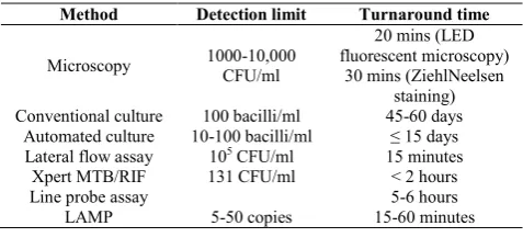
The irony of rapid diagnostic methods is that their need is elevated in high prevalence / low income countries compared to low prevalence / high income countries. However, their availability is vice versa (more availability in low prevalence / high income countries, less availability in high prevalence / low income countries) making its boon in poor countries questionable. Like two sides to a coin, both advantages and disadvantages exist to rapid diagnosis. Salient features of all diagnostic methods are compared in table 1. Flexibility in cost and availability of these tests would boost its usefulness in low and middle income countries.
Table 1 Summary of diagnostic tests for tuberculosis
Method Detection limit Turnaround time
Microscopy 1000-10,000
CFU/ml
20 mins (LED fluorescent microscopy)
30 mins (ZiehlNeelsen staining)
Conventional culture 100 bacilli/ml 45-60 days
Automated culture 10-100 bacilli/ml ≤ 15 days
Lateral flow assay 105 CFU/ml 15 minutes
Xpert MTB/RIF 131 CFU/ml < 2 hours
Line probe assay 5-6 hours
LAMP 5-50 copies 15-60 minutes
CONCLUSION
Many developments in the diagnostic methods have helped in rapid accurate diagnosis of tuberculosis. To some it is still a dream to access these rapid diagnostic facilities. Major parts of the developing world and especially in high burden countries like India, the limitation to access is more evident in the rural population. It would be of great advantage if these newer rapid diagnostics percolate into rural health facilities to alleviate the burden in this population. Cost effectiveness is the need of the hour to make effective use of these resources.
Reference
2. Parsons LM, Somosko¨vi AK, Gutierrez C, Lee E, Paramasivan CN, Abimiku A. Laboratory Diagnosis of Tuberculosis in Resource-Poor Countries: Challenges and Opportunities. ClinMicrobiol Rev. 2011;24:314-50. 3. Singhal R, Myneedu VP. Microscopy as a diagnostic
tool in pulmonary tuberculosis. Int J Mycobacteriol. 2015;4:1-6.
4. Steingart, K. R., A. Ramsay, M. Pai. Optimizing sputum smear microscopy for the diagnosis of pulmonary tuberculosis. Expert Rev Anti Infect Ther. 2007;5:327– 31.
5. Mase SR, Ramsay A, Ng V, Henry M, Hopewell PC, Cunningham J,et al. Yield of serial sputum specimen examinations in the diagnosis of pulmonary tuberculosis: A systematic review. Int J Tuberc Lung Dis 2007;11:485‑95.
6. Caulfield AJ, Wengenack NL. Diagnosis of active tuberculosis disease: From microscopy to molecular techniques. J ClinTuberc Other Mycobact Dis. 2016;4:33-43.
7. Khan MS, Dar O, Sismanidis C, Shah K, Godfrey-Faussett P. Improvement of tuberculosis case detection and reduction of discrepancies between men and women by simple sputum-submission instructions: a pragmatic randomised controlled trial. Lancet. 2007; 369:1955–60.
8. Oommen S, Banaji N. Laboratory diagnosis of tuberculosis: Advances in technology and drug susceptibility testing. Indian J Med Microbiol 2017:35;323-31.
9. Bhalla M, Sidiq Z, Sharma PP, Singhal R, Myneedu VP, Sarin R. Performance of light-emitting diode fluorescence microscope for diagnosis of tuberculosis. Int J Mycobacteriol. 2013;2:174-8.
10. Marais BJ, Brittle W, Painczyk K, Hesseling AC, Beyers N, Wasserman E et al. Use of light-emitting diode fluorescence microscopy to detect acid-fast bacilli in sputum. Clin Infect Dis. 2008;47:203-7.
11. World Health Organization, Geneva, 2007. Use of Liquid TB Culture and Drug Susceptibility Testing (DST) in Low and Middle Income Settings. Accessed on : 2/10/2018.
12. Shah NS, Moodley P, Babaria P, Moodley S, Ramtahal M, Richardson J et al. Rapid diagnosis of tuberculosis and multidrug resistance by the microscopic-observation drug-susceptibility assay. Am J RespirCrit Care Med 2011;183:1427‑33.
13. Asmar S, Drancourt M. Rapid culture-based diagnosis of pulmonary tuberculosis in developed and developing countries. Front Microbiol 2015;6:1184.
14. Sastry AS, Bhat S. In: Essentials of Medical Microbiology, 1`st ed. New Delhi: Jaypeebrothers medical publishers; 2016.
15. Soto A, Agapito J, Acun˜a-Villaordun˜a C, Solari L, Samalvides F, Gotuzzo E. Evaluation of the performance of two liquid-phase culture media for the diagnosis of pulmonary tuberculosis in a national hospital in Lima, Peru.Int J Infect Dis 2009;13:40-5. 16. New Laboratory Diagnostic Tools for Tuberculosis
Control. WHO;2008. Available from: http://www.stoptb.org/assets/documents/global/retoolin g/Diagnostic_Brochure_Print_2009_Jan_29.pdf. [Accessed on : 13/11/2018].
17. Rohner P, Ninet B, Metral C, Emler S, Auckenthaler R. Evaluation of the MB/BacT system and comparison to the BACTEC 460 system and solid media for isolation of mycobacteria from clinical specimens. J ClinMicrobiol 1997; 35:3127‑31.
18. Woods GL, Fish G, Plaunt M, Murphy T. Clinical evaluation of difco ESP culture system II for growth and detection of mycobacteria. J ClinMicrobiol 1997; 35:121‑4.
19. Caviedes L, Lee T, Gilman Rh, Sheen P, Spellman E, Lee Eh et al. Rapid, Efficient Detection and Drug Susceptibility Testing of Mycobacterium tuberculosis in Sputum by Microscopic Observation of Broth Cultures. J ClinMicrobiol 2000; 38:1203-8.
20. World Health Organization. 2008. Laboratory-based
evaluation of 19 commercially available rapid diagnostic tests for tuberculosis. Special Program for
Research and Training in Tropical Diseases.
Diagnostics evaluation series, 2. World Health Organization, Geneva, Switzerland.
21. Kanade S, Nataraj G, Suryawanshi R, Mehta P. Utility of MPT 64 antigen detection assay for rapid characterization of Mycobacteria in resource constricted setting. Indian J Tuberc 2012; 59:92-6.
22. World Health Organization. 2008. New laboratory
diagnostic tools for tuberculosis control. Stop TB Partnership: Retooling Task Force and the New
Diagnostics Working Group. World Health
Organization, Geneva, Switzerland.
23. Andrews B, Muchemwa L, Lakhi S, Bwalya M, Mabula
C, Chipii G, Heimburger DC, Bernard GR. Validation of a clinical prediction score and performance of urine lipoarabinomannan test for detecting tuberculosis bacteremia in HIV-positive patients with severe sepsis. Am J RespirCrit Care Med.189:2014: A2559.
24. MALDI-TOF Mass Spectrometry. 2017. Available
from:
http://www.biomerieux.com/en/maldi-tof-mass-spectrometry. [Last accessed on 2017 Nov 21].
25. Tiwari S, Nataraj G, Kanade S, Mehta P. Diagnosis of pediatric pulmonary tuberculosis with special reference to polymerase chain reaction based nucleic acid amplification test. Int J Mycobacteriol 2015;4:48-53. 26. WHO Global TB Programme. Automated real-time
nucleic acid amplification technology for rapid and simultaneous detection of tuberculosis and rifampicin resistance: xpert MTB/RIF assay for the diagnosis of pulmonary and extrapulmonary TB in adults and children. Geneva: World Health Organization; 2013. 27. D’souza DT, Mistry NF, Vira TS, Dholakia Y, Hoffner
S, Pasvol G et al. High levels of multidrug resistant tuberculosis in new and treatment-failure patients from the Revised National Tuberculosis Control Programme in an urban metropolis (Mumbai) in Western India. BMC Public Health 2009;9:211.
28. Lawn SD, Nicol MP. Xpert MTB/RIF assay:
Development, evaluation and implementation of a new rapid molecular diagnostic for tuberculosis and rifampicin resistance. Future Microbiol 2011;6:1067-82.
29. Teran R, de Waard JH. Recent advances in the
laboratory diagnosis of tuberculosis. eJIFCC
2015;26:295-309.
Accessed from: http://apps.who.int/iris/bitstream/10665/83142/1/WHO_ HTM_TB_2013.05_eng.pdf.
31. Tubercular Diagnostics. Molecular Line-probe Assay
for the Detection of Resistance to Second-line Anti-TB Drugs (SL-LPA) – WHO Recommendations on the Use
of the SL-LPA; 2016. Available from:
http://www.who.int/tb/Factsheet_SLLPAfinal.pdf. [Accessed on: 17/11/2018].
32. Anochie PI, Onyeneke EC, Ogu AC, Onyeozirila AC, Aluru S, Onyejepu N, Zhang J, Efere L, Adetunji MA, Bueno Sánchez JG. Recent advances in the diagnosis of Mycobacterium tuberculosis. GERMS. 2012;2:110-20. 33. Nurwidya F, Handayani D, Burhan E, Yunus F.
Molecular Diagnosis of Tuberculosis. Chonnam Med J 2018;54:1-9.
34. World Health Organization. The use of TB Interferon-Gamma Release Assays (IGRAs) in Low- and Middle-income Countries. Policy statement 2011. Accessedon
:2/10/2018, Accessed from
:http://www.who.int/tb/laboratory/policy_statements/en. 35. Huebner, R. E., M. F. Schein, and J. B. Bass, Jr. The
tuberculin skin test. Clin. Infect. Dis.1993;17:968–75. 36. Phillips M, Basa-Dalay V, Bothamley G, Cataneo RN,
Lam PK, Natividad MP et al. Breath biomarkers of active pulmonary tuberculosis, Tuberculosis2010;1-7. 37. World Health Organization. 2017. Algorithm for
laboratory diagnosis and treatment-monitoring of pulmonary tuberculosis and drug-resistant tuberculosis using state-of-the-art rapid molecular diagnostic technologies. Accessed on: 2/10/2018; Accessed from: www.euro.who.int.
How to cite this article:
Isabella Princess and RohitVadala (2019) 'Laboratory Diagnosis of Pulmonary Tuberculosis – How Rapid Can It Get?', International Journal of Current Medical And Pharmaceutical Research, 05(01), pp. 3994-3999.