University of Pennsylvania
ScholarlyCommons
Publicly Accessible Penn Dissertations
2018
Discovering Novel Hearing Loss Genes: Roles For
Esrp1 And Gas2 In Inner Ear Development And
Auditory Function
Alex Martin Rohacek
University of Pennsylvania, [email protected]
Follow this and additional works at:
https://repository.upenn.edu/edissertations
Part of the
Cell Biology Commons,
Developmental Biology Commons, and the
Molecular
Biology Commons
This paper is posted at ScholarlyCommons.https://repository.upenn.edu/edissertations/2843
For more information, please [email protected].
Recommended Citation
Rohacek, Alex Martin, "Discovering Novel Hearing Loss Genes: Roles For Esrp1 And Gas2 In Inner Ear Development And Auditory Function" (2018).Publicly Accessible Penn Dissertations. 2843.
Discovering Novel Hearing Loss Genes: Roles For Esrp1 And Gas2 In
Inner Ear Development And Auditory Function
Abstract
Hearing loss is the most common form of congenital birth defect, affecting an estimated 35 million children worldwide. To date, nearly 100 genes have been identified which
contribute to a deafness phenotype in humans, however, many cases remain in which a causative mutation has yet to be found. In addition, the exact mechanism by which hearing loss occurs in the presence of many of these mutations is still not understood. This is due, in part, to the complex nature of the development and function of the
cochlear duct, the organ of hearing. The cochlea undergoes an intricate morphogenetic development and requires the proper specification and maintenance of dozens of
different cell types in order to function correctly. In the mature duct, an interplay between
mechanotransducing sensory hair cells, supporting pillar and Dieters' cells, and generation of electrochemical potential by the stria vascularis are necessary to respond to sound stimuli. We utilized exome and RNA-sequencing experiments combined with
mouse genetics in order to discover novel genes that play roles in cochlear
development and function. Exome sequencing of families with profound hearing loss uncovered mutations in Epithelial Splicing Regulatory Protein 1 (ESRP1), a critical regulator of alternative mRNA splicing. Analysis of Esrp1 mutant mice revealed a
shortened cochlear duct, delay in hair cell differentiation and maturation, and loss of the stria vascularis due to inappropriate Fgf ligand usage, stemming from an alternatively spliced receptor, in these cells. To identify additional regulators of inner ear development
we performed an RNA-seq experiment comparing the gene expression profiles of control and Smoecko otic vesicles, which lack a cochlear duct. This generated a dataset of hundreds of cochlear enriched transcripts including Growth Arrest Specific 2 (Gas2) a
cytoskeletal binding protein with the potential to act as a regulator of cochlear development. We generated a Gas2 null mouse line and discovered that these animals have severe hearing impairment likely due to defects in microtubule organization in the
pillar cells. Taken together, these studies implicate Esrp1 and Gas2 as novel hearing loss genes that regulate aspects of cochlear development and function.
Degree Type
Dissertation
Degree Name
Doctor of Philosophy (PhD)
Graduate Group
Cell & Molecular Biology
First Advisor
Douglas J. Epstein
Keywords
Alternative Splicing, Cytoskeleton, FGF Signaling, Hearing loss, Inner Ear
Subject Categories
Cell Biology | Developmental Biology | Molecular Biology
DISCOVERING NOVEL HEARING LOSS GENES: ROLES FOR ESRP1 AND GAS2 IN INNER EAR DEVELOPMENT AND AUDITORY FUNCTION
Alex M. Rohacek A DISSERTATION
in
Cell and Molecular Biology
Presented to the Faculties of the University of Pennsylvania in
Partial Fulfillment of the Requirements for the Degree of Doctor of Philosophy
2018 Supervisor of Dissertation
__________________________________ Douglas J. Epstein, Ph.D., Professor of Genetics
Graduate Group Chairperson
__________________________________
Daniel S. Kessler, Ph.D., Associate Professor of Cell and Developmental Biology
Dissertation Committee
Stewart A Anderson, M.D., Professor of Psychiatry Russ P. Carstens, M.D., Associate Professor of Medicine
DISCOVERING NOVEL HEARING LOSS GENES: ROLES FOR ESRP1 AND GAS2 IN INNER EAR DEVELOPMENT AND AUDITORY FUNCTION
COPYRIGHT 2018
Alex Martin Rohacek
This work is licensed under the Creative Commons Attribution- NonCommercial-ShareAlike 3.0 License
To view a copy of this license, visit
iii
Dedication page
This work is dedicated to my family: my Mother, who is my most vocal supporter and my
greatest source of advice; my Father, who always pushes me to do the things I think are
impossible; my Sister, who is always my friend and ally; and our pets (both here and
who have gone), who were always there to bring me a smile. I couldn’t have done it
iv
ACKNOWLEDGMENT
This work would not have been possible without the help from many people who I’d like
to take the time to thank. My advisor, Doug Epstein, for seeing me through my graduate
work with the equal measures of pressure and guidance that I needed. My
undergraduate advisors Bryant Buchannan and Sharon Wise who encouraged me to
apply to graduate school and gave me my start in science. My lab mates who provided
me with advice and friendship over the years. And, all of my collaborators who helped
v
ABSTRACT
DISCOVERING NOVEL HEARING LOSS GENES: ROLES FOR ESRP1 AND GAS2 IN
INNER EAR DEVELOPMENT AND AUDITORY FUNCTION
Alex M Rohacek
Douglas J Epstein
Hearing loss is the most common form of congenital birth defect, affecting an estimated
35 million children worldwide. To date, nearly 100 genes have been identified which
contribute to a deafness phenotype in humans, however, many cases remain in which a
causative mutation has yet to be found. In addition, the exact mechanism by which
hearing loss occurs in the presence of many of these mutations is still not understood.
This is due, in part, to the complex nature of the development and function of the
cochlear duct, the organ of hearing. The cochlea undergoes an intricate morphogenetic
development and requires the proper specification and maintenance of dozens of
different cell types in order to function correctly. In the mature duct, an interplay between
mechanotransducing sensory hair cells, supporting pillar and Dieters’ cells, and
generation of electrochemical potential by the stria vascularis are necessary to respond
to sound stimuli. We utilized exome and RNA-sequencing experiments combined with
mouse genetics in order to discover novel genes that play roles in cochlear development
and function. Exome sequencing of families with profound hearing loss uncovered
mutations in Epithelial Splicing Regulatory Protein 1 (ESRP1), a critical regulator of
alternative mRNA splicing. Analysis of Esrp1 mutant mice revealed a shortened cochlear
duct, delay in hair cell differentiation and maturation, and loss of the stria vascularis due
to inappropriate Fgf ligand usage, stemming from an alternatively spliced receptor, in
vi
RNA-seq experiment comparing the gene expression profiles of control and Smoecko otic
vesicles, which lack a cochlear duct. This generated a dataset of hundreds of cochlear
enriched transcripts including Growth Arrest Specific 2 (Gas2) a cytoskeletal binding
protein with the potential to act as a regulator of cochlear development. We generated a
Gas2 null mouse line and discovered that these animals have severe hearing
impairment likely due to defects in microtubule organization in the pillar cells. Taken
together, these studies implicate Esrp1 and Gas2 as novel hearing loss genes that
vii
TABLE OF CONTENTS
ACKNOWLEDGMENT ... IV
ABSTRACT ... V
LIST OF TABLES ... X
LIST OF ILLUSTRATIONS... XI
CHAPTER 1: INTRODUCTION ... 1
The inner ear ... 1
Induction and patterning of the inner ear ... 1
Morphogenesis of the vestibular apparatus and cochlear duct ... 4
The structures of hearing ... 7
Development of cochlear sensory structures ... 10
Development of the cochlear lateral wall... 17
The sense of hearing and hearing loss... 19
Figures ... 22
CHAPTER 2: ESRP1 MUTATIONS CAUSE HEARING LOSS DUE TO DEFECTS IN ALTERNATIVE SPLICING THAT DISRUPT COCHLEAR DEVELOPMENT ... 27
Introduction ... 27
Alternative Splicing ... 27
Epithelial Splicing Regulatory Proteins ... 29
Rationale ... 31
Results ... 32
viii
Defects in inner ear morphogenesis and auditory hair cell differentiation in Esrp1
-/-mouse mutants ... 36
Esrp1 regulates the timing of hair cell differentiation ... 38
Esrp1 regulates the fate of nonsensory cells along the lateral cochlear wall ... 40
Altered splicing of Fgfr2 is responsible for the lateral cochlear wall defects in Esrp1 mutants ... 41
Ectopic Fgf9/Fgfr2-IIIc signaling compensates for the loss of Fgfr2-IIIb to promote cochlear morphogenesis in Esrp1-/- mutants ... 43
Discussion ... 44
Figures ... 51
Materials and Methods... 73
CHAPTER 3: GAS2 IS A SHH RESPONSIVE GENE REQUIRED FOR HEARING ... 88
Introduction ... 88
Hedgehog genes ... 88
Hedgehog signaling ... 89
Growth Arrest Specific 2 ... 91
Rationale ... 94
Results ... 95
RNA-seq reveals novel Shh targets in the developing cochlea ... 95
Gas2 is dynamically expressed in the developing and mature cochlear duct ... 98
Generation of a Gas2 knockout mouse line ... 100
ix
Gas2 mutant mice have severe hearing impairment ... 101
The pillar cell cytoskeleton is disrupted in Gas2 mutant mice ... 103
Discussion ... 106
Author Contributions ... 114
Acknowledgements... 114
Figures ... 116
Materials and Methods... 132
CHAPTER 4: CONCLUSIONS AND FUTURE DIRECTIONS ... 142
Hearing impairment and repair ... 142
Alternative splicing ... 144
Roles for Esrps in vestibular morphogenesis ... 144
Establishing roles for early otic expression patterns ... 146
Pillar cells and the sense of hearing ... 147
Conclusions ... 150
x
LIST OF TABLES
TABLE 2.1. ESRP1 DEPENDENT ALTERNATIVE SPLICING EVENTS IN THE COCHLEAR
EPITHELIUM. ... 84
TABLE 2.2 EXPRESSION VALUES OF SELECTED GENES FROM RNA-SEQ ON E16.5
CONTROL AND ESRP1-/- COCHLEAR EPITHELIUM. ... 84
TABLE 2.3. PCR PRIMERS USED TO AMPLIFY HUMAN ESRP1 AND ESRP2 EXONS. ... 85
TABLE 2.4. PRIMERS USED TO QUANTIFY CHANGES IN ALTERNATIVE SPLICING AND
GENE EXPRESSION BY RT-PCR. ... 86
xi
LIST OF ILLUSTRATIONS
FIGURE 1.1. THE STRUCTURES OF THE INNER EAR. ... 22
FIGURE 1.2. INNER EAR MORPHOGENESIS AND PATTERNING. ... 23
FIGURE 1.3. THE STRUCTURE AND CELL TYPES OF THE COCHLEAR DUCT. ... 24
FIGURE 1.4. TIMING AND REGULATION OF FACTORS CRITICAL FOR THE DEVELOPMENT
OF THE ORGAN OF CORTI. ... 25
FIGURE 1.5. THE STRUCTURES AND MECHANISMS OF HEARING. ... 26
FIGURE 2.1. ALTERNATIVE SPLICING AND EPITHELIAL SPLICE REGULATORY
PROTEINS. ... 51
FIGURE 2.2. ESRP1 MUTATIONS SEGREGATE WITH SNHL AND DISRUPT ALTERNATIVE
SPLICING. ... 52
FIGURE 2.3. ESRP1 MUTATIONS IN INDIVIDUALS WITH SNHL. ... 54
FIGURE 2.4. ESRP1 EXPRESSION IN THE DEVELOPING MOUSE COCHLEA. ... 55
FIGURE 2.5. INNER EAR MORPHOGENESIS AND AUDITORY HAIR CELL
DIFFERENTIATION ARE DISRUPTED IN ESRP1-/- MOUSE EMBRYOS. ... 56
FIGURE 2.6. HAIR CELL DIFFERENTIATION IS DELAYED AT THE APEX OF THE
COCHLEAR DUCT IN ESRP1-/- MUTANTS. ... 58
FIGURE 2.7. SPECIFICATION OF PROSENSORY PROGENITORS, SUPPORT CELLS,
VESTIBULAR HAIR CELLS AND NEURONS IS NOT COMPROMISED IN ESRP1-/- EMBRYOS.
... 59
FIGURE 2.8. SENSORY AND NONSENSORY GENE EXPRESSION PROFILES ARE
DISRUPTED IN THE COCHLEAR EPITHELIUM OF ESRP1-/- EMBRYOS. ... 60
FIGURE 2.9. HAIR CELL DIFFERENTIATION IS DELAYED IN ESRP1-/- EMBRYOS. ... 62
FIGURE 2.10. HAIR CELL DIFFERENTIATION AND MATURATION IS DELAYED IN ESRP1
xii
FIGURE 2.11. ESRP1 REGULATES THE IDENTITY OF NONSENSORY CELLS ALONG THE
LATERAL COCHLEAR WALL. ... 66
FIGURE 2.12. ALTERNATIVE SPLICING IS IMPAIRED IN THE COCHLEAR EPITHELIUM OF ESRP1-/- EMBRYOS. ... 68
FIGURE 2.13. FGF SIGNALING IS ECTOPICALLY ACTIVATED IN THE LATERAL COCHLEAR EPITHELIUM OF ESRP1 MUTANTS. ... 70
FIGURE 2.14. ECTOPIC SIGNALING THROUGH FGF9/FGFR2-IIIC IS RESPONSIBLE FOR THE LATERAL COCHLEAR WALL DEFECTS IN ESRP1 MUTANTS. ... 71
FIGURE 2.15. THE CYSTIC OTIC PHENOTYPE IN ESRP1-/- EMBRYOS IS EXACERBATED BY THE DOSE DEPENDENT LOSS OF FGF9. ... 72
FIGURE 3.1. THE HEDGEHOG SIGNALING PATHWAY. ... 116
FIGURE 3.2. RNA-SEQ ON SMOECKO MUTANTS REVEALS COCHLEAR ENRICHED TRANSCRIPTS. ... 117
FIGURE 3.3. LOSS OF COCHLEAR EXPRESSED GENES IN SMOECKO MUTANTS. ... 119
FIGURE 3.4. SHHP1 MUTANTS REVEAL SHH REGULATED GENES. ... 120
FIGURE 3.5 SOX2 AND JAG1 RESPOND TO ALTERED SHH SIGNALING. ... 121
FIGURE 3.6. GAS2 IS EXPRESSED IN THE DEVELOPING AND POSTNATAL COCHLEA. . 122
FIGURE 3.7. GENERATION OF A GAS2 NULL MOUSE LINE. ... 124
FIGURE 3.8. INNER EAR DEVELOPMENT IS UNAFFECTED BY LOSS OF GAS2. ... 125
FIGURE 3.9. GAS2 MUTANTS EXHIBIT SEVERE HEARING IMPAIRMENT. ... 127
FIGURE 3.10. GAS2 MUTANTS DISPLAY PROGRESSIVE LOSS OF MICROTUBULES FROM THE INNER PILLAR CELL HEAD. ... 129
1
Chapter 1: Introduction
Theinner ear
The inner ear is a multifunctional organ embedded in the temporal bone of the
skull (Fig. 1.1A). The dorsal structures of the inner ear comprise the vestibular
apparatus, a set of three semicircular canals (SCCs) and associated crista ampullaris
that function in the detection of three-dimensional orientation of the head, while the
utricle and saccule sense horizontal and vertical linear acceleration respectively
(Fig.1.1B). The ventral structure of the inner ear is the coiled cochlear duct which
functions in the sensing and translation of sound (Fig. 1.1B).
Induction and patterning of the inner ear
The intricate structures of the inner ear arise from a simple, common otic
precursor and undergo a complex series of morphological changes throughout
embryogenesis (Cantos et al., 2000; Kopecky et al., 2012). Inner ear development
begins with the specification of a pair of ectodermally derived placodes that form on
either side of the neural tube at the level of the hindbrain between rhombomeres 4 and 6
(Kopecky et al., 2012; Noramly and Grainger, 2002; Solomon and Fritz, 2002; Fig. 1.2A).
Induction of otic placodes is first observed at Carnegie stage 9 (CS9; ~20 days post
fertilization) in humans and embryonic day 8 (E8.0) in the mouse (Arnold and Lang,
2001; Kikuchi and Hilding, 1965; Morsli et al., 1998; Toyoda et al., 2015). Experiments in
the mouse and chicken have implicated members of the fibroblast growth factor (Fgf)
signaling family as the initiators of otic development. Mouse knockouts of Fgf3 and 10 or
Fgf3 and 8 demonstrate a complete failure in otic development, a phenotype
2
2005; Urness et al., 2010; Wright and Mansour, 2003; Yang et al., 2013; Zelarayan et
al., 2007). An arrest of early inner ear morphogenesis is also seen in mouse mutants for
Fgfr2, the receptor for Fgf3 and 10 (Pirvola et al., 2000). Conversely, ectopic expression
of FGF ligands 3 and 10 in transgenic mice, or with Fgf8 and 19 soaked beads in
chicken, leads to formation of additional, ectopic, otic placodes (Alvarez et al., 2003;
Ladher et al., 2005). These findings reveal that FGF ligands 3, 8 and 10 (mouse) or 19
(chick) emanating from the underlying mesenchyme and neural tube are critically
required for otic placode formation. Induction of an otic fate causes the ectoderm to alter
its gene expression profile. The activation of a number of transcription factors such as
Eya1, Gata3, and Pax2 as well as signaling factors including Bmp7 have been observed
to be some of the earliest definitive markers of otic identity (Lawoko-Kerali et al., 2002;
Oh et al., 1996; Xu et al., 1999).
After induction, the otic placodes form as epithelial sheets. These sheets then
invaginate to form a cup shape and finally close at the dorsal surface to become a
spherical vesicle, termed the otic vesicle, by CS13 in humans and E9.5 in mice (Arnold
and Lang, 2001; Morsli et al., 1998; Toyoda et al., 2015; Fig. 1.2B). The otic vesicle is
subsequently patterned across its dorsal-ventral (DV) and medial-lateral (ML) axes to
create gene expression territories that will specify the adult structures of the inner ear
(Fig. 1.2C-D).
A multitude of experiments in mouse and chick have implicated signals
emanating from the developing hindbrain as the primary effectors of DV pattering. The
dorsal neural tube expresses ligands of the Wnt family while Sonic hedgehog (Shh)
emanates from the ventral neural tube and notochord. The position of these morphogens
3
mediators. In chick embryos in which the hindbrain has been ablated after otic induction,
the final inner ear structure resembles a simple vesicle with no apparent patterning,
revealing the critical requirement for the hindbrain in inner ear morphogenesis (Bok et
al., 2005; Fig. 1.2C-D). Mice lacking both Wnt1 and Wnt3a demonstrate a lack of
expression of dorsally restricted genes such as Dlx5 and 6 and Gbx2 (Riccomagno et
al., 2005). Subsequently these mice fail to form any recognizable dorsal structures and
lack a defined vestibular apparatus. A similar phenotype is observed in mouse mutants
for these transcription factors with Dlx5, Dlx5/6 double, and Gbx2 mutants all displaying
SCC morphogenesis defects (Acampora et al., 1999; Lin et al., 2005; Merlo et al., 2002;
Robledo and Lufkin, 2006). Taken together, these results demonstrate the critical
requirement of Wnt signaling in vestibular specification.
In explant experiments in chick, excision and dorsal rotation of the hindbrain
abolishes the expression of dorsal otic genes while simultaneously promoting expansion
of ventrally restricted genes, such as Six1, into dorsal territories (Bok et al., 2005). A
similar phenomenon is seen in transgenic mouse (ShhP1) and chick embryos where
Shh is ectopically expressed in the dorsal otic vesicle. In ShhP1 embryos the inner ear is
malformed, lacking most vestibular structures, and, in both these and Shh transfected
chick embryos, ventrally restricted genes become expressed in the dorsal otic vesicle
(Ohta et al., 2016; Riccomagno et al., 2002). This suggests that the ventral hindbrain
specifies ventral fates, likely through Shh signaling, and that the otocyst at the vesicle
stage is still largely plastic and not yet committed to its final patterning.
Shh null mice demonstrate a nearly complete failure of otic development beyond
the vesicle stage with the inner ear lacking most of its defining structure or gene
4
largely be due to the combinatorial effects of Shh loss and an expansion of the Wnt
signaling domain, which is normally restricted by Shh signaling within the neural tube.
More sophisticated genetic approaches have revealed further roles for Shh signaling in
inner ear development. Combinatorial deletion of the Gli family transcription factors, the
downstream mediators of Shh signaling, results in shortened cochlear ducts with
reduced expression of ventrally restricted genes such as Pax2 and Otx1 and 2 (Bok et
al., 2007). Conditional deletion of the obligate Shh transducer Smoothened (Smo) within
the otic vesicle leads to a complete failure in cochlear morphogenesis and loss of
expression for almost all ventrally expressed genes (Brown and Epstein, 2011). This is
phenocopied in chick experiments in which hybridoma cells expressing Shh neutralizing
antibodies are injected near the hindbrain (Bok et al., 2005). Of note, in both of these
cases the dorsal vestibular structures are largely normal and suffer no direct
consequences from the loss of Shh signaling. These studies confirm that Shh signaling
is required for the specification of the ventral otic vesicle and subsequent development
of the cochlear duct.
Morphogenesis of the vestibular apparatus and cochlear duct
Following its initial patterning, the inner ear undergoes a complex series of
morphogenetic events to arrive at the adult structure. The vestibular apparatus develops
rapidly beginning at mouse E10.5 and human CS14 with the expansion and evagination
of the dorsal otic epithelium to form the vertical and horizontal canal pouches. The three
SCCs are sculpted from these pouches, the anterior and posterior SCCs from the
vertical pouch and the lateral SCC from the horizontal, by a process of cell movements,
apoptosis and resorption (Martin and Swanson, 1993). Proliferation of the surrounding
5
intercalate to form a single layered fusion plate (Haddon and Lewis, 1991; Pirvola et al.,
2004). The cells of the fusion plate then undergo apoptosis or are integrated into the
lateral rim of remaining epithelium to form the final canal structure (Fekete et al., 1997;
Martin and Swanson, 1993; Rakowiecki and Epstein, 2013). The resulting vestibular
structure will undergo many days or weeks of refinement but is recognizably in its
complete morphology by E14.5 or CS19.
The formation of the SCCs is governed by a host of intrinsic and extrinsic factors.
Expression of the laminin-related protein netrin 1 (Ntn1) at the fusion plate instructs
these cells to undergo apoptosis and be resorbed. Ntn1 mutant mice demonstrate an
excess of dorsal otic epithelium and an inability to form SCCs (Nishitani et al., 2017;
Salminen et al., 2000). Wnt signaling has been implicated in canal morphogenesis
through the regulation of Dlx5 and Ntn1 (Rakowiecki and Epstein, 2013). Inactivating
canonical Wnt signaling in the ear through conditional deletion of the downstream
transcriptional effecter β-catenin, after initial patterning, results in loss of Dlx5 expression
and an expansion of Ntn1 throughout the canal epithelium. The expansion of Ntn1 leads
to a loss of the anterior and posterior SCCs due to complete resorption of the vestibular
epithelium. This implicates Wnt signaling maintaining Dlx5 as the instructive cue for cell
maintenance in the presumptive horizontal and vertical SCCs. Mutants for the
transmembrane protein Lrig3 show a complete resorption of the horizontal pouch and,
thus, loss of the lateral SCC (Abraira et al., 2008). As Lrig3 expression in this canal is
complementary to Ntn1, similar to Dlx5 in the horizontal and vertical pouches, it can be
concluded that this factor is acting to maintain the cells that will give rise to the final
6
Extracellular signaling molecules of the bone morphogenic protein (Bmp), and
Fgf families have also been shown to be critical for SCC morphogenesis. A number of
Bmp ligands are expressed in the developing cristae of the SCCs including Bmp2, 4 and
7. In experiments in chicken embryos, expression of exogenous Noggin, a potent Bmp
antagonist, results in a failure of SCC formation (Chang et al., 2004a). Mice with inner
ear specific deletion of Bmp4 also present with absent vestibular organs, demonstrating
that Bmps are required for canal development (Chang et al., 2008). Fgfs have also been
shown to influence canal morphogenesis through signaling to the surrounding
mesenchyme. Mouse knockouts for Fgf9 display expanded vestibular pouches that fail
to resorb into SCCs (Pirvola et al., 2004). This is due in large part to a defect in
proliferation of the surrounding mesenchymal cells which leads to a failure to form a
fusion plate. This failure to resorb phenotype is also seen in chick embryos treated with
the Fgfr inhibitor SU5402, demonstrating the critical role for Fgf signaling in the canal
sculpting process (Chang et al., 2004b).
The morphogenesis of the cochlear duct is more prolonged than its vestibular
counterpart, completing postnatally in mouse and by gestational week 18-20 in humans.
The duct arises from a ventral evagination of the otic vesicle at E10.5 or CS14. The
initiation and outgrowth of the cochlea is dependent on Shh signaling emanated from the
ventral hindbrain, as described previously, as well as the expression of Eya1, Six1, and
Pax2 within the otic epithelium (Brown and Epstein, 2011; Ozaki et al., 2004;
Riccomagno et al., 2002; Xu et al., 1999; Zheng et al., 2003; Zou et al., 2006). Eya1 and
Six1 function cooperatively as a transcriptional coactivator complex required for
maintaining patterning and cell survival within the otic vesicle. Mouse mutants for these
7
stage and large-scale apoptosis of the inner ear epithelium (Ozaki et al., 2004; Xu et al.,
1999; Zheng et al., 2003). Pax2 is required for maintenance of cochlear outgrowth
post-initiation as evidenced by mouse knockouts that present with dramatically shortened
cochleae due to apoptosis of Pax2 expressing cells during elongation (Burton et al.,
2004). Interestingly, experiments in which compound Eya1/Six1/Pax2 heterozygous
animals were generated demonstrate that these factors function cooperatively in
cochlear outgrowth with increasingly severe phenotypes seen with decreasing gene
dosage (Zou et al., 2006). It remains unclear, however, if these interactions are direct or
how they might interplay with other regulators of cochlear outgrowth, such as Shh
signaling.
The mature cochlear duct contains one and one-half turns in the mouse and two
and one-half turns in the human. Following its initial outgrowth from the otic vesicle the
developing cochlea extends medially, towards the neural tube, then loops in the anterior
direction and begins to coil around the central spiral ganglia neurons between E12.5 and
E18.5 (Cantos et al., 2000; Kopecky et al., 2012). These stages of cochlear elongation
are dependent on convergent extension movements of sensory cells within the
epithelium and will be discussed in detail in following sections.
The structures of hearing
The internal organization of the cochlear duct is highly complex and specialized
to allow for the sensation and transmission of sound information. In cross section, the
duct is a hollow tube divided into three fluid filled chambers, from lateral to medial: the
scala vestibuli, scala media and scala tympani, separated by epithelial membranes. The
8
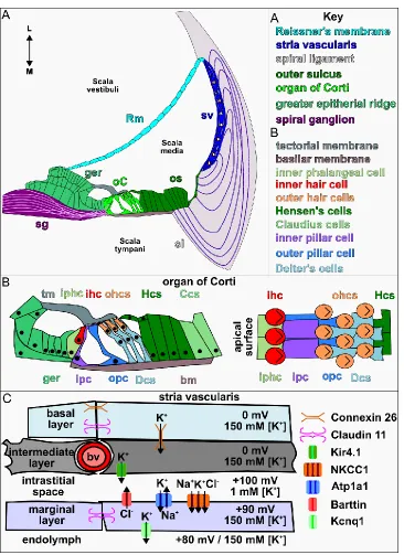
buffers the scala media. Within the scala media are the primary structures and cell
types, as well as endolymph fluid, required for hearing (Fig. 1.3A).
Lining the medial wall of the scala media is the organ of Corti which contains the
mechanosensory hair and underlying support cells which sit upon the basilar membrane
and function in the transduction of sound (Pritchard, 1878; Fig. 1.3B). The hair cells
develop from epithelial progenitors and take on neuronal properties allowing them to act
as the sensors of sound. In mammals, the hair cells are organized into 4 stereotyped
rows along the length of the cochlear duct. At the interior of the spiral a single row of
inner hair cells (IHCs) act as the primary sound transducers while the more
circumferentially located rows of outer hair cells (OHCs) function to intensify reception of
low amplitude sound. A number of actin based microvilli like projections, referred to as
stereocilia, coat the luminal surface of each hair cell in a chevron pattern. The stereocilia
are connected to each other by specialized proteins at their tips and link to mechanically
gated ion channels that open upon sound stimulation (Kachar et al., 2000; Fig. 1.5D).
Underlying and adjacent to the hair cells are a set of specialized cells, collectively
called support cells, that have additional, likely indirect, roles in hearing (Fig. 1.3A-B).
Sitting between the inner and first row of outer hair cells are a pair of pillar cells. The
pillar cells are large, column shaped, microtubule rich, cells that form the sides of an
open triangle, called the tunnel of Corti, that runs the length of the cochlea. The inner
pillar cell (IPC) sits immediately adjacent to the row of IHCs and extends a “head” across
the tunnel to connect to OHC row one. The outer pillar cell body lies adjacent to OHC
row one, while its head underlies and supports that of the IPC and extends a process to
connect with OHC row two. In the adult, the pillar cells are thought to act as structural
9
outer hair cells occurs at this point. Supporting this idea, the pillar cells are known to be
rich in acetylated- and detyrosinated-tubulin, signs of highly stable microtubules, and
display increased cellular stiffness when compared to neighboring hair cells (Arima et
al., 1986; Slepecky et al., 1995; Szarama et al., 2012a; Tannenbaum and Slepecky,
1997; Tolomeo and Holley, 1997). Experiments in gerbil cochlear explants and
mechanical modeling have shown that the pillar cells move dynamically upon sound
stimulation, likely placing these cells under shear and torsion stresses that must be
endured for proper functioning (Chan and Hudspeth, 2005; Karavitaki and Mountain,
2007; Nam and Fettiplace, 2010; Ni et al., 2016).
Sitting below the OHCs are three rows of Deiters’ cells, morphologically complex
cells that cup each OHC and send a long projection towards the luminal surface to
create a barrier between the OHCs, preventing the diffusion of ions from the endolymph.
Laterally to the OHCs and Deiters’cells are the Hensen’s and Claudius cells. These
simple, cuboidal epithelial cells function to recycle cations from the hair cells during
mechanotransduction and preserve the ionic concentration of the endolymph. The entire
organ of Corti sits between two acellular, gel-like, membranes, the basilar and tectorial
membranes, which are comprised primarily of collagen and glycoproteins. The basilar
membrane underlies the organ of Corti and bends in response to sound waves. The
tectorial membrane extends from the greater epithelial ridge and lays on top of the hair
cells providing the surface on which the stereocilia are deflected.
The lateral wall of the cochlear duct is comprised of Reissner's membrane, an
epithelial sheet separating the scala media and vestibuli, and the stria vascularis (sv), a
multilayered wall of cells responsible for generating the ionic concentrations in the
10
Spyropoulos, 1959; Fig. 1.3A,C). The stria vascularis is comprised of three layers, the
outermost basal layer interacts with cells of the spiral ligament, sharing gap junctions
claudin 11 (Cldn11) and Connexin 26 (Cx26), which bring potassium ions (K+) from the
Hensen’s and Claudius cells back into the epithelium (Kitajiri et al., 2004; Xia et al.,
1999). The central, intermediate layer, contains capillaries surrounded by specialized
melanocytes and the intrastitial fluid rich in chloride, potassium, and sodium ions. Cl- and
Na+ diffuse from the blood while K+ is pumped in from the melanocytes through Kir4.1
channels (Hibino et al., 1997). The inner most marginal layer lines the lumen of the
cochlea and contains the rectifying ion channels, Barttin (Bsnd), sodium-potassium
co-transporter (Atp1A1), and sodium–potassium–chloride co-transporter (Nkcc1) on their
basal and potassium voltage-gated channel subfamily Q member 1 (Kcnq1) on their
luminal surface (Birkenhäger et al., 2001; Flagella et al., 1999; Neyroud et al., 1997; Xia
et al., 1999; Fig. 1.3C). Atp1A1 and Nkcc1 draw K+, Na+ and Cl- from the intrastitial fluid
into the cells. Atp1A1 and Barttin then balance and remove Na+ and Cl- from the
marginal cells while Kcnq1 pumps K+ into the endolymph. This complicated orchestra of
ion movements ultimately maintains the positive potential charge within the cochlear
duct required for hearing (Fig. 1.3C).
Development of cochlear sensory structures
The complex sensory and nonsensory structures of the cochlear duct arise from
simple epithelial progenitors in the otic vesicle. The organ of Corti arises from the medial
wall of the vesicle. Early sensory progenitors can be described as early as E10.5 as a
buildup of cells in the medial wall epithelium that express the transcription factor Sox2
and notch signaling components jagged (Jag1) and lunatic fringe (Lfng) (Kiernan et al.,
11
display a dramatic reduction in sensory cells while loss of Sox2 results in a complete
absence of the organ of Corti (Kiernan et al., 2005, 2006). Furthermore, ectopic
activation of these factors, particularly Sox2, has been shown to drive otic progenitors
into a sensory fate (Liu et al., 2012a; Pan et al., 2013; Savoy-Burke et al., 2014). These
experiments demonstrate both the necessity and requirement of these genes in sensory
specification.
Following their specification and expansion, prosensory progenitors must exit the
cell cycle in order to differentiate. Starting around E12.5, expression of p27kip1 beings in
the sensory cells at the apical most extent of the cochlea and progresses towards the
base, finishing around E14.5 (Chen and Segil, 1999; Kanzaki et al., 2006; Lee et al.,
2006; Fig. 1.4A). Timely initiation of p27kip1 expression is required for proper patterning
of the organ of Corti as mouse knockouts demonstrate an overabundance of hair and
support cells, as expected for the loss of a regulator of mitosis. Interestingly, hair and
support cells do arise in these mutants and do appear to exit the cell cycle, albeit much
delayed from WT counterparts, suggesting that timely expression of p27kip1 is required
for generating proper cell numbers but not the ultimate differentiation of these cells. The
factors governing the timing of cell cycle exit remain elusive, however, recent work has
identified the RNA-binding protein Lin28b and microRNA (miRNA) let-7 as regulators of
p27kip1 expression (Golden et al., 2015). In transgenic mice, overexpression of Lin28b
results in a failure to express p27kip1 in a timely manner. The opposing phenotype, a
more rapid cell cycle exit, is seen when let-7, a target for Lin28b repression, is
overexpressed. This suggests that a critical timing window of expression for Lin28b and
12
factors must also be contributing as these models do not completely abrogate p27kip1
expression or sensory differentiation.
Once the sensory progenitors have exited the cell cycle they rapidly begin to
express differentiation markers and become specified as hair and support cells (Fig.
1.4A). The expression of the basic helix-loop-helix (bHLH) transcription factor Atonal1
Atoh1 (Math1) is the earliest marker of sensory differentiation and also progresses in a
wave like manner along the length of the cochlear duct (Bermingham et al., 1999; Chen
and Segil, 1999). Fascinatingly, this expression follows in inverse pattern to cell cycle
exit, beginning in cells at the base around E14.5 and progressing apically until E16.5.
Similarly to Sox2, Atoh1 knockout animals lack an organ of Corti as sensory cells fails to
differentiate (Bermingham et al., 1999; Pan et al., 2011). Further, overexpression of
Atoh1 is sufficient to drive presumptive support cells and cells expressing Sox2, such as
in the greater epithelial ridge, into a hair cell fate (Kawamoto et al., 2003; Woods et al.,
2004; Zheng and Gao, 2000).
How Atoh1 expression is activated in a graded fashion in the cochlea has been a
widely researched topic given its critical role in hair cell development (Fig. 1.4B).
Experiments with inactivation and ectopic expression of Sox2 have shown that it is both
necessary and sufficient for Atoh1 expression (Ahmed et al., 2012; Dabdoub et al.,
2008; Kempfle et al., 2016; Kiernan et al., 2005; Neves et al., 2012; Pan et al., 2013;
Puligilla and Kelley, 2016). After its initial activation, Atoh1 becomes autoregulatory,
binding its own promoter and activating its own transcription while becoming inhibitory to
Sox2 (Helms et al., 2000). This incoherent feedback loop is required to generate a
proper compliment of hair cells as it has been found that continued expression of Sox2
13
must be inactivated in order for hair cells to terminally differentiate as it inactivates
downstream factors, such as Pou4F3, and prevents Atoh1 autoregulation (Ahmed et al.,
2012; Dabdoub et al., 2008; Kempfle et al., 2016; Puligilla and Kelley, 2016). The
inhibitory role of Sox2 on Atoh1 is thought to be through the activation of Hey family
transcription factors Hey1 and 2, which negatively regulate Atoh1. Mouse double
knockouts for Hey1 and 2 exhibit a premature hair cell differentiation phenotype and
recent work has shown these factors bind directly to the Atoh1 promoter element,
repressing its transcriptional activity (Abdolazimi et al., 2016; Benito-Gonzalez and
Doetzlhofer, 2014).
Additional experiments have also implicated extracellular signaling from Shh and
insulin-like growth factor (Igf) in Atoh1 regulation (Fig. 1.4B). Inactivation of Shh
signaling after otic patterning, through deletion of Shh in the spiral ganglia neurons or
Smo in the cochlear duct at E12.5, leads to premature hair cell differentiation (Bok et al.,
2013; Tateya et al., 2013). A similar phenotype is observed in animals that express a
truncated, repressor form, of Gli3 which present with an expanded sensory epithelium
and ectopic hair cells (Driver et al., 2008). Ectopic activation of Shh signaling through the
use of a constitutively active Smoothened allele (SmoM2) or treating explants with Shh
ligand results in the reverse phenotype of delayed differentiation (Driver et al., 2008;
Tateya et al., 2013). In these transgenic experiments, as well as cochlear explants
treated with Shh, activation of Hey1 and 2 was found, suggesting that Shh signaling is
acting to repress Atoh1 by maintaining expression of its antagonists (Benito-Gonzalez
and Doetzlhofer, 2014; Tateya et al., 2013). In addition to Shh, inhibition of Igf signaling,
through deletion of its receptor, Igfr1, or pharmacological inhibition, also results in
14
studies, it was discovered that Sox2 was significantly upregulated, likely impairing Atoh1
expression, however a direct link between the Igf signaling pathway and Sox2 has yet to
be described.
Downstream of Atoh1, additional transcription factors, such as Brn3C/Pou4F1
and Gfi1 are activated specifically in the hair cells to further their differentiation,
maturation and are required for their survival (Erkman et al., 1996; Wallis et al., 2003;
Xiang et al., 1997). Mature hair cells express a number of specialized proteins such as
the non-muscle myosins, Myosin VI (Myo6) and Myosin VIIa (Myo7a), and tip link
proteins connecting the stereocilia, protocadherin 15 and cadherin 23 (Avraham et al.,
1995, 1997; Hasson et al., 1995; Kazmierczak et al., 2007; Siemens et al., 2004). The
timing of hair cell maturation in humans is estimated to complete by gestational week 20,
coinciding with onset of hearing function, and postnatally in mice, which begin to hear at
approximately 2 weeks of age.
Support cells are specified from a subset of the Atoh1 expressing sensory cells
and begin a separate developmental trajectory starting around E15.5. Future hair cells
will begin to express notch signaling ligands Jag2 and Delta 1 (Dll1) which activates
Notch 1 and downstream targets Hes1 and 5 in the neighboring cells (Lanford et al.,
1999; Zine et al., 2000; Fig. 1.4A). Deletion of Jag2, Dll1, Notch1, Hes1, and Hes5 lead
to varying degrees, depending on the combination of factors, of hair cell overproduction
at the expense of the support cells (Lanford et al., 1999; Tateya et al., 2011; Zheng et
al., 2000; Zine and de Ribaupierre, 2002; Zine et al., 2000, 2001). This process of lateral
inhibition, through Notch signaling, generates an alternating, “checkerboard”, pattern of
presumptive hair and support cells. The Notch activated support cells then begin to
15
et al., 2006). Interestingly, Prox1 mutants display only mild disruption of hair cell
organization and support cell development, suggesting it is not absolutely required for
their development (Fritzsch et al., 2010). Evidence from viral transduction experiments in
cochlear explants revealed that ectopic expression of Prox1 led to a repression of Atoh1
and Gfi1 and subsequent degeneration of OHCs (Kirjavainen et al., 2008). These data
support a role for Prox1 as a repressor of hair cell fate rather than as an activator of the
support cell lineage.
Differentiation of support cell progenitors into pillar and Deiters’ cells has been
shown to be dependent on Fgf signaling. Mouse knockouts for Fgf8, normally expressed
in the IHC, and Fgfr3, expressed in the support cells, demonstrate similar phenotypes
with a lack of support cell differentiation (Hayashi et al., 2007; Jacques et al., 2007a;
Mueller et al., 2002). Interestingly, the Fgfr3 knockout animals also show a fate switch of
the outer pillar cell into a Deiters’ cell, likely due to loss of responsiveness to additional
Fgf ligands, such as Fgf10 expressed from the greater epithelial ridge, and
demonstrates the strict requirement for exacting levels of Fgf signaling in support cell
fate determination (Hayashi et al., 2007; Mueller et al., 2002). Further support for this is
seen in mutants for Sprouty 2 (Spry2), an antagonist of Fgf signaling, and Fgfr3P224R,
an activating mutation in Fgfr3 that renders it responsive to additional ligands (Mansour
et al., 2009, 2013; Shim et al., 2005). These mutants result in an increased response to
Fgf signaling and the transformation of Deiters’ cells into ectopic pillar cells. Strikingly,
these phenotypes can be rescued by simply decreasing the amount of Fgf signaling
through deletion of specific ligands, Fgf8 and Spry2 or Fgf10 and Fgfr3P224R in
combination. Pillar cells continue to develop and mature until just before hearing onset,
16
complex arrays of microtubules that provide structural support for these cells begin to
form early postnatally and extend in dynamic fashion from organizing centers at the
apical and basal surfaces of the cells (Henderson et al., 1994, 1995; Mogensen et al.,
1997; Tucker et al., 1992, 1995). The acquisition of this microtubule network is
associated with alterations in the mechanical properties of these cells, such that, their
stiffness is increased over time (Szarama et al., 2012a, 2012b; Tolomeo and Holley,
1997; Zetes et al., 2012). Finally, the tunnel of Corti opens between P10 and P12, just
before hearing onset, and the pillar cells achieve their final morphology (Ito et al., 1995;
Kraus and Aulbach-Kraus, 1981).
Concomitant with their differentiation, hair and support cells undergo complex
movements to arrive at their final positions and influence the extension of the cochlear
duct. The most well studied of these movements require components of the planar cell
polarity (PCP) pathway which polarizes cells and orients them against the plane of tissue
extension. PCP components localize to the membrane of hair cells, Van Gogh-Like 2
(Vangl2) is found on the proximal side of the cell while Cadherin EGF LAG Seven-Pass
G-Type Receptor 1 (Celsr1) and Disheveled (Dvl) line the distal wall, and function to
orient the stereociliary bundles (Etheridge et al., 2008; Montcouquiol et al., 2003, 2006;
Torban et al., 2008; Wang et al., 2005, 2006). Recent work has also implicated an
interaction between G-protein coupled signaling and orientation of the kinocilium
regulated by the actions of Lgn, Bbs8, Itf29, and Daple to localize the cilium, as well as
PCP and other signaling components (Bhonker et al., 2016; May-Simera et al., 2015;
Siletti et al., 2017). Mutants for these genes exhibit similar phenotypes with misoriented
hair cells and a shortened cochlear duct that contains a buildup of hair cells at the apical
17
extension, a process by which cells migrate and reconfigure their junctions within the
plane of elongation to drive the outgrowth of a tissue. Prosensory cells initially form as a
multilayered, pseudostratified epithelium which resolves over time into the bilayered
organ of Corti. Recent work has described how prosensory cells remodel their cell
junctions into rosettes that resolve along the axis of elongation and that cell adhesion
proteins of the cadherin family, E-cadherin, N-cadherin and P120-Catenin, are involved
in both convergent extension and PCP (Chacon-Heszele et al., 2012; Driver et al.,
2017). Live cell imaging techniques have also found that hair and support cells undergo
dynamic movements, extending long processes and crawling along the basement
membrane, and that their apical migration is dependent on the cellular myosin motor,
Myosin II (Driver et al., 2017). These results paint a complex picture of sensory
development and cochlear elongation where PCP and convergent extension interact,
and potentially regulate each other, to generate the adult structure.
Development of the cochlear lateral wall
Despite the critical requirement of the stria vascularis and Reissner’s membrane
to hearing function, much less is known about their development when compared to the
organ of Corti. Proper maintenance of endocochlear potential through sv function has
been shown to be critical for both the sense of hearing as well as hair cell survival, with
nearly all the sv mutants described here demonstrating degeneration of the organ of
Corti (Ohlemiller et al., 2016; Neyroud et al., 1997; Rickheit et al., 2008; Tasaki and
Spyropoulos, 1959; Salt et al., 1987). The three layers of the sv arise from unique origins
and come together to form this complex tissue (Locher et al., 2015; Smith, 1957). The
marginal layer is derived from otic epithelium along the lateral cochlear duct and is
18
receptor β (ERR-β; Nr3b2) (Chen and Nathans, 2007). Nr3b2 ear specific knockouts
display defects in marginal cell differentiation and abnormal intermediate layers that fail
to express critical ion channels, demonstrating that this factor is required for sv
development but not its early specification. The intermediate cell layer is comprised
primarily of neural crest derived melanocytes that interdigitate with the marginal cells
and surround the intermingled capillaries to form an osmotic barrier (Freyer et al., 2011;
Hilding and Ginzberg, 1977; Locher et al., 2015; Steel and Barkway, 1989). The exact
timing of melanocyte recruitment has not been described but is known to be dependent
on hepatocyte growth factor (Hgf) signaling through receptor tyrosine kinase, c-MET
(Shibata et al., 2016). Loss of Hgf expression from the presumptive sv results in a failure
to incorporate melanocytes into the intermediate cell layer. Mouse mutants for genes
that regulate melanocyte development, such as Melanogenesis Associated Transcription
Factor (MITF) and Pax3, as well as naturally occurring albino animals, also demonstrate
breakdown in sv development and function from a lack of melanocytes (Tachibana,
2001; Cable et al., 1992; Steel and Barkway, 1989; Tachibana et al., 1992; Kim et al.,
2014; Steel and Barkway, 1989). Finally, the basal cell layer develops from the
surrounding periotic mesenchyme which condenses and takes on epithelial properties,
eventually expressing gap junction proteins shared with the spiral ligament and ion
transporters of the intermediate layer.
Reissner’s membrane develops from a thin layer of epithelial cells on the
cochlear lateral wall, immediately adjacent and lying between the organ of Corti and the
sv. Mouse knockouts for Fgf10display a complete absence of Reissner’s membrane
with little effects on other regions of the cochlea, demonstrating a critical requirement for
19
failure to develop Reissner’s membrane is found in inner ear specific knockouts for the
transcription factor Otx2, one of the earliest expressed genes on the cochlear lateral wall
(Vendrell et al., 2015). Interestingly, ear specific Otx2 mutants also display profound
defects in cochlear duct patterning with a duplicated, mirror image, organ of Corti and an
expanded sv in the domain once occupied by Reissner’s membrane. Based on these
data, it remains uncertain if Otx2is required to specify Reissner’s membrane or if the
loss of Reissner’s in these mutants is secondary to the failure to repress the domains of
the sv and sensory cells.
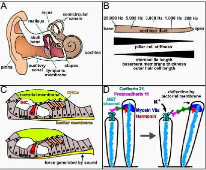
The sense of hearing and hearing loss
Sounds, at their core, are simply pressure waves that the ear detects and
translates into electrical information that is transmitted to the brain. We measure sound
by its amplitude, or loudness, in decibels (dB) and its frequency, or tone, in hertz (Hz).
Humans can hear over a wide range of frequencies and intensities, from soft breathing
at ~10Hz and 0dB to a loud siren at ~10kHz and 120dB. Interestingly, the hair cells at
each region of the cochlear duct respond only to specific frequencies, with low frequency
sound detected by cells at the apex and high frequency sound at the base, creating a
tonotopic map along the length of the duct (Fig. 1.5B). The establishment of tonotopy is
not entirely understood but is thought to arise from a combination of the properties of the
sound waves themselves, with lower frequency sound traveling further along the
cochlea, as well as increases in the length of OHC stereocilia and in the thickness and
width of the basilar membrane towards the apex of the duct.
Sound waves are collected by the outer ear and vibrate the tympanic membrane,
colloquially called the ear drum, which translates the sound mechanically through the
20
tympanic membrane moves the stapes in and out of the oval window, a small opening at
the base of the cochlear duct, and causes pressure waves to resonate through the
perilymph fluid, transmitting the sound into the cochlea. As the sound waves travel
through the cochlea they displace the perilymph fluid and cause the basilar membrane to
oscillate into the scala media (Fig. 1.5C). The oscillation of the basement membrane
causes the hair cell stereocilia to push against the tectorial membrane, deflecting them
and opening mechanically gated ion channels (Kazmierczak and Müller, 2012; Fig.
1.5D). This results in an influx of K+ into the hair cell, depolarizing it and stimulating the
release of neurotransmitter that signals to connecting neurons of the 8th cranial nerve.
Hearing impairment and deafness are the most common forms of sensory deficit
in the developed world. Loss of hearing function can arise in a multitude of ways.
Exposure to loud noise, above 120dB, direct damage and infection, normal aging and
genetic perturbations can all result in hearing loss (Furness, 2015; Venkatesh et al.,
2015). Congenital hearing impairment affects 1 in 500 newborns with approximately half
of all cases in developed countries having a genetic etiology. Single gene mutations in
over 100 different loci have been identified thus far (http://hereditaryhearingloss.org).
Mutations in the majority of these genes result in nonsyndromic sensorineural hearing
loss (SNHL), where abnormal inner ear function is the only diagnostic feature. These
mutations affect nearly every aspect of hearing function from development of sensory
cells (EYA1, POU4F3), hair cell function (MYO6, MYO7A), to maintenance of
endocochlear potential (BSND, GJB2) (Atik et al., 2015; Raviv et al., 2010; Shearer et
al., 1993; Weil et al., 1995). However, despite the large number of identified hearing loss
genes, the cause of inherited SNHL remains uncertain in many cases (Mehta et al.,
21
knowledge of inner ear development to better understand the factors that contribute to
22
Figures
Figure 1.1. The structures of the inner ear. (A) Paint fill of an E14.5 mouse embryo
displaying the anatomy and location of the inner ear within the temporal bone of the
skull. (B) Diagram of an inner ear as viewed from the posterior side with the anatomical
23
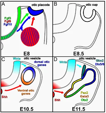
Figure 1.2. Inner ear morphogenesis and patterning. (A-D) Diagrams of mouse inner
ear development as seen on transverse section through the hindbrain at indicated
stages. (A) At E8.0 Fgf signals from the hindbrain and mesenchyme induce otic placode
formation. (B) By E8.5 the placode invaginates to form an otic cup. (C) By E10.5, the otic
cup closes at its dorsal surface and pinches off from the ectoderm to form an otic
vesicle. Hindbrain Wnt and Shh signals pattern the vesicle into dorsal and ventral gene
expression domains respectively. (D) Between E10.5 and E11.5 the otic vesicle begins
to undergo a complex series of morphogenetic changes and alterations in gene
24
Figure 1.3. The structure and cell types of the cochlear duct. (A) Diagram of a
representative transverse view of an adult cochlear duct. (B). Schematic of the organ of
Corti. (C) Schematic representation of the stria vascularis. Abbreviations: L (lateral), M
25
Figure 1.4. Timing and regulation of factors critical for the development of the
organ of Corti. (A) Diagrams of sensory development represented on an apical view of
the cochlear duct and in transverse section. Colored circles represent sensory cells at
the indicated time points along with genetic interactions. (B) Schematic of the gene
regulatory network controlling hair cell differentiation. Dashed lines represent potentially
26
Figure 1.5. The structures and mechanisms of hearing. (A) Diagram of the human
external and internal auditory system. (B) Representative schematic of the cochlear duct
displaying the range and localization of human hearing response. Changes in cell
morphology and physiology that correlate with sound frequency response are indicated
below. (C) Transverse view of the organ of Corti at rest and during movement of the
basilar membrane by sound transduction. (D) Schematic of hair cell stereocilia before
and during mechanical stimulation. Tip link proteins attached to the actin cytoskeleton
(blue spheres) of the taller stereocilia connect to the MET channels and force them open
upon deflection. Abbreviations: Hz (hertz), IHC (inner hair cell), OHC (outer hair cell),
27
Chapter 2:
ESRP1
mutations cause hearing loss due to defects
in alternative splicing that disrupt cochlear development
Introduction
Alternative Splicing
While the genomic DNA encodes nearly all of the information required for the
function of an organism it must be transcribed into a messenger RNA (mRNA) and
subsequently translated into a protein in order to enact its coded function. The direct
product of transcription is a pre-mRNA transcript which contains not only the protein
coding information, exons, but also additional stretches of intervening base pairs,
introns. To form a mature mRNA that can be translated into protein the introns must be
removed via splicing and the exons joined. Alternative splicing is an additional
posttranscriptional process that alters the exons which are retained between the pre-
and mature mRNA. The multiple types of alternative splicing events can be roughly
categorized as: cassette exons, where one or more exons are included or skipped,
mutually exclusive exons, where neighboring exons are unique to separate transcripts,
alternative 5’ or 3’ splice sites which extend the length of exons, and retained introns
(Fig. 2.1B). These events allow for a single locus to encode for multiple RNAs and
proteins, vastly increasing the complexity of a finite genome (Chen and Manley, 2009;
Fu and Ares, 2014). Recent work has discovered that ~95% of all human multi-exon
genes are alternatively spliced in a regulated manner, demonstrating that this is a nearly
ubiquitous property of protein coding genes (Pan et al., 2008; Wang et al., 2008).
The process of alternative splicing revolves around the formation of the
28
proteins that bind the pre-mRNA transcript at specific sequences and catalyze the
splicing reaction (Barabino et al., 1990; Berglund et al., 1997; Bindereif and Green,
1987; Black, 2003; Matlin and Moore, 2007; Zamore and Green, 1989). The reaction
begins with the binding of the U1 snRNP to the 5’ splice site, splicing factor 1 (SF1) to
the nucleotide which will provide the free hydroxyl group for the reaction and U2AF to
the 3’ splice site. SF1 is then replaced by U2 and the U4/U6–U5 tri-snRNP complex is
recruited to complete the spliceosome (Fig. 2.1A). Due to the relatively small size of
exons compared to introns, ~150-200bp versus thousands respectively, the binding of
these factors results in creating an exon definition where the sequence between U1 and
U2 is the intron to be spliced out (Wang et al., 2012). Long distance interaction between
the 5’ and 3’ splice site brings them into close proximity and the spliceosome complex
catalyzes the splicing reaction to remove the intervening sequence and join the exons.
Alternative splicing generally occurs when factors enhance or interfere with the
binding of U1 or U2 to their splice sites. Specialized RNA binding proteins are thought to
be the primary mediators of the inclusion or exclusion of alternatively spliced exons
based on their recruitment to cis acting elements in target transcripts (Bourgeois et al.,
1999; Chen and Manley, 2009; Fu and Ares, 2014). Alternative splicing regulators
commonly contain at least one RNA recognition motif (RRM) that binds to specific
sequences in the exons or flanking introns of target pre-mRNA transcripts. The binding
of these factors can both enhance and inhibit splicing in a context dependent manner.
Inhibition can occur through the binding of RRM containing proteins to the splice
recognition site, hampering the spliceosome complex by steric hindrance of U1 or U2 or
by direct inhibition its formation or function (Tange et al., 2001; Zhou and Lou, 2008).
29
direct the spliceosome to specific sites or facilitate formation and function of the complex
(Graveley et al., 2001; Wu and Maniatis, 1993). Additionally, binding of splice regulators
can have position dependent effects where binding upstream of an exon represses its
inclusion and binding downstream enhances inclusion.
Epithelial Splicing Regulatory Proteins
Epithelial Splicing Regulatory Proteins (Esrp) are a family of RNA binding
proteins, containing 3 RRMs, that are conserved from humans to C.elegans (Fig. 2.1C).
Mammals possess two highly homologous Esrp genes, Esrp1 and Esrp2, which share
80-90% sequence similarity in their RRM domains. The Esrps were originally
characterized in a cell-based screen for regulators of an FGFR2 splicing event
(Warzecha et al., 2009a, 2009b). FGFR2 has two primary isoforms, driven by the
inclusion of the mutually exclusive IIIB or IIIC exons, which are expressed in a cell type
specific manner and confer ligand binding preferences to the extracellular domain.
Expression of ESRP1 or 2 cDNA in mesenchymal cells drove a switch from the IIIC to
IIIB isoform of FGFR2, demonstrating the requirement for these genes in alternative
splicing (Fig. 2.1D; Warzecha et al., 2009a, 2009b). Since their initial identification,
additional genome wide studies in cell lines and mice have identified hundreds of Esrp
dependent splicing targets (Bebee et al., 2015, 2016; Cieply et al., 2016; Dittmar et al.,
2012; Fagoonee et al., 2013). Esrp1 and 2 have been found to be partially functionally
redundant, with many splicing events show a dose dependent response to loss of
individual alleles of each gene (Bebee et al., 2015). These proteins bind to 6-mers of GU
rich sequences in their target RNAs and both promote and repress splicing of all known
event types in a position dependent manner (Bebee et al., 2015; Dittmar et al., 2012).
30
as their expression is restricted to epithelial tissues throughout development and
adulthood (Bebee et al., 2015; Warzecha et al., 2009a, 2009b).
Due to their restricted expression, the Esrps have been heavily studied in the
context of epithelial-to-mesenchymal (EMT) transition. Cells undergoing EMT drastically
alter their morphology and gene expression profiles to accommodate their new identity.
Recent work has found that Esrps are downregulated during EMT which promotes a
switch to mesenchymal isoforms of alternatively spliced transcripts. In cell lines,
knockdown of Esrps results in a mesenchymal like expression pattern but not a
complete EMT and overexpression drives only a partial epithelial isoform splicing pattern
(Göttgens et al., 2016; Warzecha and Carstens, 2012; Yang et al., 2016). Thus, it
remains unclear if Esrps are drivers of EMT or an accessory pathway to assist in the
transition. Esrp function has also been studied in the context of cancer given the
contribution of an EMT component to tumor metastasis. ESRP1 was found to be
downregulated in colorectal cancer samples and mutations have been found in patients
with colitis and inflammatory bowel disease (Deloria et al., 2016; Jeong et al., 2017;
Mager et al., 2017). Several studies have also suggested that ESRP1 can act as a tumor
suppressor by inhibiting EMT and metastasis in pancreatic cancer, oral squamous cell
and non-small cell lung carcinomas (Hayakawa et al., 2017; Ishii et al., 2014; Shapiro et
al., 2011). The level of ESRPs must be tightly regulated, however, as overexpression of
this gene has also been reported to increase the metastatic properties of breast and
colorectal cancer cell lines and correlates with poor survival of breast and gastric cancer
patients (Hayakawa et al., 2017).
Esrps, and the splicing programs they regulate, are also required during
31
phenotype and defects in kidney development that manifest as a spectrum from
decreases in uretic branching and nephrons to complete kidney aplasia (Bebee et al.,
2015, 2016). Esrp2 is dispensable for development on its own, likely due to functional
redundancy with Esrp1 (Bebee et al., 2015). Double knockouts for Esrp1 and 2 present
with developmental defects in multiple organ systems in addition to the cleft-lip, palate
and kidney defects seen in the single mutants (Bebee et al., 2015). These mice are
severely reduced in size and weight and various epithelial organs are aplastic, including
the lungs and salivary glands, and the thymus is severely reduced in size. Additionally,
Esrp1/2 knockouts display forelimb agenesis with loss of multiple digits, the humerus
and radius and a skin specific deletion shows barrier defects associated with reduced
epidermal thickness and developmental defects.
Rationale
Many mammalian tissues show cell type and stage specific expression of
alternatively spliced transcripts that often fit into biologically coherent pathways (Bebee
et al., 2015; Fu and Ares, 2014; Raj and Blencowe, 2015; Traunmüller et al., 2016; Ule
et al., 2005; Vuong et al., 2016; Yang et al., 2016; Zhang et al., 2016). Consequently,
mutations that disrupt either the cis or trans regulators of alternative splicing contribute
significantly to human disease (Cieply and Carstens, 2015; Xiong et al., 2015), as well
as hearing loss in mice (Moayedi et al., 2014; Nakano et al., 2012). While many of the
key transcriptional regulators of cochlear development are known, the role that
posttranscriptional events play in the formation of inner ear structures and cell types is
less clear. A more comprehensive approach to the study of gene expression dynamics
that integrates transcriptional and posttranscriptional mechanisms is likely to improve our
32
In the current study, we performed whole exome sequencing in individuals with
profound bilateral SNHL and identified biallelic pathogenic mutations in ESRP1. Patient
derived induced pluripotent stem cells (iPSCs) showed alterations in alternative splicing,
consistent with a loss of ESRP1 function. To understand how mutations in ESRP1 might
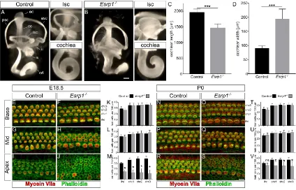
cause hearing loss we evaluated Esrp1-/- mouse embryos and uncovered defects in
inner ear morphogenesis, auditory hair cell differentiation and cell fate specification
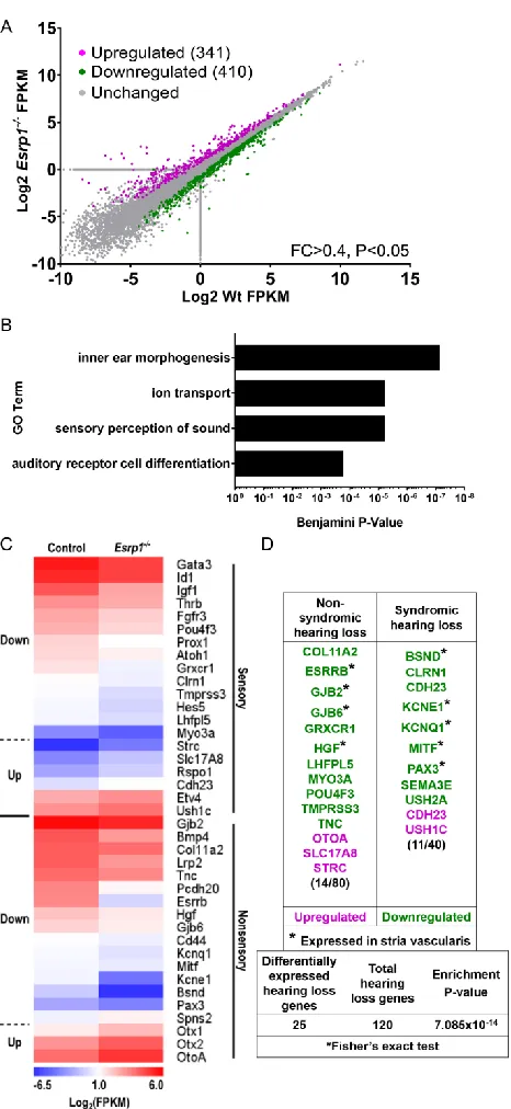
along the lateral wall of the cochlear epithelium. RNA-seq analysis revealed impaired
expression and splicing of genes associated with cochlear development and auditory
function that explain several aspects of the inner ear phenotypes in Esrp1-/- embryos. In
particular, aberrant splicing of Fgfr2 from the IIIb (epithelial) to IIIc (mesenchymal)
isoforms compromised the identity of cells along the cochlear lateral wall due to
improper Fgf9 ligand usage. Surprisingly, ectopic Fgf9/Fgfr2-IIIc signaling also
compensated for the loss of Fgfr2-IIIB to promote cochlear duct morphogenesis in
Esrp1-/- mutants. These findings implicate mutations in ESRP1 as a novel cause of
SNHL and demonstrate the complex interplay between alternative splicing, inner ear
development, and auditory function.
Results
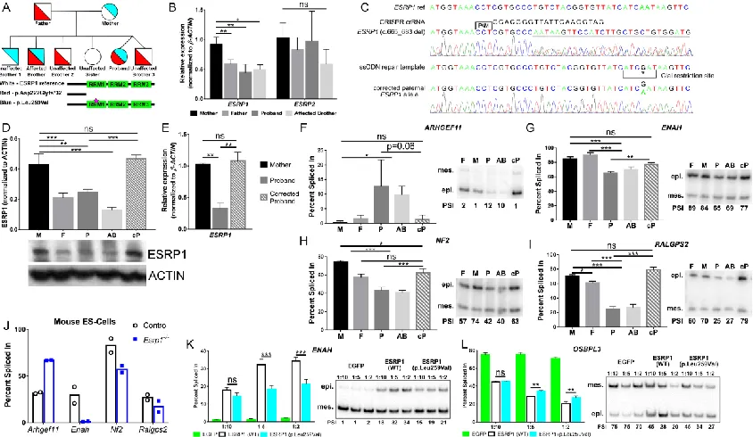
Exome sequencing reveals ESRP1 mutations in a family with SNHL
An eight-year-old female with congenital profound bilateral SNHL, born to healthy
non-consanguineous parents, was evaluated at the Division of Otolaryngology at the
Children’s Hospital of Philadelphia (Fig. 2.2A). A temporal bone CT scan of the proband
showed no abnormalities in cochlear morphology. However, an unusual vestibular
dysplasia was revealed, consisting of a rudimentary lateral semicircular canal deficient in