A Survey On Brain Tumor Detection Based On
Structural MRI Using Machine Learning And
Deep Learning Techniques.
Dhanashri Joshi, Hemlata Channe
Abstract: Structural Magnetic Resonance Image (MRI) is a useful technique to examine the internal structure of the brain. MRI is widely used for brain tumor detection as it gives a clear picture of brain soft tissues. Brain tumor identification and classification is critical and time consuming task, generally performed by radiologists. Brain tumor of different sizes and shapes can occur in any person. Extraction of exact tumor region and analysis of minute differences is difficult for humans. Digital image processing methodologies like preprocessing, segmentation and classification are useful to clinical experts for proper diagnosis of brain tumor types. This paper focuses on current trends in brain tumor detection using MRI images .Analysis of various state of the art machine learning and deep learning based methods is given. Available datasets and challenges are discussed. This extensive survey will be helpful for future research to develop better decision support system, beneficial to radiologists for accurate brain tumor diagnosis.
Index Terms: Structural MRI, brain tumor detection, Image processing, Machine learning, Image segmentation, Feature extraction
—————————— ——————————
1.
INTRODUCTION
Brain tumor is a growth of abnormal tissues inside the brain. It causes pressure in skull region and affects the normal functioning of brain. Brain tumors are classified as benign and malignant tumors. Benign tumors are non cancerous. Malignant tumors are cancerous tumors in which brain tissues split endlessly and can spread into surrounding tissues. Identification and classification of brain tumors into benign or malignant type is important for further treatment and survival of patients. Different medical image modalities like Computer Tomography(CT), Poisson Emission Tomography(PET), Magnetic Resonance Images (MRI) are used for brain tumor detection.MRI is non invasive technique which uses magnetic field and radiofrequency pulses to depict the internal body structure. Three types of MRI sequences namely T1 weighted, T2 weighted and FLAIR (Fluid Attenuated Inversion Recovery) are used for brain tumor diagnosis.
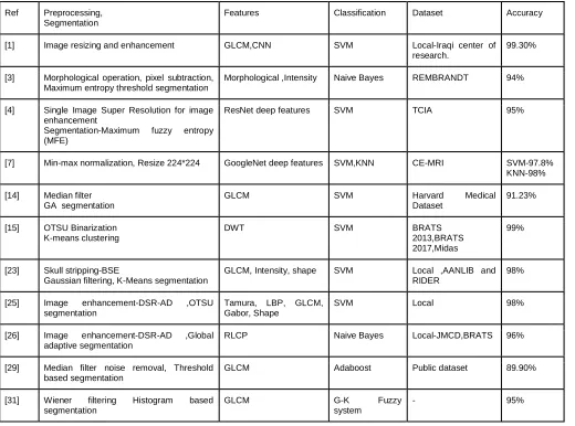
Fig.1 show different MRI sequences.
Fig. 1: T1 weighted, T2 weighted and FLAIR MRI images
The identification and detection of tumor infected area from brain MRI is critical task. Clinical experts examine medical images to identify signs and location of tumor. Due to complex nature of MRI, there is limitation for human eye to analyze the minute differences. In recent years Computer Aided Diagnosis (CAD) systems are introduced by various authors to help radiologists for accurate diagnosis. This paper focuses on review of various automatic brain tumor classification methods and highlights their strengths and limitations. Comparative analysis can be used as new research aspect for developing better brain tumor segmentation and classification system.
2
BRAIN
TUMOR
DETECTION
APPROACHES
2.1 Machine Learning Techniques
Machine learning algorithms for brain tumor detection incorporates four main stages namely Preprocessing, Segmentation, Feature Extraction and Classification.
A. Preprocessing
In medical field it is essential to get precise images for accurate observations of disease. Quality of medical images depend upon the sources of artifact acquisition such as MRI,PET,CT etc.MRI scans may contain a lot of unwanted and irrelevant parts in its actual images.MRI are influenced by Rician noise.[33] Rician noise is signal dependent and it is challenging to remove it. Image preprocessing techniques like filtering, contrast enhancement, skull stripping are used to retain original image properties.
B. Segmentation
Segmentation is used to extract Region of Interest (ROI) from digital images. It is crucial to separate out tumor region from brain MRI. Different supervised and unsupervised techniques like thresholding, soft computing, atlas based, clustering, neural network etc. exist for segmentation. Thresholding includes adaptive, global, Otsu’s, histogram based thresholding methods. Unsupervised clustering techniques include K-means, Fuzzy C means. It gives effective segmentation of brain MRI into Gray Matter (GM), White Matter (WM), Cerebrospinal Fluid(CSF).Segmentation is also performed using bio inspired algorithms like Particle Swarm ________________________
Dhanashri Joshi is currently pursuing masters degree program in computer engineering in Department of Computer Engineering, SCTR’S Pune Institute of Computer Technology, Savitribai Phule Pune University Pune, Maharashtra, India, 411043 E-mail: [email protected]
18 Optimization (PSO)[6],Genetic Algorithm(GA)[14].Advances in
segmentation shows that deep learning architectures like CNN, Mask-RNN, Unet give better performance over traditional methods.[21]
C. Feature Extraction
In feature extraction various features like shape, texture, wavelet, Gabor features are extracted from MRI. Gray-Level-Co-occurrence Matrix (GLCM) is used by most of the researchers. It is second order statistical method which can give texture features like energy, correlation, contrast etc.[5].Wavelet features are extracted using Discrete Wavelet Transform(DWT).It is applied to raw image, approximation coefficients are extracted and selected as feature vector.[19] It is observed that handcrafted features along with automatic features using deep learning models like CNN, ResNet, Capsule network has shown good performance.[1],[4].Feature reduction is achieved using PCA,GA[6],[15].
D. Classification
Brain tumors are mainly classified as benign and malignant tumors. Malignant tumors are further divided into types Glioma, Meningioma and Pituitary. WHO has given grading of Glioma into 4 different grades as shown in Fig. 2.
Fig.2 Brain Tumor Types
2.2 Deep Learning Techniques
Deep learning is generally performed by convolutional neural network which consist of input layer, output layer, hidden layers and hyper parameters. It is supervised classification method in which kernel convolves around input image to produce feature maps. DL is useful for automatic segmentation and feature extraction. Besides its popularity for medical disease diagnosis it suffers from some disadvantages like need to build complex architectures, tuning of hyper parameters, need of large amount of data for training, more training time and cost. Recent study shows that to overcome the large data availability issue extensive data augmentation methods like rotation, cropping, scaling, transformation are used. In transfer learning methods pre-trained neural network is applied on application specific dataset to extract the similar features [11].Existing transfer learning models such as VGG19,LetNet,GoogleNet,ResNet,AlexNet are used for brain tumor detection. Fig 3 shows the architecture of LetNet5.
Fig. 3: CNN LetNet5 Architecture.[34]
3 AVAILABLE
DATASETS
CAD systems developed for automatic brain tumor classification are evaluated on different publicly available databases. BrainWeb simulated brain MRI database for normal and abnormal brain is provided by McConnell Brain Imaging Centre. The Repository of Molecular Brain Neoplasia Data (REMBRANDT) containing pre-surgical multisequence MRI images of 130 patients is given by The Cancer Imaging Archive (TCIA).Database used for Medical Image Computing and Computer Assisted Intervention (MICCAI) challenge are taken from the Section of Biomedical Image Analysis (SBIA) of the Center of Biomedical Image Computing and Analytics (CBICA) at University of Pennsylvania .It is referred to Brain Tumor Segmentation (BraTS) dataset.
Table 1 Summary of available MRI datasets
4 LITERATURE
REVIEW
4.1 Brain tumor diagnosis using state of the art machine learning algorithms:
Various machine learning methods are available for brain tumor segmentation and classification through MRI in literature. Hasan et al. proposed a system for MRI brain scan classification using deep and handcrafted image features [1]. Pre-processed MRI is applied on modified GLCM for statistical feature extraction. Automatic feature extraction is done by CNN. SVM classification with a 10 fold cross-validation has shown 99.30% accuracy on 600 axial MRI scans. The proposed method has shown good performance by combining MGLCM and CNN features when compared with other transfer learning networks like AlexNet, GoogleNet. A Naive Bayes based brain tumor detection system uses threshold based maximum entropy segmentation [3]. The system is checked on REMBRANDT dataset containing 114 MRI. The proposed method has an advantage that it can detect a tumor in all possible locations of brain including the temporal lobe. A new approach for brain tumor diagnosis based on maximum fuzzy entropy segmentation and CNN is introduced in [4]. Single Image Super Resolution is applied on MRI to increase the resolution. Features are extracted from pre-trained ResNet architecture. SVM Binary classification gives 95% accuracy. Mean shift clustering technique is used for segmentation in brain tumor classification using Edge Adaptive Total Variation [5].The proposed method has two benefits, EADTV preserves edges while denoising the image and mean shift clustering updates cluster centers automatically unlike K-mean, fuzzy c-mean. An integrated design of PSO with fusion features for brain tumor detection uses Local Binary Pattern and fine-tuned capsule network for feature extraction [6]. SVM classification gives accuracy of 98.3% and 97.9% on the BRATS2018 and RIDER datasets. Proposed design has shown good results with the fusion of handcrafted and deep features. Pre-trained GoogleNet for deep feature extraction is evaluated on the CE-MRI dataset for 3 class classification into Glioma, Meningioma and Pituitary tumors using SVM and KNN classifiers with accuracy 97.8% and 98% respectively [7].Brain tumor classification approach using a multinomial logistic regression model is evaluated on BRATS 2017 dataset containing 48 images with 100% accuracy. However, the performance of the system should be verified on larger datasets [8]. An intelligent system for early assessment of brain tumor detection is suggested by Keerthana et al. [13].Noise removal and skull stripping is followed by threshold based segmentation. GLCM texture features are given to SVM for 3 class classification normal, benign and malignant tumors. The system gives good performance with GA-SVM classifier. Efficient optimization technique for brain tumor classification uses GA for tumor segmentation. GLCM texture features are given to SVM with accuracy of 91.23% [14]. Polly et al. presented system for HGG and LGG brain tumor classification using k-means segmentation [15].PCA is used to select 10 relevant features from wavelet features.SVM is used to identify normal and abnormal images. For abnormal images again SVM classifier is applied to classify HGG and LGG tumors. Proposed system shows 99% accuracy for 440 images however system need to be evaluated for large dataset with more relevant features. A method for brain tumor detection using wavelet transform uses morphological operation with threshold based approach for segmentation [19]. Amin et al. presented distinct approach for
20
Table 2: Summary of brain tumor detection using ML
4.2 Brain tumor diagnosis using Deep Learning algorithms: Brain tumor detection using deep learning algorithms is advanced field of research. Different deep learning architectures are used by researchers for automatic segmentation and classification of brain tumors. Gumaei et al. suggested Regularized Extreme Learning Method with hybrid features for brain classification [2]. A Hybrid PCA-NGIST feature extractor is used for spatial feature extraction. NGIST is a normalized feature descriptor, used to resolve the problem of image illumination and shadowing. RELM is a feed forward neural network with input, output and a single hidden layer. The proposed method is tested for 3 class classification Meningioma, Glioma, and Pituitary tumor on CE-MRI dataset with accuracy of 94.33% using 5 fold cross-validations. A light deep neural network architecture LinkNet is used for brain tumor classification in [9]. Binary classification has given 91% accuracy on publically available UCI repository dataset. Mallick et at. presented brain MRI cancer detection technique using Deep Wavelet Autoencoder based Deep Neural Network (DWA-DNN) [10].DICOM images from Rider dataset are processed to get specific image matrix and passed to Deep wavelet Autoencoder. Multi Layer Perceptron (MLP) classification has given 96% accuracy and 0.65 Kappa statistics. However in future combination of DNN with other
variations of autoencoder like denoising autoencoder, sparse autoencoder could be evaluated. Brain tumor classification system with transfer learning method is proposed in [11].MRI images are resized to 224*224 to fit VGG19 model. Block wise fine tuning of parameters like learning rate, scheduling rate, momentum is done to update the weights. System is evaluated on CE-MRI dataset with accuracy of 94.82%. The system has the disadvantage that block wise fine tuning of parameters takes 20-30 minutes to train CNN classifier. MLP is used for brain tumor classification using statistical and wavelet features [12].System is evaluated by considering statistical and DWT features separately and combined feature set. Combined feature set has shown good performance with accuracy of 96.73%. The system shows robustness of classification for large dataset containing 40,300 images. Glioma tumor detection with ANFIS classification uses Non-Sub Sampled Contourlet Transform image enhancement [16].ANFIS is used for binary classification into normal and Glioma brain tumor types on BRATS 2015 dataset The system has advantage that traditional classification algorithms like SVM, CNN gives classification error for low intensity Glioma images however ANFIS works well for both high and low intensity Glioma images .Deep neural network based system for brain tumor classification is evaluated on 66 MRI images Ref Preprocessing,
Segmentation
Features Classification Dataset Accuracy
[1] Image resizing and enhancement GLCM,CNN SVM Local-Iraqi center of research.
99.30%
[3] Morphological operation, pixel subtraction, Maximum entropy threshold segmentation
Morphological ,Intensity Naive Bayes REMBRANDT 94%
[4] Single Image Super Resolution for image enhancement
Segmentation-Maximum fuzzy entropy (MFE)
ResNet deep features SVM TCIA 95%
[7] Min-max normalization, Resize 224*224 GoogleNet deep features SVM,KNN CE-MRI SVM-97.8% KNN-98%
[14] Median filter GA segmentation
GLCM SVM Harvard Medical
Dataset
91.23%
[15] OTSU Binarization K-means clustering
DWT SVM BRATS
2013,BRATS 2017,Midas
99%
[23] Skull stripping-BSE
Gaussian filtering, K-Means segmentation
GLCM, Intensity, shape SVM Local ,AANLIB and RIDER
98%
[25] Image enhancement-DSR-AD ,OTSU segmentation
Tamura, LBP, GLCM, Gabor, Shape
SVM Local 98%
[26] Image enhancement-DSR-AD ,Global adaptive segmentation
RLCP Naive Bayes Local-JMCD,BRATS 96%
[29] Median filter noise removal, Threshold based segmentation
GLCM Adaboost Public dataset 89.90%
[31] Wiener filtering Histogram based segmentation
GLCM G-K Fuzzy
system
from AANLIB dataset. Fuzzy C-mean clustering is used for MRI segmentation.PCA is used to select relevant feature set from DWT features.7 layers DNN architecture is used for 4 class classification into classes normal, Glioblastoma, Sarcoma and Carcinoma tumors [17] .Raju et al. proposed a novel approach for brain tumor classification using Bayesian fuzzy clustering and HSC based multi SVN [18]. Feature vector consists of information theoretic, scatter and wavelet features. Proposed method has 4 level classifications with Multi SVNN. Level 1 for normal and abnormal tumors, level 2 for edema tumors, level 3 for core tumors, level 4 for advanced tumors. Weights in SVNN are optimized using HCS algorithm which is integration of Harmony search and Crow search algorithm. This approach has the advantage that Harmony search has less complexity and Crow search better converges to the global optimum in less time. Abdalla et al. presented CAD system for brain tumor detection using ANN [20]. Feed forward neural network is tested on AANLIB dataset containing 239 images with 99% accuracy using Haarlick texture features. Multigrade brain classification system using CNN is proposed in [21].Tumor is segmented using InputCascadeCNN architecture. Data samples are increased by using extensive data augmentation techniques like rotation, flip, emboss. System is evaluated on CE-MRI dataset with 94.58% accuracy using VGG19 architecture. Proposed system addresses the issue of inadequate MRI image availability by using extensive data augmentation methods. In future CAD system for brain tumor grade classification using lightweight CNN architecture could be developed for better performance. MRI based brain tumor grade classification using CNN and GA is suggested in [22].CNN with 5 convolution layers and 2 fully connected layers used to classify tumors into 3 types Glioma, Meningioma and Pituitary.CNN with 6 convolution layers and 2 fully connected layers used for four grades Glioma classification. GA is used to choose the proper parameters of CNN like no. of convolution layers, max pooling layers, no of filters, size of filters, activation function, learning rate. A novel approach for brain tumor classification using neuro fuzzy feature selection method is evaluated on BRATS dataset with 10 fuzzy rules [24].Feature extraction has been done from First-order Gray Level Statistics, GLCM, GLRL and Geometrical Shape and Size. Adaptive Neuro fuzzy classifier (ANFC) provides a good solution for high and low grade tumors classification. Linguistic hedges used for understanding fuzzy linguistic variables. A new technique for brain tumor classification using PNN is suggested in [28].K-means segmentation is followed by GLCM texture features.PNN is applied to classify brain images into 3 types benign, malignant and normal. Performance evaluation is done by comparing PNN result with other neural network architectures. A simple approach for brain tumor classification using ANN is tested on AANLIB dataset with 95% accuracy. Three colour moments mean, standard deviation and skewness are extracted from coloured MRI images [32]. Summary of different Deep Learning methods is given in Table 3
5 ANALYSIS
Brain tumor segmentation and classification is very critical and generally done by expert radiologists. Machine learning and deep learning methods can be used as decision support systems for radiologists. This study gives a summary of different state-of-the art techniques for automatic brain tumor
classification. MRI images are pre-processed using median, Gaussian, Wiener filter, histogram equalization, skull stripping methods. Segmentation is categorized into 6 types: clustering based, statistical methods, ANN, region based, threshold based edge detection methods [35].K-means, fuzzy C-means clustering, adaptive, global thresholding methods are commonly used by researchers. Segmentation using deep learning is advanced technique through which proper extraction of tumor region is achieved with greater accuracy [21]. Feature extraction is mostly performed by GLCM and DWT.GLCM gives texture features and DWT gives approximation coefficients as feature vectors. Automatic feature extraction is accomplished by deep learning architectures ResNet, capsule network [4, 6]. For dimensionality reduction PCA and bio inspired algorithms like PSO, GA are used. Selecting best features for accurate classification is a difficult task, hence hybrid approach combining different features is used for efficient classification. Classification is achieved using machine learning and deep learning algorithms. SVM with different kernels Linear, RBF, Cubic is commonly used for binary classification.CNN along with transfer learning models like VGG19, ResNet have given good classification results. Combination of fuzzy system and ANN, ANFIS shows better performance for binary classification. Standard available databases used for CAD systems are discussed in Table 1. BRATS is commonly used dataset containing T1, T2 weighted and FLAIR images. However single database doesn’t contain all types of tumors and grades for classification. Researchers have to combine the databases or need to collect MRI from local hospitals. Hence the performance of different techniques can’t be compared effectively as different data sources are used for evaluation. The standard database containing all tumor types needs to be developed, which can be used as a benchmark for further research.
6
CONCLUSION
22 Table 3: Summary of brain tumor detection using DL
REFERENCES
[1] AM. Hasan, HA. Jalab, F. Meziane , H Kahtan , AS Ahmad , ―Combining Deep and Handcrafted Image Features for MRI Brain Scan Classification,‖ IEEE Access, pp. 79959–79967, 2019.
[2] A. Gumaei, MM. Hassan, MR. Hassan, A Alelaiwi ,G. Fortino , ―A Hybrid Feature Extraction Method With Regularized Extreme Learning Machine for Brain Tumor Classification,‖IEEE Access, pp. 36266-36273, 2019. [3] HT. Zaw, N. Maneerat, KY. Win, ―Brain tumor detection
based on Naïve Bayes classification,‖ International Conference on Engineering, Applied Sciences and Technology,pp.1-4,2019
[4] E. Sert , F.Ozyurt , A.Doğantekin , " A new approach for brain tumor diagnosis system: Single image superresolution based maximum fuzzy entropy segmentation and convolutional neural network," Medical Hypothesis,pp.1-9,2019
[5] RB. Vallabhaneni, V. Rajesh,‖ Brain tumour detection using mean shift clustering and GLCM features with edge adaptive total variation denoising technique.‖ Alexandria Engineering Journal,pp.2387-2392,2018
[6] M.Sharif , J.Amin , M. Raza , M. Yasmin , SC. Satapathy,
" An Integrated Design of Particle Swarm Optimization (PSO) with Fusion of Features for Detection of Brain Tumor," Pattern Recognition Letters,pp.150-157,2020 [7] S.Deepak ,PM. Ameer,‖ Brain tumor classification using
deep CNN features via transfer learning‖, Computers in Biology and Medicine,pp.1-7,2019
[8] MH.Avizenna , I.Soesanti , I.Ardiyanto,‖ Classification of Brain Magnetic Resonance Images based on statistical features,‖ International Conference on Bioinformatics, Biotechnology, and Biomedical Engineering,pp.1-5,2018 [9] G. Hemanth , M. Janardhan , L.Sujihelen, ―Design and
Implementing Brain Tumor Detection using Machine Learning Approach,‖ Third International Conference on Trends in Electronics and Informatics,pp.1-6,2019
[10] PK. Mallick , SH.Ryu , SK.Satapathy , S.Mishra , GN.Nguyen , P. Tiwari,‖ Brain MRI Image Classification for Cancer Detection Using Deep Wavelet Autoencoder-Based Deep Neural Network,‖ IEEE Access,pp.46278-46287,2019
[11] Z. Swati , Q.Zhao , M. Kabir , F.Ali ,S. Ahmed , J.Lu,‖ Brain tumor classification for MR images using transfer learning and fine-tuning,‖ Computerized Medical Imaging and Graphics,pp.34-46,2019
Enhanced MR Image Classification Using Hybrid Statistical and Wavelets Features,‖ IEEE Access,pp.9634-9644,2018
[13] TK Keerthana , S. Xavier,‖ An Intelligent System for Early Assessment and Classification of Brain Tumor,‖ Proceedings of the 2nd International Conference on Inventive Communication and Computational Technologies,pp.1-4,2018
[14] TL. Narayana , TS.Reddy,‖ An Efficient Optimization Technique to Detect Brain Tumor from MRI Images,‖ International Conference on Smart Systems and Inventive Technology,pp.1-4,2018
[15] FP. Polly , SK Shil , MA. Hossain, A. Ayman , YM. Jang,‖ Detection and Classification of HGG and LGG Brain Tumor Using Machine Learning,‖ International conference on Information Networking,pp.813-817,2018
[16] A. Selvapandian , K. Manivannan,‖ Fusion Based Glioma Brain Tumor Detection and Segmentation using ANFIS Classification,‖ Computer Methods and Programs in Biomedicine,pp.33-38,2018
[17] H. Mohsen , E.Sayed , E. Dahshan , A. Badeeh , M.Salem,‖ Classification using deep learning neural networks for brain tumors,‖ Future Computing and Informatics Journal,pp.68-73,2018
[18] AR.Raju , P.Suresh , RR.Rao,‖ Bayesian HCS-based multi-SVNN: A classification approach for brain tumor segmentation and classification using Bayesian fuzzy clustering,‖ Biocybernetics and Biomedical Engineering,pp.646-660,2018
[19] S. Shekhar, MA. Ansari ,‖ Image Analysis for Brain Tumor Detection from MRI Images using Wavelet Transform,‖ International Conference on Power Energy, Environment and Intelligent Control,pp.1-6,2018
[20] HEM. Abdalla, MY. Esmail,‖ Brain Tumor Detection by using Artificial Neural Network,‖ International Conference on Computer, Control, Electrical, and Electronics Engineering,pp.1-6,2018
[21] M. Sajjad , S.Khan ,K. Muhammad ,W. Wu , A.Ullah , SW. Baik,‖ Multi-grade brain tumor classification using deep CNN with extensive data augmentation,‖ Journal of Computational Science,pp.174-182,2018
[22] AK. Anaraki ,A.Moosa ,F. Kazemi,‖ Magnetic resonance imaging-based brain tumor grades classification and grading via convolutional neural networks and genetic algorithms,‖ Biocybernetics and Biomedical Engineering,pp.63-74,2019
[23] J.Amin , M.Sharif , M.Yasmin , S. Fernandis ,‖ A distinctive approach in brain tumor detection and classification using MRI,‖ Pattern Recognition Letters,pp.1-10,2017
[24] S.Banerjee , S.Mitra , U.Shankar,‖ Synergetic Neuro-Fuzzy Feature Selection and Classification of Brain Tumors,‖ IEEE International Conference on Fuzzy Systems,pp.1-10,2017
[25] N. Gupta , P. Khanna,‖ A non-invasive and adaptive CAD system to detect brain tumor from T2-weighted MRIs using customized Otsu’s thresholding with prominent features and supervised learning,‖ Signal Processing: Image Communication,pp.1-9,2017
[26] N.Gupta , P.Bhatele , P. Khanna,‖ Identification of Gliomas from brain MRI through adaptive segmentation and run length of centralized patterns,‖ Journal of Computational Science,pp.1-8,2017
[27] S.Kumar , C.Dabas , G.Sunila ,‖ Classification of Brain
MRI Tumor Images: A Hybrid Approach,‖ Information Technology and Quantitative Management,pp.510-517,2017
[28] R.Lavanyadevi , M.Machakowsalya , J.NivethithaJ, N.Kumar ,‖ Brain Tumor Classification and Segmentation in MRI Images using PNN,‖ International Conference on Electrical, Instrumentation and Communication Engineering,pp.1-6,2017
[29] A.Minz , C. Mahobiya ,‖ MR Image classification using adaboost for brain tumor type,‖ IEEE 7th International Advance Computing Conference,pp.1-5,2017
[30] SA Taie , W. Ghonaim,‖ Title CSO-based algorithm with support vectormachine for brain tumor's disease diagnosis,‖ IEEE International Conference on Pervasive Computing and Communications Workshops,pp.1-5,2017 [31] AS. Shankar , A.Asokan , D. Sivakumar,‖ Brain Tumor
Classification Using Gustafson–kessel (G-k) Fuzzy Clustering Algorithm,‖ International Journal of Latest Engineering Research and Applications,pp.68-72,2016 [32] M.Nazir , F.Wahid , S.Khan ,‖ A simple and intelligent
approach for brain MRI classification,‖ Journal of Intelligent & Fuzzy Systems,pp.1127-1135,2015
[33] B. Deepa , G.Sumithra,‖ Comparative Analysis of Noise Removal Techniquesin MRI Brain Images,‖ IEEE International Conference on Computational Intelligence and Computing Research,pp.1-4,2015
[34] LetNet 5 architecture.
https://missinglink.ai/guides/convolutional-neural- networks/convolutional-neural-network-architecture-forging-pathways-future/
[35] G.Mohana , MG. Subashini,‖ MRI based medical image analysis: Survey on brain tumor grade classification,‖ Biomedical Signal Processing and Control,pp.139-161 [36] Brain tumor WHO classification.
![Fig. 3: CNN LetNet5 Architecture.[34]](https://thumb-us.123doks.com/thumbv2/123dok_us/8632662.1422370/2.612.313.575.364.572/fig-cnn-letnet-architecture.webp)