Varied clinical presentation of os odontoideum:
a case report
Karen Chrobak,
(Hons) B.H.Sc, DCa,bRyan Larson,
BSc, DCcPaula J. Stern,
BSc, DC, FCCS(C)a,da Canadian Memorial Chiropractic College, 6100 Leslie Street, Toronto, Canada
b Division of Graduate Studies, Clinical Sciences, Canadian Memorial Chiropractic College, 6100 Leslie Street, Toronto, Ontario, Canada c Private Practice, 537 Frederick St, Kitchener Ontario Canada
d Director, Graduate Education, Canadian Memorial Chiropractic College, 6100 Leslie Street, Toronto, Ontario, Canada
Disclaimers: None
Patient consent was obtained for the use of clinical information and imaging with respect to this case report. Sources of financial support: none
Corresponding Author: Dr. Karen L. Chrobak [email protected]
T: (416) 482-2340 ext. 286 F: (416) 482-2560 6100 Leslie Street, Toronto, Ontario, Canada M2H 3J1 ©JCCA 2014
Objective: To present a case of an os odontoideum and
to provide insight into the varied clinical presentations. Clinical Features: A 54 year old man presented with chronic neck pain without headache. A clinical examination was performed and the chiropractor viewed his AP and lateral radiographs. Previous flexion/ extension radiographs and MRI imaging from 2009 were requested for review. The patient was diagnosed with grade II mechanical neck pain. Treatment was rendered that day which included spinal manipulation/ mobilization. Several days later the requested imaging reports were received and described the presence of an os odontoideum.
Conclusion: In the presence of os odontoideum,
familiarity with the signs and symptoms of potential cervical instability is imperative. Health care providers
Objectif : Présenter un cas d’os odontoïde, et donner un
aperçu des différents tableaux cliniques.
Caractéristiques cliniques : Un homme de 54 ans qui
souffre de douleurs cervicales chroniques, sans maux de tête. Un examen clinique a eu lieu et le chiropraticien a vérifié sa pression artérielle et ses radiographies latérales. Des radiographies et images IRM antérieures de la flexion et l’extension, datant de 2009, ont aussi été requises pour examen. On avait diagnostiqué sur ce patient une cervicalgie mécanique de stade II. Un traitement avait été administré ce même jour, dont une manipulation / mobilisation vertébrale. Quelques jours plus tard, les rapports demandés d’imagerie sont reçus, qui font état d’un os odontoïde.
Conclusion : Il est impératif de reconnaître les signes
Introduction
Os odontoideum is the most common anomaly of the odontoid process.1 The term os odontoideum was first
coined by Giacomini in 1886 and is defined as “...an os-sicle with smooth circumferential cortical margins and no osseous continuity with the body of C2”.2 Debate within
the literature continues regarding the congenital or trau-matic etiology of os odontoideum. Regardless of etiology, both types of os odontoideum can lead to instability of C1 on C2, placing the spinal cord at significant risk of injury.3
This paper presents a case of os odontoideum in a fifty-four year old male presenting to a chiropractic clinic with longstanding neck pain. Clinical presentations, diagnosis and management are discussed. Manual therapists treating neck complaints need to be aware of this anomaly consid-ering that instability of the atlanto-axial joint secondary to an os odontoideum can have serious consequences. Case Presentation
A fifty-four year old male presented to a chiropractic clinic with a longstanding complaint of a stiff and achy neck and upper back without headache. The neck pain was focused primarily on the left and the patient reported significant right rotational restriction, especially with shoulder checking. The patient reported one previous epi-sode of neck pain in 2009 and two previous motor vehicle accidents; a roll-over accident in the 1970s and a minor accident in the 1980s. No medical attention was sought following either accident.
On examination, an alordotic cervical curve was present with mild anterior head carriage. Upper limb neurological screening was unremarkable bilaterally (upper deep
ten-don reflexes, muscle testing and sensory examination). Active and passive cervical range of motion was globally limited with pain whereas resisted cervical spine range of motion was unremarkable and graded 5/5. Static and mo-tion palpamo-tion demonstrated bilateral paraspinal muscle tension and tenderness as well as global segmental re-strictions from C3-7. The differential diagnoses after the history and physical exam included Grade II mechanical neck pain, cervical spine degenerative disc disease and cervical spondylosis.
The patient brought his cervical radiographs (AP, lat-eral) which showed moderate degenerative disc disease at the C5-C7 segments. The patient had additional radio-graphic views ordered in 2009 by his previous chiroprac-tor along with a MRI ordered by his medical docchiroprac-tor for his prior neck complaint. A request was sent to obtain the cervical spine radiographs and the MRI report. That day cervical long axis distraction mobilizations and cervical spine manipulation were administered with consent and no adverse effects were reported.
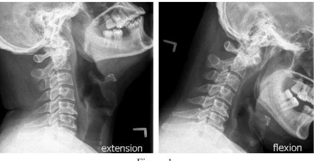
Several days later the requested radiographic and MRI reports were received by the treating chiropractor. The radiographic report, read by a chiropractic radiologist, gave the following information: “Dystopic os odontoid-eum with dynamic stenosis at C1-C2 and lateral hyper-mobility with secondary degenerative joint disease of the left lateral atlanto-axial joint. Dynamic narrowing of the spinal cord space: from 16.8 mm in extension to 15.3 mm in flexion”. (See figure 1) The MRI report stated the fol-lowing: “There is a remote fracture noted at the dens of C2. There is still some edema within this area. Suggest clinical correlation. If the fracture is recent then a CT scan
must remain diligent in their patient histories, physical exams, and imaging. This case highlights the importance of following up on imaging studies to rule out diagnoses that would involve treatment contraindications thus ensuring safe and effective treatment.
(JCCA 2014;58(3):268-272)
k e y w o r d s : os odontoideum, neck pain, cervical
anomaly, instability, chiropractic
physiques, et de l’imagerie médicale. Ce cas souligne l’importance de faire le suivi des examens d’imagerie pour écarter tout mauvais diagnostic qui engendrerait un traitement contre-indiqué, pour assurer une prise en charge sûre et efficace du patient.
(JCCA. 2014;58(3):268-272)
m o t s c l é s : os odontoïde, douleur cervicale,
of this area will be helpful to further assess the extent of healing at the fracture line”.
The patient returned to the chiropractic office a few days later and the results of the imaging reports were reviewed. The patient mentioned that the results of the 2009 imaging studies were never discussed with him. The patient was educated on the os odontoideum condition, advised that cervical spine manipulation was no longer a treatment option, and referred back to his medical doctor for further evaluation and surgical considerations. The pa-tient was not seen by an orthopaedic specialist as he was not interested in surgical intervention.
Discussion
The exact incidence and prevalence of os odontoideum is unclear within the literature as many cases remain clinic-ally silent. Considering the issues with instability and the risk of morbidity which arise in patients with os odonto-ideum, it is imperative that manual therapists are aware of potential clinical indicators of the condition. The clinical presentation of os odontoideum is quite varied within the literature and can differ significantly between patients. Although os odontoideum cannot be diagnosed without
the use of imaging, the following clinical presentations have been noted:
Asymptomatic
Many patients with os odontoideum are often delayed in their diagnosis or the os odontoideum is found as an in-cidental finding on radiographs because patients are often asymptomatic.3 The majority of asymptomatic
individ-uals are neurologically intact and only some present with incidentally discovered atlanto-axial instability.4
Neck Pain
Neck pain is one of the most common symptoms in pa-tients with a diagnosed os odontoideum. In a review of seventy eight patients identified with an os odontoideum, neck pain was the most commonly reported symptom in 64% of patients.2 It has also been reported that in some
cases, pain in the occipital or cervical region may be the only symptom.4 However, with an already high
preva-lence of neck pain in the general population and a lack of prospective studies, the link between neck pain and the presence of os odontoideum is a difficult association to make.
Figure 2.
APOM view showing an ossicle above a stump projection from the C2 centrum.
This is associated with moderate narrowing of the left lateral atlantoaxial joint with medial displacement and right lateral offset of the right C1 lateral mass.
Figure 1.
Previous flexion and extension views showing a dynamic narrowing of the spinal cord space (from C1 spinolaminar line to the posterior
Headaches
Headaches, a common condition treated by manual ther-apists, have been reported as a symptom of os odontoid-eum in the literature however not to a great extent and mostly within case reports. In a 2011 review of seventy eight patients with os odontoideum, only 2 patients pre-sented with headaches.2 The estimated lifetime
preva-lence of headache (including all headache types) has been reported to be between 93% and 98%.5 Given such a high
prevalence of headache in the general population, the as-sociation between os odontoideum and headache is un-clear.
History of Trauma
The association between os odontoideum and previous history of trauma is debated within the literature. How-ever, a large portion of evidence currently points towards a traumatic etiology in the majority of reported cases.4
There is a proposed theory that trauma may be an instigat-ing event for the development of symptoms from a pre-existing os odontoideum. In a case series by Spierings and Braakman, approximately 43% of patients presented with a history of trauma.6 Other authors maintain that previous
traumatic events may not be responsible for an os odont-oideum. These authors have proposed that a traumatic event could result in a soft tissue injury that may increase the degree of instability and thus cause a pre-existing os odontoideum to become symptomatic.7 The effect trauma
has on developing or exasperating a pre-existing os odont-oideum is still unknown. However, given the amount of evidence pointing towards a traumatic etiology, inquiring about previous trauma during a patient history may offer valuable information to warrant further investigation.
Congenital Syndromes
Os odontoideum are commonly associated with a number of congenital syndromes. It is important to keep this dif-ferential in mind in patients presenting with Down syn-drome, Klippel-Feil synsyn-drome, Morquio’s disease, mul-tiple epiphyseal dysplasia, pseudoachondroplasia, achon-droplasia, Larson syndrome, and chondrodystrophia calcificans.4 It is proposed that ligament hyperlaxity and
incomplete ossification of the odontoid process in these syndromes may predispose individuals to the develop-ment of a traumatic os odontoideum.4
Neurological Signs and Symptoms
Patients with os odontoideum can be asymptomatic however, many have also presented with a wide array of neurological symptoms. In a review of seventy eight patients with os odontoideum, eighteen patients (23%) had neurological signs or symptoms at presentation and an additional fifteen (19%) had a history of intermittent or prior neurological symptoms.2,8 Patients with an os
odontoideum may have abnormal atlanto-axial motion anteriorly, posteriorly or in both directions. Flexion of the cervical spine can cause anterior translation of C-1 leading to impingement on the dorsal aspect of the spin-al cord whereas extension can cause the anterior ring of C1 and ossicle to impinge on the ventral aspect of the cord.8 Therefore, a wide variety of neurological signs and
symptoms may present in patients with os odontoideum ranging from subtle transient myelopathy to more explicit signs such as tetraplegia, paresis, bulbar sign and central cord syndrome.4
Recommendations
The proper management of os odontoideum still remains uncertain due to the fact that it is a rare condition. The majority of the literature consists of case reports and case series making it difficult to offer evidence-based guide-lines and practice recommendations. Also, there remains a gap in knowledge of the long term natural history of untreated os odontoideum. The majority of reports indi-cate that patients tend to remain asymptomatic after a follow-up between one and seven years.2 However, one
study reported that symptomatic atlanto-axial instability can develop over time, even after a diagnosis of a ‘stable’ os odontoideum is made.9
The following recommendation has been given for patients with incidental os odontoideum: “Patients with os odontoideum, either with or without C1–2 instability, who have neither symptoms nor neurological signs may be managed with clinical and radiographic surveillance.10
However, other authors have advocated for surgical inter-vention for all patients with radiographically unstable os odontoideum, whether symptomatic or not.2 Although
Conclusion
There remains a lack of consensus within the literature regarding best practices for os odontoideum and the long term prognosis is unknown. With this uncertainty and the risk of cervical instability, it is imperative that health care professionals, particularly manual therapists who treat neck pain patients, become familiar with the signs and symptoms of potential cervical instability. These health care providers must also remain diligent in their patient histories, physical exams, and imaging studies. This case highlights the importance of following up on imaging studies to rule out diagnoses and not simply relying on the fact that they were performed. Whether or not surgical fixation is warranted is outside the scope of practice for manual therapists. However, the role of manual therapists should be to recognize signs and symptoms of os odonto-ideum, refer patients for a medical opinion and surgical consultation, and properly educate their patients on the nature and potential risks of their condition.
References
1. Fielding JW, Hensinger RN, Hawkins RJ. Os Odontoideum. J Bone Jt Surg Am. 1980;62(3):376-83.
2. Klimo P, Kan P, Rao G, Apfelbaum R, Brockmeyer D. Os odontoideum: presentation, diagnosis, and treatment in a series of 78 patients. J Neurosurg Spine. 2008;9(4):332-42.
3. Choit RL, Jamieson DH, Reilly CW. Os odontoideum: a significant radiographic finding. Pediatric Radiology. 2005;35(8):803-7.
4. Arvin B, Fournier-Gosselin MP, Fehlings MG. Os odontoideum: etiology and surgical management. Neurosurgery. 2010;66(3 Suppl):22-31.
5. Mintken PE, Metrick L, Flynn TW. Upper cervical ligament testing in a patient with os odontoideum presenting with headaches. J Ortho Sports Phys Ther. 2008;38(8):465-75.
6. Spierings EL, Braakman R. The management of
os odontoideum. Analysis of 37 cases. J Bone Jt Surg Br. 1982;64(4):422-8.
7. Brecknell JE, Malham GM. Os odontoideum: report of three cases. J Clinical Neuroscience. 2008;15(3):295-301. 8. Klimo P, Coon V, Brockmeyer D. Incidental
os odontoideum: current management strategies. Neurosurgical Focus. 2011;31(6):E10.
9. Clements WD, Mezue W, Mathew B. Os odontoideum – congenital or acquired? – that’s not the question. Injury. 1995;26(9):640-2.
10. Hadley M. Os odontoideum. Neurosurgery. 2002;50(3 Suppl):S148-55.
11. Anderson-Peacock E, Blouin J-S, Bryans R, Danis N, Furlan A, Marcoux H, et al. Chiropractic clinical practice guideline: evidence-based treatment of adult neck pain not due to whiplash. J Can Chiropr Assoc. 2005;49(3):158. 12. Qureshi MA, Afzal W, Malik AS, Ullah JS, Aebi M.
Os-odontoideum leading to atlanto-axial instability – report of surgery in four cases. J Pakistan Med Assoc. 2008;58(11):640-2.
13. Wang S, Wang C. Acquired os odontoideum: a case report and literature review. Child’s Nervous System. 2012 ;28(2):315-9.
Table 1.
Recommendations for os odontoideum
• A detailed history, physical exam and neurological exam should be completed on all neck pain patients to look for clinical indicators of instability and detect subtle myelopathies.3
• If instability is suspected, initial imaging should include cervical radiographs consisting of an open-mouth odontoid view, a lateral cervical view and flexion/extension views.3,4
• An unstable os odontoideum is an absolute contraindication to cervical spine manipulation and possibly cervical spine mobilization.11
• Atlanto-axial instability has been defined as greater than three millimeters of motion at C1-C2 on flexion/ extension films.12
• A MRI and surgical consultation is indicated if significant instability is seen on flexion/ extension radiographs or if myelopathy is detected on clinical examination.3,4,13
• All patients with an os odontoideum should be educated on potential instability.2