University of Pennsylvania
ScholarlyCommons
Publicly Accessible Penn Dissertations
1-1-2016
Bidirectional Interactions Between Mitochondrial
Function and Cell Mechanics
Judith Kandel
University of Pennsylvania, [email protected]
Follow this and additional works at:
http://repository.upenn.edu/edissertations
Part of the
Biomedical Commons
, and the
Cell Biology Commons
This paper is posted at ScholarlyCommons.http://repository.upenn.edu/edissertations/1799
For more information, please [email protected].
Recommended Citation
Kandel, Judith, "Bidirectional Interactions Between Mitochondrial Function and Cell Mechanics" (2016).Publicly Accessible Penn Dissertations. 1799.
Bidirectional Interactions Between Mitochondrial Function and Cell
Mechanics
Abstract
Energetic and structural maintenance are both critical to cellular homeostasis, and clinical disease is often characterized by alterations in both of these realms. While the manifestation of pathology in each of these fields has been extensively studied, little research has been done to characterize basic, direct interactions between mitochondrial function and cell mechanics. The experiments described in this dissertation
endeavored to address that gap, first by investigating the cytoskeletal and mechanical effects of mitochondrial dysfunction and then by considering the mitochondrial consequences of cytoarchitectural breakdown.
Mechanical integrity of the cell following mitochondrial dysfunction was investigated through multiple experimental platforms, including the quartz crystal microbalance with dissipation (QCM-D). Early work thus focused on improving the suitability of QCM-D for cell experimentation by developing a method of covalently conjugating fibronectin to QCM-D sensors. We then subjected cells to mitochondrial toxins in order to address whether and how mitochondrial dysfunction affects cell mechanics and the cytoskeleton. Cells showed characteristic rounding after long-term exposure to rotenone, an inhibitor of complex I of the mitochondrial respiratory chain. Since mitochondrial dysfunction can also be caused by genetic defects in the mitochondrial DNA (mtDNA), we also studied the cytoskeletal and mechanical variations in cells
heteroplasmic for the m.3243A>G mutation. We found a conserved, nonmonotonic relationship between m.3243A>G heteroplasmy and cell mechanics, originating in expression of actin-related genes and persisting at the levels of protein production, cytoskeletal structure, and single cell stiffness.
The second half of the dissertation considered how cytoarchitectural breakdown influences mitochondria. We first developed a novel tool for tracking individual mitochondria throughout entire cells, and then used this method to demonstrate that microtubule and microfilament depolymerization affect mitochondrial motility in opposing ways. Another set of experiments found that cytoskeletal breakdown significantly decreased mitochondrial respiration, which sometimes only occurred when mitochondria were pre-stressed by increased demands of calcium maintenance.
Together, these studies highlight direct, bidirectional interactions between mitochondrial function and cell mechanics. These findings will inform future mechanistic studies focused on a comprehensive understanding of human disease at the cellular level, which will hopefully contributing to advancing development of therapeutics.
Degree Type
Dissertation
Degree Name
Doctor of Philosophy (PhD)
Graduate Group
Bioengineering
First Advisor
David M. Eckmann
Keywords
actin, atomic force microscopy, cell mechanics, mitochondria, mitochondrial motility, mitochondrial respiration
Subject Categories
Biomedical | Cell Biology
BIDIRECTIONAL INTERACTIONS BETWEEN MITOCHONDRIAL FUNCTION
AND CELL MECHANICS
Judith Kandel
A DISSERTATION
in
Bioengineering
Presented to the Faculties of the University of Pennsylvania
In Partial Fulfillment of the Requirements for the
Degree of Doctor of Philosophy
2016
__________________________
David M. Eckmann, Ph.D, M.D, Professor of Anesthesiology and Critical Care and Bioengineering
Supervisor of Dissertation
__________________________
Jason A. Burdick, Ph.D, Professor of Bioengineering
Graduate Group Chairperson
Dissertation Committee
Marni J. Falk, M.D, Assistant Professor of Pediatrics, Children's Hospital of Philadelphia
Paul A. Janmey, Ph.D, Professor, Physiology and Institute for Medicine and Engineering
ii
ACKNOWLEDGMENTS
This work would not have been possible without the support of many people. First and
foremost, thank you to my advisor, Dr. David M. Eckmann, for your research mentorship over the
last 6 years. You have invested a tremendous amount of time, money, and energy in my
development as a scientist, and I will forever be indebted to you for that. I would also like to thank
my dissertation committee members, Drs. Paul Janmey, Marni Falk and Rob Mauck for their
support and mentorship.
There have been many other lab members, both from the Eckmann lab and beyond, who
were instrumental in the success of this research. Drs. Peter Sobolewski and Alexandra Klinger
were so giving of their time and knowledge in training me when I first came to the lab. Peter in
particular gave me my very first exposures to live imaging of mitochondria and trained me in cell
culture techniques. Dr. Hyun-Su Lee has also been dedicated to my success and has been
particularly helpful with QCM-D research and any other projects involving chemical synthesis. Dr.
M. Carme Coll Ferrer was also a wonderful friend and research mentor during her time in the lab.
I would like to thank our lab technicians, Nancy Tomcyzk, Jessica Campo, Abhay Ranganathan,
and Bruce Braender for keeping the lab running smoothly. Nancy was heavily involved in the
chemical conjugation of fibronectin onto QCM-D crystals (Chapter 3) and is co-authored on the
paper relating that work. Bruce Braender was an integral part of the QCM-D experiments
presented in Chapter 4. Dr. Meg Grady, a post-doc in our lab, has been a valuable source of
scientific feedback, particularly with AFM experiments. I would also like to thank Eric Abhold, who
joined us for a summer research project, for teaching me Western blot fundamentals.
I was lucky enough to enjoy the core facilities belonging to the Anesthesia and Critical
Care research department on the third floor of the John Morgan Building. Thank you to Dr.
Roderic Eckenhoff for his service as the Vice Chair for research in the Anesthesia department. In
addition to Eric, Brian Weiser, Kellie Woll and Tim DeYoung were amazingly helpful with Western
iii
immunofluorescence in particular. Drs. Qing Cheng Meng and Weiming Bu were great guides to
both general and specific procedural questions. Thank you also to Drs. Max Kelz and Maryellen
Eckenhoff for providing a listening ear and professional guidance when I needed it.
I have also relied on our support staff on JM3 throughout my training: Cindy Bliven, Ann Marie
Tenuto, and Randall Hardie. Thank you for doing everything from organizing potlucks to placing
orders for reagents. My work certainly would not have gone as smoothly as it did without you.
Like many interdisciplinary projects, my work has benefited from the collaboration of
many other scientists in other laboratories. Thanks to Dr. Russell Composto, a longtime
collaborator of the Eckmann lab, for his general involvement and encouragement, and his insights
particularly on the chemFN functionalization of QCM-D crystals. The AFM experiments, as well,
could not have been done without many other people. Dr. Prathima Nalam conjugated colloids
onto my AFM tips and was helpful with general AFM questions. Dr. Matt Caporizzo’s expertise in
the TIRF-AFM instrumentation saved my experiments on many different days, and Dr. Matt
Brukman of the Nano-Bio Interface Center was also always available for AFM troubleshooting.
Emmabeth Parrish Vaughn from Russ’s lab has been a great friend and collaborator in our push
to publish the kurtosis method of measuring actin stress fiber alignment (Appendix A).
Collaboration was also critical to some of our mitochondrial work. Thank you to Dr.
Douglas C. Wallace and his lab members, Drs. Martin Picard and Alessia Angelin, for their help in
bringing these projects to fruition. Martin was the one who shared the initial data showing altered
actin gene expression in the m.3243A>G cybrids with me, which developed into the great story of
mitochondrial genetics affecting single-cell mechanics told in Chapter 5. In addition to helping
with Seahorse XF Analyzer experiments (Chapter 7), Alessia initially showed me some images of
actin-stained cells in the hopes of quantifying them somehow. This work developed into the
single-cell kurtosis analysis related in Appendix A. I would also like to thank Drs. Xilma
Ortiz-Gonzalez and Tal Yardeni from the Wallace lab for their friendship and scientific collaboration.
Perhaps more important than the network of scientific collaborators contributing to this
iv
parents and grandparents (those with us and those who have passed on) for their love and
support. Now that I am a parent myself, I appreciate even more how much parents invest in their
children and recognize that I would not be who I am today without my parents and ancestors. I
would also like to acknowledge my siblings, in-laws, and siblings-in-law for their love and support
over the last few years. Additional thanks are due to my friends for being a wonderful network of
support over the last few years. I feel fortunate that there are too many of you to list here, but I
am thinking of all of you. I hope that I have given you in return a fraction of the friendship that you
have extended to me and my family.
Having children while completing a Ph.D. in experimental science is quite uncommon,
and I can definitely understand why after having done it. At the same time, my sons Ezra and
Elan are without question the best things I have ever produced, and I am so grateful to have had
them join me in this experience. There were countless times when I arrived home after a
particularly frustrating day in the lab, but was reminded by my husband and children of how
blessed and fulfilled my family life is. Becoming a parent also imbued with me with a new sense
of mission, and I truly believe that this drive has pushed me to produce more and better scientific
work. Ezra and Elan, thank you for bearing with me in this delicate balancing act between being
your mom and a scientist. I am hoping that as you grow older, you can increasingly relate to the
scientist side of me in addition to the mom side.
Finally, to my husband Ben: words cannot suffice to thank you for everything you have
done over the last 6 years while also pursuing your own Ph.D. in the process. It’s hard to believe
that we have been working towards these degrees for virtually our entire marriage. I would be
remiss if I did not acknowledge your scientific contributions to my work, including valuable
statistical input and general scientific feedback. Much more than that, though, you have been my
anchor throughout this journey, and what a journey it has been. Thank you for enriching my life in
so many immeasurable ways, and for being there to celebrate with me in my triumphs and
comfort me in my failures and frustrations. Most of all, thank you for believing in me when I didn’t
v
ABSTRACT
BIDIRECTIONAL INTERACTIONS BETWEEN MITOCHONDRIAL FUNCTION
AND CELL MECHANICS
Judith Kandel
David M. Eckmann
Energetic and structural maintenance are both critical to cellular homeostasis, and clinical
disease is often characterized by alterations in both of these realms. While the manifestation of
pathology in each of these fields has been extensively studied, little research has been done to
characterize basic, direct interactions between mitochondrial function and cell mechanics. The
experiments described in this dissertation endeavored to address that gap, first by investigating
the cytoskeletal and mechanical effects of mitochondrial dysfunction and then by considering the
mitochondrial consequences of cytoarchitectural breakdown.
Mechanical integrity of the cell following mitochondrial dysfunction was investigated
through multiple experimental platforms, including the quartz crystal microbalance with dissipation
(QCM-D). Early work thus focused on improving the suitability of QCM-D for cell experimentation
by developing a method of covalently conjugating fibronectin to QCM-D sensors. We then
subjected cells to mitochondrial toxins in order to address whether and how mitochondrial
dysfunction affects cell mechanics and the cytoskeleton. Cells showed characteristic rounding
after long-term exposure to rotenone, an inhibitor of complex I of the mitochondrial respiratory
chain. Since mitochondrial dysfunction can also be caused by genetic defects in the mitochondrial
DNA (mtDNA), we also studied the cytoskeletal and mechanical variations in cells heteroplasmic
for the m.3243A>G mutation. We found a conserved, nonmonotonic relationship between
m.3243A>G heteroplasmy and cell mechanics, originating in expression of actin-related genes
vi
The second half of the dissertation considered how cytoarchitectural breakdown
influences mitochondria. We first developed a novel tool for tracking individual mitochondria
throughout entire cells, and then used this method to demonstrate that microtubule and
microfilament depolymerization affect mitochondrial motility in opposing ways. Another set of
experiments found that cytoskeletal breakdown significantly decreased mitochondrial respiration,
which sometimes only occurred when mitochondria were pre-stressed by increased demands of
calcium maintenance.
Together, these studies highlight direct, bidirectional interactions between mitochondrial
function and cell mechanics. These findings will inform future mechanistic studies focused on a
comprehensive understanding of human disease at the cellular level, which will hopefully
vii
TABLE OF CONTENTS
ACKNOWLEDGMENTS ... II
ABSTRACT ... V
TABLE OF CONTENTS ... VII
LIST OF TABLES ... XII
LIST OF FIGURES ... XIII
CHAPTER 1 : INTRODUCTION ... 1
1.1 OVERVIEW ... 1
1.2 MITOCHONDRIA IN CELLULAR HEALTH AND DISEASE ... 2
1.2.1 The roles of mitochondria in cellular function ... 2
1.2.2 Mitochondrial dysfunction in disease ... 5
1.3 CELL MECHANICS IN HEALTH AND DISEASE ... 7
1.3.1 Overview of cell mechanics ... 7
1.3.2 Pathological changes in cell mechanics ...10
1.4 LINKAGES BETWEEN MITOCHONDRIAL AND MECHANICAL FUNCTION ..11
1.5 SUMMARY ...14
CHAPTER 2 : RESEARCH OVERVIEW ... 16
2.1 OBJECTIVE ...16
2.2 SPECIFIC AIMS, HYPOTHESES AND OUTCOMES ...16
2.2.1 Specific Aim 1: Assess whether mitochondrial dysfunction influences cytoarchitectural integrity and cell mechanics ...16
2.2.2 Specific Aim 2: Characterize the effects of cytoskeletal breakdown on mitochondrial function ...18
2.3 CHAPTER OUTLINE ...20
CHAPTER 3 : CHEMICALLY GRAFTED FIBRONECTIN FOR USE IN QCM-D
CELL STUDIES ... 22
3.1 CONTEXT AND ACKNOWLEDGEMENTS ...22
3.2 INTRODUCTION ...23
3.3 MATERIALS AND METHODS ...27
3.3.1 Surface Preparation ...27
3.3.2 Surface Characterization ...28
3.3.3 FTIR ...28
3.3.4 AFM measurements ...28
viii
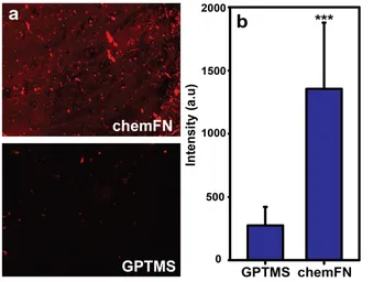
3.3.6 Fluorescence Microscopy ...31
3.3.7 Flow chamber experiments ...32
3.3.8 QCM-D experiments ...32
3.3.9 Statistics ...34
3.4 RESULTS AND DISCUSSION ...35
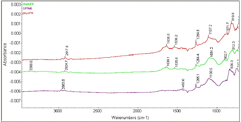
3.4.1 Immobilization of fibronectin on silicon oxide surfaces ...35
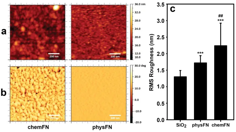
3.4.2 ChemFN and physFN surface characterization using AFM ...40
3.4.3 Biocompatibility evaluation of chemFN surfaces ...43
3.4.3.1 Cell metabolic activity and cytoskeletal structure ...43
3.4.3.2 Cell adhesion and adhesion strength ...44
3.4.3.3 Cell proliferation ...44
3.4.3.4 Calcium release in response to addition of extracellular ATP ...44
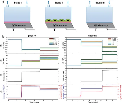
3.4.4 QCM-D ...45
3.5 CONCLUSIONS ...52
CHAPTER 4 : CYTOSKELETAL AND MECHANICAL EFFECTS OF
PHARMACOLOGICALLY INDUCED MITOCHONDRIAL DYSFUNCTION ... 53
4.1 CONTEXT AND ACKNOWLEDGEMENTS ...53
4.2 INTRODUCTION ...54
4.3 MATERIALS AND METHODS ...56
4.3.1 Cell culture and reagents ...56
4.3.2 Cell viability measurements ...57
4.3.3 Fluorescence microscopy ...58
4.3.4 Atomic force microscopy ...59
4.3.5 Quartz crystal microbalance with dissipation ...62
4.3.5.1 Sensor preparation ...62
4.3.5.2 Experimental setup ...62
4.3.5.3 Measuring cellular properties in the QCM-D ...63
4.3.5.4 Staining cells on sensors and dishes ...64
4.3.5.5 Cleaning the QCM-D ...64
4.3.6 Statistics ...65
4.4 RESULTS ...65
4.4.1 Rotenone mildly reduces cell proliferation ...65
4.4.2 Rotenone induces rearrangement of the actin cytoskeleton ...65
4.4.3 Single cell stiffness remains unchanged following treatment with mitochondrial toxins ...68
4.4.4 Challenges in assessing changes in cell mechanics using QCM-D ...70
4.5 DISCUSSION ...72
CHAPTER 5 : PROGRESSIVE INCREASE IN MTDNA 3243A>G
HETEROPLASMY IS ASSOCIATED WITH NONMONOTONIC ALTERATIONS
IN CYTOSKELETAL PROTEIN EXPRESSION AND CELL MECHANICS ... 75
5.1 CONTEXT AND ACKNOWLEDGEMENTS ...75
5.2 INTRODUCTION ...77
5.3 MATERIALS AND METHODS ...79
5.3.1 Generation of transmitochondrial cybrids ...79
ix
5.3.3 Gene expression measurements ...81
5.3.4 Cell culture ...82
5.3.5 Protein isolation and Western blotting ...83
5.3.6 Immunofluorescence and fluorescence microscopy ...84
5.3.7 Image processing and analysis ...86
5.3.8 Atomic force microscopy ...87
5.3.9 Statistics ...90
5.4 RESULTS ...90
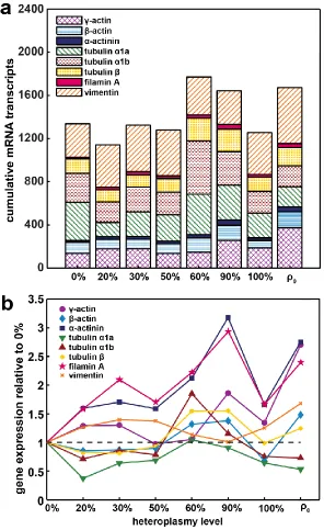
5.4.1 Gene Expression ...90
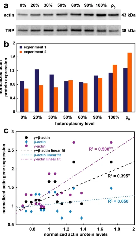
5.4.2 Protein Abundance ...93
5.4.3 Actin and Actinin Imaging ...96
5.4.4 Cell Stiffness Measurements ... 101
5.5 DISCUSSION ... 105
CHAPTER 6 : AUTOMATED DETECTION OF MITOCHONDRIAL MOTILITY
AND ITS DEPENDENCE ON CYTOARCHITECTURAL INTEGRITY ... 113
6.1 CONTEXT AND ACKNOWLEDGEMENTS ... 113
6.2 INTRODUCTION ... 114
6.3 MATERIALS AND METHODS ... 117
6.3.1 Cell culture and reagents ... 117
6.3.2 Fluorescence microscopy ... 119
6.3.3 ImageJ image pre-processing ... 120
6.3.4 Matlab algorithm ... 121
6.3.5 Statistics ... 127
6.3.6 Videos ... 127
6.4 RESULTS ... 128
6.4.1 Algorithm ... 128
6.4.2 Net distances traveled by mitochondria follow a lognormal distribution ... 128
6.4.3 CCCP decreases mitochondrial motility ... 132
6.4.4 Microtubule depolymerization decreases net distance traveled by mitochondria, while microfilament depolymerization increases it ... 134
6.5 DISCUSSION ... 137
6.6 ANALYSIS OF MITOCHONDRIAL MOTILITY IN PATIENT SAMPLES ... 143
6.6.1 Introduction ... 143
6.6.2 Materials and methods ... 143
6.6.3 Results ... 145
CHAPTER 7 : MITOCHONDRIAL RESPIRATION IS SENSITIVE TO
CYTOARCHITECTURAL BREAKDOWN ... 148
7.1 CONTEXT AND ACKNOWLEDGEMENTS ... 148
7.2 INTRODUCTION ... 149
7.3 MATERIALS AND METHODS ... 151
7.3.1 Cell culture and reagents ... 151
7.3.2 Assessing changes in mitochondrial inner membrane potential and morphology ... 152
7.3.3 Oxygen consumption measurements ... 155
7.3.4 Data analysis and statistics ... 157
x
7.4 RESULTS ... 158
7.4.1 Mitochondrial potential is unchanged following treatment with cytoskeletal toxins ... 158
7.4.2 Specific cytoskeletal toxins induce shortening of mitochondria ... 160
7.4.3 Cytoskeletal toxins caused decreased cellular basal oxygen consumption rates ... 162
7.4.4 Cytoskeletal inhibitors sensitize cells to changes in calcium homeostasis ... 165
7.5 DISCUSSION ... 167
7.6 CONCLUSIONS ... 172
CHAPTER 8 : SUMMARY, LIMITATIONS, AND FUTURE DIRECTIONS ... 174
8.1 SUMMARY ... 174
8.2 LIMITATIONS ... 175
8.2.1 General need for mechanistic studies ... 175
8.2.2 Limitations of individual studies ... 176
8.2.2.1 QCM-D experimentation with cells... 176
8.2.2.2 Clinical applicability of m.3243A>G cybrid studies ... 177
8.2.2.3 Resolution of mitochondrial motility studies ... 177
8.3 FUTURE DIRECTIONS ... 179
8.3.1 Effects of rotenone on cell mechanics and the cytoskeleton ... 179
8.3.1.1 Stress fiber alignment ... 179
8.3.1.2 Traction force microscopy ... 181
8.3.2 Mechanics of cells from patients with mitochondrial diseases ... 182
8.3.3 Influence of substrate stiffness on mitochondrial motility ... 183
8.3.4 Interactions between mitochondrial morphology and respiration ... 185
8.4 FINAL REMARKS ... 186
APPENDIX A : QUANTIFICATION OF ACTIN ALIGNMENT AS AN INDICATOR
OF CELLULAR HEALTH ... 188
A.1 BACKGROUND AND RATIONALE ... 188
A.2 MATERIALS AND METHODS ... 189
A.2.1 Image processing and Matlab object recognition ... 189
A.2.2 Measuring kurtosis ... 191
A.2.3 Statistics ... 192
A.3 RESULTS AND DISCUSSION ... 194
A.3.1 Kurtosis values are associated with cell genotype ... 194
A.3.2 Therapeutics targeting underlying disease restore actin alignment ... 194
APPENDIX B : MITOCHONDRIAL AND MECHANICAL EFFECTS OF CELL
SWELLING... 198
B.1 BACKGROUND AND RATIONALE ... 198
B.2 MATERIALS AND METHODS ... 198
B.2.1 Cell culture and reagents ... 198
B.2.2 Fluorescence microscopy ... 199
xi
B.2.4 Measuring mitochondrial motility ... 200
B.2.5 Atomic force microscopy ... 200
B.2.6 Statistics ... 201
B.3 RESULTS ... 201
B.3.1 Mitochondria shorten but do not depolarize in response to osmotic swelling ... 201
B.3.2 Mitochondrial motility is unchanged following osmotic swelling ... 203
B.3.3 Osmotic swelling causes structural changes in the cytoarchitecture ... 204
B.3.4 Cell stiffness is decreased following osmotic swelling ... 206
B.4 DISCUSSION ... 207
APPENDIX C : CUSTOM MATLAB CODES ... 209
C.1 MATLAB CODE FOR MITOCHONDRIAL TRACKING ... 209
C.2 MATLAB CODE FOR ANALYSIS OF MITOCHONDRIAL MORPHOLOGY .. 216
C.3 MATLAB CODE FOR ANALYSIS OF CELL CIRCULARITY AND ACTIN CONTENT IN M.3243A>G CYBRIDS ... 218
xii
LIST OF TABLES
Table 3.1: Parameters of QCM-D studies with low and high density cell seeding. .50
Table 5.1: Coefficients of determination and adjusted p-values showing significance of linear relationships between expression of different
cytoskeletal genes in m.3243A>G cybrids. ...92
Table 5.2: Coefficients of determination and adjusted p-values between stiffness measurements of m.3243A>G cybrids and different cytoskeletal genes. ... 104
Table 6.1: Motility parameters from all groups and p-values resulting from
pairwise K-S tests. ... 136
Table 6.2: Mitochondrial motility parameters and statistics in patient and control fibroblasts. ... 147
xiii
LIST OF FIGURES
Figure 3.1: Method of chemical conjugation of fibronectin and resulting
characterization parameters. ...36
Figure 3.2: QCM traces of chemFN and physFN coated QCM-D sensors under flow. ...37
Figure 3.3: Water contact angle measurements on a chemFN layer and two physFN layers. ...38
Figure 3.4: Fluorescence microscopy illustrating presence of fibronectin on chemFN surfaces. ...39
Figure 3.5: Fourier transform infrared spectroscopy of GPTMS, physFN and chemFN. ...40
Figure 3.6: AFM images of a GPTMS coated sensor. ...41
Figure 3.7: AFM Images of chemFN and physFN coated QCM-D sensors. ...42
Figure 3.8: Cellular characterization on chemFN and physFN sensors. ...43
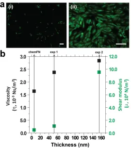
Figure 3.9: QCM-D measurements illustrating superior detection of cells by chemFN sensors as compared to physFN sensors. ...47
Figure 3.10: Viscosity and shear modulus calculated for sensors coated at different cellular densities. ...49
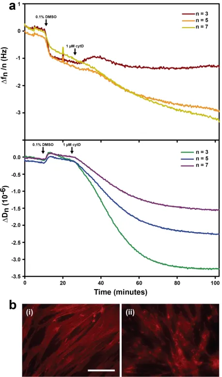
Figure 3.11: QCM-D measurements of cells treated with cytochalasin D...51
Figure 4.1: Effects of mitochondrial toxins on cell proliferation. ...66
Figure 4.2: Microtubules remain intact following treatment with mitochondrial toxins. ...66
Figure 4.3: Rotenone causes restructuring of the actin cytoskeleton in some cells. ...67
Figure 4.4: Mitochondrial toxins do not affect cell stiffness. ...69
Figure 4.5: Challenges with using QCM-D for long-term cell experimentation. ...71
Figure 5.1: Schematic illustrating the generation of the m.3243A>G cybrid cell lines. ...81
xiv
Figure 5.3: Actin protein production varies in the m.3243A>G cybrid cell lines and correlates with gene expression levels. ...95
Figure 5.4: Sample images of different m.3243A>G cells with stained microtubules and nuclei. ...97
Figure 5.5: Sample fluorescent images demonstrate cytoarchitectural
heterogeneity among several of the m.3243A>G cell lines. ...98
Figure 5.6: Sample images of m.3243A>G cells with stained actin, actinin, and nuclei. ...99
Figure 5.7: Stiffness measurements highlight mechanical differences among the m.3243A>G cybrids. ... 103
Figure 5.8: MtDNA m.3243A>G heteroplasmy affects cell mechanics. ... 106
Figure 6.1: Fluorescent images of control cells with fluorescent labeling of either microtubules or microfilaments. ... 119
Figure 6.2: Image processing of a sample cell image. ... 121
Figure 6.3: Matlab based algorithm tracks centroid locations of all objects as they travel throughout a time-lapse recording. ... 122
Figure 6.4: Descriptive data of mitochondrial objects. ... 126
Figure 6.5: Net distances traveled by mitochondria are lognormally distributed. 130
Figure 6.6: Combining cells in a given group preserves the lognormal distribution of net distances traveled by their mitochondria. ... 131
Figure 6.7: CCCP and nocodazole lower the distribution of net distances traveled by mitochondria while cytochalasin D raises it. ... 133
Figure 6.8: Cells after 30 minutes of treatment with 5 µM CCCP. ... 134
Figure 6.9: Box plots showing motility data from all groups. ... 135
Figure 6.10: Image of the 1 µM cytochalasin D-treated cell shown in Video 6.6. .. 142
Figure 6.11: Microtubules and mitochondria in fibroblasts from control and two different patients. ... 145
Figure 6.12: Mitochondrial tracking in normal and diseased patient fibroblasts. . 146
xv
Figure 7.2: Mitochondrial potential remains unchanged following treatment with
cytoskeletal toxins. ... 159
Figure 7.3: Specific cytoskeletal toxins induce shortening of mitochondria. ... 161
Figure 7.4: Number of mitochondria does not change following treatment with cytoskeletal toxins. ... 162
Figure 7.5: Cytoskeletal inhibitors affect basal mitochondrial oxygen consumption rates in intact cells. ... 164
Figure 7.6: Experiment shown in Figure 7.5a with outliers included. ... 165
Figure 7.7: Cytoskeletal inhibitors sensitize cells to changes in calcium homeostasis, leading to decreased basal mitochondrial oxygen consumption rates. ... 166
Figure 8.1: Effects of a single pixel on centroid positioning. ... 178
Figure 8.2: Actin stress fiber alignment in control and rotenone-treated bMSCs.181 Figure 8.3: Polyacrylamide gels of different stiffnesses. ... 184
Figure 8.4: Cells spread different on gels with different stiffnesses. ... 184
Figure A.1: Pre-processing of original images using ImageJ in order to highlight actin filaments. ... 190
Figure A.2: Quantification of actin filament orientation. ... 193
Figure A.3: Kurtosis values are associated with cell genotype. ... 195
Figure A.4: Scatter/boxplots showing all data. ... 196
Figure B.1: Mitochondria shorten in response to osmotic swelling. ... 202
Figure B.2: Mitochondrial net distances are unchanged following osmotic swelling. ... 203
Figure B.3: Microtubules show slight structural changes following osmotic swelling. ... 204
Figure B.4: Microfilaments disintegrate in proportion to degree of osmotic challenge. ... 205
1
CHAPTER 1: INTRODUCTION
1.1 OVERVIEW
Mitochondrial function and cellular mechanical integrity are both fundamental
regulators of mammalian cell function. All cells need energy to survive, and in most
cases mitochondrial metabolism provides that energy. Mitochondria are also involved in
other critical aspects of cellular function, such as reactive oxygen species generation
and calcium homeostasis. Cell mechanics, which involves maintaining structure as well
as responding to and generating force, is also crucial to many basic cellular processes
such as tissue morphogenesis and cell migration. Some cell types are also particularly
involved in the push and pull of mechanical forces, including endothelial and bone cells.
When studied at the cellular level, human disease often manifests as changes in both
mitochondrial and mechanical homeostasis. The experiments described in this
dissertation probe causational interactions between alterations in these two critical
aspects of cellular maintenance.
The following introduction is intended to familiarize the reader with basic aspects
of both mitochondrial function and cell mechanics in cellular pathology and
pathophysiology. Many of the topics touched upon in the following pages are themselves
the products of research spanning lifetimes, and it would be impossible to present a
comprehensive overview of each one of them. Therefore, the references provided are
frequently review articles which distill decades of findings on a given subject into a few
2
1.2 MITOCHONDRIA IN CELLULAR HEALTH AND DISEASE
1.2.1 The roles of mitochondria in cellular function
Mitochondria are fundamentally known as the “powerhouses” of the cell for their
role in producing adenosine triphosphate (ATP) to provide energy for cellular processes.
Glycolysis, which takes place in the cytosol, involves the breakdown of glucose to
pyruvate. Pyruvate enters the mitochondrial matrix, where its catabolism leads to the
release of carbon dioxide and the reduction of nicotinamide adenine dinucleotide
(NADH, in reduced form). Beginning a process known as oxidative phosphoylation
(OXPHOS), NADH then donates two electrons to complex I of the respiratory chain,
which resides in the mitochondrial inner membrane. Protons are pumped from the
mitochondrial matrix to the intermembrane space as these electrons are passed through
various components of the respiratory chain, building up an electrochemical gradient.
Oxygen serves as the final electron acceptor which combines with hydrogen ions to form
water, and the flow of protons back into the mitochondrial matrix powers ATP synthase
to produce ATP.
Beyond energy production, mitochondria are involved in many other signaling
pathways critical to cellular maintenance. The process of apoptosis, or “cellular suicide”
originates in the mitochondria. Mitochondria contain both pro- (e.g., Bax, Bak) and
anti-apoptotic proteins (e.g., Bcl-2, Bcl-w) belonging to the Bcl-2 family. When activated, the
pro-apoptotic proteins cause permeabilization of the outer mitochondrial membrane,
which releases cytochrome c and triggers the apoptotic cascade (reviewed by Brunelle
and Letai, 2009). Like many other cellular processes, apoptosis requires a delicate
balance between too much and too little: uncontrolled or inappropriate apoptosis
involves the death of healthy cells, while failure to perform apoptosis when warranted
3
Mitochondria are also key players in regulating calcium within the cell (reviewed
by Rizzuto et al., 2012) The same inner membrane potential which draws hydrogen ions
into the mitochondrial matrix in order to produce ATP also allows calcium to enter the
mitochondria. Thus, mitochondria serve as important buffers of calcium within the cell,
entering via the mitochondrial calcium uniporter and leaving via Na+/Ca2+ or H+/Ca2+
exchangers. Increases in cytosolic calcium are generally followed by increases in
mitochondrial calcium (e.g., Sobolewski et al., 2012, reviewed by Finkel et al., 2015),
which can stimulate dehydrogenases of the tricarboxylic acid cycle and increase ATP
production at low levels (Finkel et al., 2015). At sustained high concentrations, however,
continued calcium accumulation into the mitochondrial matrix causes protons to exit into
the intermembrane space, effectively uncoupling respiration and inhibiting ATP
production (Lemasters et al., 1997; Petronilli et al., 2001; Reed and Lardy, 1972; Wong
et al., 1973). Related to this, calcium overload can trigger opening of the mitochondrial
permeability transition pore (mPTP), which spans the inner and outer mitochondrial
membranes, initiating cell death (Finkel et al., 2015).
Finally, reactive oxygen species (ROS) are produced chiefly in the mitochondria
(reviewed by Valko et al., 2007) during both physiological and pathological situations. As
many as 1-2% of electrons prematurely leak out of the respiratory chain, primarily at
complexes I and III (Circu and Aw, 2010), to reduce oxygen. The resulting free radical,
superoxide (O2-.), is the most common ROS molecule produced by mitochondria and a
relatively stable one. It can be catalyzed by the manganese-dependent mitochondrial
superoxide dismutase enzyme (MnSOD) to form hydrogen peroxide (H2O2) or oxygen
(O2) (Murphy, 2009). At physiological levels, ROS can serve as “redox signaling”
molecules in critical processes such as cell growth and proliferation, activation of the
4
levels are kept in check by a delicate balance between mitochondrial ROS and
antioxidants, including MnSOD. However, elevated ROS can increase mitochondrial
outer membrane permeability (Circu and Aw, 2010; Turrens, 2003), leading to the
release of cytochrome c into the cytoplasm and triggering the apoptotic cascade.
Additionally, overabundance of superoxide can cause H2O2 to form hydroxyl radical
(OH.), or react with nitric oxide (NO.) to form peroxynitrite (ONOO-) (Turrens, 2003).
These more powerful ROS species can oxidize a wide variety of biological molecules,
including proteins and DNA, altering their chemical makeup and potentially their
functions (Turrens, 2003). Ultimately, elevated levels of ROS have been implicated in
major diseases such as Alzheimer’s disease (Markesbery, 1997), cancer (Benhar et al.,
2002), ischemia/reperfusion injury (Dhalla et al., 2000), diabetes (Newsholme et al.,
2007), and rheumatoid arthritis (Filippin et al., 2008).
Other characteristics of mitochondria beyond their function have also become
focal points of study in recent years. These include mitochondrial fusion/fission,
morphology and motility. Mitochondria are continually fusing and breaking apart, and
defects in either fusion or fission are associated with cellular pathology (Chen et al.,
2005; Ikeda et al., 2015; Ishihara et al., 2009). Relatedly, mitochondrial fragmentation
has been observed in neurodegeneration (Knott et al., 2008) and diabetes (Makino et
al., 2010) among other disorders, and occurs during apoptosis (Arnoult et al., 2005).
Other work suggests that mitochondrial fission may actually be protective in that it
potentiates removal of dysfunctional mitochondria (Twig et al., 2008), and at least one
study demonstrates that impairing fusion partially protects mitochondria from
ROS-mediated mPTP opening (Papanicolaou et al., 2012). Mitochondrial motility is also
critical to cellular health, as mitochondria tend to migrate to subcellular regions with
5
Hollenbeck, 1993) and viral assembly sites in infected cells (Rojo et al., 1998)).
Mitochondrial toxins impair mitochondrial motility (Giedt et al., 2012), and cellular models
of neurodegenerative diseases show decreased mitochondrial motility (Calkins and
Reddy, 2011; Wang et al., 2011).
Mitochondria thus represent diverse organelles which are involved in regulating
many essential aspects of cellular function including energy production, calcium
homeostasis, ROS production, and apoptosis. Furthermore, these processes are heavily
interrelated, and injury in one realm may likely manifest in other areas of mitochondrial
function as well.
1.2.2 Mitochondrial dysfunction in disease
Mitochondrial dysfunction often becomes apparent when studying disease at the
cellular level. This can manifest as basic up- or down-regulation of OXPHOS or as
changes in other aspects of mitochondrial function.
Perhaps the most well-known example of mitochondrial dysfunction in human
disease is cancer. Cancer cells famously upregulate glycolysis even in the presence of
oxygen in order to produce ATP, a phenomenon known as the Warburg effect (Warburg,
1925). However, cancer cells by no means rely universally and exclusively on such
“aerobic glycolysis” for survival and function, and many cancer cell types rely largely on
OXPHOS to meet their metabolic needs (Moreno-Sánchez et al., 2007). Recent studies
have also demonstrated increased reliance on OXPHOS in metastatic cancer cells
(LeBleu et al., 2014) and decreased viability of cancer cells under conditions of impaired
OXPHOS (Zhou et al., 2014). In addition to altered energetic pathways, cancer cells
produce more ROS than other cells types (Szatrowski and Nathan, 1991), a feature
6
Another facet of mitochondrial dysfunction in cancer is highlighted by the discovery of
increased mitochondrial DNA (mtDNA) mutations in tumor cells as compared to normal
cells (Brandon et al., 2006), particularly in protein-encoding regions of the mtDNA (He et
al., 2010). All of these aspects of mitochondrial dysfunction in cancer cells are likely
intertwined, and many studies have demonstrated connections between them.
Cardiovascular disease and the metabolic syndrome are part of another class of
diseases in which mitochondrial function is profoundly altered. Mitochondria-mediated
changes in these disorders include decreased OXPHOS and increased ROS production
(Nisoli et al., 2007; Ren et al., 2010). Ischemia has also been shown to cause
mitochondrial fragmentation, and interfering with mitochondrial fission significantly
attenuated cell death resulting from ischemia/reperfusion injury (Ong et al., 2010).
Additionally, mtDNA mutations are more prevalent in hearts from patients with coronary
artery disease in comparison to hearts from healthy patients (Corral-Debrinski et al.,
1992).
Many other widespread clinical pathologies feature mitochondrial dysfunction,
among them neurodegenerative diseases such as Parkinson’s (Hauser and Hastings,
2013) and Alzheimer’s (Wang et al., 2014) diseases. The evidence of pervasive
mitochondrial involvement in general human disease is too detailed to fully present in
this context; the interested reader is encouraged to further research this topic.
A specific class of diseases exists which originate in mtDNA mutations. These
mutations often cause defects in the electron transport chain, since mtDNA principally
encodes respiratory chain subunits. Patients suffering from mitochondrial diseases
exhibit a wide variety of clinical symptoms, ranging from optic atrophy to cardiomyopathy
(Taylor and Turnbull, 2005). This is further complicated by the fact that mtDNA mutations
7
a state termed heteroplasmy. A given mutation may cause different clinical presentation
depending on heteroplasmy level. For example, the mtDNA 3243A>G (m.3243A>G)
mutation commonly presents as diabetes and deafness at low heteroplasmy levels (van
den Ouweland et al., 1992). However, at higher (50-90%) mutation loads, patients
exhibit mitochondrial encephalomyopathy, lactic acidosis, and stroke-like episodes
(MELAS) (Goto et al., 1990), and at least one report in the literature showed very high
heteroplasmy level associated with Leigh syndrome (Koga et al., 2000), which involves
degeneration of the central nervous system and is fatal in early life. Chapter 5 of this
work includes an in-depth study of the mechanical features of cells harboring different
m.3243A>G heteroplasmy levels.
1.3 CELL MECHANICS IN HEALTH AND DISEASE
1.3.1 Overview of cell mechanics
One of the first monumental papers describing the impact of mechanical forces
on cell function was written by Pelham and Wang in 1997. They showed that cells on
stiffer substrates exhibited greater adhesion, increased spreading and more stable focal
adhesions (Pelham and Wang, 1997). Cells plated on more flexible substrates, on the
other hand, were more prone to migration than adhesion and displayed small, irregular
focal adhesions. Substrate stiffness has also been shown to be an important
determinant of stem cell differentiation among other major cellular processes (Engler et
al., 2006). Other types of forces, such as shear stress, also influence cellular
homeostasis. Cells even modify their own mechanical properties in response to external
forces. For instance, Solon et al. (Solon et al., 2007) demonstrated that substrate rigidity
8
reviewed by Kuznetsova et al., 2007), and alterations in cell mechanics can be
symptomatic of disease, as discussed below.
Microfilaments, which are composed of actin, are the cytoarchitectural
components whose role in cell mechanics is most established (Janmey and McCulloch,
2007). Breakdown of the actin cytoskeleton leads to a massive decrease in cellular
elastic modulus (Grady et al., 2016; Rotsch and Radmacher, 2000). Unlike actin, the role
of microtubules in cell mechanics is a matter of debate, with some studies showing no
direct relationship between the two (Rotsch and Radmacher, 2000; Trickey et al., 2004)
and others finding that microtubule depolymerization contributes to cellular mechanical
behavior (Barreto et al., 2013; Pelling et al., 2009). Recent work from our laboratory
suggests that the role of microtubules in cell mechanics may actually be cell type
dependent, with microtubule depolymerization exhibiting some effect on mechanics of
cancer cells but not normal cells (Grady et al., 2016). A third category of cytoskeletal
components is vimentin, whose integrity is important for preserving cytoplasmic cell
stiffness in several cell types (Wang and Stamenović, 2000; Haudenschild et al., 2011).
However, recent work distinguishes between stiffness of the cytoplasm, and stiffness of
the cell cortex, which is not affected by vimentin (Guo et al., 2013).
Focal adhesions are large complexes linking the cytoskeleton with the
extracellular matrix, and are also important players in cellular mechanosensing and force
generation (Ross et al., 2013). They are chiefly composed of integrins, heterodimeric
proteins which span the plasma membrane and contact the extracellular matrix, and
molecules such as talin or vinculin, which link integrins with the actin cytoskeleton
(Geiger et al., 2009). The roles of focal adhesion complexes include cell spreading which
in turn influences cell stiffness, as well as both generating and responding to mechanical
9
Motor proteins are also important in cytoskeletal organization, with myosin
sometimes playing a critical role in cross-linking and maintaining the actin
cytoarchitecture (Fletcher and Mullins, 2010). Myosin is also heavily involved in cell
contractility, which is the basis by which cells achieve migration.
Cell mechanics is a critical aspect of many physiological and pathological
processes. The molecular processes involved in cell migration, for example, involve
close coordination between actin-mediated membrane protrusion at the leading edge of
the cell and detachment from the substrate at the rear (Lauffenburger and Horwitz, 1996;
Ridley et al., 2003). The migratory capabilities of cells are crucial to physiological
processes such as wound healing and neutrophil activity in the immune response
(Lauffenburger and Horwitz, 1996). Epithelial-mesenchymal transition (EMT) describes a
process by cells switch from a more stationary epithelial-like phenotype to a more motile
mesenchymal-like phenotype. This transition involves major cell mechanical remodeling,
including dissolution of cell-cell junctions and reorganization of the actin cytoskeleton
(Morita et al., 2007). EMT is critical in several stages of embryonic morphogenesis,
including gastrulation and differentiation in specific cell types (Thiery et al., 2009).
Cellular mechanosensing is also involved in a wide range of physiological
signaling processes. Shear stress, for example, is well known to regulate endothelial cell
signaling via activation of ion channels, growth pathways, and changes in gene and
protein expression (reviewed by Davies, 1995). Downstream effects include blood vessel
dilation, inflammation, growth factor expression and the balance between thrombosis
and fibrinolysis (Chien et al., 1998). Furthermore, disturbances in flow patterns
predispose cells to an atherogenic phenotype (Davies, 2009). Platelet endothelial cell
adhesion molecule 1 (PECAM-1), an intercellular adhesion molecule in endothelial cells,
10
Osteocytes are another cell type which responds to mechanical forces. In a
process called adaptive remodeling, the presence of a load triggers bone maintenance
or growth, whereas the absence of load promotes bone resorption (Bonewald and
Johnson, 2008). The mechanisms by which this mechanotransduction occurs are still
under debate (Bonewald, 2011). Osteocytes also produce biochemical responses to
shear stress, as might occur in bone fluid (Bonewald, 2011).
A final example of the importance of mechanotransduction in physiological
processes is evidenced by hearing. Hair cells of the inner ear are exquisitely sensitive to
motion caused by head movements or sound waves. Resulting displacement causes
opening of mechanosensitive channels which then allows for neurotransmitters to send
auditory signals to the brain (Vollrath et al., 2007).
1.3.2 Pathological changes in cell mechanics
With the increasing development of new tools for studying mechanical properties
of cells and their surroundings, more research began to focus on how changes in cell
mechanics occur in diseased states. Most famously, it was discovered that cancer cells
are softer (Cross et al., 2007; Paszek et al., 2005; Xu et al., 2012) and produce weaker
traction forces than normal cells (Munevar et al., 2001). This observation even extends
to the comparison between highly invasive and less invasive cancer cells, with highly
invasive cancer cells showing decreased elastic moduli and more disordered actin
cytoskeletons than their less invasive counterparts (Xu et al., 2012; Swaminathan et al.,
2011). Interestingly, extracellular matrix (ECM) in tumors is much stiffer than in normal
tissue (Levental et al., 2009). This increased stiffness is induced by increased collagen
11
Cardiovascular diseases are another pervasive group of diseases which
demonstrate altered mechanics on the cellular level. For example, carotid arteries from
patients with cardiovascular disease tend to be less flexible than those from healthy
patients (Simons et al., 1999). Aortic stiffness is characteristic of hypertension and is
correlated with resulting mortality (Laurent et al., 2001). One underlying cause of aortic
stiffness is increased vascular smooth muscle cell stiffness (Sehgel et al., 2013), which
also often occurs as a consequence of aging (Qiu et al., 2010). One relatively recent
cell study also highlighted the importance of the ECM in cardiac function, showing that
the percentage of beating cardiac cells greatly decreased when cells were plated on
stiffer substrates reminiscent of fibrotic tissue (Engler et al., 2008).
Other examples of pathological changes in cell mechanics span the entire gamut
of human disease. Studies of blood disorders show increased stiffness in red blood cells
infected with malaria (Suresh et al., 2005) and abnormal red blood cells from patients
with sickle cell trait (Maciaszek and Lykotrafitis, 2011). Alterations in tissue stiffness
have also been associated with renal (Wyss et al., 2011) and liver (Georges et al., 2007)
diseases.
1.4 LINKAGES BETWEEN MITOCHONDRIAL AND MECHANICAL FUNCTION
Connections between mitochondria and the cellular cytoarchitecture have long
been explored, with particular emphasis placed on how microtubules and mitochondria
are interrelated. Structurally, mitochondrial are known to be associated with microtubules
in particular (Ball and Singer, 1982; Heggeness et al., 1978). Inhibiting microtubule
polymerization clearly impedes mitochondrial motion (Morris and Hollenbeck, 1995;
12
mitochondrial motion along microtubules (see, e.g., Brickley and Stephenson, 2011 and
Nangaku et al., 1994).
As discussed in section 1.3.1, the contribution of microtubules to cell mechanics
is debated, so linkages between mitochondria and microtubules do not necessarily
indicate a mitochondria-mechanics connection. Most work exploring structural
associations between mitochondria and microfilaments was done in yeast models
(reviewed by Boldogh and Pon, 2006), but recent research suggests that actin may be
important for mitochondrial fission in particular (Korobova et al., 2013). Furthermore,
studies as far back as 1995 showed that impairing microfilament formation markedly
reduced directional transport of mitochondria towards the growth cone in neurons
(Morris and Hollenbeck, 1995).
Functional connections between mitochondria and cell mechanics are largely
inferred from observations of heterogeneity in mitochondrial localization or potential.
While mitochondrial performance varies among a given cell population (Huang et al.,
2004) and even within a single cell (Collins et al., 2002), several influential papers have
shown that highly energized mitochondria actually migrate to cellular regions where
energy needs are particular high. This is demonstrated, for example, by intense
mitochondrial movement along neurons to regions such as synapses and active growth
cones (Hollenbeck and Saxton, 2005), or mitochondrial clustering around viral assembly
sites in infected cells (Rojo et al., 1998). The principle of energetic requirement dictating
mitochondrial localization may explain several related observations showing increased
localization or elevated inner membrane potential of mitochondria within cellular regions
associated with mechanosensing. For instance, it has been shown that lymphocyte
migration is dependent on mitochondrial ATP and is associated with mitochondrial
13
uropod (Jannat et al., 2010), and mitochondrial clustering in this region suggests that
energetic needs are particularly high there. Greater energetic need due to increased
mechanical activity may also explain the reason that mitochondria with higher potentials
are often more concentrated at the cell periphery, which we and others have observed,
since traction forces tend to be concentrated there due to directional cell migration
(Collins et al., 2002). The proposition that mitochondrial potential correlates with
increased mechanical energy demanded by cell migration supports work done as early
as 1981, when Johnson et al. (Johnson et al., 1981) showed that mitochondrial potential
was highest at the migrating edge of an epithelial sheet in a wound-healing model.
Directional signaling between mitochondria and cell mechanics has been
explored to some extent as it relates to specific cellular phenomena. Anoikis is perhaps
the most widely studied example in this regard, and describes a unique type of apoptosis
resulting from cell detachment from the extracellular matrix. As reviewed by Reddig and
Juliano (Reddig and Juliano, 2005), the molecular pathways involved in anoikis are in
direct opposition to those involving cell growth in some studied cell lines. Several
different pro-apoptotic mitochondrial proteins, including Bim and Bax, have been shown
to become activated upon loss of integrin engagement with the substrate. Zhang et al.
also showed that integrin activation increases the expression levels of Bcl-2, an
antiapoptotic mitochondrial protein (Zhang et al., 1995). ROS are also involved in the
deleterious cell signaling which follows cell detachment from the substrate. Li et al. note
that this process is accompanied by a strong burst in ROS (Li et al., 1999), a finding later
supported by the observation that α5β1 integrin inhibition resulted in an ROS increase
(Werner and Werb, 2002).
Other studies investigating direct relationships between mitochondrial function
14
production by mitochondria in response to high shear stress (Kudo et al., 2000), as well
as differential caspase activity depending on substrate stiffness (Zhang et al., 2011).
Several recent studies have also specifically investigated mitochondrial requirements for
tumor invasiveness (Caino et al., 2015; Desai et al., 2013). These studies suggest a
complex interplay between mitochondrial and mechanical function in cells. However, a
foundational characterization of this relationship is lacking. The experiments described in
this thesis therefore attempt to characterize fundamental directional signaling between
mitochondria and cell mechanics.
1.5 SUMMARY
Both mitochondrial function and cell mechanics are critically involved in cellular
physiology and pathology. Mitochondria are centrally involved in energy production,
calcium homeostasis, ROS production, and apoptosis. A vast number of human
diseases are characterized by changes in one or more of these fundamental
mitochondrial roles. The mechanical integrity of the cell is mediated primarily by the actin
cytoskeleton and proteins linking actin with the extracellular matrix. Cellular mechanical
properties as well as mechanosensing and force generation are important in many
physiological processes, and disturbances in cell mechanics are hallmarks of cell and
tissue dysfunction.
Despite vast research on both mitochondrial and mechanical function in cells and
implied connections between them, fundamental questions about this interplay have yet
to be answered. How does cytoskeletal breakdown affect mitochondrial motility,
structure, and function? Conversely, how does impaired mitochondrial function influence
15
research presented herein, and is it my hope that this work provokes further
16
CHAPTER 2: RESEARCH OVERVIEW
2.1 OBJECTIVE
As Chapter 1 presented, both mitochondrial and mechanical function are critical
regulators of cellular homeostasis. Clinical pathologies persistently show coinciding
alterations in both of these realms, but a foundational characterization of their interaction
is lacking in the literature. The objective of this dissertation was to elucidate basic
components of the causational relationships between mitochondrial and mechanical
function, with the broader goal of shedding light on how the dynamics of these
relationships may be exploited for therapeutic purposes.
2.2 SPECIFIC AIMS, HYPOTHESES AND OUTCOMES
The global hypothesis of this dissertation is that mitochondrial function and the
cellular mechanostructure exhibit direct interactions with one another. As such, I
endeavored to investigate the dynamics of this relationship in both directions, comprising
Specific Aims 1 and 2. Each of these aims is further divided into sub-aims which address
particular questions borne out of the more general Specific Aims.
2.2.1 Specific Aim 1: Assess whether mitochondrial dysfunction influences
cytoarchitectural integrity and cell mechanics
Specific Aim 1a: Characterize the effects of pharamacologically-induced mitochondrial dysfunction on the cytoskeleton and cell mechanics
Motivation: Based on the often concurrent dysfunction in mitochondria and cell
mechanics, we wanted to investigate directional effects of mitochondrial toxicity on cell
17
doses of the complex I inhibitor rotenone and complex III inhibitor antimycin A for
periods of 24 hours. We then examined whether these cells showed alterations in
cytoskeletal structure and mechanics.
Hypothesis: Because mitochondria are critical regulators of many basic aspects of cell
function, pharmacological inhibition of mitochondria will cause cytoskeletal and
mechanical alterations in cells.
Outcomes: We found a slight but significant decrease in viability of cells subjected to 2
µM rotenone. Fluorescence microscopy revealed that while the microtubule structures
remained largely intact in treated cells, rotenone-treated cells frequently showed a
rounded morphology which was evident in actin cytoskeletal restructuring. However, this
reorganization was not reflected by any detectable changes in cell mechanical
properties. Future work should examine whether cell rounding induced by rotenone
changes the distribution and magnitude of force generation in these cells.
Specific Aim 1b: Describe the cytoskeletal and mechanical features of cells with genetically dysfunctional mitochondria
Motivation: Mitochondria contain their own DNA which largely encodes components of
the respiratory chain. The percentage of mutant mtDNA copies per cell can vary, a state
termed heteroplasmy, and can influence clinical presentation of a given mitochondrial
disease. A famous example of this is the m.3243A>G mutation (the most common
genetic cause of mitochondrial encephelomyopathy, lactic acidosis and stroke-like
episodes, MELAS syndrome). Previous work using m.3243A>G cybrid cells showed
heteroplasmy-induced changes of expression of many nuclear genes. This study
18
expression of cytoskeletal genes, and resulting cytoskeletal structure and cell
mechanics.
Hypothesis: Cytoskeletal gene expression, protein production, and cell mechanical
properties will vary among cell lines harboring different heteroplasmy levels of the
m.3243A>G mutation.
Outcomes: We showed that expression of various cytoskeletal genes varied among cell
lines harboring different heteroplasmy levels in a manner strikingly uncorrelated with
heteroplasmy level. This particular pattern persisted in measures of protein production,
quantification of fluorescent cytoskeletal images, and measurements of cell stiffness. We
believe this to be the first demonstration of mechanical effects of mtDNA mutation. The
nonmonotonic pattern of effect suggests a complex regulatory circuit between
m.3243A>G mutation and expression of genes encoding production of cytoskeletal
proteins.
2.2.2 Specific Aim 2: Characterize the effects of cytoskeletal breakdown on
mitochondrial function
Specific Aim 2a: Demonstrate how cytoskeletal toxins alter motility of individual mitochondria on the whole-cell level
Motivation: Mitochondrial motility is subject to increasing study, and has been shown in
many cases to correlate with mitochondrial function. We wished to characterize the
effects of cytoskeletal breakdown on mitochondrial motility. However, tools to
characterize mitochondrial motility tend to be limited to a small number of well-resolved
mitochondria or to a whole-cell motility “index” which does not relate to motion of
19
motility of individual mitochondria across the entire cell and then use it to characterize
changes in mitochondrial motility resulting from cytoskeletal disintegration.
Hypothesis: Motility of individual mitochondria can be tracked over a two-dimensional
time-lapse recording of an entire cell. The distribution of mitochondrial tracks will change
to reflect impeded or accelerated mitochondrial motion resulting from cytoskeletal
breakdown.
Outcomes: Our method of mitochondrial tracking first involved image-preprocessing in
order to best resolve mitochondria. Then we created a custom Matlab algorithm which
tracked individual mitochondria on a frame-by-frame basis over time. We found that in a
given cell or group of cells, net distances traveled by mitochondria followed a lognormal
distribution. The lognormal distribution provided a metric by which different groups of
cells could be compared to one another. Cells treated with nocodazole to inhibit
microtubule polymerization showed a leftward shift of the lognormal distribution,
indicating shorter net distances traveled. Microfilament disintegration via cytochalasin D,
on the other hand, resulted in greater net distances traveled by mitochondria. This study
thus demonstrated that cytoskeletal integrity is critical to maintenance of normal
mitochondrial motility.
Specific Aim 2b: Assess changes in mitochondrial inner membrane potential, morphology, and oxygen consumption following cytoskeletal depolymerization
Motivation: Our findings on the effects of cytoskeletal toxins on mitochondrial motility led
us to question whether cytoskeletal breakdown also affected other mitochondrial
parameters including oxygen consumption. Oxygen consumption is a basic indicator of
mitochondrial function, but to our knowledge had not been measured followed
20
human pathologies, characterizing the direct effects of cytoskeletal breakdown on
mitochondrial function may suggest signaling pathways between mitochondrial and
mechanical dysfunction in cells.
Hypothesis: Cytoskeletal breakdown will alter mitochondrial oxygen consumption in
intact, adherent cells.
Outcomes: We found that although they did not reduce mitochondrial inner membrane
potential, cytoskeletal toxins induced significant decreases in basal mitochondrial
respiration. In some cases, basal respiration was only affected after cells were
pretreated with the calcium ionophore A23187 in order to stress mitochondrial function.
In most cases, mitochondrial morphology remained unaffected, but extreme
microfilament depolymerization or combined intermediate doses of microtubule and
microfilament toxins resulted in decreased mitochondrial lengths. Interestingly, these two
particular exposures did not affect mitochondrial respiration in cells not sensitized with
A23187, indicating an interplay between mitochondrial morphology and respiration. In all
cases, inducing maximal respiration diminished differences between control and
experimental groups, suggesting that reduced basal respiration is a largely adaptive
rather than pathological symptom of cytoskeletal impairment.
2.3 CHAPTER OUTLINE
The thesis is organized into eight chapters and three appendices. Chapter 1,
which preceded this chapter, gave a general introduction to familiarize the reader with
mitochondrial and mechanical function in cellular physiology and pathophysiology.
Chapter 3 is a prelude to Chapter 4, presenting a method for chemically coating QCM-D
sensors with fibronectin in order to optimize the QCM-D setup for cell mechanical
21
pharmacologically induced mitochondrial dysfunction on cell mechanics and the
cytoskeleton (Specific Aim 1a). Chapter 5 considers Specific Aim 1b by examining
cytoskeletal and mechanical effects of genetically-induced mitochondrial dysfunction.
Directional signaling from mitochondria to the cytoskeleton and cell mechanics
are discussed beginning in Chapter 6. Chapter 6, which relates to Specific Aim 2a,
presents a novel approach to characterizing mitochondrial motility and employs it to
measure changes in mitochondrial motility following cytoskeletal disintegration. Specific
Aim 2b is then addressed in Chapter 7, which considers other mitochondrial effects of
cytoskeletal breakdown with particular emphasis on mitochondrial oxygen consumption.
Conclusions, limitations and future directions are presented in Chapter 8.
Appendices A and B present shorter, related projects which did not entirely fit
into the general dissertation structure. Appendix A considers mitochondrial and
mechanical effects of cell swelling. Appendix B presents a novel approach to measuring
actin stress fiber alignment in cells, and uses a specific example to demonstrate that
alignment correlates with other measures of cellular health. Appendix C contains
important MATLAB codes constructed for analysis of mitochondrial and cytoskeletal
22
CHAPTER 3: CHEMICALLY GRAFTED FIBRONECTIN FOR USE
IN QCM-D CELL STUDIES
3.1 CONTEXT AND ACKNOWLEDGEMENTS
The intention of the work presented in this chapter was to enable use of the
QCM-D for later studies in cell mechanics, with a particular goal to investigate changes
in cell mechanics resulting from mitochondrial dysfunction. In traditional cell culture
methods, surfaces for cell seeding are often coated with a physisorbed layer of
fibronectin. Coating a QCM-D sensor in this way with fibronectin led to a layer which was
not stable under flow and potentially interfered with the ability of QCM-D to detect
mechanical changes in the cells seeded on the surface. To address these problems, we
developed a method of covalently conjugating fibronectin to surfaces, including QCM-D
sensors, which resulted in a thin and stable fibronectin layer. We demonstrate that cells
adhere to this layer and behave similarly to cells on traditionally physisorbed fibronectin
layers. Furthermore, QCM-D experimentation is more predictable and sensitive when
sensors are coated with a chemically rather than physically attached layer of fibronectin.
This work was published in 2014 in Biosensors and Bioelectronics under the title “Chemically grafted fibronectin for use in QCM-D cell studies” by Judith Kandel, Hyun-Su
Lee, Peter Sobolewski, Nancy Tomczyk, Russell J. Composto and David M. Eckmann.
The first two authors equally contributed to the work. Hyun-Su was primarily responsible
for developing the coating method while I designed and executed cell-related
experiments. Nancy Tomczyk served as a technician in our laboratory who assisted with