Identification of two novel elements involved in human
MUC1 gene expression
in vivo
Toshiyuki Shiraga,1*David Smith,1,2*Hugh N Nuthall,1**Michael A Hollingsworth,2and Ann Harris1
1
Paediatric Molecular Genetics, Institute of Molecular Medicine, University of Oxford, John Radcliffe Hospital, Oxford, UK
2
Eppley Institute, University of Nebraska Medical Center, Omaha, NE, USA
Accepted January 10, 2002
Abstract
Background: MUC1, a membrane-tethered glycoprotein that is expressed on a number of epithelial cell types
in vivo, is over-expressed in adenocarcinomas and thought to play a significant role in tumour progression and metas-tasis. Hence, elucidation of the mechanisms of regulation of MUC1gene expression is of considerable biological im-portance. Our aim was to evaluate regulation of MUC1 ex-pression in vivo.
Materials and Methods: DNase I hypersensitive sites (DHS) were mapped in chromatin from human cell lines and human MUC1transgenic mice. MUC1 expression was evaluated by RT-PCR and Northern blots.
Results:We identified two novel DHS in the MUC1 pro-moter at750 bp and250 bp from the transcriptional start site. These DHS were detected in human cell lines
Address correspondence and reprint requests to: Ann Harris, Paediatric Molecular Genetics, Institute of Molecular Medicine, University of Oxford, John Radcliffe Hospital, Oxford, OX3 9DS, UK; fax: 44-1865-222626; e-mail: [email protected]. * Contributed equally.
** Current address, Montreal Neurological Institute.
and in a human MUC1transgene in mice. The750 DHS was apparent in many cell types irrespective of the level of
MUC1expression but the250 DHS was only evident in cells that express MUC1 and its intensity correlated with the abundance of MUC1 transcripts. The250 DHS be-came undetectable in cell lines representing a transition from colon adenoma to carcinoma, commensurate with a significant reduction in MUC1expression.
Conclusions: The750 and250 regions are conserved between the human MUC1and mouse Muc1genes and may be associated with functionally important genetic ele-ments. The DHS at250 is in the vicinity of previously defined purine/pyrimidine mirror repeat elements that may form intramolecular H-DNA structures, which can al-ter the accessibility of chromatin to regulatory proteins.
Introduction
MUC1 is a cell surface-associated mucin glycopro-tein that is highly overexpressed and differentially glycosylated by various adenocarcinomas (1). The MUC1 protein plays a role in the biological proper-ties of tumour progression, especially the process of metastasis.
The promoter of the MUC1 gene has been par-tially characterized and some cis elements that are important for basal promoter activity have been identified (2–4). Sequential deletions, specific dele-tions and site specific mutadele-tions of sequences 5’ to the MUC1 gene revealed that at least 600 bp of up-stream sequence was required for maximal promoter activity in transient transfections (2,3). This effect may be partly due to deletion of an Sp1 site located approximately 570 bp from the transcription start site, an AP-3 site adjacent to this, or several other potential cis elements just downstream. DNase I footprint and/or gel shift analysis revealed several putative elements in the 5’ region that may be
The regulatory mechanisms that result in overex-pression of MUC1 in tumours have not been estab-lished. Activation of Signal Transducer and Activator of Transcription (STATs) proteins are probably im-portant because the MUC1promoter contains a func-tional STAT3/1 element that is responsive to IL-6 and g-interferon in reporter gene assays (8). In addition, MUC1 expression in human mammary cell lines is regulated by the c-ErbB2 and ras signalling path-ways (9).
Our aim was to search in vivofor novel regulatory elements within the MUC1gene and its 5’ and 3’ flank-ing regions by investigatflank-ing DNase I hypersensitive sites (DHS), which are often associated with these el-ements. DHS were evaluated in human cell lines that express or do not express the gene, in a colonic cell line showing progression from adenoma to carcinoma (10) and in transgenic mice carrying a human MUC1 genomic construct. The transgenic mice have a 10.6 kb SacII fragment of genomic DNA that includes 1.6 kb of upstream sequence and 1.9 kb of downstream se-quence (11) and show tissue-specific expression of MUC1similar to that seen in humans (12).
We defined two novel DNase I hypersensitive sites in the MUC1promoter. These two DHS are de-tected in vivoin cell lines and in certain tissues from MUC1transgenic mice that express the human trans-gene. Further, the appearance of one of these DHS correlates with high levels of MUC1 transcription in certain carcinoma cell lines. This DHS may be asso-ciated with a regulatory element that causes elevated MUC1 transcription in some primary cell types and tumour cell lines.
Materials and Methods
Cell Culture
The following cell lines were used; HPAF (13), Caco2 (14), HT29 (15) and MCF7 (16) were cultured in DMEM; the lymphoblastoid cell line 37566 was cul-tured in RPMI 1640 and the AA/C1 and AA/C1/ SB10C colonic cell lines in DMEM supplemented with 1 g/ml hydrocortisone and 0.2 units/ml insu-lin (10).
Transgenic Mice
The generation of C57/BL6 mice carrying the 10.6 kB Sac II fragment of the human MUC1 gene are de-scribed elsewhere (11,12). Control mice were wild type C57/BL6. Mouse tissues were collected imme-diately after death and either placed in liquid nitro-gen for RNA extraction or processed directly for chromatin extraction.
Extraction of Chromatin
Chromatin was extracted from transgenic and nor-mal mouse tissues and from cell lines as described previously (17–19). For all tissues chromatin was extracted from the whole organ without further mi-crodissection.
34 Molecular Medicine, Volume 8, Number 1, January 2002
RNA Extraction, Reverse Transcriptase-PCR (RT-PCR) and Northern Blots
Total RNA was extracted from transgenic and nor-mal mouse tissues and from cell lines by standard methods (20). All tissues were evaluated for tran-scription of human MUC1 and mouse Muc1mRNA by standard methods of RT-PCR (Superscript). PCR pa-rameters were 95C 1 min, 50C 2 min, 72C 5 min for 30 cycles. The locations of primers used for RT-PCR were MUC1HMA 5’ ACTACTACCAAGA-GCTG 3’ (J05582: 3264–3280; M84683: 1339–1355) and MUC1HMB 5’ CTCATAGGATGGTAGGT3’ (J05582: 3693–3677; M84683: 1762–1746). The 429 bp human cDNA product is cleaved by Dra III into 273 and 156 bp fragments and the 424 bp murine cDNA product by Eag I into 261 and 163 bp fragments. These data are not truly quantitative as the MUC1HMA primer, though matching the human MUC1sequence exactly has a 1 base mismatch with the murine gene. The MUC1HMB primers are 100% matched to human and mouse gene sequences. The b-actin primers were b3’ ATGCCATCCTGCGTCTG-GACCTGGC and b5’ AGCATTTGCGGTGCGA-CATGGAGGG producing a 607 bp fragment from mouse RNA. RT-PCR for MUC1expression from hu-man cell lines was carried out as described previ-ously (21). Northern blots of total RNA from each cell line were carried out by standard methods and were probed with 5’MUC1 probe described below.
DNase I Hypersensitivity Assays
Chromatin from mouse tissues and cell lines was probed for DNase I hypersensitive regions by stan-dard methods (22). The probes used for DHS South-ern blots were 5’MUC1 (bases 1–345 of the MUC1 cDNA), PB352 a 352 base pair Pvu I/BamH I frag-ment of the MUC1 cDNA (J05582: 3748–4100), BS308 (BamH I/Sac I M61170: 6633–6941), AB350 (Afl II/Bsm I M61170: 3255–3600) and BS280 (BspLU11 I/Sac I U16175: 4675–4953). AB350 hy-bridized to human genomic DNA only and not to mouse genomic DNA under the conditions used and BS280 hybridized to mouse but not human DNA.
Results
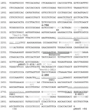
A number of previous studies have been carried out on the MUC1 promoter in vitro. Important sequence motifs identified by the previous studies in the 5’ re-gion of the MUC1gene are shown in Fig. 1.
Identification of DHS in the MUC1 Gene in Human Cell Lines
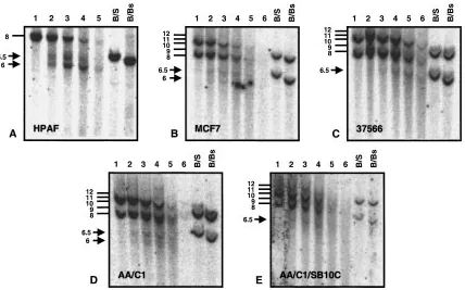
in HPAF where only 1 genomic BamH I fragment hy-bridises to the 5’MUC1 probe, as the cell line is ho-mozygous or hemizygous for MUC1 (Fig. 3A), all other cell lines analysed showed 2 genomic frag-ments presumably due to variation in the MUC1 tandem repeat number on the 2 alleles. Chromatin isolated from the breast carcinoma cell line MCF7 contained the same two DHS though at a reduced in-tensity (Fig. 3B). The750 bp site was also seen in the lymphoblastoid cell line (37566) (Fig. 3C) and the colon carcinoma cell line HT29 (data not shown).
Evaluation of the750 and250 DHS in a Colon Adenoma to Carcinoma Transition
The AA/C1 (adenoma) and AA/C1/SB10C (carci-noma) cell lines represent models of cancer progres-sion in the human colon (10). The AA/C1 line is a premalignant variant of a human colonic adenoma cell line PC/AA derived from a familial polyposis coli patient. The AA/C1/SB10C line was generated by treating AA/C1 with agents that induce differen-tiation and carcinogenesis. Though MUC1 is ex-pressed at a low levels in normal human colonic fragment were probed for DHS using the 5’MUC1,
PB352 and BS308 probes shown in Figure 2A. Two DHS were identified in the 5’ promoter region of MUC1 in chromatin from the HPAF pancreatic ade-nocarcinoma cell line. Figure 2B shows an 8 kb BamH I genomic fragment hybridising to the 5’MUC1 probe and subfragments at 6.5 and 6 kb that correspond to DHS at about750 bp and250 bp with respect to the transcriptional start site. BsrG I (x69118: 2520) and Sac I (x69118: 2154) cleavage sites at351 and 717 respectively from the tran-scription start site enable confirmation of the ap-proximate location of the DHS by the BamH I/SacI and BamH I/BsrG I double digestion (Fig. 2). These DHS were also seen with the PB352 probe. Screen-ing of DNA lyScreen-ing 3’ to the BamH I site at M61170:6633 failed to reveal any additional DHS in HPAF or lymphoblastoid (37566) cell line chro-matin.
Additional cell lines were evaluated to establish whether the750 and250 DHS were seen only in chromatin from the HPAF pancreatic adenocarci-noma or were a more general phenomenon. Unlike
Fig. 1. The sequence of the immediate 5’ promoter region of the human MUC1gene.
36 Molecular Medicine, Volume 8, Number 1, January 2002
epithelium and colon carcinoma-derived cell lines (23,24) these 2 cell lines have been shown to ex-press MUC1 glycoprotein (25). Evaluation of the 750 and250 DHS in these 2 cell lines showed both DHS in the adenoma (AA/C1) (Fig. 3D) while only the750 DHS was evident in the carcinoma cell line (AA/C1/SB10C) (Fig. 3E).
Expression of the Human MUC1 Gene in Human Cell Lines There have been many reports on the relative ex-pression levels of MUC1 mRNA in different cell lines. To evaluate the potential significance of our data on the presence of the 750 and250 DHS all cell lines were tested by a semi-quantitative RT-PCR assay for MUC1 and by northern analysis. Fig. 4A shows RT-PCR data for MUC1 mRNA ex-pression (656 bp product) relative to a housekeep-ing gene glucocerebrosidase (572 bp product). As expected high levels of MUC1 mRNA are seen in the HPAF line and MCF7 while the MUC1gene expression is barely detectable in the lymphoblas-toid cell line (37566). MUC1 expression in the AA/C1 cell line is significantly less than in HPAF and further reduced in the AA/C1/SB10C cell line.
These data were confirmed and extended by north-ern analysis (Fig. 4B), which showed that the lev-els of MUC1 mRNA in HPAF are significantly higher than in MCF7, and that MUC1 mRNA was undetectable in the 37566, AA/C1 and AA/C1/ SB10C cell lines.
Presence of the750 and250 DHS Correlates with MUC1 Expression
The750 DHS was weakly evident in all cell lines analysed irrespective of MUC1 expression levels. In contrast, presence of the250 DHS correlated with MUC1expression levels. The250 DHS was not de-tected in the lymphoblastoid cell line (37566) in which very low levels of MUC1 mRNA were seen by RT-PCR. In MCF7, which expresses moderate amounts of MUC1 mRNA, the750 and250 DHS are of approximately equal intensity. In HPAF, which expresses very high levels of MUC1 mRNA the250 DHS is of much greater intensity than the 750 DHS. In cells representing the adenoma (AA/C1) to carcinoma (AA/C1/SB10C) transition, which is accompanied by a decrease in MUC1 mRNA, the250 DHS disappears.
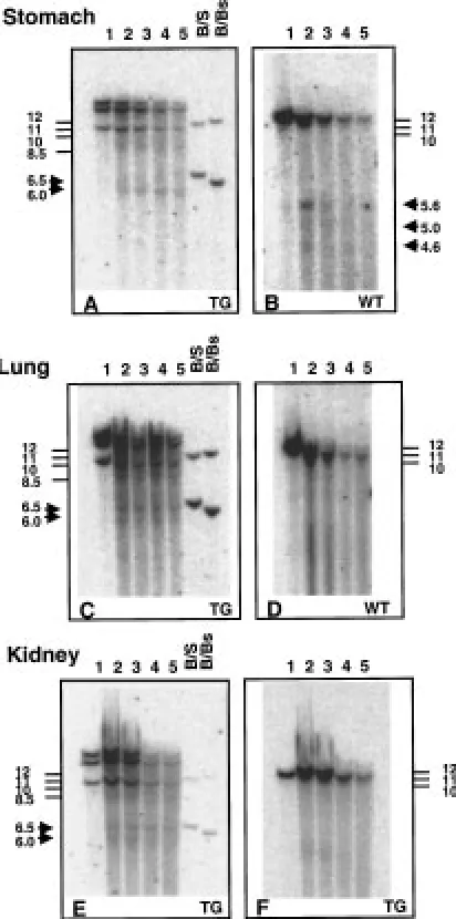
Fig. 2. DNase I hypersensitive sites at750 bp and250 bp with respect to the transcriptional start site of the human
specific probe, hybridising to the murine Muc1gene as shown in panel B. (The mouse-specific DHS are not evident in chromatin from lung and kidney in which the murine Muc1 gene is expressed, possibly due to insufficient sensitivity to detect chromatin changes in only a small percentage of cells in the tissue sample.) The250 DHS is more evident than that at750 bp (Fig. 5A) in chromatin from trans-genic mouse stomach. The two DHS are of equal intensity in lung chromatin (Fig. 5C) and in kidney (Fig. 5E). Both DHS were also seen in small in-testine and pancreas at low intensity (data not shown).
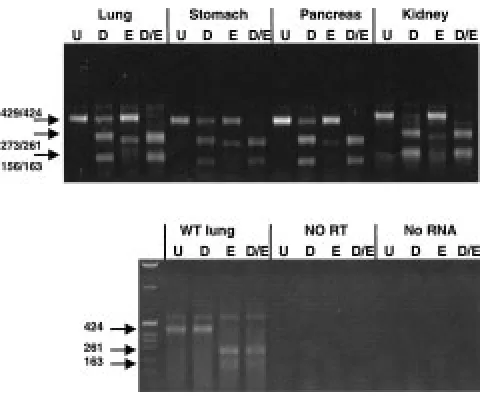
Expression of the Human MUC1 Gene in Transgenic Mice Figure 6 shows expression of the MUC1 transgene and endogenous muc1using RT-PCR on RNA from transgenic mice. The primers MUC1HMA and MUC1HMB amplify a fragment of 429 bp from the human gene that is cleaved by Dra III into (frag-ments of 273 and 156 bp) and 424 bp from the murine gene that is cleaved by EagI into 261 and 163 bp fragments. Cleavage of the RT-PCR product with the appropriate enzyme enables discrimina-tion of the human and mouse cDNAs. The assay is not truly quantitative as the PCR reactions are likely to be of unequal efficiency due to a 1 base pair mismatch with the mouse sequence in the Identification of DHS in the Human MUC1
Gene in Transgenic Mice
Chromatin was extracted from tissues of the MUC1 transgenic mice and evaluated for DHS in the human transgene. Figure 5 shows DNase I treated chromatin from transgenic mouse stomach, lung and kidney. A probe specific for human MUC1(within intron 1 and part of exon 2 of the gene) hybridised to 3 BamH I frag-ments in transgenic mice carrying the 10.6 kb Sac II fragment of human MUC1 (Fig. 5A,C,E) instead of the expected single fragment. This suggests there are 2 or more copies of the human gene integrated into these mice. In contrast, a probe specific for the murine muc1gene (also within intron 1 and part of exon 2 of the murine gene) hybridised to a single BamH I frag-ment of about 12 kb, as expected (Fig. 5B,D,F). The human-specific probe used in Fig. 5 is predicted to hybridise to a BamH I fragment of about 9.2 kb based on the restriction fragment map of the human MUC110.6 kb Sac II fragment (11). This is likely to be the smallest of the BamH I fragments, allowing for slight variation between the migration of 1 kb ladder molecular weight markers and DNase I treated, restriction enzyme digested chromatin. Figure 5A,C,E show appearance of the750 and250 DHS as 6.5 and 6 kb subfragments of the human MUC1genomic fragments. Different DHS (detected as 5.6, 5.0 and 4.6 kb subfragments) are seen with the mouse
Fig. 3. Detection of the750 and250 DHS in chromatin from cell lines. Southern blot of DNase I digested chromatin from
38 Molecular Medicine, Volume 8, Number 1, January 2002
MUC1HMA primer. However, expression of the human MUC1 gene is seen in lung, stomach, pan-creas and kidney (Fig. 6), and also in small intes-tine, bladder, mammary gland and ovary (data not shown).
Comparison of the Human MUC1 and Murine muc1 Gene Promoters
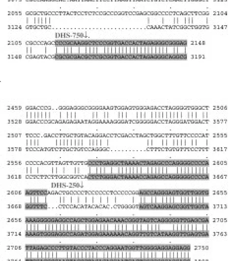
The 5’ untranslated (X69118: 1851–2941) region of MUC1 shows approximately 74% identity between human and mouse. Of particular interest are the re-gions shown in Fig. 7 which correspond to the A) DHS750 and B) DHS250. There is high homology between the human and mouse genes immediately 3’ to the750 DHS which may reflect functional conservation in this region. The250 DHS region is flanked by regions of high human/mouse sequence conservation both 5’ and 3’ to the site. Since map-ping of DHS by Southern blotting is not exact, even with restriction enzyme sites to confirm the loca-tions, these regions of high homology between the human and mouse genes may represent functional importance.
Discussion
DNase I hypersensitive sites in chromatin are often associated with regulatory elements and so can be informative in the identification of control elements that are active in vivo. Regulation of expression of the MUC1 gene has been studied by in vitro methods;
Fig. 4. Expression of MUC1in the cell lines analysed in figure 3. (A) RT-PCR using primers for MUC1that produce a 656 bp fragment and for glucocerebrosidase that produce a 572 bp fragment.(B) Northern blot of RNA from cell lines, probed with 5’MUC1.
in other tissues in which MUC1transgene expression was high, such as kidney and lung. These data sug-gest that the mouse contains the necessary transcrip-tion factors to interact with the important sequences generating the DHS. It was of interest to compare the250 and750 regions of the mouse and human genes for regions of cross-species homology that might be functionally important. A region of about 34 base pairs of high homology mapped to the pre-dicted core of the750 DHS. A short region of high homology 5’ and a more extended region immedi-ately 3’ to the predicted location of the250 DHS were also evident.
These regions of high cross-species conservation were re-evaluated to see if they coincided with im-portant regulatory motifs previously identified in the promoter of the MUC1 gene. The750 DHS is close to a predicted half oestrogen response (ER) el-ement and a GC box/Sp1 site (2) though neither of these have been shown to be functionally important in MUC1expression.
The250 DHS corresponds exactly to the loca-tion of a region of non-random purine/pyrimidine strand asymmetry (purine/pyrmidine mirror repeat elements-PMR) identified in the MUC1 promoter (M-PMR2) (26), and is proximal to another PMR el-ement (M-PMR3) located at approximately 130. M-PMR3 is evident in both the human MUC1 and mouse Muc1 genes but M-PMR2 is not conserved. These non-random sequences, which contain per-fect or nearly perper-fect mirror repeat elements, may be associated with the formation of H-DNA (in-tramolecular triple helical) conformations. The M-PMR3 element was shown previously to adopt a relatively uncommon H-DNA conformation (Hy5 isomer) in vitro (27). M-PMR2 did not exhibit S1 sensitivity (evidence of H-DNA character) in vitro (26) when investigated in a plasmid that contained both PMR2 and PMR3. However, the M-PMR2 element has not been examined in isolation and is predicted to be capable of forming H-DNA structures under conditions of high superhelical ten-sion. The previous experiments that examined M-PMR2 and M-PMR3 together (26) were conducted under conditions of standard plasmid superhelical density. The energy from superhelical tension that is required for producing H-DNA conformations was probably absorbed by the formation of H-DNA at the M-PMR3 element in those experiments and may not have been sufficient for formation of a second structure at M-PMR2 (which is shorter and may re-quire more energy). Thus, it remains possible that M-PMR2 could form an H-DNA structure in vivo un-der conditions of superhelical density that would be predicted to occur in chromatin upstream of a gene undergoing active transcription. The finding of a DHS at250 that correlates with transcriptional activity supports the hypothesis that altered DNA conformations in this region are associated with transcriptional activity of the MUC1 gene. It is also however, the mechanisms that confer tissue
speci-ficity on gene expression in vivohave not been fully elucidated. We identified two novel DHS in the MUC1 gene promoter region at250 and750 bp with respect to the major transcriptional start site. These sites were first seen in the HPAF pancreatic adenocarcinoma cell line that expresses high level of endogenous MUC1.
Further evaluation of these DHS showed that the 750 site was present in many cell types, both ep-ithelial and non-epep-ithelial and its intensity did not correlate with levels of MUC1 expression. In con-trast, the250 DHS was only seen in cell types that express relatively high levels of MUC1and its inten-sity showed a strong correlation with the abundance of MUC1 mRNA. Of particular interest was the ob-servation that MUC1 expression levels were reduced and the250 DHS became undetectable on South-ern blots in cells that model the adenoma to carci-noma transition (AA/C1 to AA/C1/SB10C colonic epithelial cell lines).
Detection of DHS in a human gene in transgenic mice can provide strong support for their importance in vivo. Mice transgenic for the human MUC1 gene carried on a 10.6 kb SacII fragment including 5’ and 3’ flanking DNA, show a very similar MUC1 expres-sion pattern to that seen for the endogenous muc1 (12). The 750 and250 DHS were detected in chro-matin extracted from certain tissues of these trans-genic mice. The relative intensity of the250 and 750 DHS was not constant, with the250 DHS being most evident in stomach, though also present
Fig. 6. Detection of the expression of the human MUC1
40 Molecular Medicine, Volume 8, Number 1, January 2002
possible that unknown transcription factors are as-sociated with this element and that these cause the observed DHS. Although the M-PMR3 element was previously shown to not influence transcriptional activity of promoter reporter constructs (28), the M-PMR2 element has not been investigated to date. Hence, the role of M-PMR2 in regulation of MUC1 gene expression remains intriguing but unclear.
In summary, we have identified two novel DHS in the MUC1 promoter. One DHS at750 was de-tected irrespective of transcriptional activity of the gene. A second DHS at250 that was associated with transcriptional activity of the gene mapped near previously defined purine/pyrimidine mirror repeat elements that may form intramolecular H-DNA structures. Further elucidation and evaluation of the DNA sequence encompassing the250 DHS regions is warranted to elucidate the mechanisms of action of potential regulatory elements located within this region of the MUC1promoter.
Acknowledgments
We are grateful to Sandra Gendler for transgenic mice and Shinichi Sameshima for help with mouse surgery; also to Christos Paraskeva for the AA/C1 and AA/C1/SB10C cell lines. This work was funded by NIH grant CA79580, the Cystic Fibrosis Trust, UK and a Wellcome Biomedical collaboration grant.
References
1. Hanisch FG. (2001) O-glycosylation of the mucin type. Biol. Chem. 382:143–149.
2. Abe M, Kufe D. (1993) Characterization of cis-acting ele-ments regulating transcription of the human DF3 breast carcinoma-associated antigen (MUC1) gene. Proc. Natl. Acad. Sci. USA90: 282–286.
3. Kovarik A, Peat N, Wilson D, et al. (1993) Analysis of the tissue-specific promoter of the MUC1 gene. J. Biol. Chem. 268: 9917–9926.
4. Shirotani K, Taylor Papadimitriou J, Gendler SJ, Irimura T. (1994) Transcriptional regulation of the MUC1 mucin gene in colon carcinoma cells by a soluble factor. Identification of a regulatory element. J. Biol. Chem. 269: 15030–15035.
5. Irimura T, McIsaac AM, Carlson D, et al. (1990) Soluble fac-tor in normal tissues that stimulates high-molecular-weight sialoglycoprotein production by human colon carcinoma cells. Cancer Res. 50:3331–3338.
6. Morris JR, Taylor Papadimitriou J. (2001) The Sp1 transcrip-tion factor regulates cell type-specific transcriptranscrip-tion of MUC1.
DNA Cell Biol. 20:133–139.
7. Parry G, Li J, Stubbs J, et al. (1992) Studies of Muc-1 mucin expression and polarity in the mouse mammary gland demon-strate developmental regulation of Muc-1 glycosylation and establish the hormonal basis for mRNA expression. J. Cell Sci. 101: 191–199.
8. Gaemers IC, Vos HL, Volders HH, et al. (2001) A stat-responsive element in the promoter of the episialin/MUC1 gene is involved in its overexpression in carcinoma cells.
J. Biol. Chem. 276:6191–6199.
9. Scibetta AG, Albanese I, Morris J, et al. (2001) Regulation of MUC1 expression in human mammary cell lines by the c-ErbB2 and ras signaling pathways. DNA Cell Biol. 20:265–274. 10. Williams AC, Harper SJ, Paraskeva C. (1990) Neoplastic transformation of a human colonic epithelial cell line: in vitro evidence for the adenoma to carcinoma sequence. Cancer Res.
50:4724–4730.
11. Peat N, Gendler SJ, Lalani N, et al. (1992) Tissue-specific ex-pression of a human polymorphic epithelial mucin (MUC1) in transgenic mice. Cancer Res. 52:1954–1960.
12. Rowse GJ, Tempero RM, VanLith ML, et al. (1998) Tolerance and immunity to MUC1 in a human MUC1 transgenic murine model. Cancer Res. 58:315–321.
13. Kim YW, Kern HF, Mullins TD, et al. (1989) Characterization of clones of a human pancreatic adenocarcinoma cell line representing different stages of differentiation. Pancreas. 4: 353–362.
14. Fogh J, Wright WC, Loveless JD. (1977) Absence of HeLa cell contamination in 169 cell lines derived from human tumors.
J. Natl. Cancer Inst. 58:209–214.
15. Huet C, Sahuquillo Merino C, et al. (1987) Absorptive and mucus-secreting subclones isolated from a multipotent intestinal cell line (HT-29) provide new models for cell polarity and terminal differentiation. J. Cell Biol. 105:345–357. 16. Soule HD, Vazguez J, Long A, (1973) A human cell line from a pleural effusion derived from a breast carcinoma. J. Natl. Cancer Inst. 51: 1409–1416.
17. Smith AN, Barth ML, McDowell, et al. (1996) A regulatory element in intron 1 of the cystic fibrosis transmembrane con-ductance regulator gene. J. Biol. Chem. 271:9947–9954.
A
B
Fig. 7. Comparison of the human MUC1and murine Muc1
18. Moulin DS, Manson AL, Nuthall HN, et al. (1999) In vivo analysis of DNase I hypersensitive sites in the human CFTR gene. Mol. Med. 5:211–223.
19. Becker P, Renkawitz R, Schutz G. (1984) Tissue-specific DNase I hypersensitive sites in the 5’-flanking sequences of the tryptophan oxygenase and the tyrosine aminotransferase genes. EMBO J. 3:2015–2020.
20. Chirgwin JM, Przybyla AE, MacDonald RJ, Rutter WJ. (1979) Isolation of biologically active ribonucleic acid from sources enriched in ribonuclease. Biochemistry 18:5294–5299. 21. Chambers JA, Harris A. (1993) Expression of the cystic fibro-sis gene and the major pancreatic mucin gene MUC1 in hu-man ductal epithelial cells. J. Cell Sci. 107:413–424.
22. Higgs DR, Wood WG, Jarman AP, et al. (1990) A major posi-tive regulatory region located far upstream of the human alpha-globin gene locus. Genes Dev. 4:1588–1601.
23. Ogata S, Uehara H, Chen A, Itzkowitz S. (1992) Mucin gene expression in colonic tissues and cell lines. Cancer Res. 52: 5971–5978.
24. Hollingsworth MA, Strawhecker JM, Caffrey TC, Mack DR. (1994) Expression of MUC1 MUC2 MUC3 and MUC4 mucin mRNAs in human pancreatic and intestinal tumor cell lines.
Int. J. Cancer. 57:198–203.
25. Vavasseur F, Dole K, Yang J, et al. (1994) O-glycan biosyn-thesis in human colorectal adenoma cells during progression to cancer.Eur. J. Biochem. 222:415–424.
26. Hollingsworth MA, Closken C, Harris A, et al. (1994) A nu-clear factor that binds purine-rich single-stranded oligonu-cleotides derived from S1-sensitive elements upstream of the CFTR gene and the MUC1 gene. Nucleic Acids Res. 22:1138– 1146.
27. Nelson KL, Becker NA, Pahwa GS, et al. (1996) Potential for H-DNA in the Human MUC1 mucin gene promoter. J. Biol. Chem. 271:18061–18067.