0022-538X/92/010001-05$02.00/0
Copyright© 1992, American Society for Microbiology
UV
Activation
of
Human
Immunodeficiency Virus
Gene
Expression in Transgenic Mice
JONATHAN VOGEL,1 MARIO CEPEDA,1
ERWINTSCHACHLER,2 LAURA A.NAPOLITANO,1
AND GILBERT
JAY'*
Laboratory of Virology,
Jerome H. Holland Laboratory, American Red Cross, Rockville, Maryland20855,1
andDepartment
of
Dermatology,
University
of
ViennaMedical
School, Vienna,
Austria2
Received 13 June 1991/Accepted 24 September 1991
Humanimmunodeficiency virus (HIV) infection is associated with a clinical latency of as long as 10 years before the development of disease. One explanation for this delay is the requirement of cofactors such as other DNA or RNA viruses, cytokines critical forimmune modulation, or environmental UV light. At least in tissue culturestudies, these agents are capable of inducing HIV gene expression in cell lines which either harbor the entire viral genome or contain a reporter gene under the control of the viral long terminal repeat regulatory region. The role of these cofactors in terminating clinical latency and inducing disease has been difficult to ascertain because
of
the lack of an appropriate animal model. We now report that UV light can markedly induce HIV gene expression in transgenic mice carrying both the cis-acting (long terminal repeat) and trans-acting (thetatgene) elements which are essential for viral transactivation andreplication in infected cells. Ourfinding may explain the clinical observations that cutaneous lesions in HIV-infected individuals are often seen in thesunlight exposed areas of the skin, including the face and neck.The
clinical latency associated with human
immunodefi-ciency virus (HIV)
canbe
defined
as thelong lag time
between the
initial infection with
HIVand
thedevelopment
of
clinically apparent disease (18, 20, 26). Because of the
potential therapeutic value of prolonging
the latentperiod
and
preventing
thedevelopment of active disease after HIV
infection, attention
has beenfocused
on the underlyingmechanisms
which hasten the
endof clinical latency.
In vitrostudies have been used
tostudy how cofactors of HIV
infection may influence the progression of disease. Such
cofactors
include
coinfecting
DNAviruses (17, 25),
cyto-kines
critical for immune modulation (10, 27, 45), and
environmental UV light (28, 35, 36, 40, 41). These cofactors
are
capable of inducing
HIVexpression
incell lines which
either harbor HIV and express low but detectable levels of
HIV
orcontain
anindicator gene controlled by the HIV long
terminal repeat (LTR)
regulatory region. Consequently,
these
cofactors could increase
the HIVtiter by increasing
HIV
replication and eventually
deplete thetarget
CD4+ cellswhich are infected with
HIV. Ingeneral, the role of these
cofactors in the
progression
of disease is
difficult
todeter-mine because of the lack of
agood in
vivo model with which
they
canbe tested.
Approaches used
tostudy latency
inindividuals include
anexamination of which tissues and cell
types
contain
HIV(15, 23, 38, 44)
andwhich of
thoseinfected cells
areactively expressing
HIVgenes
during
different stages
of
clinical disease (4, 8, 12, 13, 24, 31). Even
during clinical latency,
HIV canbe isolated
from
serum andperipheral
blood
mononuclear cells
(8, 13)
and
HIVis
present
in
peripheral CD4+
Tlymphocytes (24, 31).
There-fore,
HIVgene
expression
and
replication
continueduring
the
clinical
latentphase,
and a truemicrobiological latency
with
quiescent
HIVmayexist
in some HIVinfected
cells butcertainly
notin
all(26).
Tobetter understand how various
cofactors influence
HIVexpression
in theclinically
latentperiod,
anappropriate
*
Corresponding
author.animal model with all of the relevant cell-cell
interactions
would
have clearadvantages over many of the
available
tissue culture systems.
We chose to use in ourstudies
transgenic mice
containing the
HIVtype
1tatgene
under thecontrol of
the HIVtype
1LTR(43). This animal model
has theadvantage of
containing both
thecis-acting
(LTR) and thetrans-acting (tat)
elementwhich
arenecessary for
expression and transactivation of HIV genes. The presence
of both of these
regulatory elements should
approximate the
requirements of intact
HIVfor viral
geneexpression in the
infected
cell (34) and allow us to analyzetheir
interactions
with various cofactors. Given
the presence of HIV in theepidermis of the skin
(37, 38)and
thefact
that UVlight
canactivate the
HIV LTRin tissue culture cells
(28, 35, 36, 40,41),
anobvious
concern inHIV-infected
individuals
wouldbe
thefinding
thatUV
raysinsunlight
canactivate
HIV in theexposed skin.
Thisactivation
of
HIV may account for the cutaneouslesions
seen inHIV-infected individuals in
sunlight-exposed
areasof
theskin
(19, 21, 29, 30). Our
transgenic mice contain
the LTR-tat genein their skin
andallow
us to testfor
UVactivation of
HIV geneexpression
invivo.
We
have
previously
demonstrated that
the LTR-tattrans-genic mice selectively
express thetatgene in theskin
(43).
Wenow
demonstrate
thatthis
expression is localized
totheepidermal portion of the skin and
canbe
markedly
buttransiently
induced
by
UV
light
at avariety
of
different wavelengths.MATERIALS ANDMETHODS
Derivationoftransgenicmice.The
generation
oftransgenic
mice
containing
the LTR-tat transgenehas beenpreviously
described
(43).
Twoindependent
founder
linesdesignated
E10 and F2 wereused in these
experiments.
Separation
of skin intoepidermal
and dermalportions.
Shaved dorsal
(back)
and ear skin were removed fromsacrificed
oranesthetizedanimals,
and allunderlying
fat and musclewere removed. The skin is treated with 0.5 to 1.0%1
on November 10, 2019 by guest
http://jvi.asm.org/
trypsin in
phosphate-buffered saline without calcium
ormagnesium
for 30min
at37°C
in
a5% CO2 environment (32).
The
epidermal
portionis
thenseparated
from the
underlying
dermis, and both
components areprocessed for
RNA.Preparation of RNA.
RNA
wasisolated from these tissues
by the guanidinium isothiocyanate-CsCl procedure, using
aTekmar
tissuemizer probe for tissue disruption. Northern
(RNA) blot
analysis
wasperformed
aspreviously described
(43).
UV
light
treatment.Both
UVC (200
to290
nm) and UVB
(290
to320
nm) aswell
asnatural
sunlight
wereused
toirradiate the animals.
Asingle dose of UVC
light
of 0.2
mW/cm2
for 10
min wasprovided
with
aSylvania germicidal
lamp (Thomas
Scientific, Swedesboro, N.J.) which had
apeak
emission of 254
nm.The
single dose of UVB irradiation
was
provided by
twoFS-40
sunlamps(Westinghouse), which
have
apeak
emission
at313
nmwith
anirradiance of 0.2
mW/cm for
1 to 2h. All dose
measurementswereperformed
with
a UVXRadiometer
(UVP Inc.,San
Gabriel,
Calif.).
The
mice
werealso
exposed
tonatural
sunlight during
October
mornings with
asurface irradiance
of 0.5
to0.6
mW/cm2. Despite rigid
temperaturecontrol, only 15- and
30-min
exposures weretolerated by the mice.
Following anesthesia and removal of dorsal fur with
electric
clippers, small skin biopsies of dorsal skin and
ear weretaken
immediately before
UVirradiation and
at theindicated time intervals following UV irradiation.
Histological
procedures.Tissue
samples of skin
wereplaced in
a10% buffered formalin for
24h,
embedded in
paraffin, sectioned, and stained in
hematoxylin and eosin.
RESULTS
Epidermal localization of LTR-tat
expression. We have
previously demonstrated that the LTR-tat
transgenic mice
selectively
expressthe
tat genein the skin
(43). To better
understand both how
cofactors
canaffect
theexpression
of
the LTR-tat
geneand
toidentify the
biological
effects of
tat geneexpression in these mice, it is important
tofirst
deter-mine where
tatis
expressed in the skin. To do
so, weseparated the
skin into the
overlying epidermal and
under-lying dermal
portions by
treatmentwith
trypsin (32). As
shown
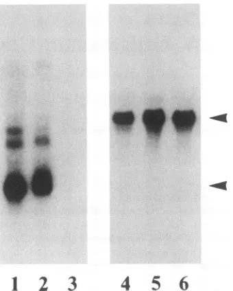
in the Northern blot
assayof
poly(A)+
RNA(Fig.
1),
expression
of the LTR-tat
gene occursalmost
exclusively in
the
epidermal
portion of the skin (lane 2), with little
ornoexpression
found
inthe
underlying
dermal
layer
(lane 3).
UV
light
induction of LTR-tat expression.To
determine
whether
UV
light could induce higher levels of
LTR-tatexpression,
the
mice
wereexposed
tosingle doses
of both
UVC
(254 nm) and UVB (290
to320 nm)
light, and both
earand shaved dorsal
(back)
skin biopsies
weretaken
for
analysis.
We
chose
asingle dose
of UV light that would
not causeepidermal
ordermaldamage
asassessed
onhematox-ylin-and-eosin staining of skin biopsies taken
at 24and 48 hafter
irradiation. This would avoid the potential
complica-tions caused
by
aninflammatory
cellinfiltrate
and its atten-dantcytokines which could potentially
induce tat expression(10, 27, 45).
For UVC
irradiation, the nondestructive dose was an exposureof
0.2-mW/cm2
irradiance for a time period of no morethan 15
min.Compared
with the backgroundexpres-sion
(Fig. 2A, lanes
1and
3), a 10-min dose of UVC light(0.12
J/cm2)
significantly induced
tat mRNA expression(lanes
2 and4) taken
8 hafter
irradiation. Low levels ofbackground
tatexpression
weredetected
since only small
amounts
of total
RNAwere usedfor
Northern blot assay;-,. :...1,f
-....
1_.
I,PI
1
2
3
4
5
6
FIG. 1. Localization of LTR-tat gene expression in the skin. Total skin(lane 1), epidermis(lane 2), and dermis (lane3) from a transgenicmouse wereassayed fortatexpression by Northernblot hybridization analysisofpoly(A)+RNA.Thecorresponding
p-actin
controls to indicate the relative input amounts of RNA from the various samples are shown in lanes4through 6. The toparrowhead indicates the
P-actin
mRNA, and bottom arrowhead indicates the 0.8-kbtattranscript. The minor components migratingbehind the 0.8-kbtattranscriptsare seenonlyinonetransgenicmouselineand most likely were derived from cryptic RNA start sites in the transgeneatthesiteofintegration.this will accommodate the dramatic induction
seen uponirradiation. There
wasnoinduction
of
expression
in
control
mice which had
biopsies
taken
at 0and 8
hbut
did
notreceive
UVC
irradiation.
Since the
Tatprotein
is
apotenttransactivator
of
LTR-driven gene
expression
(34), it is
important
tostudy
thekinetics of induction
by UVC
light
todetermine
whether,
once
activated,
expression
of
the LTR-tat gene maybe
sustained
over along
period
of
time
atahigh
level
by
the
Tatprotein.
When ear ordorsal
skin
biopsies
weretaken
at9and
21
hfollowing
UVC irradiation
(Fig.
2B, lanes
2and
3,
respectively),
expression
wassignificantly
induced
at9 h but
has
already started
tofall
back toward the 0-h baseline
(lane
1) after
21h. This
observation
washighly
reproducible
in
separate
experiments in which additional time points
wereincluded. The
suggestion
from this
study is that the induction
of
expression by
asingle
dose
of
UVC is transient and
cannot
be maintained
by Tat, despite
the fact thatsuch
transactivation
can occurin murine
cells(14).
In
general, the
ozonelayer in
theatmosphere
preventspenetration of UVC
irradiation,
and
individuals
on the earth'ssurface will
notbe
exposed
toUVC light
unless thereis
damage
to the ozonelayer
(3,11). However,
UVBirradiation
does
penetrate the ozonelayer
andconsequently
would
be
a more relevantrisk factor for
thoseindividuals
infected with
HIV.We also find that
asingle dose
of UVB
irradiation
wascapable of
inducing
asignificant
amountof
tat
expression
in both
earand dorsal
skin
8 hafter
treatment(Fig. 3,
lanes 2 and4,
respectively) relative
to theback-ground expression
at0hour
(lanes
1 and3,
respectively).
The
single UVB
doseconsisted
of
a 1- to2-h
exposure aton November 10, 2019 by guest
http://jvi.asm.org/
[image:2.612.354.524.81.293.2]A
B
S
1
2
3
4
5
6
7
8
1
2
3
4
5
6
FIG. 2. UVC treatment of the LTR-tat transgenic mice. (A) Induction of LTR-tat gene expression in the skin by UVC (254 nm) irradiation. Small biopsies of shaved dorsal skin and ear skin were taken immediately before UVC treatment (lanes 1 and 3, respectively) and 8 h posttreatment(lanes 2 and 4, respectively). The corresponding
P-actin
controls are shown in lanes5through 8. (B) Kinetics of UVC induction of LTR-tat gene expression. Serial dorsal skin biopsies were taken immediately before UVC irradiation (lane 1) and 9 and 21 h postirradiation (lanes 2 and 3, respectively). The correspondingI-actincontrols are shown in lanes 4 through 6.Northern blothybridization analysis was performedontotalRNA.The top arrowhead indicates the1-actinmRNA (theband immediately below represents muscle actin RNA from residual muscle tissue in the skin biopsies), and the bottom arrowhead indicates the 0.8-kb tat transcript.0.2-mW/cm2 irradiance
(0.7 tlamps which
have a peak emicomparable
tothose given
chr(
tumors(2, 9)
and
to studyimn
By
comparison,
natural sunlig;surface has
anirradiance of
a cm2 at310
nm.Although
naturby mice,
we sawinduction of
following
a30-min
exposure(i
.4
1
2
3
4
FIG. 3. UVB induction of LT transgenicmice. Smallbiopsiesof taken immediately before a 2-h
respectively)andat8 hposttreatn
The level ofinductionwasabout 10-fold in the back skin. The co
shown in lanes5through8.
o 1.4
J/cm2), using
two sun-.s,inn at I1l nm- Thic dnvt- kcDISCUSSION
aaivuall to7mice to ind
uce
sia Wehave
demonstratedthat
the HIV tatgene,
under the)nically
tomice
toinduce skin
controlof
the HIV LTR,is
selectivelyexpressed
in the ht as measured on thetpproximately
earths
sepidermal portion
of theskin
of our transgenicmice and that0.5
to
0.6
mW/
thisexpression
canbetransiently
inducedtoamuch
higher
a1 sunlight is poorly tolerated level by UV light. These observations contrast with thosetat
expressionin ear
skin 8 h which assayed HIV LTR-directed expression of reportertata
not shown). genes in the absence of the tat transactivator (14, 16).Although
thosestudies
also noted expressionin
theepider-mal
portion of
theskin, they also found
expression in many othertissues, including thymus,
eye, spleen,small intestine,liver,
and heart (14, 16). It is likely that this widespreadexpression
represents the basal constitutive activity of the LTRin theabsence of
the Tattransactivator,
with detection madepossible
by sensitive
methods such as thechloram-phenicol acetyltransferase
assay.Additionally,
the use of anindicator
gene to assess theability of
UVlight
toinduce
LTR-directed
expression in theabsence of
Tat(7) is
problematic because
themagnitude
andduration of
UVlight induction
cannoteasily
bedetermined.The
steady-state
levelof
a mRNAspecies in
thecell is
dependent
onboth
its rateof
synthesis
and its rate of-OK
turnover;
thestability of the transcript for
theindicator
genein
theepidermis
was notstudied (7). Analysis
of
the tat mRNAis, therefore,
necessary toaccurately determine
the extentand half-life of
UVlight induction.
The mechanisms of
activation
by
UV irradiation in our animal model invivo
arelikely
tobe
complex.
In vitro studiesof UV induction of the HIV LTR havesuggested
that5
6
7
8
changes
in chromatin structure may increasetranscription
by
allowing
betteraccessofnecessarytranscriptional
factors'R-tat
expression in the skin of (28).The cis-acting NFKB element in the HIV LTR may also Fearand shaved dorsal skinwereUVB treatment (lanes 1 and 3
play
a roleIn
UVactivation
oftranscription,
assuggested In ient(lanes2 and4,respectively),
transient
transfection assays
(36).3-foldinthe ear andgreaterthan Tounderstand the effects of UV light in an in vivo animal
rresponding
,-actin controls aremodel,
one must also consider themany different potential targetcells and how UVlight
may affect them and interfereon November 10, 2019 by guest
http://jvi.asm.org/
[image:3.612.104.526.85.263.2] [image:3.612.73.294.489.666.2]withthe complex communications thatexistbetweenthem. The murine epidermis is predominantly made up
of
kerati-nocytes but also contains smallerpopulations ofbone
mar-row-derived
Thy-1+
dendriticepidermal
cells and Langer-hans cells (6, 33, 39). In humans, the latter are known to harbor HIV (37, 38). Theeffect of UVlightonskin in mice
has been extensively studied, withparticular attentionto
its
effects on the ability of the skin to present
antigens
andinfluence the immune system (2, 3, 22). UV light has been found to affect Langerhans cells and cause a
decrease
intheir antigen-presenting ability. The
keratinocytes
mayalsorespond to UV irradiation with the release ofa variety
of
different cytokines, including interleukin-1,
interleukin-6,
tumornecrosis factor alpha, and
intercellular adhesion
mol-ecule1 (5). Thesefactors mayinduceavarietyof
responsesin theskin orin othertissues.
The effect ofUVlight in this transgenicanimal modelmay
betotransientlyincrease the level oftat expression in cells in which it is already being expressed at low levels or to activate its expression in cell types in which it is not
normally expressed. Our previous findings in these mice
suggestthat expression ofTat in theepidermal cells mayin
turn induce the secretion of factors which can affect the
proliferation of specific cells in the dermis, leading to the development of Kaposi's sarcoma (43), or in the
liver,
contributingtotheetiologyof hepatocellularcarcinoma (42). Given that the UVeffecton tatexpressionis only
transient,
itwouldbe ofinterestto determine whetherrepeated expo-sures to UV light will exacerbate the
dermal lesions.
This study is currently under way.The implications ofour findings for individuals
infected
with HIVareprovocative. Therecertainlyis the
suggestion
thatincidentUVirradiation couldinduce HIV
expression in
theskin of infected individuals. Thesingle dosesof UV
light
used in these animals are large but attainable in
outdoor
sunlight. However, the induction oftat
expression in
this model is transient, and repeated exposures to UVlight
wouldberequiredtosustainahigh levelof Tatprotein in the
involved cells. This animal model shouldallow us to deter-mine both the biochemical and functional consequences
of
UVirradiationon HIVexpression.
ACKNOWLEDGMENTS
This workwas supported byNIH grantsCA53633 and CA52408.
WethankLisa Ruiz and LucieRainone for helpinpreparation of
themanuscript.
REFERENCES
1. Aberer, W., N. Romani, A. Elbe, and G.Stingl. 1986. Effectsof physiochemical agents on murine epidermal Langerhans cells
andThy-i-positive dendriticepidermal cells. J. Immunol. 136: 1210-1216.
2. Alcalay,J.,J. N. Craig,and M. L.Kripke. 1989. Alterations in Langerhanscellsand Thy-i+ dendriticepidermalcellsinmurine epidermis during the evolution of ultraviolet radiation-induced skincancers. CancerRes. 49:4591-4596.
3. Baadsgaard, 0. 1991. In vivo ultraviolet irradiation of human
skin results in profound perturbation of the immune system.
Arch. Dermatol. 127:99-109.
4. Baltimore,D., and M. B. Feinberg. 1989. HIVrevealed: toward
anatural history ofthe infection. N. Engl. J. Med.
321:1673-1675.(Editorial.)
5. Barker, J. N. W. N., R. S. Mitra, C. E. M. Griffiths, V.M.Dixit, andB. J.Nickoloff. 1991. Keratinocytes as initiators of
inflam-mation. Lancet 337:211-214.
6. Bergstresser, P. R., R. E. Tigelaar, J. H. Dees, and J. W.
Streilein. 1983. Thy-1 antigen-bearing dendritic cells
populate
murineepidermis. J. Invest. Dermatol. 81:286-288.
7. Cavard, C.,A.Zider, M. Vernet, M.Bennoun, S.Saragosti,G. Grimber, and P. Briand. 1990. In vivo activationbyultraviolet raysof the humanimmunodeficiencyvirus type 1longterminal repeat. J. Clin. Invest. 86:1369-1374.
8. Coombs, R. W., A. C. Collies, J. P. Allain, B. Nikora, M. Leuther, G. F. Gjerset, andL.Corey. 1989. Plasma viremiain human immunodeficiency virus infection. N. Engl. J. Med. 321:1626-1631.
9. deGruiji, F. R.,andJ.C. van der Leun. 1991. Developmentof skin tumorsin hairless mice afterdiscontinuation of ultraviolet irradiation. Cancer Res. 51:979-984.
10. Folks, T. M., J.Justement,A.Kinter,C. A.Dinarello,and A.S. Fauci. 1987.Cytokine-inducedexpressionof HIV-1 ina chron-icallyinfected promonocyte cell line. Science 238:800-804. 11. Frederick, J. E., and H. E. Snell. 1988. Ultraviolet radiation
levelsduring the antarcticspring. Science 241:438-440. 12. Harper, M. E., L.M.Marselle,R. C.Gallo,and F.Wong-Staal.
1986. Detection oflymphocytes expressing human T-lympho-tropic virus typeIIIinlymphnodes andperipheral blood from infectedindividualsbyin situhybridization. Proc. Natl. Acad. Sci. USA 83:772-776.
13. Ho, D. D., T. Moudgil, and M. Alam. 1989. Quantitation of humanimmunodeficiency virus type1in the blood of infected persons. N. Engl. J. Med. 321:1621-1625.
14. Khillan, J. S.,K.C. Deen, S.H.Yu,R. W.Sweet,M.Rosenberg, and H. Westphal. 1988.Gene transactivation mediatedbythetat
gene of human immunodeficiency virus in transgenic mice. Nucleic Acids Res. 16:1423-1430.
15. Koenig, S.,H.E.Orenstein, J.M.Gendelman,M. C. DalCanto, G. H. Pezeshkpour, M. Yungbluth, F. Janotto, A. Aksamit, M. A.Martin,and A.S. Fauci.1986. Detectionof AIDS virus in macrophages in brain tissue fromAIDSpatientswith encepha-lopathy. Science 233:1089-1093.
16. Leonard, J., J. S. Khillan, H. E. Gendelman, A. Adachi, S. Lorenzo, M. A. Martin, and M. S. Meltzer. 1989. The human immunodeficiency virus long terminal repeat is
preferentially
expressed in Langerhans cells in transgenic mice. AIDS Res. Hum. Retroviruses 5:421-430.
17. Lifson, A. R., G. W. Rutherford, and H. W. Jaffe. 1988. The natural history of humanimmunodeficiency virus infection. J. Infect. Dis. 158:1360-1367.
18. Lui, K.-J., W. W. Darrow, andG. W. Rutherford III. 1988. A model-basedestimate ofthe meanincubationperiodfor AIDS in homosexual men. Science 240:1333-1335.
19. Mathes,B.M.,and M. C. Douglass. 1985. Seborrheicdermatitis inpatients with acquired immunodeficiency syndrome. J. Am. Acad. Dermatol. 13:947-951.
20. Medley, G. F.,R.M.Anderson, D. R.Cox,and L. Billard. 1987. Incubation period of AIDS inpatients infected via blood
trans-fusion. Nature(London) 328:719-721.
21. Mitsuyasu, R. T. 1988. AIDS-related Kaposi's sarcoma: a review of itspathogenesis andtreatment. BloodRev. 2:222-231. 22. Morrison, W. L. 1989. Effects of ultraviolet radiation on the immune system in humans. Photochem. Photobiol. 50:515-524.
23. Nelson, J. A.,C. A.Wiley, C.Reynolds-Kohler, C. E.Reese, W.
Margaretten, andJ. A. Levy. 1988. Human immunodeficiency virus detected inbowel epithelium frompatients with gastroin-testinal symptoms. Lanceti:259-262.
24. Psallidopoulos, M. C., S. M. Schnittman, L. M. Thompson III, M.Baseler, A. S.Fauci, A. C. Lane, and N. P. Salzman. 1989. Integrated proviral human immunodeficiency virus type 1 is present inCD4+ peripheral blood
lymphocytes
inhealthy sero-positive individuals. J. Virol. 63:4626-4631.25. Rosenberg,Z.F.,andA. S. Fauci.1989. Inductionofexpression on HIV in latently or chronically infected cells. AIDS Res. Hum. Retroviruses5:1-4.
26. Rosenberg, Z. F., and A. S. Fauci. 1990. Activation of latent HIVinfection. J. NIH Res. 2(6):41-45.
27. Rosenberg, Z. F., and A. S. Fauci. 1990. Immunopathogenic mechanisms of HIV infection: cytokine induction of HIV
on November 10, 2019 by guest
http://jvi.asm.org/
expression. Immunol. Today 11:176-180.
28. Sadai, M. R., E. Tschachler, K. Valerie, M. Rosenberg, B. K. Feller, G. N.Paviakis, M. E. Klofman, and F. Wong-Staal. 1990. Activation of tat-defective human immunodeficiency virus by ultraviolet light. New Biol. 2:479-486.
29. Sadick, N. S., N. S. McNutt, and M. H. Kaplan. 1990. Papulos-quamousdermatosis of AIDS. J. Am. Acad. Dermatol. 22:1270-1277.
30. Safai, B., K. G. Johnson, P. L. Myskowski, B. Koziner, S. Y. Yang, S.Cunningham-Rundles, J. H. Godbold, and B. Dupont. 1985.The natural history ofKaposi's sarcoma in the acquired immunodeficiency syndrome. Ann. Intern. Med. 103:744-750.
31. Schmittman, S. M., M. C. Psailidopoulos, H. C. Lane, L. Thompson, M. Baseler, F. Massari, C. H. Fox, N. P. Salzman, and A. S. Fauci. 1989. The reservoir for HIV-1 in human peripheral blood is a T cell that maintains expression of CD4. Science 245:305-308.
32. Schuler, G., and R. M. Steinman. 1985. Murine epidermal Langerhan's cells mature into potentimmunostimulatory den-dritic cells in vitro. J. Exp. Med. 161:526-546.
33. Shimada, S., andS. I.Katz. 1988.The skinas animmunologic organ.Arch. Pathol. Lab. Med. 112:231-234.
34. Sodroski, J., C. Rosen, F. Wong-Staal, S. Z. Salahuddin, M. Popovic, S. Arya, R. C. Galo, and W. A. Haseltine. 1985. Trans-acting transcriptional regulation of human T-cell leuke-miavirus type III long terminal repeat. Science 227:171-173. 35. Stanley, S. K.,T.M.Folks,and A. S.Fauci. 1989.Inductionof
expression of human immunodeficiency virus ina chronically infected promonocytic cell line by ultraviolet irradiation. AIDS Res.Hum. Retroviruses 5:375-384.
36. Stein, B., M. Kramer, H. J. Rahmsdorf, H. Ponta, and P. Herrlich. 1989. UV-induced transcription from the human im-munodeficiencyvirus type 1 (HIV-1) long terminal repeat and UV-induced secretion ofan extracellular factor that induces
HIV-1 transcription in nonirradiated cells. J. Virol. 63:4540-4544.
37. Stingl, G., K. Rappersberger, E. Tschachler, S. Gartner, V. Groh,D. L.Mann,K.Wolff,andM.Popovic. 1990.Langerhans cells in HIV-1 infection. J. Am. Acad. Dermatol. 22:1210-1217.
38. Tschachler, E.,V.Groh, M.Popovic,D.L.Mann,K.Konrad,
B.Safai,L.Eron,F. D.Veronese,K.Wolff,andG.Stingl. 1987. Epidermal Langerhanscells-a target forHTLV-III/LAV infec-tion. J. Invest. Dermatol. 88:233-237.
39. Tschachler,E., G. Schuler,andJ. Hutterer. 1983. Expression of Thy-1 antigen by murineepidermal cells. J. Invest. Dermatol.
81:282-285.
40. Valerie,K.,A.Delers, C. Bruck, C. Thiriart, H. Rosenberg, C. Debouck,and M. Rosenberg. 1988. Activation of human immu-nodeficiency virus type 1 by DNA damage in human cells. Nature(London)333:78-81.
41. Valerie, K., and M. Rosenberg. 1990. Chromatin structure
implicated in activation of HIV-1 gene expression by ultra-violetlight. New Biol. 2:712-718.
42. Vogel, J.,S. H.Hinrichs,L. A.Napolitano, L. Ngo, and G.Jay. Submitted forpublication.
43. Vogel,J., S.H. Hinrichs, R. K. Reynolds,P.A.Luciw, and G. Jay. 1988. The HIVtatgeneinduces dermal lesionsresembling Kaposi'ssarcomaintransgenic mice. Nature (London) 335:606-611.
44. Wiley, C. A., R.D.Schrier, J. A. Nelson, P. W. Lampert, and M.B. A. Oldstone. 1986. Cellular localization of human immu-nodeficiency virus infection within the brains of acquired im-mune deficiency syndrome patients. Proc. Natl. Acad. Sci. USA83:7089-7093.
45. Zagury, D., J. Bernard, R.Leonard, R.Cheynier, M. Feldman, P. S. Sarin, and R. C. GaBo. 1986. Long-term cultures of HTLV-III-infected T cells: amodel of cytopathology of T-cell depletion in AIDS. Science 231:850-853.