RESEARCH ARTICLE
CATEGORIZATION OF BACKGROUND PARENCHYMAL ENHANCEMENT:
REPEATABILITY AND REPRODUCIBILITY
1
Elisabetta
Giannotti, *
2Khodor Haidar Hassan,
1Abdulcadir Dalmar,
1Diego De Benedetto,
1
Giulia Bicchierai,
1Cecilia Boeri,
1Ermanno Vanzi,
3Andy Evans and
1Jacopo Nori
1
Diagnostic Senology Unit AOU Careggi Florence
2
Department of Biology and Bioinformatics, Lebanese University, Faculty of Sciences, Hadat-Lebanon
3
Division of Imaging and Technology, Ninewells Hospital and Medical School, University of Dundee
ARTICLE INFO
ABSTRACT
Background: Background parenchymal enhancement (BPE) on breast Magnetic Resonance Imaging
(MRI) is the amount of enhancing fibroglandular tissue, BPE influences both MRI interpretation and possibly risk of breast cancer. If increased BPE proves to be an important risk factor, then it has the potential to serve as a bio-marker of breast cancer risk.Before BPE can be introduced into clinical practice, data confirming the repeatability and reproducibility of its measurement is required. .
PURPOSE The aim of this study is to assess repeatability and reproducibility of BPE measurements by expert and non expert breast radiologists.
Material and Method: BPE measurements were made retrospectively on a consecutive series of 51
clinically indicated MRI’sperformed. All examinations were independently assessed for BPE by two expert readers and an inexperienced reader and assigned a category 1-4 in accordance with the proposed BIRADS MRI categorisation.
Results: The inter-reader agreement between expert readers obtained was substantial (k=0.75 p< 0,01).
Agreement between expert and non expert readers was moderate (k=0,494 p< 0,01 and k=0,55 p< 0,01). The intra-reader agreement between the three readers was strong (ICC 0.94). The intra-reader agreement for the expert readers was substantial (k=0.78 k=0.68 p< 0,01).
Conclusion: Our results show a high intra and inter-reader reproducibility, between expert readers.
Lower reproducibility for non-expert radiologists suggests the presence of a learning curve and training requirements for categorization of BPE.
Copyright©2016, ElisabettaGiannotti et al. This is an open access article distributed under the Creative Commons Attribution License, which permits unrestricted use, distribution, and reproduction in any medium, provided the original work is properly cited.
INTRODUCTION
Background parenchymal enhancement (BPE) on breast Magnetic Resonance Imaging (MRI) is the amount of enhancing fibroglandular tissue, or non fatty, non cystic breast parenchyma (King), seen on the first contrast-enhanced series in a standard DCE MRI examination. BPE reflects the vascularity of the fibroglandular tissue and has been shown to vary with hormonal changes, being highest during weeks 1 and 4 and lowest during week 2 (muller, Kuhl radiology 1997) (Hussain, Kuhl, King radiology 2012 MousaOksa, Engwoky, King V 2012 breast journ, pfeiderer, harevey, delille, graham, hussainmelasether AJR 2014). The amount of BPE refers qualitatively to the volume and intensity of the enhancement of
*Corresponding author:Khodor Haidar Hassan,
Department of Biology and Bioinformatics, Lebanese University, Faculty of Sciences, Hadat-Lebanon.
normal breast tissue after intravenous contrast administration (morrisradiolclinnort am 2007). Evidence suggests that BPE correlates negatively with subject age and increases with greater hormonal activity (muller, pfeiderer, kuhl radiology 1997, delille). One study suggested that BPE does not correlate with mammographic density, although the results clearly showed that both of these parameters were reduced in postmenopausal women as compared with premenopausal women (Cubuk). Although BPE has been shown not to directly correlate with breast density (Cubuk), BPE is similar to breast density in that it should be considered a physiological variable which affects both interpretation and risk of breast cancer. (Melsaether 2014). Four recent studies suggest increased BPE may also affect both reading accuracy(De martini , Hambly) and risk (King radiology 2011, Dontchos). However this is an emerging topic and some studies have not shown these correlations (AJR 2014 Melsaether). If increased BPE proves to be an important risk factor, then it has the
ISSN: 0976-3376
Asian Journal of Science and Technology Vol. 07, Issue, 07, pp.3187-3191, July,2016Available Online at http://www.journalajst.com
ASIAN JOURNAL OF
SCIENCE AND TECHNOLOGY
Article History:
Received 18th April, 2016
Received in revised form 19th May, 2016
Accepted 21st June, 2016
Published online 30th July, 2016
Key words:
potential to serve as an additional tool for risk stratification, especially in high-risk women undergoing breast MR imaging screening, and has possibly for monitoring chemoprevention (King radiology 2011).
However, there is very little data available in the literature on BPE, especially regarding the repeatability of categorisation of BPE, so before BPE measurements can be implemented into routine clinical practice, it is crucial to measure the repeatability of BPE measurements. The aim of this study is to asses repeatability (intra) and reproducibility (inter-reader agreement) of BPE measurements between expert and non expert breast radiologists, using a retrospective evaluation of a consecutive series of breast MRI performed for a variety of cinical indications. If BPE measurement is proved to be reproducibile then it could be used for risk stratification in the future.
MATERIALS AND METHODS
Categorisation reproducibility
To assess inter-and intra-reader repeatability, BPE
measurements were made retrospectively on a consecutive series of 51MRI scans (mean age 51 years, range 28-78years), performed for local staging or as a baseline prior to neoadjuvant chemotherapy (NAC) (n=48 (94%)) or for screening (n=3 (6%)) at our institution (Diagnostic Senology Unit AOU Careggi Florence) between July 2014 and October 2014. All premenopausal women were studied during the second week (8-14) of their menstrual cycle. Exceptions were rare; however we do not have specific data to determine the percentage of women who underwent an MRI examination outside this window. This retrospective study was approved by our institutional re-view board. Women undergoing MRI for implant assessment or interim or final assessment of NACT were excluded. The latter group was excluded to ensure the absence of post treatment effects. BPE assessment was made of the contralateral breast to avoid the influence of tumour neo-vascularity. If the patient had a personal history of breast conservation therapy less than 5 years earlier the evaluation was made on the contralateral breast. 10 patients were excluded, 5 were having neoadiuvant chemotherapy, one had a contralateral tumor and 4 underwent MRI for implant assessment.
All breast MRI examinations were independently assessed for BPE by each of two expert readers and an inexperienced reader. Each radiologist was blinded to the other readers’ assessments and to the assessment included in the original report. At each assessment the order of examinations was varied by random computer sorting and each reader was blinded to his prior interpretation. Expert readers repeated the analysis after a minimum interval of 2 weeks to further reduce bias. For each MRI examinations BPE was retrospectively recorded and assigned a category 1-4 in accordance with the proposed BIRADS MRI categories (De Martini, morris rad clin 2007); where 1 indicates minimal enhancement (< = 25% enhancement of glandular tissue); 2 mild enhancement
(25-50%enhancement of glandular tissue); 3 moderate
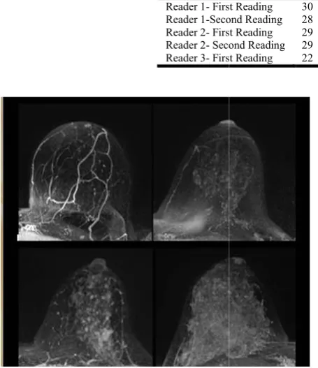
enhancement (51-75% enhancement of glandular tissue); 4 severe enhancement (≥ 75% enhacement of glandular tissue). (Figure 1).
A combination of unenhanced, initial contrast-enhanced, subtraction and maximum-intensity-projection images was typically used to determine BPE according to the literature (King, De Martini, Hambly, Melsaether)
MRI acquisition/technique
MRI images were performed at 1.5 Tesla (MagnetomAera Siemens Healthcare) with the patient placed in a head-first prone position with a dedicated breast coil (4 channels).The standard imaging protocol includes localizing sequences followed by:
Axial T1 weighted sequence Conventional GRE FID
imaging (Flash) 3D with fat suppression Dixon 2 point (TR 6.84 ms / TE 2.39 - 4.77 ms ) slice thickness 2.5 mm; FoV 330 mm; Flip Angle 20 degrees;
Axial T2 weighted Rapid Acquisition SE with fat
suppression adiabatic RF pulse SPAIR (TR 3300ms / 114.0 ms) slice thickness 3.5 mm; FoV 320 mm; Flip angle 150 degrees
Axial Rapid Acquisition GRE FID imaging 3D (T1
weighted sequence Flash 3D) fat saturated (TR 4.67 ms / 1.91 ms) slice thickness 1.5 mm FoV 320 mm; Flip Angle 35 degrees
After localizer images, T1w Rapid acquisition GRE FID imaging with fat saturation DIXON 2 point and Rapid Acquisition SE T2w with fat suppression adiabatic RF pulse SPAIR without use of contrast medium were performed. The third and last sequence, Rapid acquisition GRE FID imaging 3D Dyn VIBE is used as dynamic study of the intravenous administration of contrast, with a time resolution of 1:30 minutes. The contrast agent is administered 0.2 mmol per kilogram of body weight (Multihance, Bracco). The sequence Dyn VIBE is composed of 5 measure of 1:30 minutes each one, which fill k-space with sequencial encoding and the whole study volume is acquired 5 times. The first is acquired without contrastand the next 4 acquisitions post contrast injection to allow subtraction. Image acquisition begins after the administration of contrast material and a saline bolus, which is enjected at the beginning of the second volume acquisition at 1:30 minutes. Therefore, the first contrast-enhanced sequence was obtained approximately 90 to 180 seconds and the last one at 6 to 7:30 minutes.
Image analysis
Image analysis was performed using a high-resolution PACS workstation. The images were anonymized and randomly presented to readers to minimize bias. All image analysis was performed by two experienced breast radiologist (5 years reporting breast MRI) and an inexperienced breast sonographer with no experience on reading breast MRI.
Statistical analysis
Simple kappa coefficients were used to assess inter and intra-reader agreement. Kappa was interpreted as an indication that agreement was slight when 0-0.2, fair when 0.2-0.4, moderate when 0.4 to 0.6, substantial when 0.6 to 0.8 and near perfect when greater than 0.8 in accordance with Landis an Koch (Landis). BPE agreement between the three readers was
assessed with intra-class correlation coefficients (ICC) and scatterplots, (Bland altaman and international
analyses, the results were considered to indicate a statistically significant difference if associated with a
p-0.05.
RESULTS
The number of breast MRI examinations assessed as BPE category by each of the three readers at each time are showed in Table 1.
Figure 1. Examples of varying amounts of BPE, as prospectively assessed qualitatively. Axial postcontrast maximum intensity projection MR images show (a) minimal, (b) mild, (c) moderate,
and (d) marked BPE
Figure 2. ICC Multiple dot plot for intra reader agreemente between three readers
Table 1. Number of breast MRI examination assessed as BPE category by each of
Readers
Reader 1- First Reading Reader 1-Second Reading Reader 2- First Reading Reader 2- Second Reading Reader 3- First Reading
class correlation coefficients (ICC) and altaman and international, 1994). For all idered to indicate a statistically -value of less than
The number of breast MRI examinations assessed as BPE each time are showed
Examples of varying amounts of BPE, as prospectively maximum intensity projection MR images show (a) minimal, (b) mild, (c) moderate,
Figure 2. ICC Multiple dot plot for intra reader agreemente
Both expert readers did two evaluations, in order to study also their intra-reader agreement. Having 4 total readings we tried all the combinations and got a medium value of the 4 results. The inter-reader agreement obtained was substantial (k=0.75 p< 0, 01). To evaluate the intra
three readers ICC was used and we got as a medium value between the 4 combinations an ICC of 0.94 (
coefficient was also used to evaluate the inter agreement between the first reading of expert readers and non expert readers, all got moderate agreement, particularly between reader expert 1 and the inexpert reader was k=0,494
p< 0,01 while between reader expert 2 and the inexpert reader k=0,55 p< 0,01. The intra-reader agreement was evaluated only for the expert readers and both had substantial results (k=0.78 k=0.68 p< 0, 01).
DISCUSSION
BPE appears to influence the rate of abnormal interpretation and may correlate with breast cancer risk, but currently there is very little data regarding this field. Accurate and standard assessment of BPE has important clinical implications in terms of examinations outcome and risk stratification (melsaether), but before BPE measurements can be implemented into routine clinical practice, it is crucial to assess the reproducibility of BPE measurements.
show a high intra and inter between expert readers. The
substantial for both expert readers; the inter
is better between expert readers than between expert and non expert readers; This lower reproducibility for non radiologists suggests the presence of a learni
presence of training requirements for categorization of BPE.
In a recent study of Melsaether () repeated assessment at three time point, before and 1 and 3 weeks after training and they found that with training inter-reader agreement inc
fair (k=0.36) to moderate (k=0.48). Improvement was sustained at 3 weeks after training (k=0.45) for ordinal BPE categories and collapsed BPE categories (k=0.36 sustained at 0.47) analysis. BPE assessment were collapsed into low (BPE categories minimal and mild) and high (BPE categories moderate and severe). They conclude that inter reader agreement was fair among breast radiologist but achieved sustained improvement with training, showing that training changed the way readers evaluated BPE
the importance of education and inclusion of standardized BPE categories in a reference atlas. Regarding intra agreement, it improved for three of four readers to moderate, near perfect, and near perfect (k=0.65,0.98 and 0.95) between time 2 and 3 compared with fair, moderate and near perfect (k =0.45,0.58 and 0.84) between time 1 and 2. In their study a
Number of breast MRI examination assessed as BPE category by each of three readers at each time are showed
Minimal (1)
Mild (2)
Moderate (3)
Severe (4)
Tot
First Reading 30 7 7 7 51
Second Reading 28 11 5 7 51
First Reading 29 10 6 6 51
Second Reading 29 7 6 9 51
First Reading 22 15 8 6 51
Both expert readers did two evaluations, in order to study also reader agreement. Having 4 total readings we tried all the combinations and got a medium value of the 4 results. reader agreement obtained was substantial (k=0.75 To evaluate the intra-reader agreement between the ICC was used and we got as a medium value between the 4 combinations an ICC of 0.94 (Figure 2). K coefficient was also used to evaluate the inter-reader agreement between the first reading of expert readers and non expert readers, all got moderate agreement, particularly between reader expert 1 and the inexpert reader was k=0,494
while between reader expert 2 and the inexpert reader reader agreement was evaluated only for the expert readers and both had substantial results
BPE appears to influence the rate of abnormal MRI interpretation and may correlate with breast cancer risk, but currently there is very little data regarding this field. Accurate and standard assessment of BPE has important clinical implications in terms of examinations outcome and risk n (melsaether), but before BPE measurements can be implemented into routine clinical practice, it is crucial to assess the reproducibility of BPE measurements. Our results show a high intra and inter-reader agreement, especially between expert readers. The intra-reader agreement is substantial for both expert readers; the inter-reader agreement is better between expert readers than between expert and non expert readers; This lower reproducibility for non-expert radiologists suggests the presence of a learning curve and the presence of training requirements for categorization of BPE.
In a recent study of Melsaether () repeated assessment at three time point, before and 1 and 3 weeks after training and they reader agreement increased from fair (k=0.36) to moderate (k=0.48). Improvement was sustained at 3 weeks after training (k=0.45) for ordinal BPE categories and collapsed BPE categories (k=0.36-0.50 sustained at 0.47) analysis. BPE assessment were collapsed ories minimal and mild) and high (BPE categories moderate and severe). They conclude that inter-reader agreement was fair among breast radiologist but achieved sustained improvement with training, showing that training changed the way readers evaluated BPE highlighting the importance of education and inclusion of standardized BPE categories in a reference atlas. Regarding intra-reader agreement, it improved for three of four readers to moderate, near perfect, and near perfect (k=0.65,0.98 and 0.95) between time 2 and 3 compared with fair, moderate and near perfect (k =0.45,0.58 and 0.84) between time 1 and 2. In their study a
change in intra-reader agreement occurred in all but one reader. This change was greater in the less experienced readers indicating that these readers were the most receptive to training. Inexperienced readers showed the most improvement with training, reflecting the importance of dedicating training. In another study King et al () report inter-reader agreement for BPE and reported an inter-reader agreement (k) for BPE level between two readers of 0.47 for ordinal BPE and 0.57 for collapsed BPE. The same study reported intra-reader agreement (k) for one reader of 0.62 for ordinal BPE and 0.69 for collapsed BPE.
Several studies have investigated inter reader agreement for
mammographic density and Melsaether et al () reviewed 4
studies (Redondo, Berg, Bernardi, Ciatto) and found moderate to substantial inter-reader agreement (K=0.43-0.73) and intra-reader agreement (k=0.58 -0.71). Considering that the evaluation of breast density could be comparable to evaluation of BPE we can consider our results acceptable and reproducible. Similarly to breast density at mammography, the amount of fibro glandular tissue (FGT) seen on MRI images and the level of background parenchymal enhancement (BPE) at MR imaging after contrast material administration are features of normal breast tissue (King radiology 2011). Breast density and BPE are not clearly linked, many have wondered whether the tissue characteristic of BPE may have a similar important effect on the diagnostic accuracy of breast MRI. (De Martini) BPE could cause false negatives obscuring malignancies or could result in false positives mimicking the appearance of breast cancers (De Martini).
Some recent studies suggest that increased BPE may also affect both reading (DeMartini, Hambly) and risk of developing a breast cancer (King radiology, Dontchos). This is an emerging topic that has been reported variably (AJR 2014
Melsaether). Hambly et al () and DeMartini et al () did not
find an increased incidence of cancer with increased BPE, but
King et al and Dontchos et al have suggested a relationship
between BPE and breast cancer risk. Hambly et al () evaluated the effect of BPE in short-term follow up and cancer detection rates in 250 baseline high-risk screening MRI. They found that mild, moderate and marked BPE are associated with a significantly higher rate of short term follow-up than minimal BPE. There is no significant difference in biopsy rate or positive predictive value of biopsy.
De Martini et al () analyzed 736 MRI examinations and found that BPE was influenced by age and that increased BPE was associated with higher abnormal interpretation for MRI, in
keeping with the results of Hambly et al. () Moderate or
marked BPE resulted in more assessment leading to additional imaging or biopsy but that these initial finding did not impact performance outcome when compared with women with
minimal or mild BPE (De Martini). King et al analyzed ()
1275 MRI’s and found that increased BPE at breast MRI is associated with greatly increased odds of breast cancer, greater than MR imaging FGT. These odds may be as great as those associated with mammographic density. Dontchtos et al found that women at high risk of developing breast cancer undergoing screening breast MR imaging who have mild, moderate, or marked background parenchymal enhancement were nine times more likely to develop breast cancer within a mean follow-up interval of 5.6 years 6 1.3 [standard deviation]
than were those with minimal BPE in their cohort (odds ratio = 9.0; 95% confidence interval: 1.1, 71.0). This study has some limitations. The firstly is its retrospective nature, we had small number of patients and readers, and we did not analyzed the role of a training to improve the agreement. We used qualitative measures, according to our clinical practice, because no reliable quantitative measure is available. We were not able to evaluate the relationship between BPE and other variables such as mammographic breast density, menopausal status or timing relative to the menstrual cycle in premenopausal women.
Conclusion
Further studies to elucidate the relationship between the role of BPE are needed especially to evaluate relationship between BPE and breast cancer risk, as BPE is a promising imaging bio- marker of breast cancer risk and could refine clinical risk assessment models to aid individualization of screening strategies. Our study shows good reproducibility of BPE categorisation, particularly between expert readers. Specific training, and an atlas of BPE categories could improve BPE reproducibility, and facilitate the introduction of BPE categorization in routine clinical practice. The scientific guarantor of this publication is Dr Jacopo Nori. Noneof the authors of this manuscript disclose any conflicts of interest. We would like to thank Dr StefanoChiti and Dr Simona Covizzoli AOU Careggi for technical support.
REFERENCES
Berg, W.A., Campassi, C., Langenberg, P., Sexton, M.J. 2000.
Breast Imaging Reporting and Data System:
interandintraobserver variability in feature analysis and final assessment. AJR, 174:1769–1777
Bernardi, D., Pellegrini, M., Di, Michele, S., et al. 2012.
Interobserver agreement in breast radiological density
attribution according to BI-RADS quantitative
classification. RadiolMed (Torino), 117:519–528
Bland, J.M., Altman, D.G. 1986. Statistical methods for assessing agreement between two methods of clinical measurement. Lancet. Feb 8;1(8476):307-10.
Ciatto, S., Houssami, N., Apruzzese, A. et al. 2005.
Categorizing breast mammographic density: intraand
interobserver reproducibility of BI-RADS density
categories. Breast 2005; 14:269–275
Cubuk, R., Tasali, N., Narin, B., Keskiner, F, Celik, L., Guney,
S. 2010. Correlation between breast density in
mammography and background enhancement in MR mammography. Radiol Med (Torino), 115 (3): 434 – 441 Delille, J.P., Slanetz, P.J., Yeh, E.D., Kopans, D.B., Halpern,
E.F., Garrido, L. 2005. Hormone replacement therapy in postmenopausal women: breast tissue perfusion determined
with MR imaging—initial observations. Radiology, 235
(1): 36 – 41.
Delille, J.P., Slanetz, P.J., Yeh, E.D., Kopans, D.B., Halpern, E.F., Garrido, L. 2005. Hormone replacement therapy in postmenopausal women: breast tissue perfusion determined
with MR imaging—initial observations. Radiology,
235:36–41
DeMartini, W.B., Liu, F., Peacock, S., Eby, P.R., Gutierrez, R.L., Lehman, C.D. 2012. Background parenchymal enhancement on breast MRI: impact on diagnostic
performance. AJR Am J Roentgenol. Apr; 198(4): W373-80.
Eng-Wong, J., Orzano-Birgani, J., Chow, C.K. et al. 2008.
Effect of raloxifene on mammographic density and breast magnetic resonance imaging in premenopausal women at
increased risk for breast cancer. Cancer Epidemiol
Biomarkers Prev, 17:1696–1701
Graham, S.J., Stanchev, P.L., Lloyd-Smith, J.O., Bronskill, M.J., Plewes, D.B. 1995. Changes in fibroglandular volume and water content of breast tissue during the menstrual cycle observed by MR imaging at 1.5 T. J Magn Reson Imaging, 5:695–701
Hambly, N.M., Liberman, L., Dershaw, D.D., Brennan, S., Morris, E.A. 2011. Background parenchymal enhancement on baseline screening breast MRI: impact on biopsy rate
and short-interval follow-up. AJR Am J Roentgenol. Jan;
196(1):218-24.
Harvey, J.A., Santen, R.J., Petroni, G.R., et al. 2008.
Histologic changes in the breast with menopausal hormone therapy use: correlation with breast density, estrogen receptor, progesterone receptor, and proliferation indices. Menopause, 15:67–73
Hussain, Z., Roberts, N., Whitehouse, G.H., García-Fiñana, M., Percy, D. 1999. Estimation of breast volume and its variation during the menstrual cycle using MRI and stereology. Br J Radiol., 72:236–245
International Organization of Standardization 1994. Accuracy (Trueness and Precision) of Measurement Methods and Results—Part I: General Principles and Definitions (ISO 5725-1). ISO, Geneva, Switzerland.
King, V., Brooks, J.D., Bernstein, J.L., Reiner, A.S., Pike, M.C., Morris, E.A. 2011. Background parenchymal enhancement at breast MR imaging and breast cancer risk. Radiology. 2011 Jul; 260(1):50-60.
King, V., Goldfarb, S.B., Brooks, J.D., et al. 2012.Effect of
aromatase inhibitors on background parenchymal
enhancement and amount of fibroglandular tissue at breast MR imaging. Radiology 2012; 264:670–678
King, V., Kaplan, J.B., Pike, M.C., et al. 2012. Impact of
tamoxifen on fibroglandular tissue, background
parenchymal enhancement, and cysts on breast magnetic resonance imaging. Breast J., 18:527–534
Kuhl, C.K., Bieling, H.B., Gieseke, J., et al. 1997. Healthy
premenopausal breast parenchyma in dynamic contrast-enhanced MR imaging of the breast: normal contrast medium enhancement and cyclical-phase dependency. Radiology, 203 (1): 137 – 144 .
Kuhl, C.K., Bieling, H.B., Gieseke, J., et al. 1997. Healthy
premenopausal breast parenchyma in dynamic contrast-
enhanced MR imaging of the breast: normal contrast medium enhancement and cyclicalphase dependency. Radiology, 203:137–144
Kuhl, C.K., Bieling, H.B., Gieseke, J., Kreft, B.P., Sommer,
T., Lutterbey, G., Schild, H.H. 1997. Healthy
premenopausal breast parenchyma in dynamic contrast-enhanced MR imaging of the breast: normal contrast medium enhancement and cyclical-phase dependency. Radiology. Apr; 203(1):137-44.
Landis, J.R., Koch, G.G. 1977. The measurement of observer agreement for categorical data. Biometrics. Mar; 33(1): 159-74.
Melsaether, A., McDermott, M., Gupta, D., Pysarenko, K., Shaylor, S.D., Moy, L. 2014. Inter- and intrareader agreement for categorization of background parenchymal
enhancement at baseline and after training. AJR Am J
Roentgenol. 2014 Jul; 203(1): 209-15. doi:
10.2214/AJR.13.10952.
Morris, E.A. 2007. Diagnostic breast MRimaging:
currentstatus and future directions. Radiol Clin North Am. Sep; 45(5):863-80, vii. Review.
Morris, E.A. 2010. Diagnosticbreast M Rimaging: current
status and future directions. Magn Reson Imaging Clin N
Am. 2010 Feb; 18(1):57-74.
Mousa, N.A., Eiada, R., Crystal, P., Nayot, D., Casper, R.F. 2012. The effect of acute aromatase inhibition on breast parenchymal enhancement in magnetic resonance imaging: a prospective pilot clinical trial. Menopause,19:420–425 Müller-Schimpfle, M., Ohmenhser, K., Stoll, P., Dietz, K.,
Claussen, C.D. 1997. Menstrual cycle and age: influence on parenchymal contrast medium enhancement in MR imaging of the breast. Radiology. 1Apr; 203(1):145-9. Oksa, S., Parkkola, R., Luukkaala, T., Mäenpää, J. 2009.
Breast magnetic resonance imaging findings in women treated with toremifene for premenstrual mastalgia. ActaRadiol, 50:984–989
Pfleiderer, S.O., Marx, C., Vagner, J., Franke, R.P., Reichenbach, J.R., Kaiser, W, A. 2005. Magnetic resonance-guided large-core breast biopsy inside a 1.5-T magnetic resonance scanner using an automatic system: in vitro experiments and preliminary clinical experience in four patients. Invest Radiol. Jul; 40(7):458-63.
Pfleiderer, S.O., Sachse, S., Sauner, D., et al. 2004. Changes in magnetic resonance mammography due to hormone replacement therapy. Breast Cancer Res., 6:R232–R238
Redondo, A., Comas, M., Macià, F., et al. 2012. Inter- and
intraradiologist variability in the BI-RADS assessment and breast density categories for screening mammograms. Br J Radiol, 85:1465–1470