Original Article
CODEN: IJPNL6
QUANTITATIVE EVALUATION OF PLASMA PROTEINS, LIPID PROFILE, LIVER
AND KIDNEY FUNCTION PARAMETERS AFTER CHRONIC ADMINISTRATION OF
“PRADARANTAK LOUHA” TO MALE SPRAGUE –DAWLEY RATS
Mariyam Akter
1,2,Chinmoy Kumar Sen
1, Mohammad Salim Hossain
1, M Shahabuddin Kabir
Choudhuri
31
Department of Pharmacy, Noakhali Science and Technology University, Sonapur-3814,
Noakhali, Bangladesh.
2
Department of Developmental and Regenerative Biology, Graduate School of Medical Science,
Nagoya City University, 1, Kawasumi, Mizuho-cho, Mizuho-ku. Nagoya-shi, Aichi 467-8601,
Japan.
3
Department of Pharmacy, Jahangirnagar University, Savar, Dhaka-1342, Bangladesh.
*Corresponding author e-mail:
[email protected], [email protected]
Received on: 01-12-2016; Revised on: 17-04-2017; Accepted on: 27-05-2017
ABSTRACT
Background: Pradarantak Louha (PDL), a herbomineral Ayurvedic medicine has been used as a traditional medicine in the treatment of leucorrhoea for many years. Objectives: To evaluate the effect of Pradarantak Louha on major body organs. We assessed the possibility of side-effects after long term administration of Pradarantak Louha. Materials and Methods: To evaluate the effect of PDL, it was administered to the rats at a dose of 400 mg/kg for 54 days. Result: PDL does not change the plasma proteins (Total protein, albumin, and globulin) significantly. The change of bilirubin content was also not significant. In case of kidney function parameters, statistical significant increase was noted in both the creatinine content (p value: 0.015) and urea content (p value: 0.012). To assess the effect of PDL on cardiovascular health, lipid profile of rats were assayed and no significant changes were found. Conclusion: The outcome of this study implies that PDL is safe for our body but care should be taken when it is administered for long term and when it is accompanied by any kidney complications.
Keywords: Pradarantak Louha, Ayurvedic Medicine, Creatinine, Uric Acid, Plasma Protein.
INTRODUCTION
Traditional medicinal systems are the earliest
enlightenment in health care system of mankind. In
the very recent past, the use of traditional medicines
is growing worldwide both in developed and
developing countries.[1] Ayurveda, which means
science of long life, is at least a 5,000-year-old
system of traditional medicine (1500–1000 BC)
designed to promote good health and longevity rather
than to fight disease and was practiced by physicians
and surgeons (called vaidya). Until 700 BC, this
science was orally discussed between sages and
physicians. Thereafter, two different textbooks were
assembled: one by ‘‘Charaka’’ is called Charaka Samhita and the other by ‘‘Sushruta’’ is called
SushrutaSamhita. Whereas Charaka Samhita deals
International Journal of Pharmacy
with the etiology, symptomatology, pathology,
prognosis, and medical management of disease,
SushrutaSamhita deals with various surgical
instruments and procedures.[2] Pradarantak Louha
(PDL) is included (pages 325-326) in the Bangladesh
National Formulary of Ayurvedic Medicine 1992
(Approved by the Government of Bangladesh vide
Ministry of Health and Family Welfare Memo No.
Health-1/Unani-2/89/ (Part-1) 116 dated 3-6-1991). It
is a traditional Ayurvedic preparation widely used by
the rural and ethnic people of Bangladesh to treat
leucorrhea. It is a preparation of various metals,
non-metals, animal constituents and medicinal herbs
[Table 1, Table 2]. Safety of traditional medicine
system is time tested and it is believed to be spiritual
and known not to produce toxic effects. But no
objective verifiable data exists to support many such
claims. It is conceivable that a single herb extract or a
pure active chemical constituent may cause some
adverse effects under certain conditions and dose
levels. For example, SushrutaSamhita describes the
use of guggul (Commiphoramukul) for a wide variety
of conditions, including rheumatism and obesity. But
it has been shown to produce some anticoagulant
effect under certain conditions. [3] In addition, there
are lot of works and discussions going on globally
about heavy metals and toxicity of heavy metal
poisoning such as mercury, lead, arsenic etc. In 2003,
a survey says that Ayurvedic theory attributes
important therapeutic roles to mercury and lead and
that perhaps 35-40% of medicines in the Ayurvedic
formulary contain at least one metal. The intrigue
phenomenon of its manufacturing converts these into
complex mineral forms which are effective and
nontoxic. However, improper
processing/manufacturing of Ayurvedic medicines
may result into severe toxicity.[4]Several studies done
in other countries have had similar findings.[5,6]
Another main component of Ayurvedic medicines is
medicinal herbs as medicinal plants can be directly
used as healing agent and their phytochemicals also
serve as lead compound for developing potential
drugs to cure various diseases in human.
[7,8]Furthermore, there are several reports which state
on the potential toxicity of the phyto products.
Contamination of these products by pesticides,
herbicides, naturally occurring toxins, microbes or
adulteration by means of synthetic substitutes is a
cause for concern. Toxicity manifestations include
hepatotoxicity, nephrotoxicity, and neurotoxicity,
hematological, mutagenic and cardiovascular
toxicities. [9] Due to all the above concerns nowadays
in the present era it has become very important to
understand Ayurvedic medicines by carrying out
certain safety studies. The outcome of those studies
will be helpful for a clear judgment and revalidation
of the safety and efficacy of Ayurvedic medicines in
living organisms. Pharmacological evaluation of PDL
would provide proper proof of safety of major organs
like kidney, liver and heart. Since there is no strong
clinical data about the safety of PDL for major body
organs at the current situation mentioned above, it
was decided to undertake a detailed evaluation about
the effect of PDL on major body organs.
MATERIALS AND METHODS
Ayurvedic formulation: For the current study Pradarantak Lauha (PDL) was collected from Sree
Kundeswari Aushadhalaya Ltd, Chittagong,
Dose and Route of administration: In this study we administered the drug per oral route at a dose of 400
mg/kg of the body weight daily. Also, Ketamine were
administered intra-peritoneally (500 mg/kg i.p.) for
anesthesia purposes.
Experimental animals: For this research work, healthy albino rats (Rattusnovergicus:
Sprague-Dawley strain,) eight-week old of male rats were
used. These animals were weighed about 180±20 g.
The rats were bred and maintained at the Animal
House of the Department of Pharmacy, Jahangirnagar
University under standard laboratory conditions
(relative humidity 55–65%, room temperature 25.0 ±
2.0°C, and 12 h light-dark cycle). The animals were
randomly assigned to control and treatment groups
(10 rats per group) and housed in clear plastic cages
containing wood shavings for bedding. At the end of
the experimental period of 54 days and after
overnight fasting, at 9:00a.m, the animals were
sacrificed to get a blood sample. Before sacrifice
ketamine (500mg/kg) were administered
intraperitonially for anaesthetized the animals.
Immediately, after sacrificing the animals’ blood
sample was collected from the post vena cava and
then was transferred to the tubes having heparin
without any delay.
Biochemical test: To collect the intended plasma and to remove red blood cells, the collected samples of
blood were centrifuged at 4,000 g for 10 min using
bench top centrifuge (MSE Minor, England). After
separation, serum was collected using dry Pasteur
pipette and stored in the refrigerator for analysis. The
analysis of all the biochemical parameter was
accomplished within 24 h of sample collection. After
collection of plasma different methods were applied
for bio-chemical tests. All the reagents and kits used
for the biochemical tests are purchased as ready to
use form and all test samples were prepared
according to instruction guide of Human GmbH,
Wiesbaden, Germany. The absorbances of all the test
samples were determined using Humalyzer, Model
No-3000 (Human GmbH, Wiesbaden, Germany).
Statistical analysis: The group data are expressed as Mean ± SEM (Standard Error of the Mean). Unpaired
"t" tests were done for statistical significance. SPSS
(Ver. 20) for Windows was applied for the analysis
of data. Differences between groups were considered
significant at p< 0.05, 0.01 and 0.001.
RESULT
After 54 days of PDL administration, biochemical
parameters critical for normal and balanced
physiological condition were considered. To assess
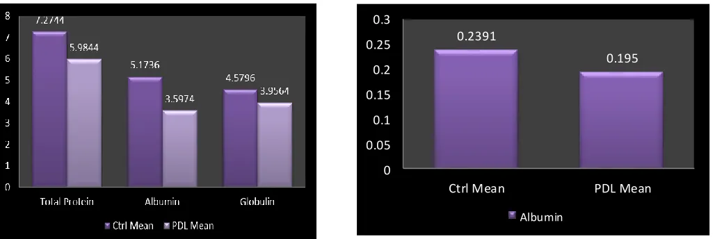
the effect of PDL on blood proteins, change in total
protein, albumin and globulin contents were
measured but there was no statistically significant
change [Table 3]. The change in bilirubin content
was also measured and the decrease of bilirubin level
after PDL administration was also not significant
[Table 4]. To evaluate the effect of PDL on kidneys,
measurement of creatinine, urea and uric acid level
results in statistically significant increase in both
creatinine (↑60.4167%; p: 0.015*) and urea (↑30.8694%; 0.012*). But the change in uric acid was
not significant [Table 5]. Lipid profiles including
triglycerides, total cholesterol, very low density
lipoprotein, low density lipoprotein and high density
lipoprotein was measured before and after PDL
administration. But there was no statistically
DISCUSSION
Effect on plasma protein contents: The level of total protein in the blood is normally a relatively
stable value, reflecting a balance in loss of old
protein molecules and production of new protein
molecules. [10-13] After chronic administration of PDL
preparation in the male rats the total protein content
in the plasma was decreased (17.7334%. decr.) and it
was not significantly different from its corresponding
control value (p=0.119). The decrease of albumin and
globulin content was 30.466 % and 13.608 %
respectively. Both changes were not significantly
different from their corresponding control values. In
this study, there was no significant difference in
plasma protein level between experimental group
animals and control group animals shown in table 3
& figure 1.
Effect on liver: Blood bilirubin test measures the amount of bilirubin in the blood in order to evaluate
liver function or to help diagnose anemia caused by
the increased destruction of RBCs (hemolytic
anemia). [14] After administration of PDL to male rats
for 54 days, bilirubin level was decreased by
18.4441% in the plasma in comparison to their
control group but it was not statistically significant
shown in table 4 & figure 2. The plasma albumin
content is another indicator of liver health as it is a
protein made specifically by the liver. In this study,
the decrease of albumin content was also not
statistically significant. Study of bilirubin content and
albumin content in blood reflects that PDL does not
alter the normal physiologic condition of liver.
Effect on kidney: Creatinine is usually a good indicator of how well the kidneys are working.[16] The
concentration of urea in the serum is useful in
prediction of different types of health problems, like
kidney disease or failure, blockage of the urinary
tract by a kidney stone, congestive heart failure,
dehydration, fever, shock and bleeding in the
digestive tract. Low levels are also seen in trauma,
surgery, opioids, malnutrition, and anabolic steroid
use [19,22], BUN (Blood urea nitrogen) is affected by
tubular reabsorption of urea and several non renal
factors like diet and urea cycle enzymes. Serum uric
acid level is used to detect high levels of this
compound in the blood in order to help diagnose gout
and kidney failure. In this study PDL causes
statistically highly significant (p=0.015) increase in
the creatinine (60.417% incr.) content in plasma and
an increase of urea level (30.8694% incr.) in the
plasma was noted in comparison to their control
group, the increase was statistically significant
(p=0.012) shown in table 5 and figure 3. Also, it was
observed that there was a negligible increase in the
plasma uric acid content (1.806% incr.) in the PDL
treated male rats, and this increase obviously was not
significant (p=0.862). This study shows a statistically
significant increase in the creatinine content and urea
content in plasma in PDL treated male rats. So, it
indicates that prolong administration of PDL may
cause nephrotoxicity. These results suggest that
administration of PDL should be carefully monitored
to confirm proper functioning of kidneys.
Effect on lipid profile: Triglyceride and cholesterol is different from most tests in that it is not used to
diagnose or monitor a disease. Both are used to
estimate risk of developing a disease specifically
heart disease. [23-26] In this study the change in
triglyceride and total cholesterol level of PDL treated
rat is not significant shown in table 6 & figure 4. Due
to the very high tendency of LDL to block the artery
cholesterol in the blood, the LDL cholesterol is
considered the most important form in determining
risk of heart disease. In the present study, decrease
was noted in the triglyceride level (11.7462 % decr.),
and HDL (43.5008 % decr.) content in the plasma of
the PDL treated male rats. In both cases, the decrease
was not statistically significant; triglyceride
(p=0.869), and HDL (p=0.621) whereas, there was
increase in the total cholesterol (10.9886 % incr.),
VLDL (44.5378 % incr.) and LDL (15.1560 % incr.).
But none of those increases was statistically
significant; total cholesterol (p=0.084), VLDL
(p=0.209), LDL (p=0.201) (Table 6 and figure 4).
After chronic administration of Pradarantak Louha
(PDL) for 54 days, the increase of LDL was not
significant in PDL treated male rats and also HDL
was decreased not significantly [Table 6]. These non
significant changes predict that the cardiovascular
health of PDL treated rats are good enough.[27] In
addition, Increased levels of VLDL-cholesterol, have
been found to be associated with increased risk of
heart disease and stroke. PDL treated rats show no
significant change in VLDL level. Accumulating all
these above mentioned statistical changes in PDL
treated male rats compared to control group indicates
that cardiovascular health of PDL treated rats is
sound and well. In the current study, we observed the
effects of PDL on major body organs. It was found
that PDL does not cause any significant change of the
liver and cardiovascular system. But prolong
exposure of PDL should be carefully monitored
because in this study, PDL has increased both
creatinine and urea statistically significantly. Care
should also be taken when PDL is treated with
impaired renal function. This kind of study should
also be continued to establish clinical and
pharmacological data of Ayurvedic medicines. In
addition, based on this kind of study further more
detailed study may be planned to observe the efficacy
and safety of Ayurvedic medicines in the cellular and
tissue level.
CONCLUSION
Being one of the most ancient healing systems,
Ayurvedic medicine has been practiced for years. But
still there is no sufficient reliable scientific data about
the safety and efficacy of Ayurvedic medicines. In
the present study, to assess safety of Pradarantak
Louha and effect of this Ayurvedic medicine on
major body organs, some important biochemical
parameters were measured with and without PDL
administration. Result of the present study showed
that PDL is safe for almost all the major body organs
but care should be taken while it is associated with
nephropathy. To understand Ayurvedic safety
principles and efficacy science more deeply, this type
Table1: List of plants and animal constituents used in the formulation of Pradarantak Louha.
Plants Part Used English/Common Name Scientific Name Family
Sunthi Dry root Dry ginger Zingiber
officinale
Zingiberaceae
Marica Fruit Pepper black Piper nigrum Piperaceae
Pippali Fruit Long papper, Pipli Piper longum Piperaceae
Haritaki Fruits Almond tree Terminalia chebula Combretaceae
Devadaru Leaves,
Heartwood,
Devadaru Cedrus deodara Pinaceae
Bibhitaka Fruit Belliric myrobalans Terminalia bellirica
Combretaceae
Amalaki Fruit Amla Emblica
officinalis
Phyllanthaceae
Citra (citraka) Root Citra Plumbago
zeylanica
Plumbaginaceae
Vidanga Fruit False black pepper Embelia
ribes
Primulaceae
Vaca Leaves,
Rhizomes
Sweet flag, Calamus Acorus calamus
Araceae
Havusa (hapusa) Fruit Juniper plant Juniperus communis Cupressaceae Palaka (kustha) Dried root Saw-wort, Snow lotus Saussurea
lappa
Asteraceae
Patha Root Abuta Cissampelos pariera Menispermaceae
Ela Seed Cardamom Elettaria
cardamomum
Zingiberaceae
Sankha (bhasma) Shell Sea snails Turbinella pyrum Turbinellidae Cavika (cavya) Root, Fruit Wild pepper Piper methysticum Piperaceae Vrddhadaraka Root Elephant creeper,Guguli Argyreia Speciosa Convolvulaceae
Note: All ingredients are used as 1 part.
Table 2: List of minerals used in the formulation of Pradarantak Louha.
Minerals English /Common Name Scientific Name
Lauha (bhasma) Iron calyx Calcined ferrum
Tamra (bhasma) Ash, Copper Cuprum
Haritala (bhasma) Arsenic trisulphide
Vanga (bhasma) Tin calyx Calcined stannum
Abhra (abhrakabhasma) Powdered talc Mica oxide
Vida lavana Ammonium salt Combination of Sodium chloride, Sodium sulphate, Alumina, Magnesia, Ferric oxide and Ferric sulphide
Sauvarcala Black salt Combination of Sodium chloride with some Sulphur content.
Audbhida lavana Combination of Sodium chloride, Sulphide
and Sodium bicarbonate
Samudra lavana Sea salt Sodium chloride
Saindhava lavana Rock salt Potassium chloride
Table 3: Effect of PDL on Total Serum Protein, Albumin, Globulin and Albumin/Globulin contents (g/dl) in male rats.
Parameters Mean ±SEM %Changes p Value
Control Test
Total Protien 7.2744±0.69553 5.9844±0.28955 ↓17.7334% 0.119
Albumin 5.1736±0.72772 3.5974±0.20049 ↓30.46582% 0.084
Globulin 4.5796±0.98631 3.9564±0.75582 ↓13.6084% 0.623
Table 4: Effect of PDL on Bilirubin content (mg/dl) in male rats.
Parameters Mean ±SEM % Changes P Value
Control Test
Bilirubin 0.2391±0.05808 0.1950±0.05777 ↓18.4441% 0.117*
Table 5: Effect of PDL on Creatinine, Urea, Urea/Creatinine and Uric Acid contents (mg/dl) in male rats.
Parameters Mean ±SEM % Changes P Value
Control Test
Creatinine 1.6000±0.09686 2.5667±0.32222 ↑60.4167% 0.015**
Urea 22.4199±3.59719 29.3408±2.33467 ↑30.8694% 0.012*
Uric acid 3.1078±0.18156 3.1639±0.25222 ↑1.80645% 0.862
Table 6: Effect of PDL on Triglycerides, Total cholesterol, VLDL, LDL, HDL, TCHO/HDL and LDL/HDL contents (mg/dl) in male rats
Parameters Mean ±SEM % Changes p Value
Control Test
Triglycerides (TG) 52.3684±5.23961 46.2171±4.46509 ↓11.7462% 0.869 Total Cholesterol
(TCHO)
60.6582±1.53422 67.3237±1.85046 ↑10.9886% 0.084
VLDL 4.4737±1.04792 6.4662±1.07258 ↑44.5378% 0.209
LDL 10.8135±3.45427 12.4524±2.46251 ↑15.1560% 0.201
HDL 48.9642±1.11841 27.6644±0.54007 ↓43.5008% 0.621
Figure 1: Graphical presentation of total protein profile test Figure 2: Graphical presentation of liver function test
Figure 3: Graphical presentation of kidney function test Figure 4: Graphical presentation of lipid profile test
REFERENCES
1. WHO Traditional Medicine Strategy 2002-2005. Geneva; 2002, Publication number
WHO/EDM/TRM/2002.1.
2. Thakar VJ. Ayu, 2010; 31: 400-402. doi: 10.4103/0974-8520.82024
3. Bordia A, Chuttani SK. Indian J. Med. Res, 1979; 70: 992-996.
4. Saper RB, Kales SN, Paquin J, Burns MJ, Eisenberg DM, Davis RB. JAMA, 2004; 292: 2868 – 2873.
doi:10.1001/jama.292.23.2868
5. Saper RB, Phillips RS, Sehgal A, Khouri N, Davis RB, Paquin J. JAMA, 2008; 300: 915-23. doi:
10.1001/jama.300.8.915
0.2391
0.195
0 0.05 0.1 0.15 0.2 0.25 0.3
Ctrl Mean PDL Mean
Albumin
52.3684
60.6582
4.4737
10.8135
48.9642 46.2171
67.3237
6.4662
12.4524
27.6644
0 10 20 30 40 50 60 70 80
Triglycerides TCHO VLDL LDL HDL
Ctrl Mean PDL Mean
1.6
22.4199
3.1078 2.5667
29.3408
3.1639
0 5 10 15 20 25 30 35
Creatinine Urea Uric Acid
6. Hore P, Ahmed M, Ehrlich J, Ng C, Steffen L, Sedlar S. Lead Poisoning in Pregnant Women Who Used
Ayurvedic Medications from India — New York City, 2011–2012. Morbidity and Mortality Weekly
Report, Centers for Disease Control and Prevention, New York City; 2012, 61, pp. 641-646.
7. Kamboj VP. Current Science, 2000; 78: 35–39.
8. Verma S, Singh SP. Veterinary World, 2008; 1:347–350. doi: 10.5455/vetworld.2008.347-350
9. Bunchorntavakul C, Reddy KR. Aliment Pharmacol Ther, 2013; 37: 3–17. doi:10.1111/apt.12109
10. Nicholson JP, Wolmarans MR, Park GR. Br J Anaesth, 2000; 85: 599–610.doi: 10.1093/bja/85.4.599
11. Naganna B. Textbook of Biochemistry and Human Biology. Plasma proteins. Talwar GP, Srivastava LM
(eds). 3rd ed., New Delhi; Hall of India Private Ltd: 2003, pp. 62-72. ISBN: 10-8120319656
12. Klein S.Goldman`s Cecil Medicine. Protein-energy malnutrition. Goldman l, Schater AI (eds). 24th ed.,
Philadelphia; Saunders Elsevier: 2012; pp. 1388. ISBN: 978-1-4377-1604-7
13. Busher JT.Clinical Methods,The History, Physical, and Laboratory Examinations. Serum Albumin and
Globulin. Walker HK, Hall WD, Hurst JW (eds). 3rd ed., Georgia: Emory University School of Medicine:
1990. Chapter 101. ISBN-10: 0-409-90077-X
14. Tygstrup N. J. Gastroenterol. Hepatol, 1990; 5: 468–682. DOI: 10.1111/j.1440-1746.1990.tb01426.x
15. Zuo Y, Wang C, Zhou J, Sachdeva A, Ruelos VC. Anal Sci, 2008; 24: 1589-92.
16. Rule AD, LarsonTS, Bergstralh EJ, Slezak JM, Jacobsen SJ, Cosio FG. Ann Intern Med, 2004; 141:929–
37. doi:10.7326/0003-4819-141-12-200412210-00009
17. Edmund L, David J. Tietz Textbook of clinical chemistry and molecular diagnostics. Carl AB, Edward R,
David E (eds). Kidney function tests. 4th ed., New Delhi; Elsevier: 2006, pp. 797–808. ISBN:
978-0-7216-0189-2
18. Corbett JV, Banks AD. Laboratory tests and diagnostic procedures with nursing diagnoses. 8th ed., Prentice
Hall: 2013. ISBN-10: 0132373327
19. Pagana KD, Pagana TJ. Mosby's Manual of Diagnostic and Laboratory Tests. 5th ed., Canada; St. Louis
Mosby, Inc: 2014.
20. Rosner MH, Bolton WK. Am J Kidney Dis, 2006; 47: 174-83. doi:10.1053/j.ajkd.2005.08.038
21. Schrier RW. Circ Heart Fail, 2008; 1: 2-5. doi: 10.1161/CIRCHEARTFAILURE.108.770834
22. Conchol MB, Shlipak MG, Katz R, Sarnak MJ, Newman AB, Siscovick DS. Am J Kidney Dis, 2007;
50:239–247.
23. Miller M, Stone NJ, Ballantyne C, Bittner V, Criqui MH, Ginsberg HN. Circulation, 2011; 123:
2292-2333.doi: 10.1161/CIR.0b013e3182160726
24. Ademuyiwa O, Ugbaja RN, Idumebor F, Adebawo O. Lipids Health Dis, 2005; 4: 19.
25. McBride PE. Journal of American Medical Association, 2007; 298: 336– 338.
26. Colpo A. Journal of American Physicians and Surgeons, 2005; 10: 83-89.
27. Lichtenstein AH, Appel LJ, Brands M, Carnethon M, Daniels S, Franklin B. Circulation, 2006; 114: 82–